chlorophyll organization and function in green photosynthetic bacteria
TRANSCRIPT

Photochemistry and Photobiology, 1998, 67(1): 61-75
Invited Review
Chlorophyll Organization and Function in Green Photosynthetic Bacteria*
John M. Olsont Department of Biochemistry and Molecular Biology, University of Massachusetts, Amherst, MA, USA
Received 28 August 1997; accepted 15 October 1997
INTRODUCTION
The green photosynthetic bacteria are characterized by the presence of chlorosomes appressed to the cytoplasmic side of the cytoplasmic membrane. The chlorosomes are filled with bacteriochlorophyll (BChl)$ c, d or e molecules in a highly aggregated state. The truly “green” bacteria contain mainly BChl c or d ; while the others look orange or brown because of a high content of carotenoid. From a phylogenetic point of view the green bacteria are really two separate “phyla” based on 16s rRNA (1-3), reaction center (RC) type and physiology. It is truly remarkable that such differ- ent types of bacteria (green filamentous bacteria and green sulfur bacteria) contain such similar light-harvesting entities as chlorosomes.
Green filamentous bacteria (Chloroflexaceae) contain a quinone-type RC similar to those found in purple bacteria (Proteobacteria), whereas the green sulfur bacteria (Chloro- biaceae) contain an iron-sulfur-type RC similar to those found in heliobacteria and in photosystem I of cyanobacteria and chloroplasts. The filamentous bacteria live either as fac- ultative photoautotrophs that grow in relatively bright light or as respiring chemoheterotrophs. They are found predom- inantly in hot springs, often in mixed population with cy- anobacteria that provide organic carbon compounds for them. Most of our knowledge about the filamentous bacteria at the molecular level comes from one species, Chlorojlexus aurantiacus. A second species, Chlorojlexus aggregans, has recently been isolated and characterized by Hanada et al.
The sulfur bacteria are obligate photoautotrophs and strict anaerobes that grow in dim light in sulfide-rich environ-
14).
*Dedicated to the memory of Philip Thornber (1934-1996). tTo whom correspondence should be addressed at: Department of
Biochemistry and Molecular Biology, Lederle Graduate Research Center, University of Massachusetts, Box 34505, Amherst, MA 01003-4505, USA. Fax: 413-545-3291; e-mail: [email protected]
$AAbhreviarions: B, BChl, bacteriochlorophyll; BChlide, bacterio- chlorophyllide; BPh, H, bacteriopheophytin; F, iron-sulfur center; F-chlorosome, chlorosome from a green filamentous bacterium; FMO, Fenna-Matthews-Olson; FT, Fourier transform; LD, linear dichroism; MQ, menaquinone; P, pigment; PSU, photosynthetic unit; RC, reaction center; S-chlorosome, chlorosome from a green sulfur bacterium.
0 1998 American Society for Photobiology 003 1-8655/98 $5.00+0.00
ments. These conditions are found in effluents of sulfur springs and in the chemocline of stratified lakes and in ma- rine habitats. In one extreme case green sulfur bacteria can be found living at a depth of 80 m in the Black Sea (5) . Our knowledge about the sulfur bacteria comes from several spe- cies including Chlorobium limicola, Chlorobium phaeovi- brioides, Chlorobium tepidum, Chlorobium vibrioforme, Pe- lodictyon luteolum and Prosthecochloris aestuarii.
CHLOROPHYLLS The chlorophylls found in green bacteria are of two types, the so-called chlorosome chlorophylls (called chlorobium chlorophylls in the older literature) and BChl a (see Fig. 1). Bacteriochlorophyll a is the same molecule found in most purple bacteria; it is found in all RCs of green bacteria, in a membrane-bound antenna complex (filamentous bacteria) and in the water-soluble Fenna-Matthews-Olson (FM0)- protein (sulfur bacteria). The main esterifying alcohol is phy- tol. Chlorosome chlorophylls are a family of chlorophylls (BChl c, d and e) each of which contains an hydroxyethyl group at position 3. Only BChl c is found in the filamentous bacteria where the main esterifying alcohol is stearol. The homologs of BChl c tend to have the same alkylation pattern but differ in the type of esterifying alcohol (6,7). Sulfur bac- teria may contain BChl c and/or d or e (sometimes BChl c and d occur together in the same organism). The main es- terifying alcohol is farnesol. In some green sulfur bacteria there are at least four chemically distinct homologs of BChl c that differ in the degree of alkylation on the chlorin ring at positions 8 and 12 but have the same monomer absorption spectrum. The homolog distribution depends on the illumi- nation conditions during growth. As the light intensity is lowered, the degree of alkylation increases, and at the same time there is a red shift in the absorption spectrum of the oligomeric antenna system (8,s). The homologs appear to be distributed uniformly within the antenna (10).
The photosynthetic unit (PSU) of a typical green filamen- tous bacterium such as Cf: aurantiacus may contain 100- 200 BChl c molecules (in a chlorosome) and about 10 BChl a molecules feeding excitation energy to a single quinone- type RC embedded in the cytoplasmic membrane (1 1).
The PSU of a green sulfur bacterium may contain 1000- 2000 chlorosome chlorophyll molecules and about 100 BChl a molecules feeding excitation energy to a single iron-sul- fur-type RC embedded in the cytoplasmic membrane. The
61

62 John M. Olson
Figure 1. Structures of (A) BChl a and (B) the chlorosome chlo- rophylls (BChl c, d and e ) (163). For BChl a R = phytyl. For BChl c from C' auruntiucus R , = R, = R, = methyl, R, = ethyl, and R, = stearyl (major), phytyl (minor), geranylgeraniol (minor), cetyl (minor) or oleyl (minor) (161,162). In green sulfur bacteria BChl c is a mixture of several homologs in which R, = R, = methyl, R, = ethyl, n-propyl or isobutyl, R, = ethyl or methyl and R, = far- nesyl (91%), phytyl (3%), cetyl (3%) or tetrahydrogeranylgeranyl (2%) (169-171). Bacteriochlorophyll d is also a mixture in which R, = H, R, = methyl, R3 = ethyl, n-propyl, isobutyl or neopentyl, R, = methyl or ethyl and R, = farnesyl (major) (169,170). Bacter- iochlorophyll e is another mixture in which R , = methyl, R, = formyl, R, = ethyl, n-propyl or isobutyl, R4 = ethyl and R, = farnesyl (major) (166,171).
PSU of a sulfur bacterium can easily be 10 times as large as the PSU of a filamentous bacterium.
REACTION CENTERS Filamentous bacteria
The RC of green filamentous bacteria (e .g . Cf: aurantiacus) is quite similar to that of purple bacteria with two branches of cofactors labeled A (L) and B (M). The primary electron donor is called P865 and consists of a homodimer of BChl a as in purple bacteria. The intermediate electron carrier
m ( L r u / . / l / J kj L branch Leu
Figure 2. Schematic diagram showing the pigment-protein inter- actions of the structural model for the primary donor microenviron- ment of the Cf. uurantiacus RC and the assignments of the acetyl and keto carbonyl groups as deduced by FT Raman spectroscopy, the proposed protein sequence alignment (19) and assuming struc- tural analogy with the primary donor of Rb. sphaeroides. Also shown are possible neighboring residues for the primary donor of Cf: auruntiacus, and those in italics correspond to those of R6. sphaeroides RC (20). Reproduced with permission from Ivancich el al. (20).
(HA) is a bacteriopheophytin (BPh) a molecule, and the two final acceptors ( Q A and QB) are menaquinone molecules. However in green filamentous bacteria one of the two ac- cessory BChl a molecules found in purple bacteria is re- placed by a BPh a molecule. The overall stoichiometry of BCh1:BPh is therefore 3:3 instead of 4:2 as in purple bac- terial RC. Unlike the RC of most purple bacteria, the RC of the filamentous bacteria contain no carotenoid (12-16).
The electron carriers (cofactors) are held in a fixed con- figuration in the RC by interactions with two different 35 kDa polypeptides (A and B) each having sequence identities of about 40% with the L and M subunits of purple bacteria (17-19). All these polypeptides appear to have five trans-
Table 1. bacteria
Redox potentials of cofactors in reaction centers of green
Filamentous bacteria Sulfur bacteria -
Cofactor Em (mv) Cofactor Em (mV)
+240/250 P865 +360/386/420 P840 (32,156,157) (pH 8.1) (158,159) (PH 6.8)
HA (BPh a) -650 A, (BChl 663) - 1000" (30) (pH f 8.1)
MQA -5O/-210 A, (quinone?) -8OO* (156,157) (pH = 8.1)
-110 F, (Fe-S center) -700* (pH = 8.1)
MQB (30)
-5401-560 F A & ( 160,161 ) (pH 9.9-10.5)
*Approximate values for the RC of photosystem I (162).

Photochemistry and Photobiology, 1998, 67(1) 63
membrane helices and similar binding sites for the various cofactors. The one exception is polypeptide B that contains a Leu residue in place of the His (M180) in Rhodopseudo- monas viridis. This difference explains why Rps. viridis binds an accessory BChl h while Cf: aurantiacus can only bind an accessory BPh a molecule. Because there is no ev- idence for an H subunit, the RC of green filamentous bac- teria is the smallest functional RC known.
The primary donor P865 has been characterized by near- infrared Fourier transform (FT) (pre)resonance Raman spec- troscopy. In the proposed model (Fig. 2) the primary donor is a BChl a dimer with one axial ligand to each BChl, and one H bond between BChl, and Tyr (Ml87). The redox potential is predicted to be ca 100 mV lower than that of P870 from Rhodobacter sphaeroides, and in P865' the charge localization is estimated to be 65% in favor of one BChl as compared to 80% for Rhodobacter sphaeroides (20).
From the redox potentials of the various RC cofactors listed in Table 1 it can be seen that the RC of green fila- mentous bacteria operates between substantially more posi- tive redox levels than does the RC of green sulfur bacteria.
The initial photochemical reaction is a charge separation in which an electron is transferred from P865* to BA (ac- cessory BChl a ) or to HA (BPh a ) within 7 ps at 296 K (9 ps at 320 K) (21). Some workers believe that the electron is first localized on BA and then is transferred to HA (22,23). Others believe that the electron is transferred directly from P865* to HA (24). The next step from HA to menaquinone, (MQA) requires 300400 ps (25-29), and the final step from MQA to MQB requires about 400 k s (30) or 1.3 ms (31). The quantum yield of charge separation (P+MQ,-) was orig- inally reported to be 0.6 (32), but the most recent value is 1.0 (33).
A comprehensive chapter on the RC of Cj: auraittiacus has been written by Feick et al. (30). Recently this RC has been crystallized in a triclinic form that diffracts up to 3.2 A in two directions and 4.0 A in the third direction (34).
Sulfur bacteria
The RC of green sulfur bacteria is similar to that of helio- bacteria and to the RC of photosystem I in cyanobacteria and chloroplasts. The primary electron donor is called P840 and consists of a highly symmetric dimer of BChl a mole- cules (35-38) in a different configuration than found in pur- ple bacteria (39). Also, the radical cation P840' shows a symmetrical distribution of electron spin density in contrast to P870' and P960' in purple bacteria (38,4041). However, the structure of P840' in terms of the resonance interactions between the two BChl a molecules in P840' from FTIR spectroscopy is thought to be close to that of P870' and P960' in purple bacterial RC (42,43). The first intermediate electron carrier (A,) is a chlorophyll molecule (BChl 663) (44) similar to chlorophyll a (45,46). The next electron car- rier (A,) may be a menaquinone-7 molecule (B. Kjm, N.- U. Frigaard, H. L. Nielsen, J. Golbeck, F. Yang and H. V. Scheller, unpublished data), but the assignment is definitely controversial (47,48). Three Fe-S centers called F,, FA and F, in analogy with photosystem I serve as terminal electron acceptors (37). In the overall reaction the RC accepts an
751
813 3 1 I I I
Wavelength ( nm)
Figure 3. Absorption spectra of the RC from C j auruntiacus at 20 K (dashed curve) and the putative RC core complex from Cb. tep- idum at 6 K (solid curve). The absorbance scales for the two spectra are different. The RC from C' auruntiacus contains 6 chromo- phores, while the RC core complex contains 24-30 chromophores (55). The data for the RC at 20 K (57) is essentially the same as that for 4 K (16). Adapted from Parot et ul. (57) and from Francke et al. (56).
electron from a membrane-bound cytochrome on the peri- plasmic side of the membrane and transfers it to a soluble ferredoxin on the cytoplasmic side of the membrane. Redox values for some of the electron carriers are listed in Table 1.
Most of the cofactors are held in the RC by interactions with two identical 65 kDa subunits (PscA) (49,50) contain- ing 11 putative transmembrane helices as do the large sub- units (PsaA, PsaB) of photosystem I. The PscA subunit fur- ther resembles PsaA and PsaB in conserving the putative binding sites for P840P700, Ao, A, and F,. A 32 kDa sub- unit (PscB) binds the centers F A and F B in the RC of sulfur bacteria. The electron transfer time from P840 to A,, is less than 10 ps and from A, to A, is 0.5-0.6 ns (22,32,51). By analogy with the RC of photosystem I we might expect that the transfer time from A, to Fx would be about 200 ns (52,53).
When the RC of sulfur bacteria is isolated from the cy- toplasmic membrane, there are usually a few molecules of BChl a-protein (FMO-protein) bound to the RC core com- plex. Very recently new procedures have been developed for the isolation of FMO RC core complexes from P. aestuarii (54). Three different preparations were obtained containing about 3, 1 and 0 FMO trimers per RC core complex, re- spectively. The RC core complex with no FMO trimer con- tained a large subunit (PscA, M, = 64 kDa) and a smaller subunit (PscB, 32 m a ) but no cytochrome. The BChl a (24- 30 molecules) (55) and BChl 663 were present in a ratio of 3.5: 1. All preparations were photochemically active both at room temperature and at cryogenic temperature. They all had a shoulder in the Qr region of the absorption spectrum that developed into a separate band at 837 nm upon cooling (compare with Fig. 3). Illumination caused an oxidation of P840. At room temperature in the RC core complex essen- tially all of the P840' produced in a flash was rereduced within several seconds in the dark showing that the FeS cen- ters were fully functional.
The Fe-S-type RC of sulfur bacteria is significantly dif- ferent from the quinone-type RC of filamentous bacteria both in cofactor content and subunit composition. The dif-

64 John M. Olson
ferences in pigment organization are especially clear in the comparison of absorption spectra of the two RC. As shown in Fig. 3, the reduced RC-core spectrum for a sulfur bacte- rium (Cb. tepidum) at low temperature shows narrow bands at 797, 808, 818 and 837 nm with a pronounced shoulder at ca 834 nm (56), whereas the reduced spectrum for a fila- mentous bacterium (Cf: aurantiacus) at low temperature shows a broad band at 887-890 nm, relatively narrow bands at 813 nm and 757 nm and a shoulder at 787 nm (12,16,57).
The RC of both kinds of green bacteria contains bound cytochromes that serve as electron donors to P86.5' or P840+. In filamentous bacteria the cytochrome subunit (c- 554) contains four hemes (58-61), but in sulfur bacteria it is not entirely clear whether the cytochrome subunit contains one (c-551) or four (c-553) hemes (62-66).
A comprehensive chapter on the RC of green sulfur bac- teria has been written by Feiler and Hauska (47).
BACTERIOCHLOROPHYLL +PROTEIN COMPLEXES In Cf: aurantiacus the RC in the cytoplasmic membrane is surrounded by pigment-protein complexes (B806-866 an- tenna complexes) similar to the LH2 antenna found in purple bacteria. Each complex consists of an a - and a P-polypeptide in a ratio of 1: 1 . The a-polypeptide (44 residues, 4.9 kDa) and the @-polypeptide (51 residues) both show a three-do- main structure similar to the domain structure of the antenna polypeptides of purple bacteria and sequence homologies (27-30%) to the light-harvesting a- and @-polypeptides of purple bacteria (67-70). Furthermore a His residue is found within the hydrophobic a-helical domain of each polypep- tide. By analogy with the antenna complexes of purple bac- teria we may suppose that each polypeptide binds one BChl a molecule to the centrally located His residue.
In marked contrast to the filamentous bacteria the sulfur bacteria contain only one intrinsic pigment-protein complex: the RC core complex with 11 membrane-spanning a-helices. In addition the sulfur bacteria contain a unique water-soluble BChl a-protein that is located between the cytoplasmic membrane and the chlorosomes. This protein has been named the FMO-protein after those who first determined its structure. It is a trimer with 46.5 kDa subunits, each of which contain seven BChl a molecules. Five of the BChl a molecules are coordinated to His residues, and the other two are coordinated to a carbonyl oxygen and a water molecule, respectively. Each subunit is surrounded by a string bag of polypeptide most of which is in the P-sheet conformation (7,71,72). The FMO proteins have been sequenced from P. aestuarii (73) and Cb. tepidum (74) and found to be 78% identical and 88% similar.
A more complete description of the FMO proteins is given by Blankenship et al. (7) and Li et al. (72).
CHLOROSOMES Structural features
Chlorosomes are mostly made up of chlorophyll, but lipid, protein. carotenoid and quinone (75) are also present. The size and especially the thickness of chlorosomes depend on the species of cells, on the stage of development and on
A I- 30 nm+ CsmA
Rod element
Envelope
BChl a baseplate protein
Membrane
RC B806-866 complex
B 1-50nrn I f m A
T I
25 nm
.
CsmB
Rod element
Envelope
BChl a baseplate protein
FMO protein
RC core complex
Figure 4. Cross-sectional views of antenna system models for (A) Cf: aurantiacus and (B) green sulfur bacteria. Relatively small chlo- rosomes are shown in both cases. The model for the F-chlorosome (A) is based on the electron microscopy of Staehelin et al. (76). and the model for the S-chlorosome and its attachment site (B) is based on Staehelin et al. (78). The FMO trimers are represented as oblate ellipsoids of revolution, 5.7 nm X 8.3 nm (172).
growth conditions. Typically the chlorosomes from Chlo- rojlexus (designated F-chlorosomes for filamentous) are ap- proximately 100 nm long, 20-40 nm wide and 10-20 nm high (76,77). The chlorosomes from the sulfur bacteria (des- ignated S-chlorosomes for sulfur) can be considerably larger with lengths from 70 to 180 nm and widths from 30 to 60 nm (77,78). As shown in Fig. 4 all chlorosomes consist of a core and a 2-3 nm-thick envelope. The core consists of rod elements made up largely of aggregated chlorosome chlorophyll, while the envelope is thought to be made up of a monolayer of mostly galactolipid and some protein. Each chlorosome may contain 10-30 rod elements. The rod ele- ments of S-chlorosomes are 10 nm in diameter (78) and may contain a central hole of about 3 nm (79), whereas the rod elements of F-chlorosomes are only 5.2-6 nm in diameter (76). It is not known what controls the diameter of rod el- ements, but one or more chlorosome proteins are suspected. Structures resembling rod elements have been observed in preparations from trypsinized S-chlorosomes (S. Chavoshy and J. Ormerod, unpublished data).
The core of both types of chlorosomes also contains a relatively small amount of BChl a (B790/794). The molar ratio of chlorosome chlorophyll to BChl a (B794) in S-chlo- rosomes is about 90/1 (80), whereas in F-chlorosomes it is typically about 25/1 (81). The CD spectrum of B794 in S- chlorosomes is conservative and indicates the possibility of a BChl a dimer (80).
Green filamentous bacteria grown anaerobically contain the carotenoids, p-carotene, y-carotene and HO-y-carotene (82). Most green sulfur bacteria with BChl c or d contain

Photochemistry and Photobiology, 1998, 67(1) 65
3 nm, while the envelope layer adjacent to the cytoplasm shows no substructure (76).
Spectral properties
The spectral properties of a chlorosome are determined by the chlorophylls it contains. In Fig. 5 the absorption and fluorescence emission spectra of F-chlorosomes demonstrate the presence of a large amount of BChl c, a small amount of BChl a and energy transfer from BChl c to BChl a (88). The absorption and fluorescence peaks at 745 and 750 nm, respectively, belong to BChl c, while the peaks at 795 and 800 nm belong to BChl a. The CD spectrum (not shown) demonstrates a rather strong exciton interaction among the BChl c molecules. All these properties of BChl c in both F- and ~-ch~orosomes are comp~ete~y different from those of monomeric BChl c dissolved in polar solvents. Monomeric BChl c has absorption and fluorescence peaks at ca 660 and
Figure 5. Absorption and fluorescence emission spectra of F-chlo- rosomes. Fluorescence was excited at 460 nm. Adapted from Brune et al. (88).
chlorobactene and OH-chlorobactene (83,84), while bacteria with BChl e contain isorenieratene and p-isorenieratene (85).
A marked structural difference between the two types of chlorosomes is in the contact region between the chlorosome and the attachment site on the cytoplasmic membrane. In the sulfur bacteria electron microscopy indicates that the attach- ment site is a 5-6 mm-thick crystalline structure (78). The ridges of the lattice make an angle of between 40" and 60" with the long axis of the chlorosome and have a repeating distance of -6 nm. It is assumed that the FMO protein as- sociated with the attachment site provides the mechanical and functional coupling between the pigments in the chlo- rosome and the RC within the cytoplasmic membrane. Stae- helin et al. (78) suggested that the attachment site consists of FMO proteins organized as a planar lattice, and Olson (86) further proposed a two-dimensional crystal of trigonal space group P3 ,. This arrangement of FMO trimers nicely accounts for the 6 nm repeating distance between the ridges of the lattice and is consistent with a thickness of 5-6 nm above the cytoplasmic membrane. In the proposed arrange- ment the 3, screw axis of the trimers makes an angle of 25" with the plane of the membrane.
From linear dichroism (LD) measurements of intact cells, membrane fragments and FMO proteins from P. aestuarii, Swarthoff et al. (87) concluded that on the average the 3, axes of FMO proteins bound to the cytoplasmic membrane make angles between 55" and 90" with the membrane. Re- cent studies of LD and CD of FMO proteins bound to cy- toplasmic membrane fragments from Cb. tepidum indicate that the three-fold axis of the FMO protein is perpendicular to the membrane plane (A. N. Melkozernov, J. M. Olson and R. E. Blankenship, unpublished data). These findings do not fit in with the proposed two-dimensional crystal structure for the attachment site and the observed 6 nm spacing. In Fig. 4 the attachment site for the S-chlorosome is shown as a monolayer of FMO trimers with three-fold axes perpen-
665 nm and an absorption spectrum very much like that of monomeric Chl a. The CD spectrum shows only a very small negative peak at 660 nm (89). The spectral evidence sug- gests that in chlorosomes BChl c exists in some kind of aggregated state.
Krasnovsky and his coworkers were among the first to demonstrate that dried films of pure chlorosome chlorophylls formed aggregates with spectral properties similar to those of chlorosomes (90). Smith et al. (91) showed that farnesyl BChl c can be induced to form a 748 nm aggregate with an absorption spectrum very similar to the S-chlorosome spec- trum just by dissolving BChl c in CH2C12 and diluting the mixture in hexane. Olson and Pedersen (89) further showed that 3-S-( l-hydroxyethyl)-8-isobutyl- 12-methyl/ethyl farne- syl BChl c in CCl, forms an aggregate whose absorption and CD spectra resemble those of chlorosomes. In water-satu- rated CCI, the aggregates were shown to consist of 20-40 BChl c molecules (92). Finally Hirota et al. (93) showed that BChl c-lipid aggregates were formed when a protein- free extract of S-chlorosomes was diluted in aqueous me- dium. These aggregates were ellipsoidal bodies (130 X 86 nm) that showed absorption and fluorescence spectra similar to those of intact S-chlorosomes. The LD in aggregates ori- ented in a stretched polyacrylamide gel was as strong as that in chlorosomes. Miller et al. (94) prepared aggregates from F-chlorosomes and showed that the aggregates had absorp- tion and CD spectra like those of intact chlorosomes. Elec- tron microscopy showed that these aggregates formed gran- ules (40-270 nm in diameter) consisting of an amorphous core of nonpolar lipids covered by a monolayer of polar lipids, but there was no evidence of rod element formation (L. A. Staehelin, J. Meehl, M. Miller and J. M. Olson, 1993, unpublished data). The absence of proteins from these model systems supports the idea that chlorosome chlorophylls form aggregates by themselves, but protein may be required for incorporation of aggregates into rod elements.
- - dicular to the membrane. The ellipsoids of revolution rep- resenting FMO trimers give the correct thickness for the at- tachment site but do not account for the 6 nm spacing ob- served by electron microscopy.
No FMO protein is found in filamentous bacteria. The membrane-associated envelope layer of F-chlorosomes is marked by very fine striations with a repeat distance of 2.5-
Role of proteins
A most significant characteristic of both F- and S-chloro- somes is the very low protein content in relation to the amount of chlorosome chlorophyll found. In F-chlorosomes the mass ratio of protein to chlorophyll has been reported to be 2.2 (7,95), and in S-chlorosomes this ratio has been found

66 John M. Olson
Figure 6. Structural models for the 740 nm BChl c oligomer with five-coordinate Mg and involving both the 3-hydroxyethyl and 13'- keto groups. (A) Antiparallel chain model. (B) Stepped, parallel chain model. Reproduced with permission from Brune et al. ( 1 10).
to be 1.2 (79) and 1.7 (96). But in a recent study of highly purified chlorosomes from Cb. tepidum Chung et al. (97) found the ratio to be only about 0.5. This ratio varied from 0.45 to 0.55 as a function of growth under light intensities from 60 to 250 FE m-* s-I. In comparison, the protein-to- BChl a ratio for the FMO protein is 6.3 (7) and for the €3866-800 antenna complex is 5.8 or 3.9, depending on whether the complex binds two or three BChl a molecules.
F-chlorosomes (Cf: aurantiacus) contain three major pro- teins (CsmA, CsmM and CsmN) with relative molecular masses (M,) of 3700, 11 000 and 18 000 and a minor com- ponent with M, = 5800 (95). The true mass of CsmA was later found to be 5.7 kDa (98). S-chlorosomes also contain three major proteins with molecular masses of 6.2 kDa (CsmA), 7.5 kDa (CsmB) and 14.5 kDa (CsmC) and a total of seven minor components (97,99,100). Five of these pro- teins (CsmA, CsmB, CsmC, CsmD and CsmE) are known to be localized in the chlorosome envelope (99,100). The sequence identity between the 5.7 kDa protein from C' au- rantiacus and the 6.2 kDa proteins from various sulfur bac- teria is 30% and clearly indicates homology of structure and function. Both proteins are therefore designated CsmA.
The question of organization of the chlorosome chloro- phylls has been a controversial subject for at least 10 years. In 1984 Feick and Fuller (95) proposed a model for Chlo- roflexus chlorosomes in which the BChl c molecules were organized on a framework formed by the 5.7 kDa polypep- tide. Wechsler et al. (101) determined the amino acid se- quence for the 5.7 kDa protein and proposed seven possible binding sites for the chlorophylls. However, none of the sug- gested binding sites are conserved in the homologous 6.2 kDa proteins isolated from various green sulfur bacteria (102,103). Also in spite of intense effort no one has yet isolated a complex between BChl c and the putative BChl c-binding proteins. Furthermore, Wullink et al. (104) showed that antibodies prepared against the 5.7 kDa poly- peptide in Chloroflexus react with the chlorosome surface, indicating that the 5.7 kDa protein is not embedded inside the rod elements of the chlorosome as predicted by the mod- el of Feick and Fuller. Based on the primary structures of the various CsmA proteins it appears reasonable to propose that all the proteins consist of two a-helical stretches with a f.3-bend in the middle (105). The conserved triplet GHW is responsible for the bend, and the His residue might be in- volved in binding a BChl c molecule at the periphery of the chlorosome core (106) even though hydropathy plots of the CsmA proteins (7) indicate that the GHW region is relatively hydrophilic. The lengths of the folded CsmA polypeptides
Figure 7. Energy-minimized aggregate of 40 methyl BChlides d arranged in five stacks each of 5, 10, 10, 10 and 5 chlorins, in partial views from the outside of the tentative tubular structure (above) and the top (below). The bottom representation indicates the sector of the tubular structure (diameter ca 5.4 nm for the Mg-Mg distance) that is obtained on extension of the 40-mer aggregate. H bonds are indicated by dashed lines. Reproduced with permission from Holz- warth and Schaffner (1 14).
are about right for spanning the chlorosome envelope, and the conserved INxNAY region is also relatively hydrophilic. This is consistent with the idea that the INxNAY region is on the outside of the envelope.
The CsmA proteins appear to be the only proteins that might be directly involved with the organization of chloro- some chlorophylls, but they possess only one possible bind- ing site for chlorophyll. This means that only one out of every seven BChl c molecules in F-chlorosomes could have a direct interaction with CsmA protein. Little is known about the function of the other chlorosome proteins. The CsmB protein (99) is found only in S-chlorosomes, so it might have something to do with binding the chlorosome to the FMO

Photochemistry and Photobiology, 1998, 67(1) 67
protein between the chlorosome and the cytoplasmic mem- brane (106). The minor 5.8 kDa protein in F-chlorosomes might be the BChl a-binding protein (B790) ( 9 9 , but this has been disputed by Lehmann et al. (107) and Foidl et al. (108). Other proteins may be responsible for maintaining the native shape of chlorosomes, because treatment of F-chlo- rosomes with 0.2% lithium dodecyl sulfate causes them to assume a more nearly spherical shape without changing their absorption spectrum (109). These proteins might also be in- volved in binding the chlorosome to the attachment site on the cytoplasmic membrane (D. Bryant, personal observa- tion).
Models of chlorophyll organization in chlorosomes
When researchers realized the many problems inherent in the concept of chlorophyll-proteins as applied to the chlo- rosome antenna, many turned to the concept of chlorophyll- chlorophyll interactions and began to build models based on the unique structure of BChl c, d and e, all of which contain a hydroxyl group in the 3' position as shown in Fig. 1.
Brune et trl. (1 10) proposed two models (Fig. 6) consistent with absorbance, FTIR and resonance Raman data. In both these models the Mg atom of BChl c is five-coordinated. In one model (Fig. 6A) the BChl molecules are arranged in antiparallel chains with the molecules in each chain linked by H bonds between the 3'-hydroxyl and the 13' keto groups. The BChl molecules in opposite chains are attached by hydroxyl ligation of the central Mg atoms. In the other model (Fig. 6B) the interacting BChl c molecules are ar- ranged in parallel, stepped chains. Each BChl molecule is linked to the next by 3'-hydroxyl-to-Mg ligation and to the one after next by 3'-hydroxyl-to-13' keto H bonding. Most of the recent large-scale models of pigment organization are similar to one or the other of these two models.
Nozawa and coworkers (1 11,112) and Matsuura et al. (1 13) have put forth rod element models that are basically a three-dimensional extension into a tube of the antiparallel chain model. The most impressive of the large-scale struc- tural models (Fig. 7) has been developed by Holzwarth and Schaffner (1 14). Using computer molecular modeling and energy minimization, they built up aggregates of up to 40 molecules of bacteriochlorophyllide (BChlide) d. The local geometry around each chromophore is essentially that of the parallel chain model, but the large-scale structure is a tube with a diameter of 5.4 nm. In this model the BChl macro- cycles are perpendicular to the surface of the tube, and the hydrocarbon tails are oriented toward the outside of the tube, leaving a hole down the center.
In the models of Nozawa and coworkers the chlorophyll macrocyles are oriented so that the tails are arranged toward the inside of the tube (1 I 1 ) or with alternate tails toward the inside and outside (1 12). In the model of Matsuura and co- workers ( 1 13) the tails are also arranged toward the inside of the tube. There is however a big problem with the tails extending toward the inside of the tube. The tubes in F- chlorosomes are substantially smaller in diameter than those in S-chlorosomes. In addition the chlorophyll tails in F-chlo- rosomes are ?h larger than the tails in S-chlorosomes. So it is doubtful that there is enough space inside an F-chlorosome to accommodate the larger stearyl tails. In the model of
Holzwarth and Schaffner (1 14), there is no problem with how to pack the tails. Each rod element is covered with hydrophobic tails that can promote interactions between rod elements and between rod elements and envelope lipids.
In a recent model proposed by Mizoguchi et al. (1 15) the chlorophyll macrocycles are stacked to form a one-dimen- sional inclined column in which the y-axis of each macro- cycle is parallel to the long axis of the column.
Buck and Struve (1 16) showed that the tclbular excitation models mentioned above can account for the Q, absorption spectrum of a spectrally homogeneous BChl c antenna (e.g. F-chlorosomes) if the following conditions are satisfied:
1. The transition moments are oriented at <54.7" to the aggregate axis in order for the aggregate spectrum to concentrate dipole strength to the red of the monomer transition at ca 670 nm.
2. Significant symmetry breaking is introduced to yield a multiplet of dipole-allowed bandheads (strongest lines) with a realistic intensity distribution.
3. Energy spacings between major bandheads are appre- ciably less than ca 100 cm-' in order to reproduce the broad featureless nonresonant hole observed by Fetisova and Mauring (1 17).
The simulated absorption spectra (but not E,,,) are relatively insensitive to the aggregate length when the length is large, because in this limit the satellite bands merge with the band- head.
The tubular model of Buck and Struve ( 1 16) like that of Fetisova and coworkers ( 1 18.1 19) consists of several J-ag- gregates on the surface of a cylinder. A J-aggregate is one in which the pigments are arranged in a straight one-dimen- sional array with their transition moments mutually parallel. To get a realistic BChl c oligomer spectrum strong cross interactions are needed between adjacent J-aggregates; oth- erwise one gets only a weakly perturbed J-aggregate spec- trum. In the model of Buck and Struve the total BChl c spectrum is a superposition of several J-aggregate spectra with weights that depend on their number and the interac- tions among them. Each individual J-spectrum consists of a lowest-energy bandhead and a sequence of weaker satellite bands that merge into the bandhead in the limit of large J- aggregate size.
Although it is not yet possible to present a definitive struc- ture for the BChl aggregates in the chlorosome antenna, there are some features that can be enumerated (7):
1. Chlorosomes contain BChl c, d or e oligomers in which the basic geometry is determined by BChl-BChl inter- actions. Some oligomers might possibly be associated with one or more proteins.
2. The overwhelming majority of Mg atoms in the BChl c, d or e molecules are five-coordinate in chlorosomes.
3. The 13'-carbonyl of each BChl c, d or e interacts strong- ly with a functional group on another BChl.
4. The 3'-hydroxyethyl group participates in oligomer for- mation and is likely to be coordinated directly to a Mg atom (second BChl c, d or e molecule) and H-bonded to the 13'-carbonyl of a third BChl c, d or e molecule.
5. The BChl c, d or e molecules are close together and oriented so that there is a strong excitonic interaction.

68 John M. Olson
6. The BChl c, d or e molecules exhibit long-range order with their Q, transitions parallel to the long axis of the chlorosome.
In both F-chlorosomes and S-chlorosomes two spectral forms of aggregated BChl c have been proposed based on LD spectra (10,113,120). The two forms have peaks at 727 and 744 nm in F-chlorosomes and at 740 and 760 nm in S- chlorosomes. However the wavelength dependence of LD can be explained with a homogeneous antenna in which dif- ferent exciton components have different polarization.
Additional information about chlorosomes can be found in a chapter by Oelze and Golecki (77), a chapter by Blan- kenship et al. (7) and a review by Tamiaki (121).
ENERGY TRANSFER Overall pathway
The role of carotenoid in filamentous and sulfur bacteria is somewhat variable, but the overall pathway of excitation en- ergy transfer upon irradiation with blue light can still be written as follows:
Carotenoid (chlorosome) + BChl c, d or e (chlorosome)
BChl a (chlorosome) 4 BChl a (membrane)
BChl a (RC).
+ +
The chlorosome light-harvesting system is an example of an energy funnel in which each successive pigment pool has progressively red-shifted absorption and fluorescence emis- sion spectra. The descending energy levels thus provide an excitation gradient into the RC (7).
Energy transfer can be conveniently divided into two types. The first type is energy transfer between closely spaced molecules in a particular pigment pool, and the sec- ond type is energy transfer between different pigment pools that are not so closely spaced. Energy transfer between BChl c molecules in the chlorosome is an example of the first type, and energy transfer between the BChl c pool and the BChl a pool in the chlorosome is an example of the second type. The first type often takes place by a relatively fast exchange mechanism, while the second type usually takes place by a relatively slow dipole-dipole mechanism that obeys the rules of Forster energy transfer. According to these rules, the life- time (7) of the excited state in the donor molecule is given by the following expression
T = ( k , + k2 + k3)-l
where k , is the rate constant for fluorescence emission, k2 is the rate constant for radiationless deactivation, and k, is the net rate constant for energy transfer to the acceptor mole- cule. Sometimes there is back transfer from acceptor to do- nor, especially when the acceptor is an RC. We shall assume that this expression is also valid for energy transfer between pools of molecules in photosynthesis. Holcomb and Knox (122) have shown that the microscopic rate constant for do- nor and acceptor molecules at the periphery of adjacent pools can be estimated from the macroscopic rate between pools (compartments) according to the following equation:
F = Ni X (SK') X f
where F is the peripheral intermolecular rate, N, is the num- ber of molecules in the donor compartment, 5 is the dynam- ical factor, r' is the effective number of transfer pathways between compartments, and f is the macroscopic intercom- partmental rate of energy transfer. The value of the dynam- ical factor depends upon geometrical factors.
Researchers studying energy transfer often measure the lifetimes of excited states in both donor and acceptor mol- ecules by changes in absorbance or fluorescence. If the de- cay of the exited state in the donor matches the appearance of the excited state in the acceptor, it is assumed that k, > k , + k2, and therefore T = k3-I.
Sybesma and Olson (123) first showed that excitation en- ergy is transferred from BChl c to BChl a in green sulfur bacteria, and the overall transfer efficiency in P. aestuarii was estimated to be 33% at 294 K. Otte (124) later estimated an efficiency of 40% for excitation transfer from the chlo- rosomal BChl u to the FMO protein at 6 K. This latter step of the process is different in the filamentous bacteria than in the sulfur bacteria because of the large difference between the B806-B866 antenna complex of filamentous bacteria and the FMO protein.
Vos et al. (125) found that in Cb. lirnicola the transfer of excitation energy among BChl c molecules is not restricted by energy barriers, but that the excitation can move freely through the entire chlorosome (ca 10000 BChl c molecules) at physiological temperatures. A chlorosome appears to function as a common antenna for five to seven RC from which energy transfer back to the chlorosome can take place. In F-chlorosomes on the other hand annihilation studies sug- gested that the excitation energy is restricted to a domain of about 30 BChl c molecules (126).
Several groups have camed out studies of energy trapping in green bacteria using time-resolved fluorescence and ab- sorption methods. Freshly isolated whole cells, isolated com- plexes and pure oligomeric pigments have been investigated. The data over all are consistent with a sequential energy transfer pathway from BChl c, d or e in the core of the chlorosome to BChl a in the baseplate and then to the mem- brane-associated BChl a complexes (B806-B866 or FMO protein) and finally to the RC primary electron donor (P865 or P840). However, a complete picture has not yet emerged because of many apparent inconsistencies in the data (7).
Time-resolved measurements of chlorosomes with plane- polarized light generally strengthen the concepts of chloro- phyll organization already presented in this article. There is little depolarization of excited states within the lifetime of BChl c, d or e (126-130), but transfer to the baseplate BChl a (B792) leads to a substantial depolarization because the two pigments differ markedly in orientation (1 13). The fluo- rescence decay of B792 in Cf: auruntiacus (about 40 ps at room temperature) corresponds to the rise of the fluores- cence emission of the B808-866 antenna complex in the membrane (131,132). The energy transfer time from B808 to B866 is about 5-6 ps (133,134). This is similar to the transfer time for the B806850 complex in purple bacteria. The energy transfer time from B866 to reduced P865 in the RC has been measured as 43 ps by Causgrove et al. (131) and as 70-90 ps by Muller et al. (132). Comparable data for green sulfur bacteria have not yet been obtained.
Mimuro et al. (120) showed that 1-hexanol (25% satura-

Photochemistty and Photobiology, 1998, 67(1) 69
tion) inhibits energy transfer from BChl a (B793) in F-chlo- rosomes to the B806-866 complexes in the membrane. They also suggested a specific interaction between BChl c with an absorption maximum at 771 nm and B793. In a model for the antenna system a far-red absorber, B888, was proposed as an addition to the B806-866 complex, and both functional (A793 and F813) and nonfunctional (A813 and F826) BChl a was assigned to the baseplate.
Energy transfer within chlorosomes
In Cf: aurantiacus the BChl c excited state was first observed to decay with a lifetime of 10-15 ps (7,126,132,135,136). This excited-state decay is matched by a rise component in the baseplate BChl a (B792) in most studies (131,136). More recently Savikhin et al. (137,138) found that the BChl c + BChl a energy transfer is well simulated with biexponential kinetics with lifetimes of 2-3 and 10-11 ps. According to the energy transfer model shown below,
k , k2 (forward rate constants) BChl c ++ BChl c t) BChl a
kl k3 (reverse rate constants)
the lifetime data are best fitted by values of 0.2 ps-' for k, and k2. The values of k, and k, are linked by the Boltzmann factor, exp (-AElkt), where AE is the energy separation be- tween the lowest BChl c exciton component (752 nm) and B795. Very recently Savikhin et al. (139) also observed downhill energy transfer within light-harvesting BChl c oligomers in intact F-chlorosomes.
It is important to note that the BChl c aggregate in cells grown under low light (6 pmol rn-, s-I) has a significantly different structure than does the aggregate in cells grown under high light (60 pmol m-2 s - ' ) and that the decay time of the BChl c excited state increases from 7 ps in low-light chlorosomes to 15 ps in high-light chlorosomes (140).
Fetisova et al. (1 19) showed by picosecond fluorescence spectroscopy that the BChl c excitation lifetimes show a quasilinear dependence on chlorosome size as predicted by their model of cylindrical exciton migration within the chlo- rosome. Time constant values for excitation energy transfer between BChl c aggregates as well as to baseplate BChl a were calculated to be 27 and 14 ps, respectively. The hop- ping time between the antenna aggregates (rod elements) was calculated to be 4 ps for the middle layers, 7 ps for the upper layer and 14 ps for one layer of rods.
Fetisova er al. (1 18) also developed a theory of excitation energy transfer inside the chlorosome. Six exciton-coupled BChl chains (rod elements) with interchain distances of about 2 nm were claimed to yield the key spectral features of natural chlorosomes, i.e. the exciton level structure re- vealed by spectral hole-burning experiments and polarization of all the levels parallel to the long axis of the chlorosome. However, the 2 nm separation between chains leads to in- teractions between J-aggregates that are apparently too weak to explain the chlorosome absorption spectrum. A possible mechanism of excitation energy transfer with the chlorosome is the formation of a cylindrical exciton, delocalized over a tubular aggregate of BChl c chains and Forster-type transfer of such a cylindrical exciton between nearest-neighbor rod elements as well as to baseplate BChl a.
In green sulfur bacteria the lifetimes of chlorosome chlo- rophyll excited states (30-100 ps) are usually longer than in Cf aurantiacus (127,130,131,141) but may be shorter when certain redox-activated quenchers are oxidized (142,143).
Control by redox potential
Energy transfer in the green sulfur bacteria is apparently regulated by the redox potential in vivo (142). This effect has been observed in whole cells, isolated membranes and purified chlorosomes. Under oxidizing conditions the overall energy transfer efficiency drops from nearly 100% to 10% or less (7). Redox titration of fluorescence in isolated chlo- rosomes gives a midpoint potential of -146 mV at pH 7.0 with a pH dependence of -59 mV per pH unit (143).
The excited-state lifetimes of the chlorosome chlorophylls are controlled by the redox potential. At low potential there is close to 100% energy transfer from BChl c, d or e to BChl a (B795) in the baseplate (142), and the excited-state life- time is 50-100 ps as mentioned previously. At high redox potential energy transfer is reduced to 10-20% (142), and the lifetime is reduced to 17 ps (143). In addition, there is no characteristic rise in the B795 fluorescence (143). The excited-state lifetime in the FMO protein is similarly reduced from 2 ns to 60 ps at high redox levels (144). Van Noort ef al. (145) have recently shown that the excited-state quench- ing in the chlorosome may be due to BChl c radicals. Blan- kenship et al. (7) postulate that the formation of quenchers at high redox potentials protects the bacteria from lethal pho- tooxidation reactions (e.g. superoxide formation) resulting from the reduction of ferredoxin in the presence of oxygen. These redox effects are largely missing from the green fil- amentous bacteria, whose RC do not reduce ferredoxin.
Both F-chlorosomes and S-chlorosomes contain isopren- oid quinones in an approximate molar ratio of 1 quinone for every 10 chlorosorne chlorophylls (75). The S-chlorosomes contain about 70% 1 '-oxomenaquinone (chlorobiumquinone) and lesser amounts of MQ-7. The F-chlorosomes contain mostly MQ-10 and no oxomenaquinone at all. When oxo- menaquinone or MQ is added to an artificial BChl c aggre- gate, the fluorescence properties of the BChl c change sig- nificantly. The fluorescence yield under reducing conditions is increased, and the yield under oxidizing conditions is de- creased. Oxomenaquinone is definitely more effective than MQ. These results suggest that quinones play an important but not exclusive role in regulating fluorescence and energy transfer in chlorosomes under oxic conditions.
Energy transfer within the FMO protein and to the RC core complex
Inside the trimer from P. aestuarii the 21 BChl a molecules are so close together that they interact as a single exciton in the absorption and CD spectra (146). In the trimer from Cb. tepidurn at room temperature there is a -2 ps anisotropy decay component that has no counterpart in the isotropic absorption difference kinetics (137,147). Such a component suggests an energy transfer process between seven-pigment (or more localized) states with identical difference spectra but different orientation. From the three-fold symmetry of the trimer the -2 ps anisotropy component would corre- spond to a -6 ps transfer time between identical subunits.
70 John M. Olson
Gulbinas et al. (148) confirmed from a study of singlet- singlet annihilation that the transfer time between subunits in the trimer is 7 ps at room temperature. It had previously been shown that the mean time of energy migration between BChl a molecules within the subunit is on the order of 1 0 s 900 fs (137,147) and that excitation equilibration leads to a localization of the excitation on BChl a #7 (146,149). It seems reasonable that energy transfer between subunits should take place mainly between BChl a #7 molecules (150).
Quite unexpectedly, the efficiency of excitation transfer from the FMO protein to the core complex was found to be only 35% at 6 K in the EMO-RC core complex from Cb. tepidum (56). Previously, efficiencies of about 20% at 6 K and room temperature had been observed in isolated mem- branes of P. aestuarii (124,151). These low efficiencies of energy transfer into the RC do not fit our current concept of how the excitation pathway should work.
Summary
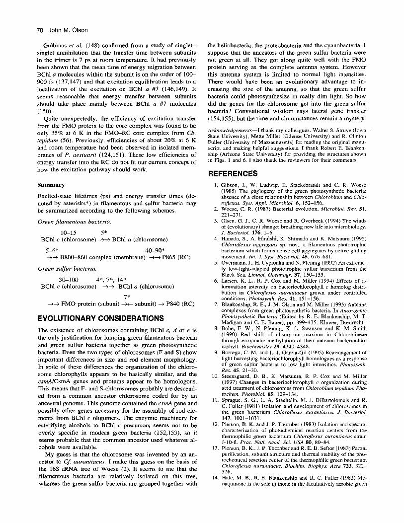
Excited-state lifetimes (ps) and energy transfer times (de- noted by asterisks*) in filamentous and sulfur bacteria may be summarized according to the following schemes.
Green filamentous bacteria.
10-15 5* BChl c (chlorosome) -++ BChl a (chlorosome)
5-6* 40-90* ++ B800-860 complex (membrane) ++ P865 (RC)
Green suljur bacteria.
30-100 4*, 7*, 14* BChi c (chlorosome) -+-+ BChl a (chlorosome)
7* ++ FMO protein (subunit ++ subunit) -+ P840 (RC)
EVOLUTIONARY CONSIDERATIONS The existence of chlorosomes containing BChl c, d or e is the only justification for lumping green filamentous bacteria and green sulfur bacteria together as green photosynthetic bacteria. Even the two types of chlorosomes (F and S) show important differences in size and rod element morphology. In spite of these differences the organization of the chloro- some chlorophylls appears to be basically similar, and the csmAICsmA genes and proteins appear to be homologous. This means that F- and S-chlorosomes probably are descend- ed from a common ancestor chlorosome coded for by an ancestral genome. This genome contained the csmA gene and possibly other genes necessary for the assembly of rod ele- ments from BChl c oligomers. The enzymic machinery for esterifying alcohols to BChl c precursors seems not to be overly specific in modem green bacteria (152,153), so it seems probable that the common ancestor used whatever al- cohols were available.
My guess is that the chlorosome was invented by an an- cestor to Cf: aurantiacus. I make this guess on the basis of the 16.5 rRNA tree of Woese (2). It seems to me that the filamentous bacteria are relatively isolated on this tree, whereas the green sulfur bacteria are grouped together with
the heliobacteria, the proteobacteria and the cyanobacteria. I suppose that the ancestors of the green sulfur bacteria were not green at all. They got along quite well with the FMO protein serving as the complete antenna system. However this antenna system is limited to normal light intensities. There would have been an evolutionary advantage to in- creasing the size of the antenna, so that the green sulfur bacteria could photosynthesize in really dim light. So how did the genes for the chlorosome get into the green sulfur bacteria? Conventional wisdom says lateral gene transfer (154,155), but the time and circumstances remain a mystery.
Acknowledgements-I thank my colleagues, Walter S. Struve (Iowa State University), Mette Miller (Odense University) and R. Clinton Fuller (University of Massachusetts) for reading the original manu- script and making helpful suggestions. I thank Robert E. Blanken- ship (Arizona State University) for providing the structures shown in Figs. 1 and 6. I also thank the reviewers for their comments.
REFERENCES I .
2.
3.
4.
5.
6.
7.
8.
9.
10.
11.
12.
13
14.
Gibson, J., W. Ludwig, E. Stackebrandt and C. R. Woese (1985) The phylogeny of the green photosynthetic bacteria: absence of a close relationship between Chlorobium and Chlo- rojexus. Syst. Appl. Microbiol. 6, 152-156. Woese, C. R. (1987) Bacterial evolution. Microbiol. Rev. 51, 221-271. Olsen, G. J., C. R. Woese and R. Overbeek (1994) The winds of (evolutionary) change: breathing new life into microbiology. J. Bacteriol. 176, 1-6. Hanada, S., A. Hiraishi, K. Shimada and K. Matsuura (1995) Chlorojexus aggregans sp. nov., a filamentous phototrophic bacterium which forms dense cell aggregates by active gliding movement. Int. J . Sysr. Bacteriol. 45, 676-681. Overmann, J., H. Cypionka and N. Pfennig (1992) An extreme- ly low-light-adapted phototrophic sulfur bacterium from the Black Sea. Limnol. Oceanogr. 37, 150-155. Larsen, K. L., R. P. Cox and M. Miller (1994) Effects of il- lumination intensity on bacteriochlorophyll c homolog distri- bution in ChloroJexus aurantiacus grown under controlled conditions. Photosynth. Res. 41, 151-156. Blankenship, R. E., J. M. Olson and M. Miller (1995) Antenna complexes from green photosynthetic bacteria. In Anoxygenic Photosynthetic Bacteria (Edited by R. E. Blankenship, M. T. Madigan and C. E. Bauer), pp. 399-435. Kluwer, Dordrecht. Bobe, F. W., N. Pfennig, K. L. Swanson and K. M. Smith (1990) Red shift of absorption maxima in Chlorobiineae through enzymatic methylation of their antenna bacteriochlo- rophyll. Biochemistry 29, 4340-4348. Borrego, C. M. and L. J. Garcia-Gil (1995) Rearrangement of light harvesting bacteriochlorophyll homologues as a response of green sulfur bacteria to low light intensities. Photosynth. Res. 45, 21-30. Steensgaard, D. B., K. Matsuura, R. P. Cox and M. Miller (1997) Changes in bacteriochlorophyll c organization during acid treatment of chlorosomes from Chlorobium tepidum. Pho- tochem. Phorobiol. 65, 129- 134. Sprague, S. G., L. A. Staehelin, M. J. DiBartolomeis and R. C. Fuller (1981) Isolation and development of chlorosomes in the green bacterium Chlorofiexus aurantiacus. J . Bacteriol.
Pierson, B. K. and J. P. Thornber (1983) Isolation and spectral characterization of photochemical reaction centers from the thermophiiic green bacterium Chlorojexus auruntiacus strain J-10-fl. Proc. Natl. Acad. Sci. USA 80, 80-84. Pierson, B. K., I. P. Thornber and R. E. B. Seftor (1983) Partial purification, subunit structure and thermal stability of the pho- tochemical reaction center of the thermophilic green bacterium Chlorojexus aurantiacus. Biochim. Biophys. Acra 723, 322- 326. Hale, M. B., R. E. Blankenship and R. C. Fuller (1983) Me- naquinone is the sole quinone in the facultatively aerobic green
147, 1021-1031.

Photochemistry and Photobiology, 1998, 67(1) 71
photosynthetic bacterium Chlorojexus aurantiacus. Biochim. Biophys. Actu 723, 376-382.
15. Vasmel, H. and J. Amesz (1983) Photoreduction of rnenaqui- none in the reaction center of the green photosynthetic bacte- rium Chlorojlexus aurantiacus. Biochim. Biophys. Actu 724, 118-122.
16. Vasmel, H., R. F. Meiburg, H. J. M. Kramer, L. J. de Vos and J. Amesz (1983) Optical properties of the photosynthetic re- action center of Chlorojlexus aurantiucus at low temperature. Biochim. Biophys. Acta 724, 333-339.
17. Ovchinnikov, Yu. A,, N. G. Abdulaev, A. S. Zolotarev, B. E. Shmukler, A. A. Zargarov, M. A. Kutuzov, I. N. Telezhinskaya and N. B. Levina (1988) Photosynthetic reaction centre of Chlorqflexus aurantiacus. I. Primary structure of L-subunit. FEBS Lett. 231, 237-242.
18. Ovchinnikov, Yu. A., N. G. Abdulaev, B. E. Shmukler, A. A. Zargarov, M. A. Kutuzov, 1. N. Telezhinskaya, N. B. Levina and A. S. Zolotarev (1988) Photosynthetic reaction centre of Chlorojlexus aurantiacus. Primary structure of M-subunit.
19. Shiozawa, J . A,, F. Lottspeich, D. Oesterhelt and R. Feick ( 1989) The primary structure of the Chlorojlexus aurantiacus reaction-center polypeptides. Eur. J. Biochem. 180, 75-84.
20. Ivancich, A., R. Feick, A. Ertlmaier and T. A. Mattioli (1996) Structure and protein binding interactions of the primary donor of the Chlorojlexus aurantiacus reaction center. Biochemistry 35, 6126-6135.
21. Becker, M., V. Nagarajan, D. Middendorf, W. W. Parsons, J. E. Martin and R. E. Blankenship (1991) Temperature depen- dence of the initial electron-transfer kinetics in photosynthetic reaction centers of Chlorofiexus aurantiacus. Biochim. Bio- phys. Aclu 1057, 299-3 12.
22. Shuvalov, V. A., H. Vasmel, J. Amesz and L. N. M. Duysens (1986) Picosecond spectroscopy of the charge separation in reaction centers of Chlorojexus aurantiacus with selective ex- citation of the primary electron donor. Biochim. Biophys. Acta 851, 361-368.
23. Ark, T., S. Schmidt, W. Kaiser, C. Lauterwasser, M. Meyer, H. Scheer and W. Zinth (1993) The accessory bacteriochlo- rophyll: a real electron carrier in primary photosynthesis. Proc. Natl. Acad. Sci. USA 90, 1 1757-1 1761.
24. Kirmaier, C. and D. Holten (1991) An assessment of the mech- anism of initial electron rransfer in bacterial reaction centers. Biochemistry 30, 609-61 3.
25. Blankenship, R. E., R. Feick, B. D. Bruce, C. Kirmaier, D. Holten and R. C. Fuller (1983) Primary photochemistry in the facultative green photosynthetic bacterium Chlorojlexus aurun- tiacus. J. Cell Biol. 22, 25 1-26 1 .
26. Kirmaier, C., D. Holten, R. Feick and R. E. Blankenship (1 983) Picosecond measurements of the primary photochemi- cal events in reaction centers isolated from the facultative green photosynthetic bacterium Chlorojrxus aurantiacus. FEBS Lett. 158, 73-76.
27. Kirmaier, C., D. Holten, L. J. Mancino and R. E. Blankenship ( 1984) Picosecond photodichroism studies on reaction centers from the green photosynthetic bacterium Chlorojlexus auran- tiacus. Biochim. Biophys. Acta 765, 138-146.
28. Kirmaier, C., R. E. Blankenship and D. Holten (1986) For- mation and decay of radical pair state P'I- in Chlorojexus auruntiacws reaction centers. Biochim. Biophys. Acta 850, 275-285.
29. Nuijs, A. M., H. Vasmel, L. N. M. Duysens and J . Amesz ( 1986) Antenna and reaction-center processes upon picosec- ond-Rash excitation of membranes of the green photosynthetic bacterium Chlorojlexus aurantiacus. Biochim. Biophys. Acta 849, 3 16-324.
30. Feick, R., J . A. Shiozawa and A. Ertlmaier (1995) Biochemical and spectroscopic properties of the reaction center of the green filamentous bacterium, Chlorojlexus aurantiacus. In Anoxygen- ic Photosynthetic Bacteria (Edited by R. E. Blankenship, M. T. Madigan and C. E. Bauer), pp. 699-708. Kluwer, Dordrecht.
31. Blankenship, R. E., J. T. Trost and L. J. Mancino (1988) Prop- erties of reaction centers from the green photosynthetic bac- terium Chlorojlexus aurantiucus. In Structure of Bacterial Re-
FEBS Lett. 232, 364-368.
action Centers (Edited by J. Breton and A. Vermeglio), pp. 119-127. Plenum, New York.
32. Shuvalov, V. A., A. Ya. Shkuropatov, S. M. Kulakova, M. A. Ismailov and V. A. Shkuropatova (1986) Photoreactions of bacteriopheophytins and bacteriochlorophylls in reaction cen- ters of Rhodopseudomonas sphaeroides and Chlorofiexus au- rantiacus. Biochim. Biophys. Acta 849, 337-346.
33. Volk, M., G. Scheidel, A. Ogrodnik, R. Feick and M. E. Mich- el-Beyerle (1991) High quantum yield of charge separation in reaction centers of Chlorojlexus aurantiacus. Biochim. Bio-
34. Feick, R., A. Ertlmaier and U. Ermler (1996) Crystallization and X-ray analysis of the reaction center from the thermophilic green bacterium Chlorojlexus aurantiacus. FEBS Lett. 396, 1 6 1- 164.
35. Swarthoff, T., P. Gast and A. J. Hoff (1981) Photooxidation and triplet formation of the primary electron donor of the green photosynthetic bacterium Prosthecochloris aestuarii, observed with ESR spectroscopy. FEBS Lett. 127, 83-86.
36. Wasielewski, M. R., U. H. Smith and J. R. Norris (1982) ESR study of the primary electron donor in highly "C-enriched Chlorobium limicola f. thiosulfatophilum. FEBS Lett. 149,
37. Nitschke, W., U. Feiler and A. Rutherford (1990) Photosyn- thetic reaction center of green bacteria studied by EPR. Bio- chemistry 29, 3834-3842.
38. Feiler, U., D. Albouy, B. Robert and T. A. Mattioli (1995) Symmetric structural features and binding site of the primary electron donor in the reaction center of Chlorobium. Biochem- istry 34, 11099-1 1105.
39. Olson, J . M., M. Miller and J. D'Olieslager (1995) The asym- metry of P t in bacterial reaction centers revealed by circular dichroism spectroscopy. Biochemistry 34, 15230-1 5234.
40. Rigby, S. E. J., R. Thapar, M. C. W. Evans and P. Heathcote (1994) The electronic structure of P840", the primary donor of the Chlorobium limicola f. sp. thiosulphatophilum photo- synthetic reaction centre. FEBS Lett. 350, 24-28.
41. Bratt, P. J., I. P. Muhiuddin, M. C. W. Evans and P. Heathcote (1996) I4N electron spin echo envelope modulation (ESEEM) spectroscopy of the cation radical P840", the primary electron donor of the Chlorobium limicola reaction center. Photochem. Photobiol. 64, 20-25.
42. Nabedryk, E., W. Leibl and J. Breton (1996) FTIR spectros- copy of primary donor photooxidation in photosystem 1, He- liobacillus mobilis, and Chlorobium limicola. Comparison with purple bacteria. Photosynth. Res. 48, 301-308.
43. Noguchi, T., N. Kusumoto, Y. Inoue and H. Sakurai (1996) Electronic and vibrational structure of the radical cation of P- 840 in the putative homodimeric reaction center from Chlo- robium tepidum as studied by FTIR spectroscopy. Biochemis- try 35, 15428-15435.
44. van de Meeut, E. J., M. Kobayashi, C. Erkelens, P. van Veelen, S. Otte, K. Inoue, T. Watanabe and J. Amesz (1992) The nature of the primary electron acceptor in green sulfur bacteria. Biochim. Biophys. Actu 1102, 371-378.
45. Braumann, T., H. Vasmel, L. H. Grimme and J . Amesz (1986) Pigment composition of the photosynthetic membrane and re- action center of the green bacterium Prosthecochloris aestu- arii. Biochim. Biophys. Acta 848, 83-9 1.
46. Feiler, U., D. Albouy, C. Pourcet, T. A. Mattioli, M. Lutz and B. Robert (1994) Structure and binding site of the primary electron acceptor in the reaction center of Chlorobiurn. Bio- chemistry 33, 7594-7599.
47. Feiler, U. and G. Hauska (1995) The reaction center from green sulfur bacteria. In Anox.ygenic Photosynthetic Bacteria (Edited by R. E. Blankenship, M. T. Madigan and C. E. Bauer), pp. 665-685. Kluwer, Dordrecht.
48. Frankenberg, N., C. Hagar-Braun, U. Feiler, M. Fuhnnann, H. Rogl, N. Schneebauer, N. Nelson and G. Hauska (1996) P-840 reaction centers from Chlorobium tepidurn-uinone analysis and functional reconstitution into lipid vesicles. Photochem. Photobiol. 64, 14-19.
49. Buttner, M., D.-L. Xie, H. Nelson, W. Pinther, G. Hauska and N. Nelson (1992) The photosystem I-like P840-reaction center
phys. Acta 1058, 217-224.
138- 140.
72 John M. Olson
of green S-bacteria is a homodimer. Biochim. Biophys. Acra
50. Biittner, M., D.-L. Xie, H. Nelson, W. Pinther, G. Hauska and N. Nelson (1992) Photosynthetic reaction center genes in green sulfur bacteria and in photosystem 1 are related. Proc. Natl. Acad. Sci. USA 89, 8 135-8 139.
51. Nuijs, A. M., H. Vasmel, H. L. P. Joppe, L. N. M. Duysens and J. Amesz (1985) Excited states and primary charge sepa- ration in the pigment system of the green photosynthetic bac- terium Prosthecochloris aestuarii as studied by picosecond ab- sorbance difference spectroscopy. Biochim. Biophys. Acta 807, 24-34.
52. Golbeck, J. H. (1993) Shared thematic elements in photochem- ical reaction centers. Proc. Narl. Acad. Sci. USA 90, 1642- 1646.
53. Evans, M. C. W. and G. Bredekamp (1990) The structure and function of the photosystem I reaction centre. Physiol. Plant. 79, 415420.
54. Francke, C., H. P. Permentier, E. M. Franken, S. Neerken and J. Amesz (1997) Isolation and properties of photochemically active reaction center complexes from the green sulfur bacte- rium Prosrhecochloris aestuarii. Biochemistry 36, 14 167- 14172.
55. Oh-oka, H., S. Kakutani, S. Kamei, H. Matsubara, M. Iwaki and S. Itoh ( 1 995) Highly purified photosynthetic reaction cen- ter (PscAkytochrome cS5& complex of the green sulfur bac- terium Chlorobium limicola. Biochemistry 34, 1309 1-13097.
56. Francke, C., S. C. M. Otte, M. Miller, J. Amesz and J. M. Olson (1996) Energy transfer from carotenoid and FMO-pro- tein in subcellular preparations from green sulfur bacteria. Spectroscopic characterization of an FMO-reaction center core complex at low temperature. Photosynth. Res. SO, 71-77.
57. Parot, P., N. Delmas, D. Garcia and A. Vermeglio (1985) Structure of Chloroflexus aurantiacus reaction center: photo- selection at low temperature. Biochim. Biophys. Acta 809, 137- 140.
58. Freeman, J. C. and R. E. Blankenship (1990) Isolation and characterization of the membrane-bound cytochrome c-554 from the thermophilic green photosynthetic bacterium Chlo- roflexus aurantiacus. Photosynrh. Res. 23, 29-38.
59. Meyer, T. E., G. Tollin, M. A. Cusanovich, J. C. Freeman and R. E. Blankenship (1989) In vitro kinetics of reduction of cy- tochrome css4 isolated from the reaction center of the green phototrophic bacterium, Chlorojexus aurantiacus. Arch. Bio- chem. Biophys. 272, 254-261.
60. Heibel, G., K. Griebenow and P. Hildebrandt (1991) Structural studies of cytochrome c-554 from Chloroflexus aurantiacus by resonance Raman spectroscopy techniques. Biochim. Biophys. Acra 1060, 196-202.
61. Dracheva, S., J. A. Williams, G. Van Driessche, J. J. Van Beeumen and R. E. Blankenship (1991) The primary structure of cytochrome c-554 from the green photosynthetic bacterium Chlorojexus aurantiacus. Biochemistry 30, 1145 1-1 1458.
62. Feiler, U. (1991) Untersuchung am Photosystem von verschie- denen photosynthetischen Bakterien. Thesis, University Frank- fort.
63. Feiler, U., W. Nitschke and H. Michel (1992) Characterization of an improved reaction center preparation from the photosyn- thetic green sulfur bacterium Chlorobium containing the FeS centers FA and F, and a bound cytochrome subunit. Biochem- istry 31, 2608-2614.
64. Okkels, J . S., B. Kjler, 0. Hansson, I. Svensen, B. L. Moeller and H. V. Scheller (1992) A membrane-bound monoheme cy- tochrome c-551 of a novel type is the immediate electron donor to P840 of the Chlorobium vibrioforme photosynthetic reaction center complex. J. Biol. Chem. 267, 21139-21 145.
65. Hagar-Braun, C., D.-L. Xie, U. Jarosch, E. Herold, M. Biittner, R. Zimmerman, R. Deutzmann, G. Hauska and N. Nelson (1995) Stable photobleaching of P840 in Chlorobium reaction center preparations: presence of the 42-kDa bacteriochloro- phyll a protein and a 17-kDa polypeptide. Biochemistry 34, 96 17-9624.
66. Oh-oka, H., S. Kamei, H. Matsubara, M. Iwaki and S. Itoh (1995) Two molecules of cytochrome c function as the electron
1101, 154-156. donors to P840 in the reaction center complex isolated from a green sulfur bacterium, Chlorobium tepidum. FEBS Lett. 365, 30-34.
67. Wechsler, T., R. Brunisholz, F. Suter, R. C. Fuller and H. Zuber (1985) The complete amino acid sequence of a bacter- iochlorophyll a binding polypeptide isolated from the cyto- plasmic membrane of the green photosynthetic bacterium ChloroJlexus aurantiacus. FEBS Lett. 191, 34-38.
68. Wechsler, T. D., R. A. Brunisholz, G. Frank, F. Suter and H. Zuber (1987) The complete amino acid sequence of the anten- na polypeptide B806-866-P from the cytoplasmic membrane of the green bacterium Chloroflexus aurantiacus. FEES Lett.
69. Wechsler, T. D., R. A. Brunisholz, G. Frank and H. Zuber (1991) Isolation and protein chemical characterization of the B806-866 antenna complex of the green thermophilic bacte- rium Chlorojexus aurantiacus. J . Photochem. Photobiol. B Biol. 8, 189-197.
70. Zuber, H. and A. Brunisholz (1991) Structure and function of antenna polypeptides and chlorophyll-protein complexes: prin- ciples and variability. In Chlorophylls (Edited by H. Scheer), pp. 627-703. CRC Press, Boca Raton, FL.
71. Matthews, B. W., R. E. Fenna, M. C. Bolognesi, M. F. Schmid and J. M. Olson (1979) Structure of a bacteriochlorophyll a- protein from the green photosynthetic bacterium Prostheco- chloris aestuarii. J . Mol. Biol. 131, 259-285.
72. Li, Y.-F., W. Zhou, R. E. Blankenship and J. P. Allen (1997) Crystal structure of the bacteriochlorophyll a protein from Chlorobium tepidum. J. Mol. Biol. 272, 456-471.
73. Daurat-Larroque, S. T., K. Brew and R. E. Fenna (1986) The complete amino acid sequence of a bacteriochlorophyll a-pro- tein from Prosthecochloris aestuarii. J . Biol. Chem. 261,
74. Dracheva, S., J. C. Williams and R. E. Blankenship (1992) Cloning and sequencing of the FMO-protein gene from Chlo- robium tepidum. In Research in Photosynthesis, Vol. I (Edited by N. Murata), pp. 53-56. Kluwer, Dordrecht.
75. Frigaard, N.-U., S. Takaichi, M. Hirota, K. Shimada and K. Matsuura (1997) Quinones in chlorosomes of green sulfur bac- teria and their role in the redox-dependent fluorescence studied in chlorosome-like bacteriochlorophyll c aggregates. Arch. Mi- crobiol. 167, 343-349.
76. Staehelin, L. A,, J. R. Golecki, R. C. Fuller and G. Drews (1978) Visualization of the supramolecular architecture of chlorosomes (Chlorobium type vesicles) in freeze-fractured cells of Chlorojexus aurantiacus. Arch. Microbiol. 119, 269- 277.
77. Oelze, J. and J. R. Golecki (1995) Membranes and chloro- somes of green bacteria: structure, composition and develop- ment. In Anoxygenic Photosynrheric Bacteria (Edited by R E. Blankenship, M. T. Madigan and C. E. Bauer), pp. 259-278. Kluwer, Dordrecht.
78. Staehelin, L. A,, J. R. Golecki and G. Drews (1980) Supra- molecular organization of chlorosomes (Chlorobium vesicles) and of their membrane attachment sites in Chlorobium limi- cola. Biochim. Biophys. Acta 589, 30-45.
79. Cruden, D. L. and R. Y. Stanier (1970) The characterization of Chlorobium vesicles and membranes isolated from green bacteria. Arch. Mikrobiol. 72, 115-134.
80. Gerola, P. D. and J. M. Olson (1986) A new bacteriochloro- phyll a-protein complex associated with chlorosomes of green sulfur bacteria. Biochim. Biohpys. Acta 848, 69-76.
81. Feick, R. G., M. Fitzpatrick and R. C. Fuller (1982) Isolation and characterization of cytoplasmic membranes and chloro- somes from the green bacterium Chlorojexus auranriacus. J. Bacteriol. 150, 905-9 15.
82. Pierson, B. K. and R. W. Castenholz (1995) Taxonomy and physiology of filamentous anoxygenic phototrophs. In Anoxy- genic Photosynthetic Bacteria (Edited by R. E. Blankenship, M. T. Madigan and C. E. Bauer), pp. 3 1 4 7 . Kluwer, Dor- drecht.
83. Gloe, A,, N. Pfennig, H. Brockman, Jr. and W. Trowitsch (1 975) A new bacteriochlorophyll from brown-colored Chlo- robiaceae. Arch. Microbiol. 102, 103-109.
210, 189-194.
3607-36 15.

84. Schmidt, K. (1978) Biosynthesis of carotenoids. In The Pho- tosynthetic Bacteria (Edited by R. K. Clayton and W. R. Sis- trom), pp. 729-750. Plenum Press, New York.
85. Liaaen-Jensen, S . (1965) Bacterial carotenoids. XVIII. Aryl- carotenes from Phaeobium. Acta Chem. Srand. 19, 1025-1030.
86. Olson, J. M. (1980) Chlorophyll organization in green photo- synthetic bacteria. Biochim. Biophvs. Acta 594, 33-5 1 .
87. Swarthoff, T., B. G. de Grooth, R. F. Meiburg, C. P. Rijgers- berg and J . Arnesz (1980) Orientation of pigments and pig- ment-protein complexes in the green photosynthetic bacterium Prosthecochloris aestuarii. Biochim. Biophys. Acta 593, 5 1- 59.
88. Brune, D. C., P. D. Gerola and I. M. Olson (1990) Circular dichroism of green bacterial chlorosomes. Photosynth. Res. 24, 253-263.
89. Olson, J. M. and J. P. Pedersen (1990) Bacteriochlorophyll c monomers, dimers, and higher aggregates in dichloromethane, chloroform, and carbon tetrachloride. Photosynth. Res. 25, 25- 37.
90. Krasnovsky, A. A. and M. L. Bystrova (1980) Self-assembly of chlorophyll aggregated structures. BiaSystems 12, 18 1-194.
91. Smith, K. M., L. A. Kehres and J. Fajer (1983) Aggregation of the bacteriochlorophylls c, d, and e. Models for the antenna chlorophylls of green and brown photosynthetic bacteria. J. Am. Chem. Soc. 105, 1387-1389.
92. Uehard, K. and J. M. Olson (1992) Aggregation of bacterio- chlorophyll c homologs to dimers, tetramers, and polymers in water-saturated carbon tetrachloride. Photosynth. Res. 33, 25 1- 257.
93. Hirota, M., T. Moriyama, K. Shimada, M. Miller, J. M. Olson and K. Matsuura (1992) High degree of organization of bac- teriochlorophyll c in chlorosome-like aggregates spontaneously assembled in aqueous solution. Biochim. Biophys. Acta 1099,
94. Miller, M., T. Gillbro and J. M. Olson (1993) Aqueous aggre- gates of hacteriochlorophyll c as a model for pigment organi- zation in chlorosomes. Photochem. Photobiol. 57, 98-102.
95. Feick, R. G. and R. C. Fuller (1984) Topography of the pho- tosynthetic apparatus of Chloroflexus aurantiacus. Biochemis- try 23, 3693-3700.
96. Schmidt, K. (1980) A comparative study on the composition of chlorosomes (Chlorobium vesicles) and cytoplasmic mem- branes from Chlorojlexus aurantiacus strain Ok-70-fl and Chforobium limicola f. thiosu~at~philum strain 6230. Arch. Microbiol. 124, 2 1-3 1 .
97. Chung, S., G. Frank, H. Zuber and D. A. Bryant (1994) Genes encoding two chlorosome components from the green sulfur bacteria Chforobium vibrioforme strain 8327D and Chloro- bium tepidum. Photosynth. Res. 41, 261-275.
98. Theroux, S . J., T. E. Redlinger, R. C. Fuller and S . J. Robinson ( 1 990) Gene encoding the 5.7-kilodalton chlorosome protein of Chloroflexus aurantiacus: regulated message levels and a predicted carboxy-terminal protein extention. J. Bacteriol. 172, 449711504.
99. Chung, S. and D. A. Bryant (1996) Characterization of csmB genes, encoding a 7.5-kDa protein of the chlorosome envelope, from the green sulfur bacteria Chlorobium vibrioforme 8327D and Chlorobium tepidum. Arch. Microbiol. 166, 234-244.
100. Chung, S . and D. A. Bryant (1996) Characterization of the csmD and csmE genes from Chlorobium tepidum. The CsmA, CsmC, CsmD, and CsmE proteins are components of the chlo- rosome envelope. Photosynth. Res. 50, 41-59.
101. Wechsler, T., F. Suter, R. C. Fuller and H. Zuber (1985) The complete amino acid sequence of the bacteriochlorophyll c binding polypeptide from chlorosomes of the green photosyn- thetic bacterium Chlorojlexus aurantiacus. FEBS Lett. 181,
102. Wagner-Huber, R., R. Brunisholz, G. Frank and H. Zuber (1 988) The BChlc/e-binding polypeptides from chlorosomes of green photosynthetic bacteria. FEBS Lett. 239, 8-12.
103. Wagner-Huber, R., U. Fischer, R. Brunisholz, M. Rumbeli, G. Frank and H. Zuber (1990) The primary structure of the pre- sumable BChl d-binding polypeptide of Chlorobium vibriofor- me f . thiosulfatophilum. Z. Natutforsch. 45c, 8 18-822.
27 1-274.
17 3- I 78.
Photochemistry and Photobiology, 1998, 67(1) 73
104. Wullink, W., J. Knudsen, J. M. Olson, T. E. Redlinger and E. F. J. Van Bruggen (1991) Localization of polypeptides in iso- lated chlorosomes from green phototrophic bacteria by im- muno-gold labeling electron microscopy. Biochim. Biophys.
105. Gerola, P. D., P. Hejrup, J. Knudsen, P. Roepstorff and J. M. Olson (1988) The bacteriochlorophyll c-binding protein from chlorosomes of Chlorobium limicola f. thiosu&tophilum. In Green Photosynthetic Bacteria (Edited by J . M. Olson, J. G. Ormerod, J. Amesz, E. Stackebrandt and H. G. Triiper), pp. 43-52. Plenum Press, New York.
106. Hojrup, P., P. Gerola, H. F. Hansen, J. M. Mikkelsen, A. E. Shahed, J. Knudsen, P. Roepstorff and J . M. Olson (1991) The amino acid sequence of a major protein component in the light harvesting complex of the green photosynthetic bacterium Chlorobium limicola f. thiosulfatophilum. Biochim. Biophys. Acta 1077, 220-224.
107. Lehmann, R. P., R. A. Brunisholz and H. Zuber (1994) Struc- tural differences in chlorosomes from Chlorojfexus aurantiacus grown under different conditions support the BChl c-binding function of the 5.7 kDa polypeptide. FEBS Lett. 342, 3 19-324.
108. Foidl, M., J. R. Golecki and J. Oelze (1994) Bacteriochloro- phyll c formation and chlorosome development in Chlorojfexus aurantiacus. Photosynth. Res. 41, 145-150.
109. Miller, M., D. Simpson and T. E. Redlinger (1993) The effect of detergent on the structure and composition of chlorosomes isolated from Chlorojlexus aurantiacus. Photosynth. Res. 35,
110. Brune, D. C., G. H. King and R. E. Blankenship (1988) Inter- actions between bacteriochlorophyll c molecules in oligomers and in chlorosomes of green photosynthetic bacteria. In Pho- tosynthetic Light-Harvesting Systems (Edited by H. Scheer and S. Schneider), pp. 141-151. Walter de Gruyter, Berlin.
11 1 . Nozawa, T., K. Ohtomo, M. Suzuki, Y. Morishita and M. T. Madigan ( 1993) Structures and organization of bacteriochlo- rophyll c's in chlorosomes from a new thermophilic bacterium Chlorobium tepidum. Bull. Chem. SOC. Jpn. 66, 23 1-237.
112. Nozawa, T., K. Ohtomo, M. Suzuki, H. Nakagawa, Y. Shika- ma, H. Konami and Z-Y. Wang (1994) Structures of chloro- somes and aggregated BChl c in Chlorobium tepidum from solid state high resolution CP/MAS I3C NMR. Photosynth. Res. 41, 21 1-223.
113. Matsuura, K., M. Hirota, K. Shimada and M. Mimuro (1993) Spectral forms and orientation of bacteriochlorophylls c and N
in chlorosomes of the green photosynthetic bacterium Chlo- rojlexus aurantiacus. Photochem. Photobiol. 57, 92-97.
114. Holzwarth, A. R. and K. Schaffner (1994) On the structure of bacteriochlorophyll molecular aggregates in the chlorosomes of green bacteria. A molecular modelling study. Photosynth. Res. 41, 225-233.
115. Mizoguchi, T., K. Matsuura, K. Shimada and Y. Koyama (1996) The structure of the aggregate form of bacteriochloro- phyll c showing the Q, absorption above 740 nm: a 'H-NMR study. Chem. Phys. Lett. 260, 153-158.
116. Buck, D. R. and W. S. Struve (1996) Tubular exciton models for BChl c antennae in chlorosomes from green photosynthetic bacteria. Photosynth. Res. 48, 367-377.
117. Fetisova, 2. and K. Mauring (1992) Experimental evidence of oligomeric organization of antenna bacteriochlorophyll c in green bacterium Chlorojlexus aurantiac'us by spectral hole burning. FEBS Lett. 307, 37 1-374.
118. Fetisova, Z., A. Freiberg, K. Mauring, V. Novoderezhkin, A. Taisova and K. Timpmann (1996) Excitation energy transfer in chlorosomes of green bacteria: theoretical and experimental studies. Biophys. J. 71, 995-1010.
119. Fetisova, Z., A. Freiberg, V. Novoderezhkin, A. Taisova and K. Timpmann (1996) Antenna size dependent exciton dynam- ics in the chlorosomal antenna of the green bacterium Chlo- rojlexus aurantiacus. FEBS Let. 383, 233-236.
120. Mimuro, M., Y. Nishimura, I. Yamazaki, M. Kobayashi, Z. Y. Wang, T. Nozawa, K. Shimada and K. Matsuura (1996) Ex- citation energy transfer in the green photosynthetic bacterium Chlorojlexus auranriacus: a specific effect of 1-hexanol on the
Act0 1060, 97-105.
275-283.

74 John M. Olson
optical properties of baseplate and energy transfer processes. Photosynth. Res. 48, 263-270.
121. Tamiaki, H. (1996) Supramolecular structure in extra membra- neous antennae of green photosynthetic bacteria. Coord. Chem. Rev. 148, 183-197.
122. Holcomb, C. T. and R. S. Knox (1996) The relationship of intercompartmental transfer rate constants to those of an un- derlying physical model. Photosynrh. Res. 50, 1 17-1 3 1.
123. Sybesma, C. and J. M. Olson (1963) Transfer of chlorophyll excitation energy in green photosynthetic bacteria. P roc. Natl. Acad. Sci. USA 49, 248-253.
124. Otte, S. C. M. (1992) Pigment systems of photosynthetic bac- teria and photosystem I1 of green plants. Doctoral Thesis, Uni- versity of Leiden, The Netherlands.
125. Vos, M., A. M. Nuijs, R. van Grondelle, R. J. van Dorssen, P. D. Gerola and J. Amesz (1987) Excitation transfer in chloro- somes of green photosynthetic bacteria. Biochim. Biophys. Acra 891, 275-285.
126. Miller, M., R. P. Cox and T. Gillbro (1991) Energy transfer kinetics in chlorosomes from Chlorojlexus aurantiacus: studies using picosecond absorbance spectroscopy. Biochim. Biophys.
127. Fetisova, Z. G., A. M. Freiberg and K. E. Timpmann (1988) Long-range molecular order as an efficient strategy for light harvesting in photosynthesis. Nature 334, 633-634.
128. Freiberg, A. M., K. E. Timpmann and Z. G. Fetisova (1988) Excitation energy transfer in living cells of the green bacterium Chlorobium limicola studied by picosecond fluorescence spec- troscopy. In Green Photosynthetic Bacteria (Edited by J. M. Olson, J. G. Ormerod, J. Amesz, E. Stackebrandt and H. G. Triiper), pp. 81-90. Plenum Press, New York.
129. Mimuro, M., M. Hirota, Y. Nishimura, T. Moriyama, I. Ya- mazaki, K. Shimada and K. Matsuura (1994) Molecular or- ganization of bacteriochlorophyll in chlorosomes of the green photosynthetic bacterium Chlorojlexus aurantiacus: studies of fluorescence depolarization accompanied by energy transfer processes. Photosynth. Res. 41, 181-191.
130. Gillbro, T., A. Sandstrom, V. Sundstrom and J. M. Olson ( 1 988) Picosecond energy transfer kinetics in chlorosomes and bacteriochlorophyll a-proteins of Chlorobium limicola. In Green Photosynthetic Bacteria (Edited by J. M. Olson, J. G. Ormerod, J. Amesz, E. Stackebrandt and H. G. Truper), pp. 91-96. Plenum Press, New York.
131. Causgrove, T. P., D. C. Brune, J. Wang, B. P. Wittmershaus and R. E. Blankenship (1990) Energy transfer kinetics in whole cells and isolated chlorosomes of green photosynthetic bacte- ria. Photosynth. Res. 26, 39-48.
132. Muller, M. G. , K. Griebenow and A. R. Holzwarth (1993) Picosecond energy transfer and trapping kinetics in living cells of the green bacterium Chlorojlexus aurantiacus. Biochim. Biophys. Acta 1144, 161-169.
133. Vasmel, H., R. J. van Dorsen, G. J. de Vos and J. Amesz (1986) Pigment organization and energy transfer in the green photosynthetic bacterium Chlorojlexus aurantiacus I. The cy- toplasmic membrane. Photosynth. Res. 7, 28 1-294.
134. Griebenow, K., M. G. Muller and A. R. Holzwarth (1991) Pigment organization and energy transfer in green bacteria. 3. Picosecond energy transfer kinetics within the B806-866 bac- teriochlorophyll a antenna complex isolated from Chlorojlexus aurantiacus. Biochim. Biophys. Acta 1059, 226-232.
135. Mimuro, M., T. Nozawa, N. Tamai, K. Shimada, I. Yamazaki, S. Lin, R. S. Knox, B. P. Wittmershaus, D. C. Brune and R. E. Blankenship (1989) Excitation energy flow in chlorosome antennas of green photosynthetic bacteria. J. Phys. Chem. 93,
136. Holzwarth, A. R., M. G. Miiller and K. Griebenow (1990) Picosecond energy transfer kinetics between pigment pools in different preparations of chlorosomes from the green bacterium Chlorojexus aurantiacus Ok-70-fl. J. Photochem. Photobiol. B 5, 457465.
137. Savikhin S., W. Zhou, R. E. Blankenship and W. S. Struve (1 994) Femtosecond energy transfer and spectral equilibration in bacteriochlorophyll a-protein antenna trimers from the green bacterium Chlorobium tepidum. Biophys. J. 66, 110-1 13.
Acta 1057, 187-194.
7503-7509.
138. Savikhin, S., Y. Zhu, R. E. Blankenship and W. S. Struve (1996) Ultrafast energy transfer in chlorosomes from the green photosynthetic bacterium Chlorojexus aurantiacus. J. Phys. Chem. 100, 3320-3322.
139. Savikhin, S., Y. Zhu, R. E. Blankenship and W. S. Struve (1996) Intraband energy transfers in the BChl c antenna of chlorosomes from the green photosynthetic bacterium Chlorojlexus aurantiacus. J . Phys. Chem. 100, 17978- 17980.
140. Ma, Y.-Z., R. P. Cox, T. Gillbro and M. Miller (1996) Bac- teriochlorophyll organization and energy transfer kinetics in chlorosomes from Chlorojlexus auruntiacus depend on the light regime during growth. Photosynth. Res. 47, 157-165.
141. Causgrove, T. P., D. C. Brune and R. E. Blankenship (1992) Forster energy transfer in chlorosomes of green photosynthetic bacteria. J. Photochem. Photobiol. B Biol. 15, 171-179.
142. Wang, J., D. C. Brune and R. E. Blankenship (1990) Effects of oxidants and reductants on the efficiency of excitation trans- fer in green photosynthetic bacteria. Biochim. Biophys. Acta 1015, 457-463.
143. Blankenship, R. E., P. Cheng, T. P. Causgrove, D. C. Brune, S. H.-H. Wang, J.-U. Choh and J. Wang (1993) Redox regu- lation of energy transfer efficiency in antennas of green pho- tosynthetic bacteria. Photochem. Photobiol. 57, 103-1 07.
144. Zhou, W., R. LoBrutto, S. Lin and R. E. Blankenship (1994) Redox effects on the bacteriochlorophyll a-containing Fenna- Matthews-Olson protein from Chlorobium tepidum. Photo- synth. Res. 41, 89-96.
45. van Noort, P. I., Y. Zhu, R. LoBrutto and R. E. Blankenship (1997) Redox effects on the excited-state lifetime in chloro- somes and bacteriochlorophyll c oligomers. Biophys. J. 72,
46. Pearlstein, R. M. (1992) Theory of the optical spectra of the bacteriochlorophyll a antenna protein trimer from Prosthe- cochloris aestuarii. Photosynth. Res. 31, 21 3-226.
47. Savikhin, S. and W. S. Struve (1994) Ultrafast energy transfer in FMO trimers from the green bacterium Chlorobium tepi- dum. Biochemistry 33, 1 1200-1 1208.
148. Gulbinas, V., L. Valkunas, D. Kuciauskas, E. Katilius, V. Liu- olia, W. Zhou and R. E. Blankenship (1996) Singlet-singlet annihilation and local heating in FMO complexes. J . Phys. Chem. 100, 17950-17956.
149. Lu, X. Y. and R. M. Pearlstein (1993) Simulations of Pros- thecochloris bacteriochlorophyll a-protein optical spectra im- proved by parametric computer search. Photochem. Photobiol.
150. van Mourik, F., R. R. Venvijst, J. M. Mulder and R. van Gron- delle (1994) Singlet-triplet spectroscopy of the light-harvesting BChl a complex of Prosthecochloris aesruurii. The nature of the low-energy 825 nm transition. J. Phys. Chem. 98, 10307- 10312.
151. Kramer, H., T. J. Aartsma and J. Amesz (1996) Excited states and charge separation in membranes of the green sulphur bac- terium Prosthecochloris aestuarii. Photochem. Photobiol. 64,
152. Larsen, K. L., R. P. Cox and M. Miller (1994) Effects of il- lumination intensity on bacteriochlorophyll c homolog distri- bution in Chlorojlexus aurantiacus grown under controlled conditions. Photosynth. Res. 41, 151-156.
153. Steensgaard, D. B., R. P. Cox and M. Miller (1996) Manipu- lation of the bacteriochlorophyll c homolog distribution in the green sulfur bacterium Chlorobium tepidum. Photosynth. Res.
154. Pierson, B. K. and J. M. Olson (1989) Evolution of photosyn- thesis in anoxygenic photosynthetic prokaryotes. In Microbial Mats: Physiological Ecology of Benthic Microbial Communi- ties (Edited by Y. Cohen and E. Rosenberg), pp. 402-427. American Society of Microbiology, Washington, DC.
155. Blankenship, R. E. (1992) Origin and early evolution of pho- tosynthesis. Photosynth. Res. 33, 91-1 11.
156. Bruce, B. D., R. C. Fuller and R. E. Blankenship (1982) Pri- mary photochemistry in the facultatively aerobic green pho- tosynthetic bacterium Chlorojlexus aurantiacus. Proc. Natl. Acad. Sci. USA 79, 6532-6536.
316-325.
57, 86-91.
26-3 1.
48, 385-393.

157. Venturoli, G . and D. Zannoni (1988) Oxidation-reduction ther- modynamics of the acceptor quinone complex in whole-mem- brane fragments from Chlorojlexus auranriacus. Eur. J . Bio- chem. 178, 503-509.
158. Fowler, C. F., N. A. Nugent and R. C. Fuller (1971) The iso- lation and characterization of a photochemically active com- plex from Chloropseudomonas ethylica. Proc. Natl. Acad. Sci. USA 68, 2278-2282.
159. Prince, R. C. and J. M. Olson (1976) Some thermodynamic and kinetic properties of the primary photochemical reactants in a complex from a green photosynthetic bacterium. Biochim. Biophys. Acta 423, 357-362.
160. Olson, J. M., R. C. Prince and D. C. Brune (1976) Reaction- center complexes from green bacteria. Brookhaven Symp. Biol.
161. Swarthoff, T., P. Cast, A. J. Hoff and J. Amesz (1981) An optical and ESR investigation on the acceptor side of the re- action center of the green photosynthetic bacterium Prosthe- cochloris uestuarii. FEBS Lett. 130, 93-98.
162. Lockau, W. and W. Nitschke (1993) Photosystem 1 and its bacterial counterparts. Physiol. Plant. 88, 372-38 1.
163. Senge, M. 0. and K. M. Smith (199.5) Biosynthesis and struc- tures of the bacteriochlorophylls. In Anoxygenic Photosynthetic Bacteria (Edited by R. E. Blankenship, M. T. Madigan and C. E. Bauer), pp. 137-151. Kluwer, Dordrecht.
164. Risch, N., H. Brockmann, Jr. and A. Gloe (1979) Strukturauf- klarung von neuartigen Bacteriochlorophyllen aus Chlorojlexus auranriacus. Liehigs Ann. Chem., pp. 408-41 8.
28, 238-245.
Photochemistry and Photobiology, 1998, 67(1) 75
165. Fages, F., N. Griebenow, K. Griebenow, A. R. Holzwarth and K. Schaffner ( I 990) Characterization of light-harvesting pig- ments of Chlorojlexus aurantiacus. Two new chlorophylls: oleyl (octadec-9-enyl) and cetyl (hexadecanyl) bacteriochlo- rophyllides-c. J. Chem. SOC. Perkin Trans. 1, 279 1-2797.
66. Jones, 0. T. G. (1978) Biosynthesis of porphyrins, hemes, and chlorophylls. In The Photosynthetic Bacteria (Edited by R. K. Clayton and W. R. Sistrom), pp. 751-777. Plenum, New York.
67. Caple, M. B., H. Chow and C. E. Strouse (1978) Photosyn- thetic pigments of green sulfur bacteria. The esterifying alco- hols of bacteriochlorophylls c from Chlorobium limicola. J . Biol. Chem. 253, 6730-6737.
68. Smith, K. M., L. A. Kehres and H. D. Tabba (1980) Structures of the bacteriochlorophyll c homologues: solution to a long- standing problem. J . Am. Chem. SOC. 102, 7149-7151.
169. Smith, K. M., D. A. Goff, J. Fajer and K. M. Barkigia (1982) Chirality and structures of bacteriochlorophylls d. J . Am. Chem. SOC. 104, 3747-3749.
170. Smith, K. M., D. A. Goff, J. Fajer and K. M. Barkigia (1983) Isolation and characterization of two new bacteriochlorophyll d bearing neopentyl substituents. J. Am. Chem. SOC. 105, 1674-1 676.
171. Brockmann, H., Jr. (1976) Bacteriochlorophyll e: structure and stereochemistry of a new type of chlorophyll from Chlorobi- aceae. Phil. Trans. R. SOC. Lond. B 273, 277-285.
172. Fenna, R. E., B. W. Matthews, J. M. Olson and E. K. Shaw (1974) Structure of a bacteriochlorophyll-protein from the green photosynthetic bacterium Chlorobium limicola: crystal- lographic evidence for a trimer. J. Mol. Biol. 84, 23 1-240.