chest x rays
DESCRIPTION
collection of 100 chest x rays of common conditionsTRANSCRIPT
100 Chest X Rays for Study Group
by Dr. Suneet Khurana
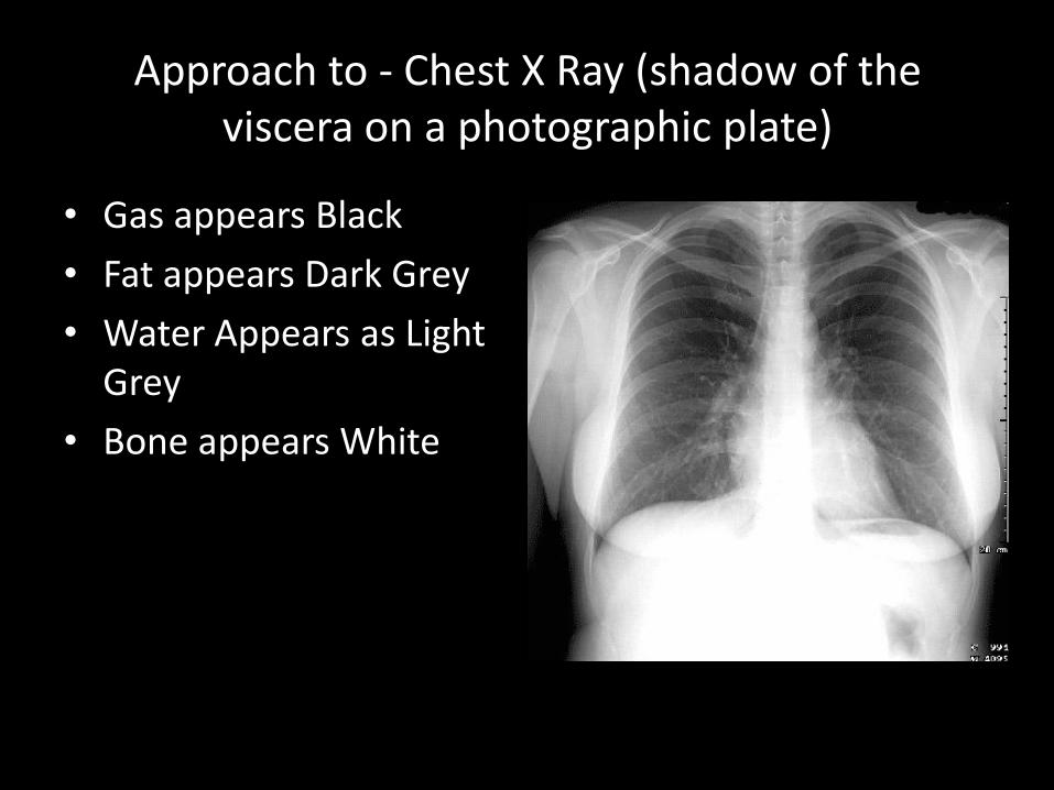
Approach to - Chest X Ray (shadow of the viscera on a photographic plate)
• Gas appears Black
• Fat appears Dark Grey
• Water Appears as Light Grey
• Bone appears White
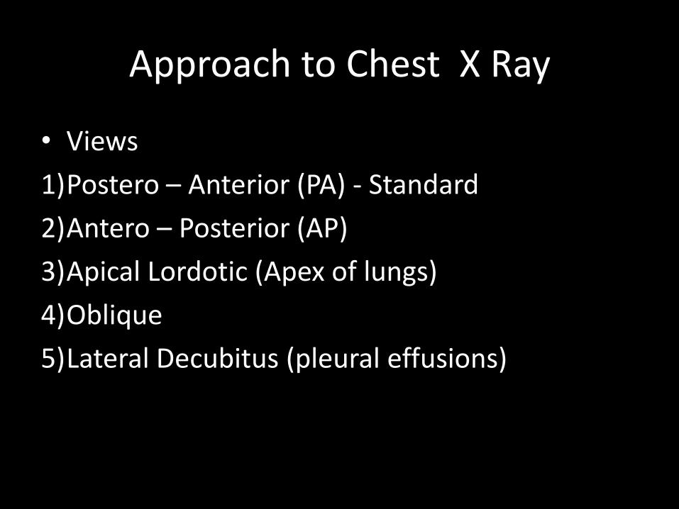
Approach to Chest X Ray
• Views
1)Postero – Anterior (PA) - Standard
2)Antero – Posterior (AP)
3)Apical Lordotic (Apex of lungs)
4)Oblique
5)Lateral Decubitus (pleural effusions)
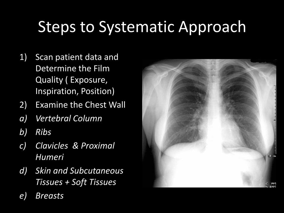
Steps to Systematic Approach
1) Scan patient data and Determine the Film Quality ( Exposure, Inspiration, Position)
2) Examine the Chest Wall
a) Vertebral Column
b) Ribs
c) Clavicles & Proximal Humeri
d) Skin and Subcutaneous Tissues + Soft Tissues
e) Breasts
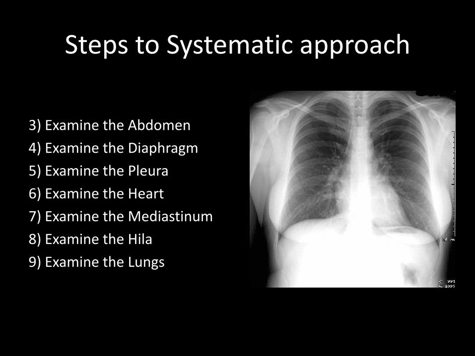
Steps to Systematic approach
3) Examine the Abdomen
4) Examine the Diaphragm
5) Examine the Pleura
6) Examine the Heart
7) Examine the Mediastinum
8) Examine the Hila
9) Examine the Lungs
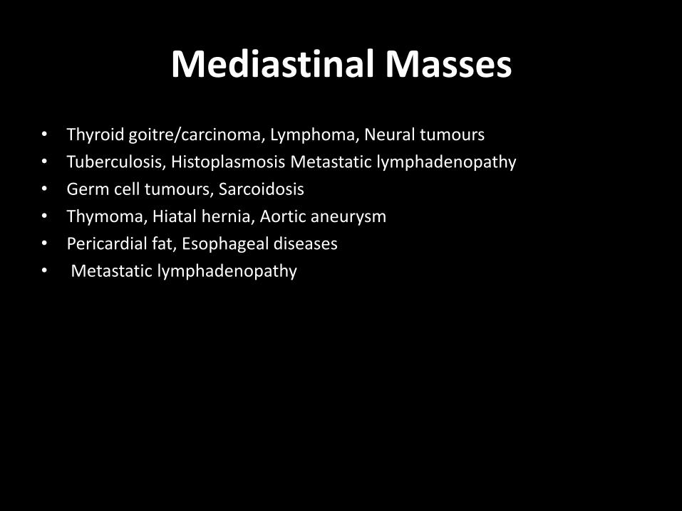
Mediastinal Masses
• Thyroid goitre/carcinoma, Lymphoma, Neural tumours
• Tuberculosis, Histoplasmosis Metastatic lymphadenopathy
• Germ cell tumours, Sarcoidosis
• Thymoma, Hiatal hernia, Aortic aneurysm
• Pericardial fat, Esophageal diseases
• Metastatic lymphadenopathy
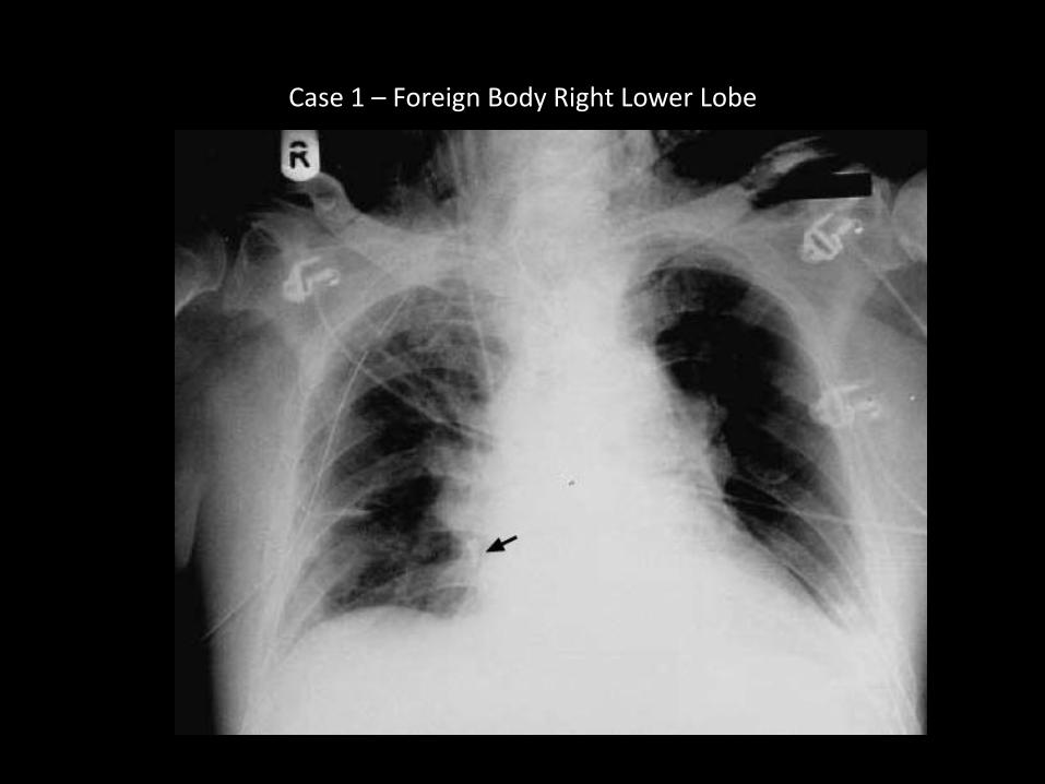
Case 1 – Foreign Body Right Lower Lobe
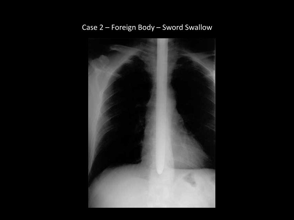
Case 2 – Foreign Body – Sword Swallow
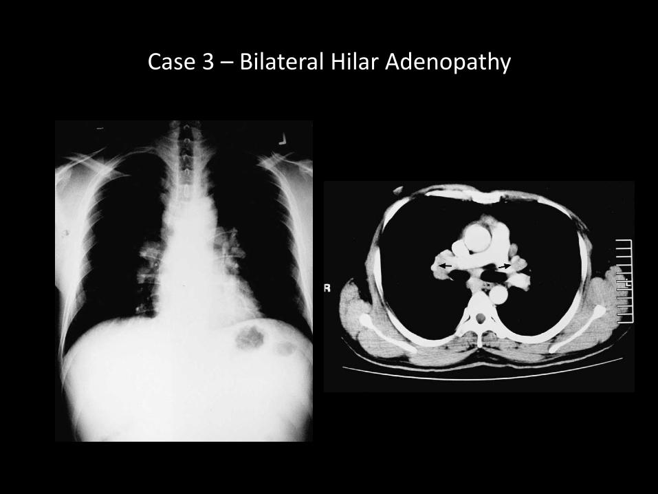
Case 3 – Bilateral Hilar Adenopathy
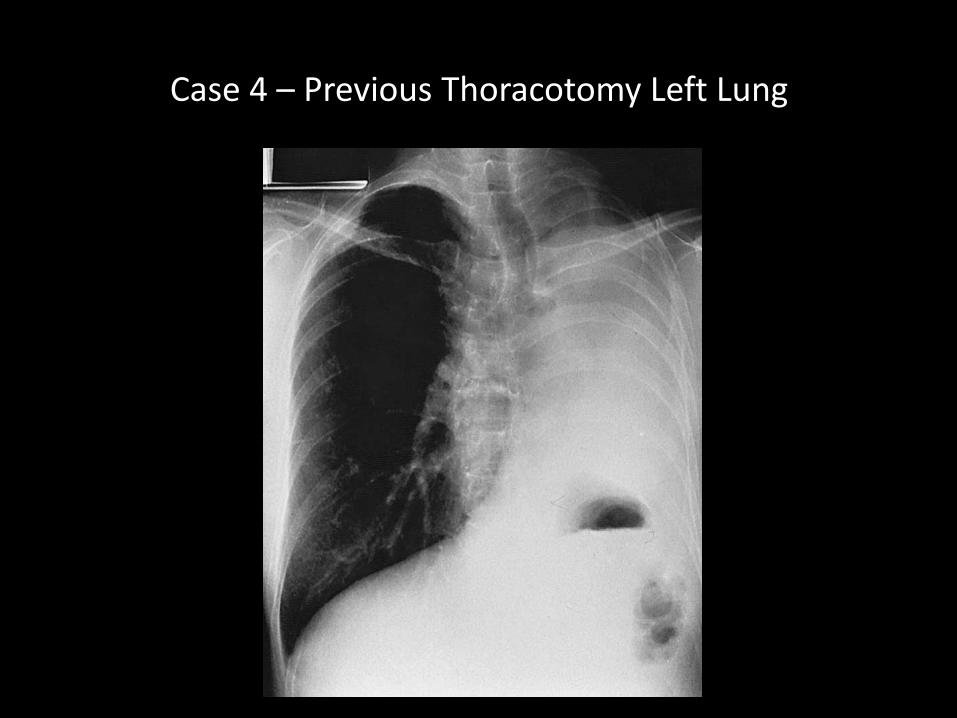
Case 4 – Previous Thoracotomy Left Lung
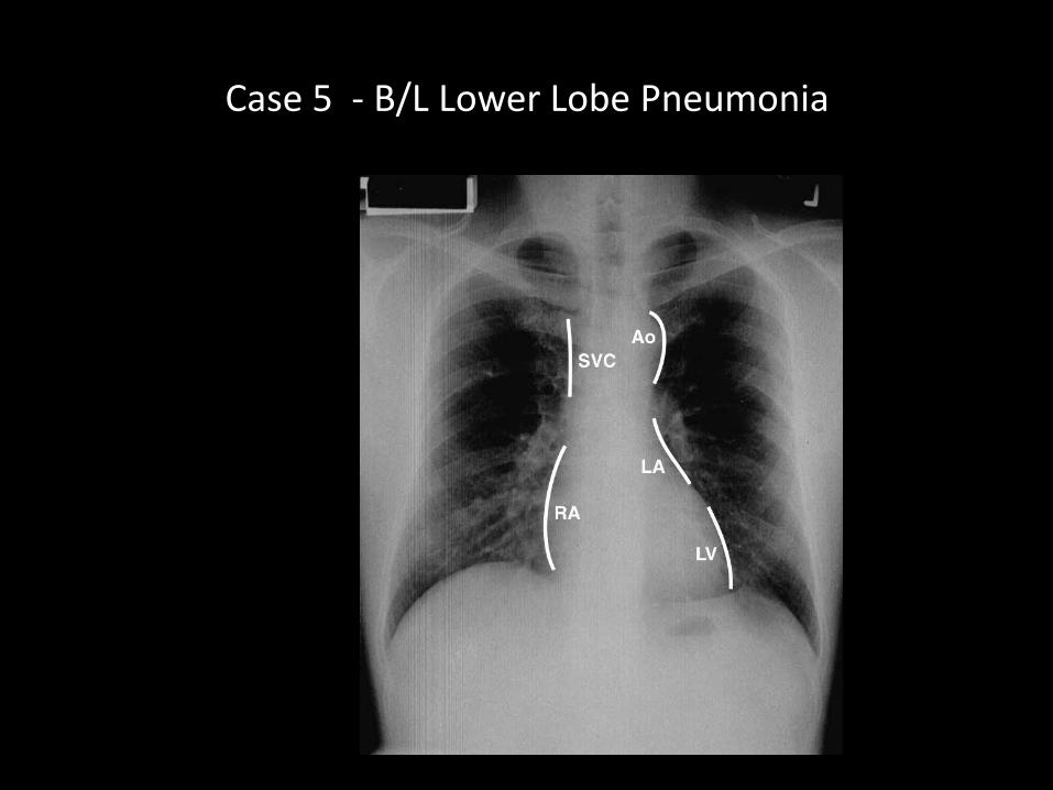
Case 5 - B/L Lower Lobe Pneumonia
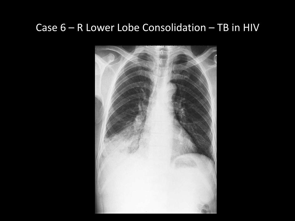
Case 6 – R Lower Lobe Consolidation – TB in HIV
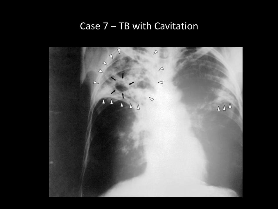
Case 7 – TB with Cavitation

Case 8 – Ruptured Liver Abscess in Pleural Cavity
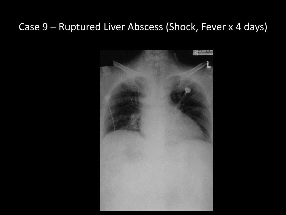
Case 9 – Ruptured Liver Abscess (Shock, Fever x 4 days)
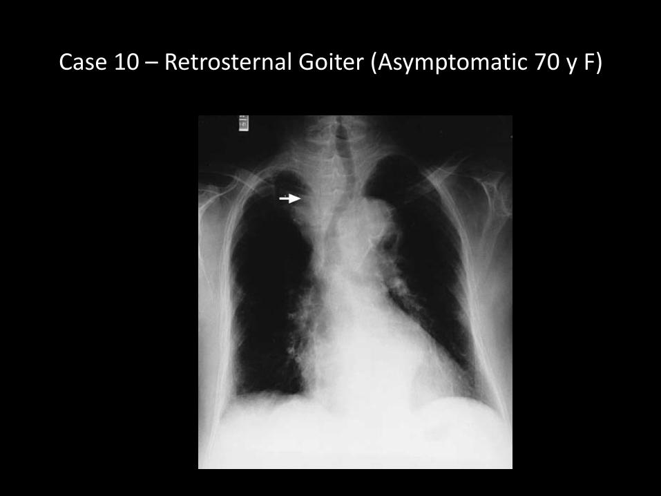
Case 10 – Retrosternal Goiter (Asymptomatic 70 y F)
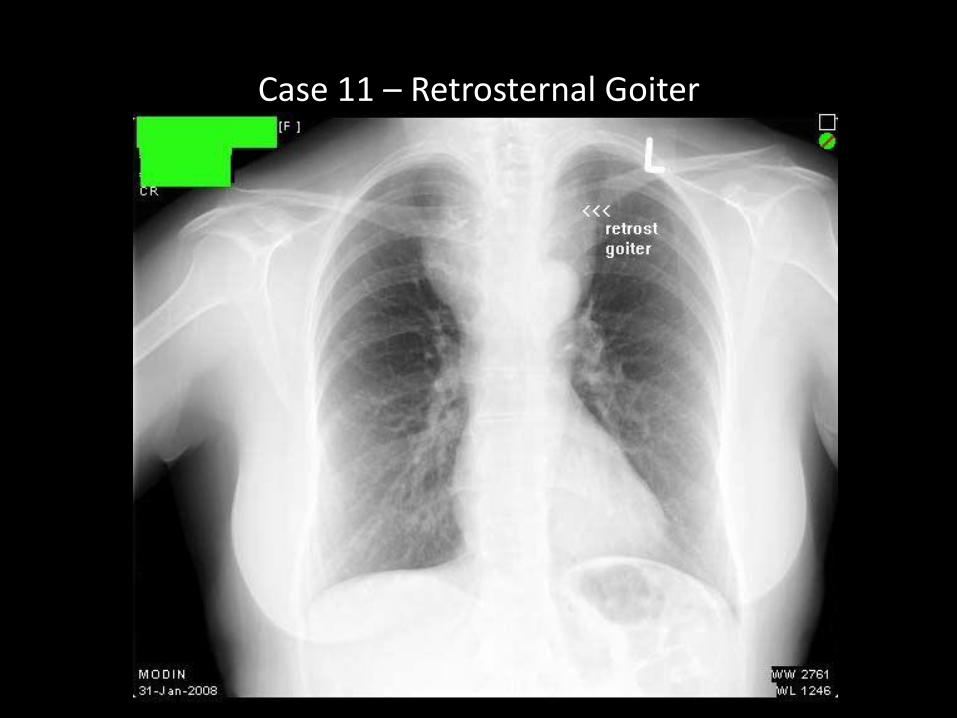
Case 11 – Retrosternal Goiter
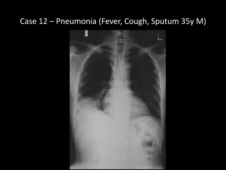
Case 12 – Pneumonia (Fever, Cough, Sputum 35y M)
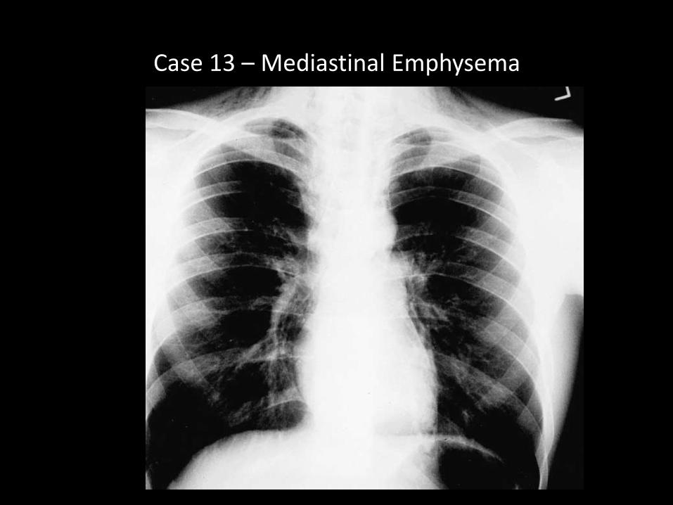
Case 13 – Mediastinal Emphysema
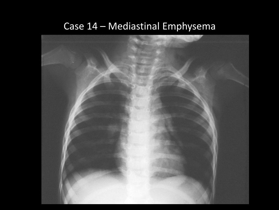
Case 14 – Mediastinal Emphysema
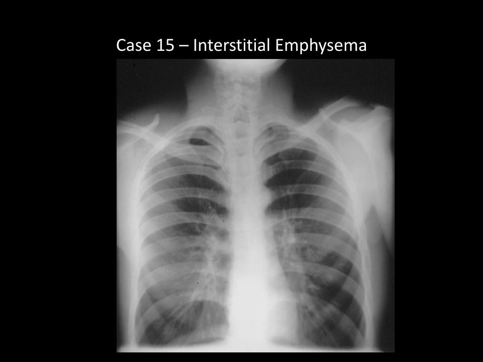
Case 15 – Interstitial Emphysema
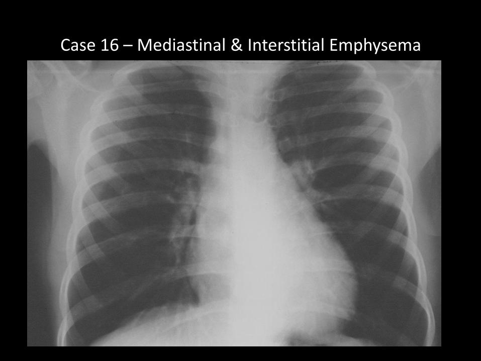
Case 16 – Mediastinal & Interstitial Emphysema
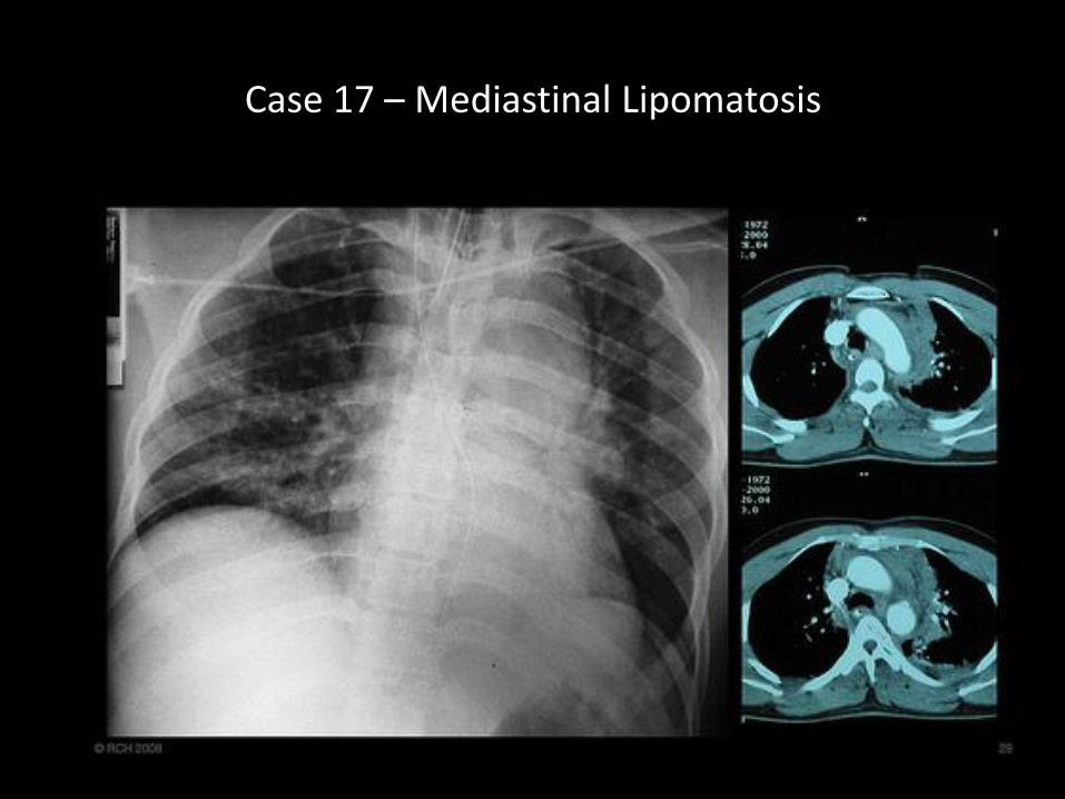
Case 17 – Mediastinal Lipomatosis

Case 18 – Anomalous Pulmonary Venous Drainage
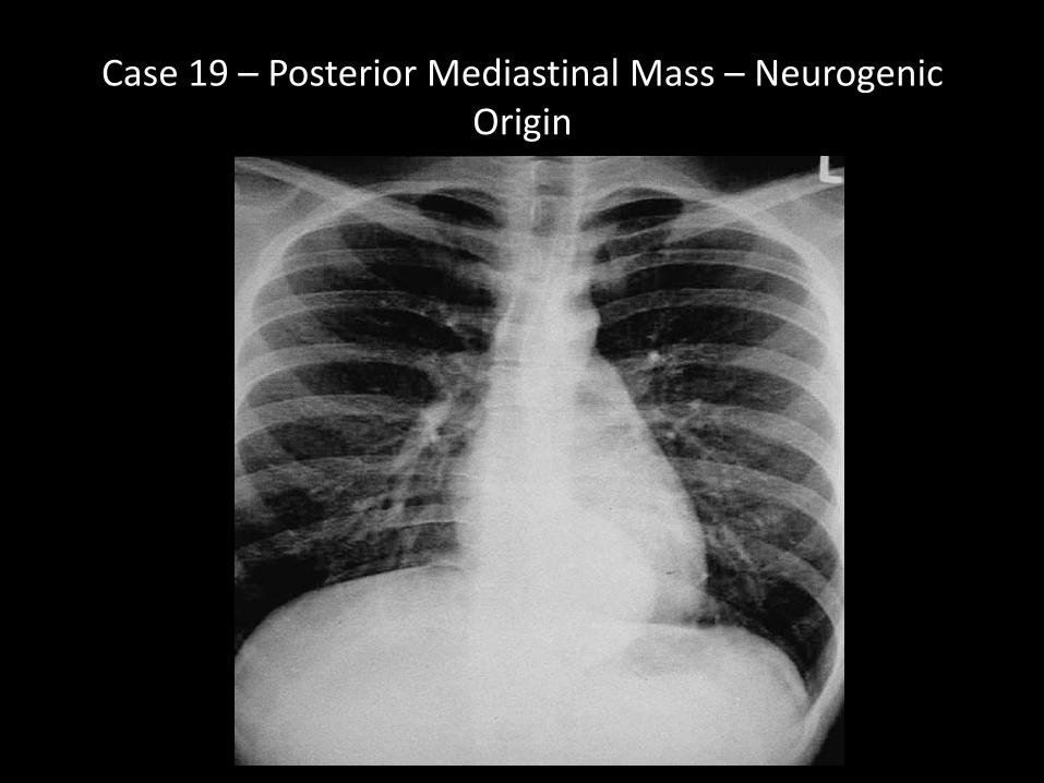
Case 19 – Posterior Mediastinal Mass – Neurogenic Origin
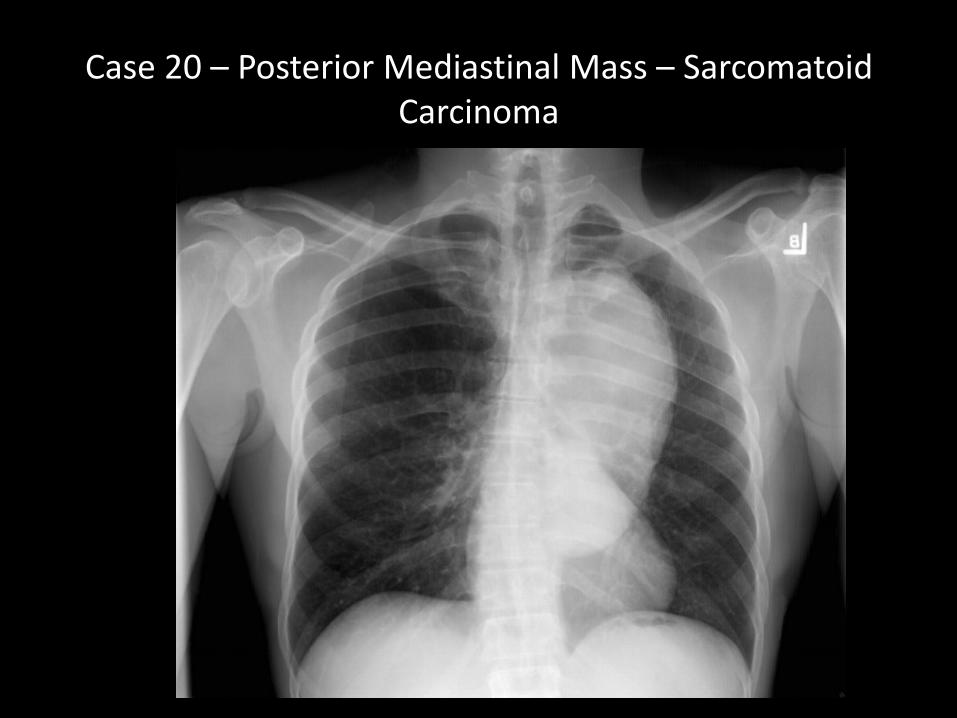
Case 20 – Posterior Mediastinal Mass – Sarcomatoid Carcinoma
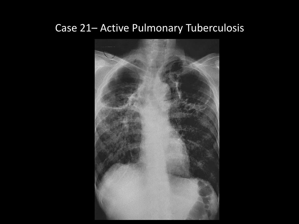
Case 21– Active Pulmonary Tuberculosis
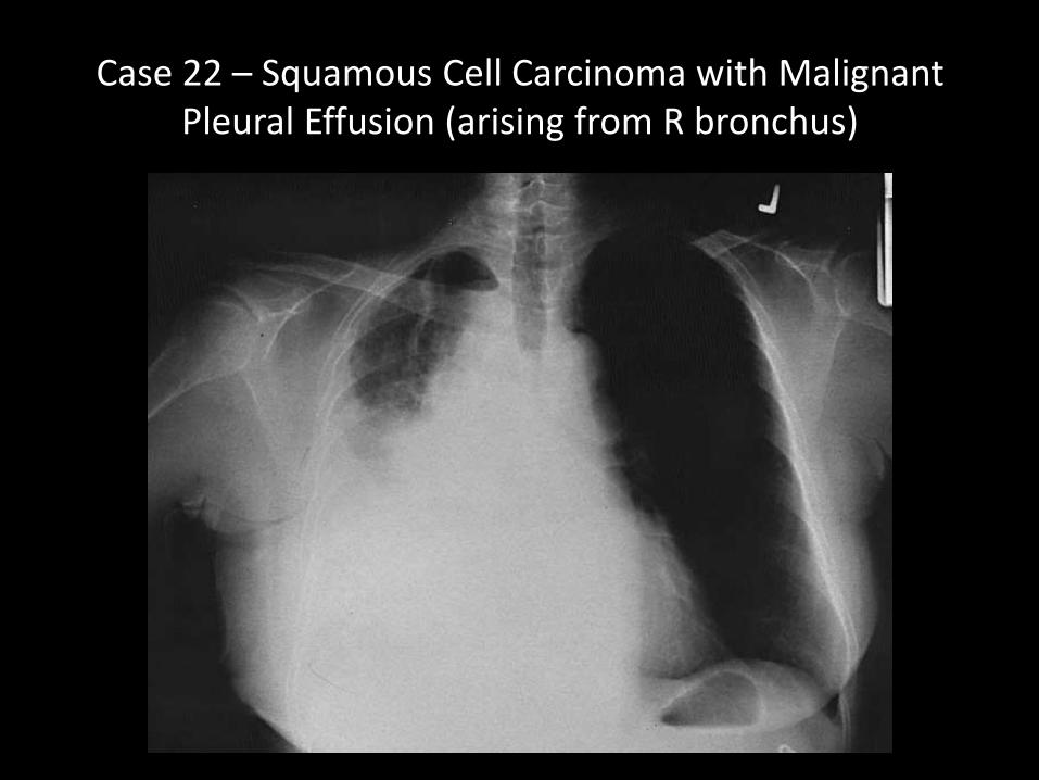
Case 22 – Squamous Cell Carcinoma with Malignant Pleural Effusion (arising from R bronchus)
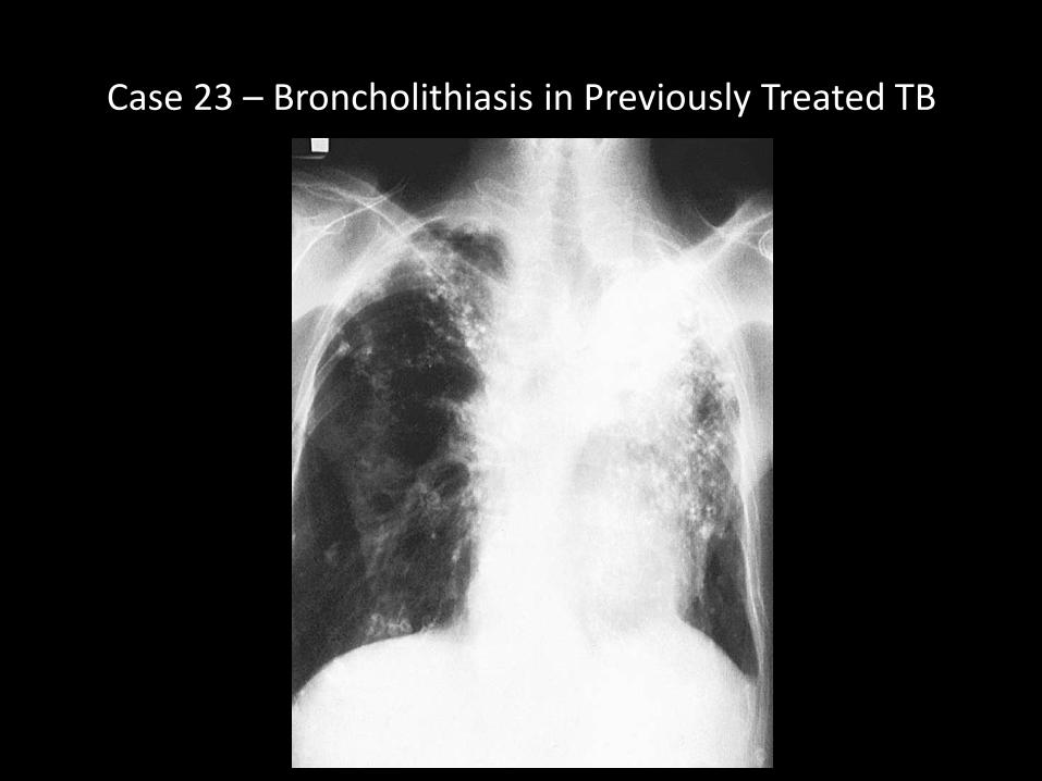
Case 23 – Broncholithiasis in Previously Treated TB

Case 24 – Displaced Pulmonary Artery Catheter (MI management in CCU)
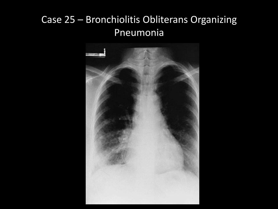
Case 25 – Bronchiolitis Obliterans Organizing Pneumonia
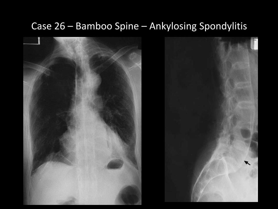
Case 26 – Bamboo Spine – Ankylosing Spondylitis
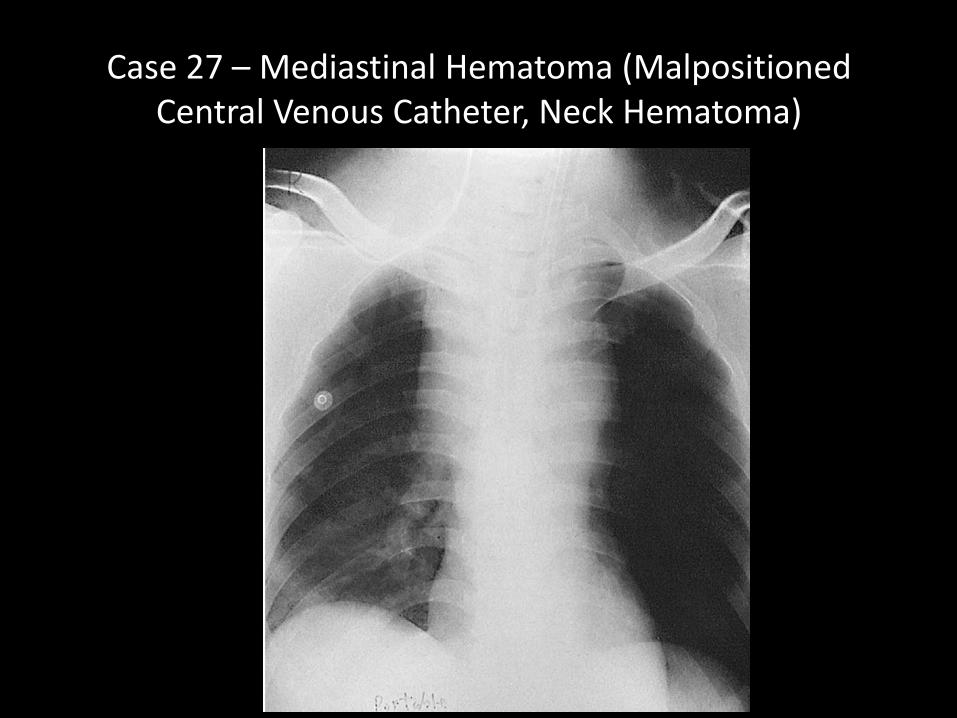
Case 27 – Mediastinal Hematoma (Malpositioned Central Venous Catheter, Neck Hematoma)
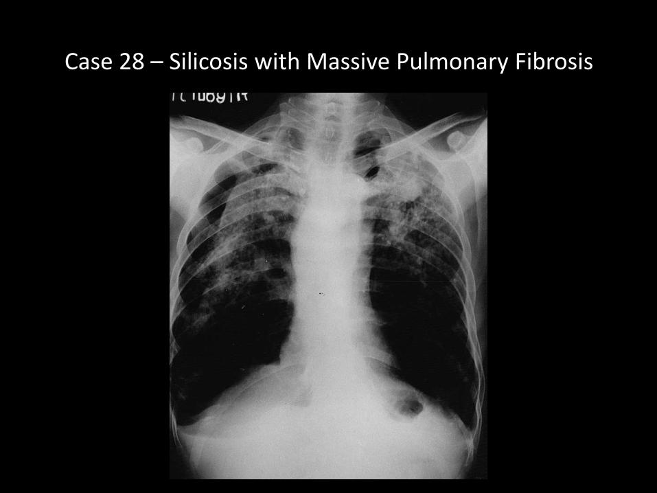
Case 28 – Silicosis with Massive Pulmonary Fibrosis
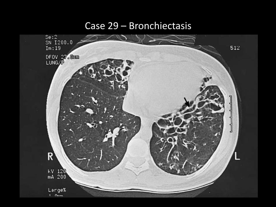
Case 29 – Bronchiectasis

Case 30 – AML with Pulmonary Angio – Invasive Aspergillosis
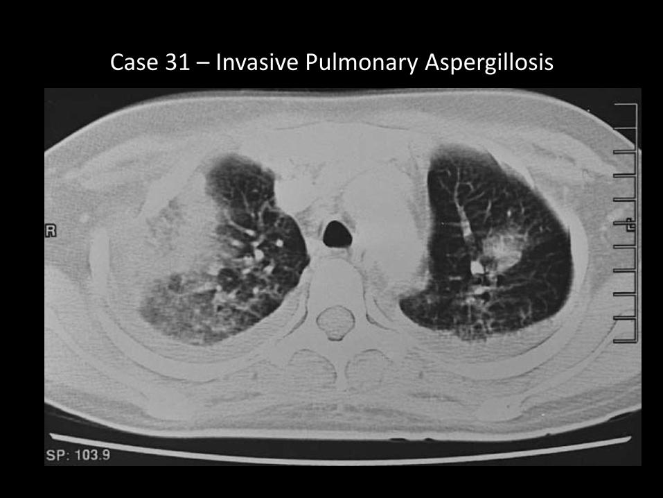
Case 31 – Invasive Pulmonary Aspergillosis

Case 32 – Right Upper Lobe Collapse (Golden’ Sign)

Case 33 – Westmark Sign of Acute Pulmonary Embolism
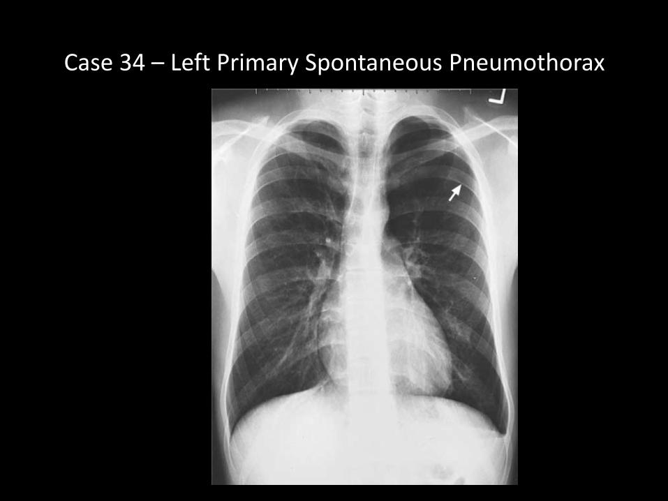
Case 34 – Left Primary Spontaneous Pneumothorax

Case 87 – Radiation Fibrosis of R Lung

Case 31 – Miliary Tuberculosis
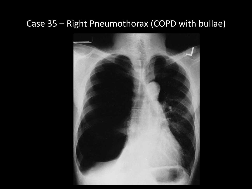
Case 35 – Right Pneumothorax (COPD with bullae)

Case 36 – 100 Pack yr (5y Dyspnea) COPD (hyperinflation)

Case 37 – B/L First Rib Fractures

Case 38 – Mediastinal Lipomatosis
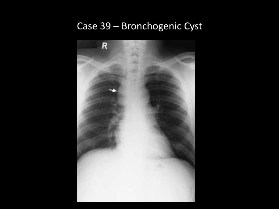
Case 39 – Bronchogenic Cyst
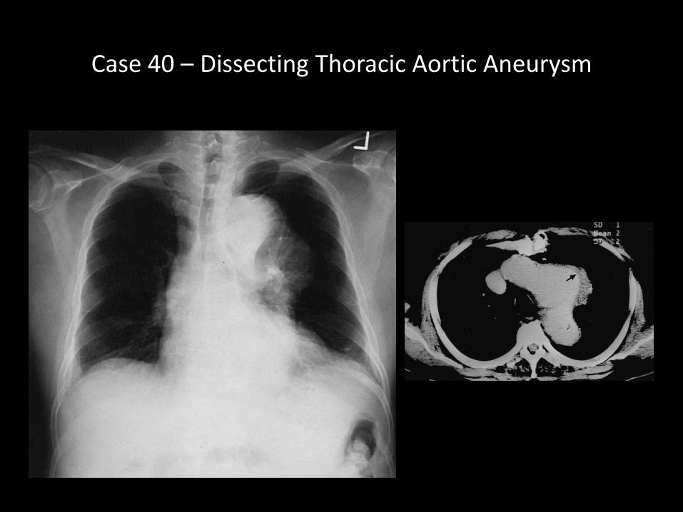
Case 40 – Dissecting Thoracic Aortic Aneurysm
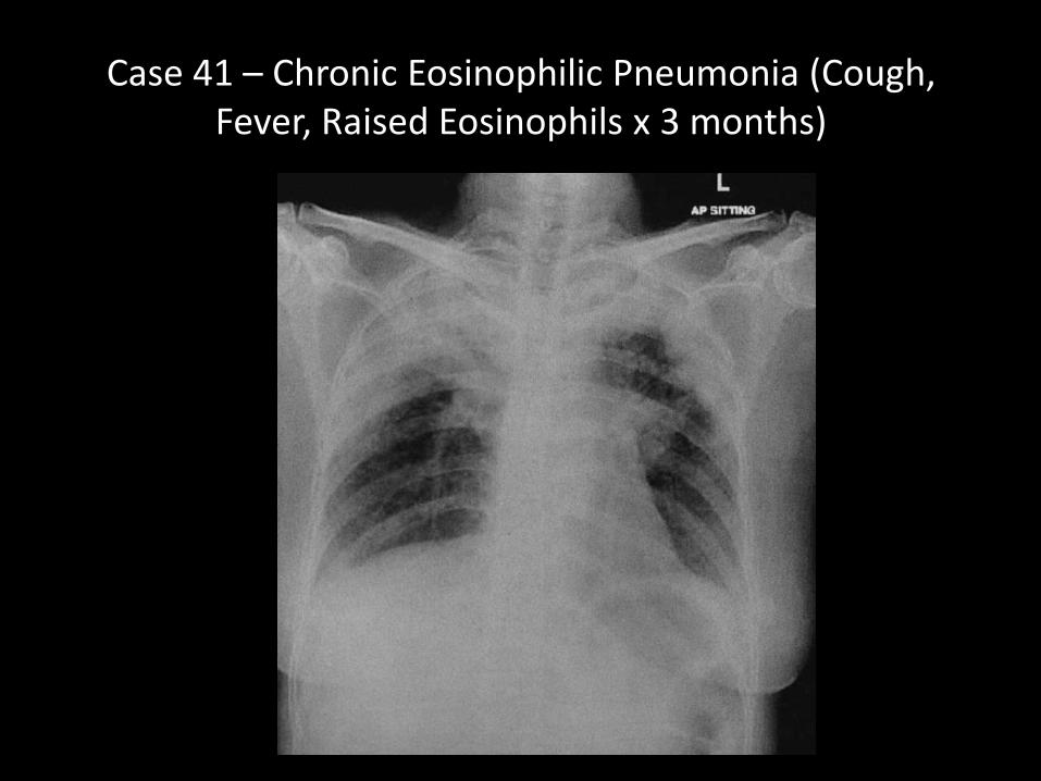
Case 41 – Chronic Eosinophilic Pneumonia (Cough, Fever, Raised Eosinophils x 3 months)
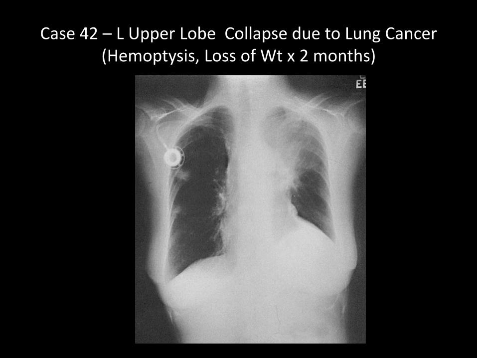
Case 42 – L Upper Lobe Collapse due to Lung Cancer (Hemoptysis, Loss of Wt x 2 months)
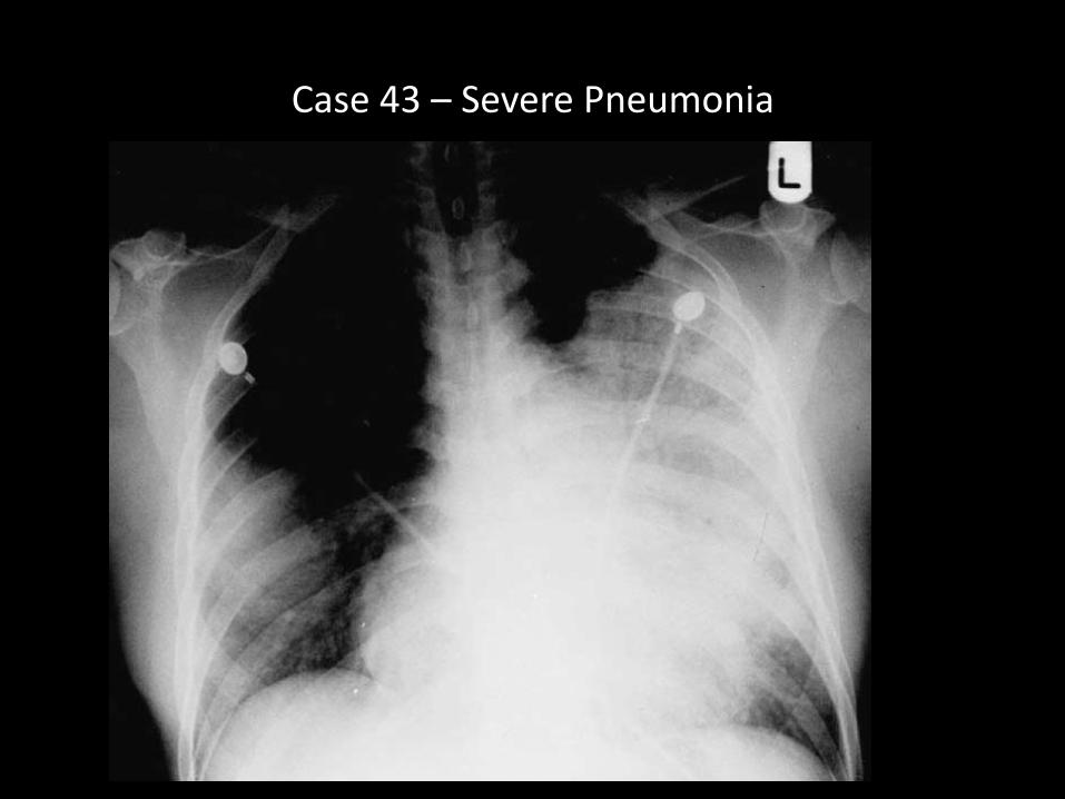
Case 43 – Severe Pneumonia
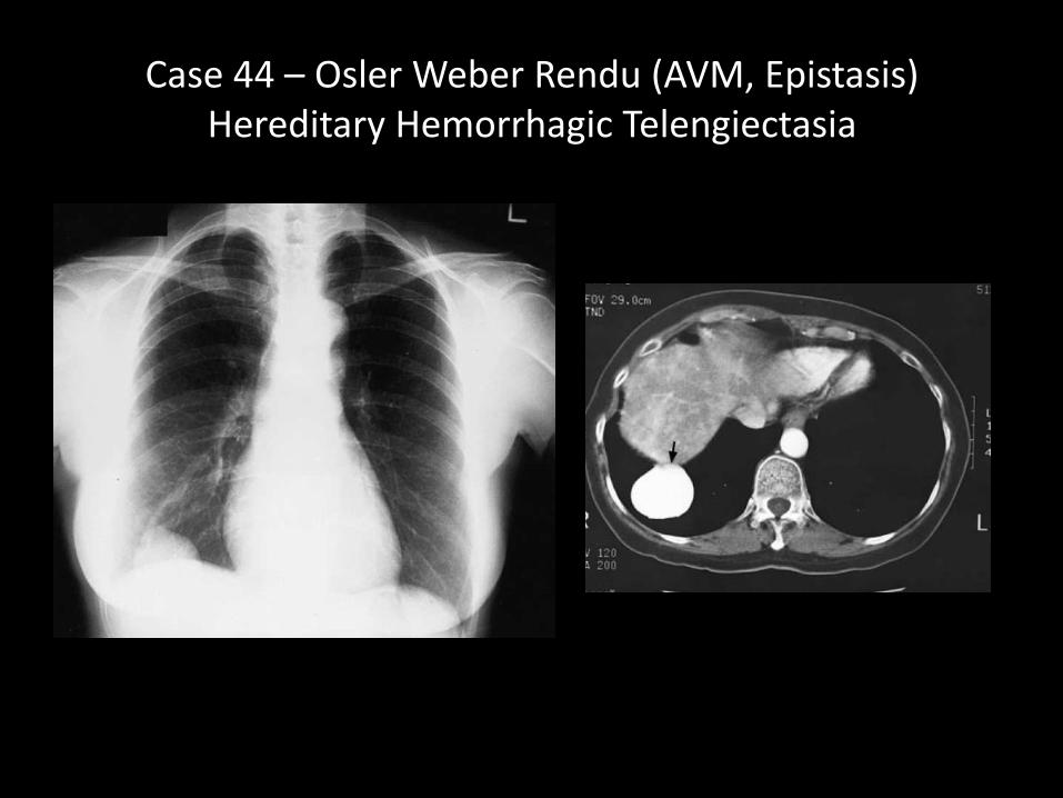
Case 44 – Osler Weber Rendu (AVM, Epistasis)Hereditary Hemorrhagic Telengiectasia
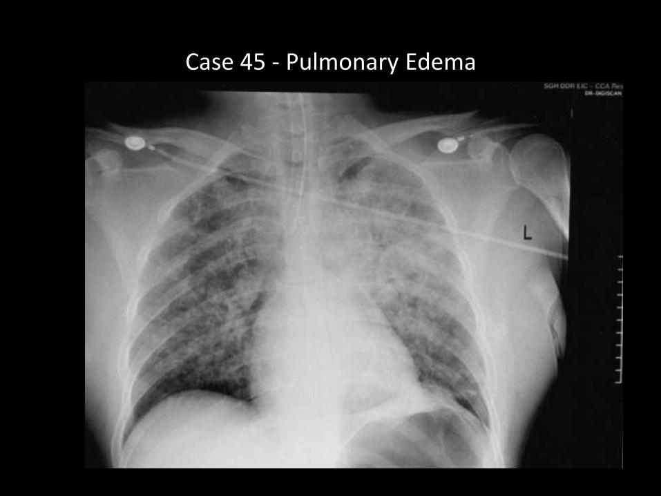
Case 45 - Pulmonary Edema
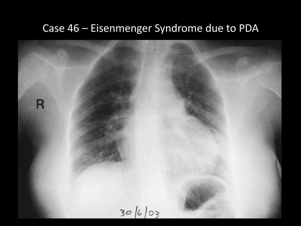
Case 46 – Eisenmenger Syndrome due to PDA
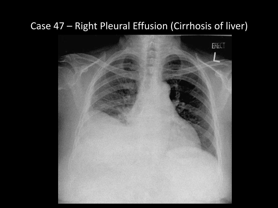
Case 47 – Right Pleural Effusion (Cirrhosis of liver)

Case 48 - Hamartoma
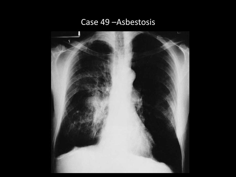
Case 49 –Asbestosis
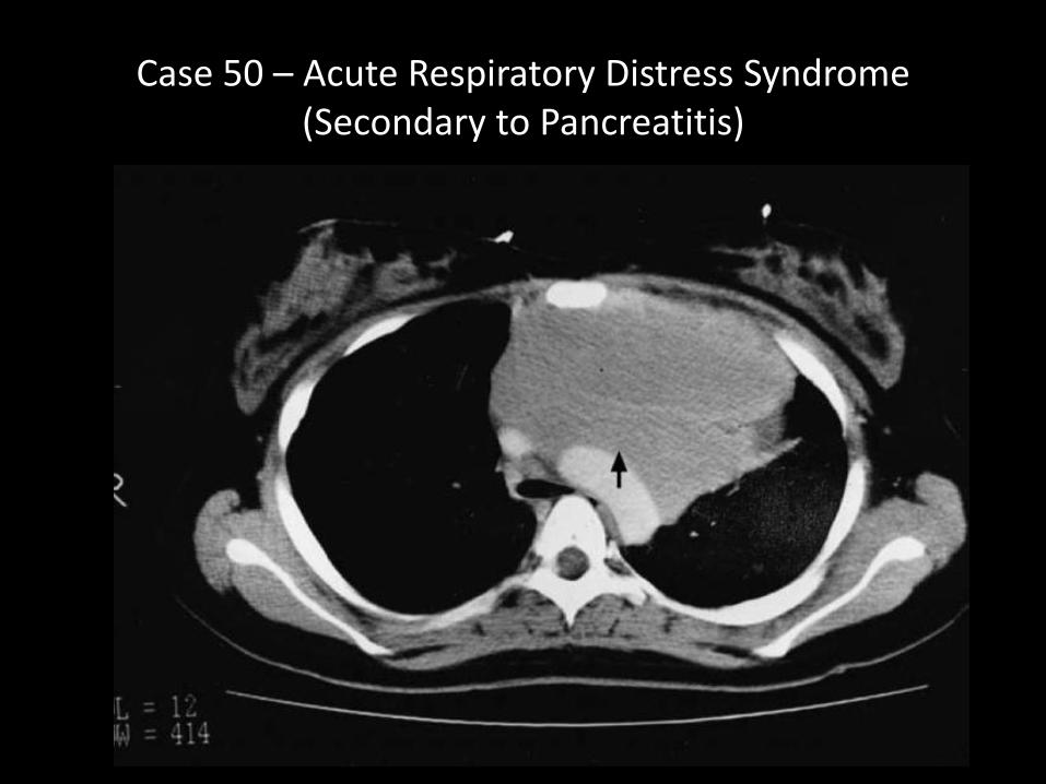
Case 50 – Acute Respiratory Distress Syndrome (Secondary to Pancreatitis)
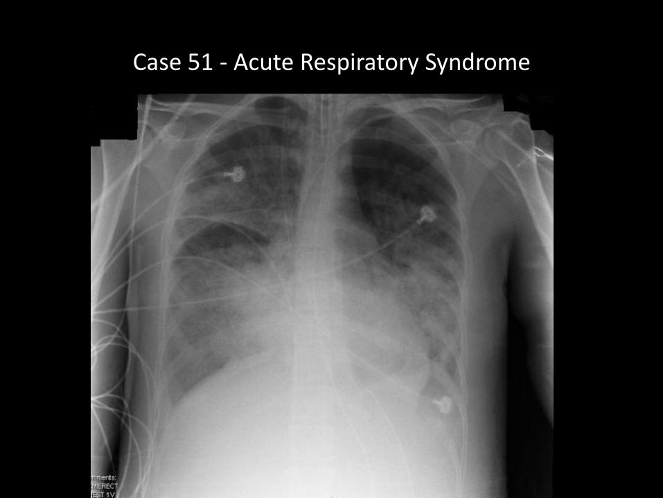
Case 51 - Acute Respiratory Syndrome
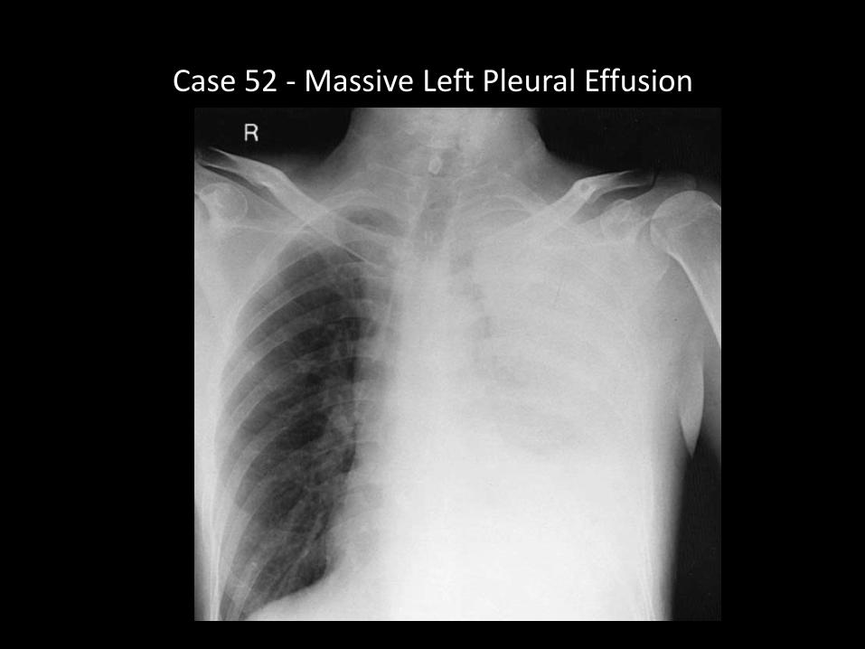
Case 52 - Massive Left Pleural Effusion
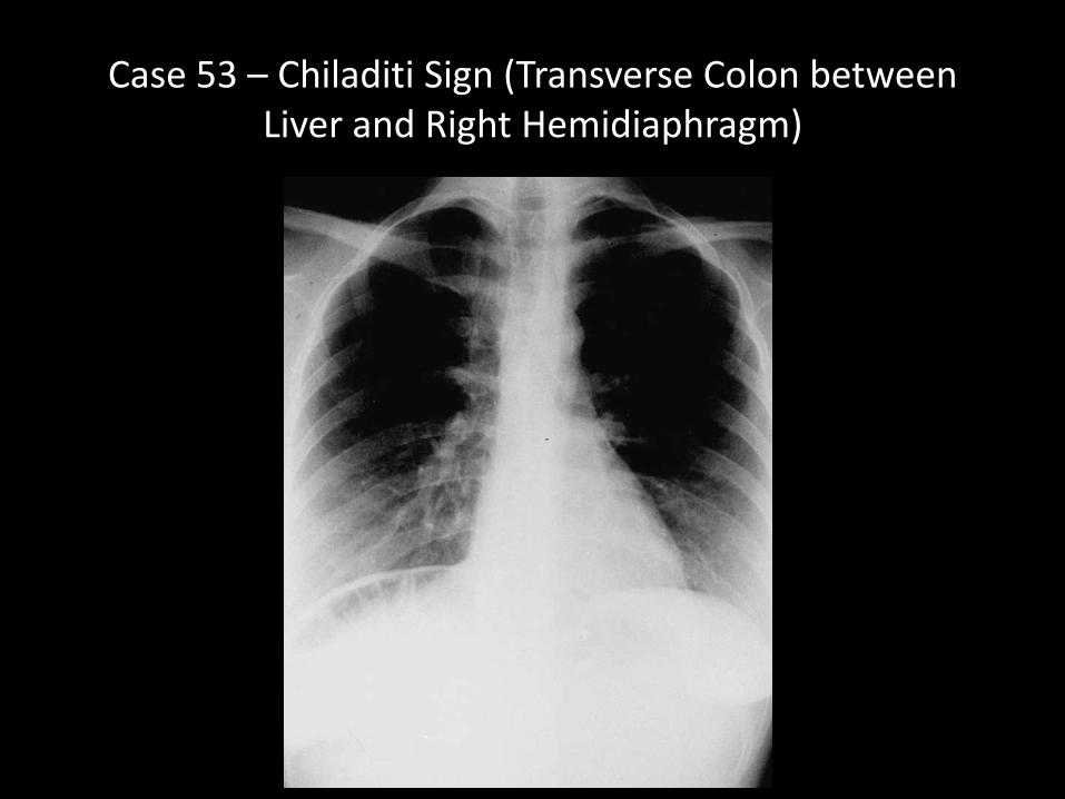
Case 53 – Chiladiti Sign (Transverse Colon between Liver and Right Hemidiaphragm)
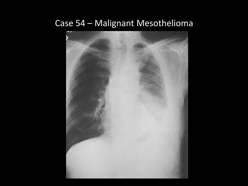
Case 54 – Malignant Mesothelioma
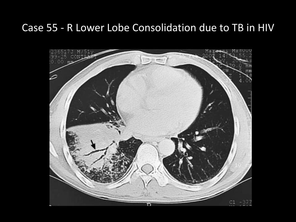
Case 55 - R Lower Lobe Consolidation due to TB in HIV
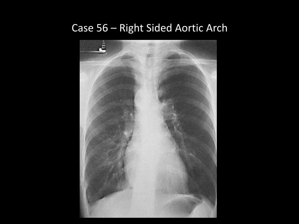
Case 56 – Right Sided Aortic Arch
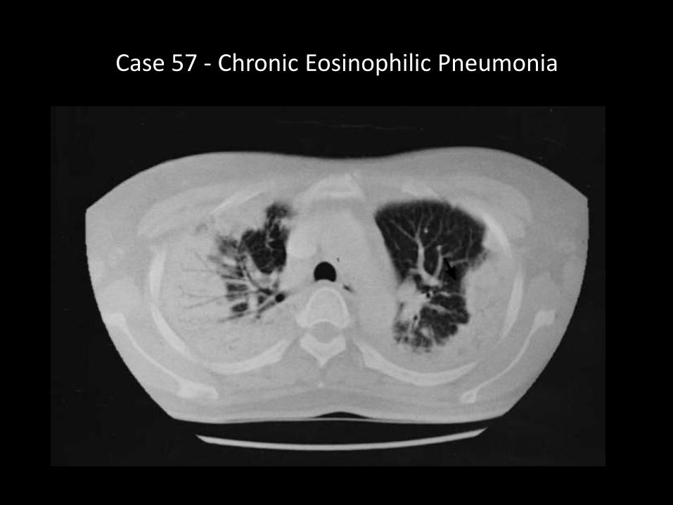
Case 57 - Chronic Eosinophilic Pneumonia
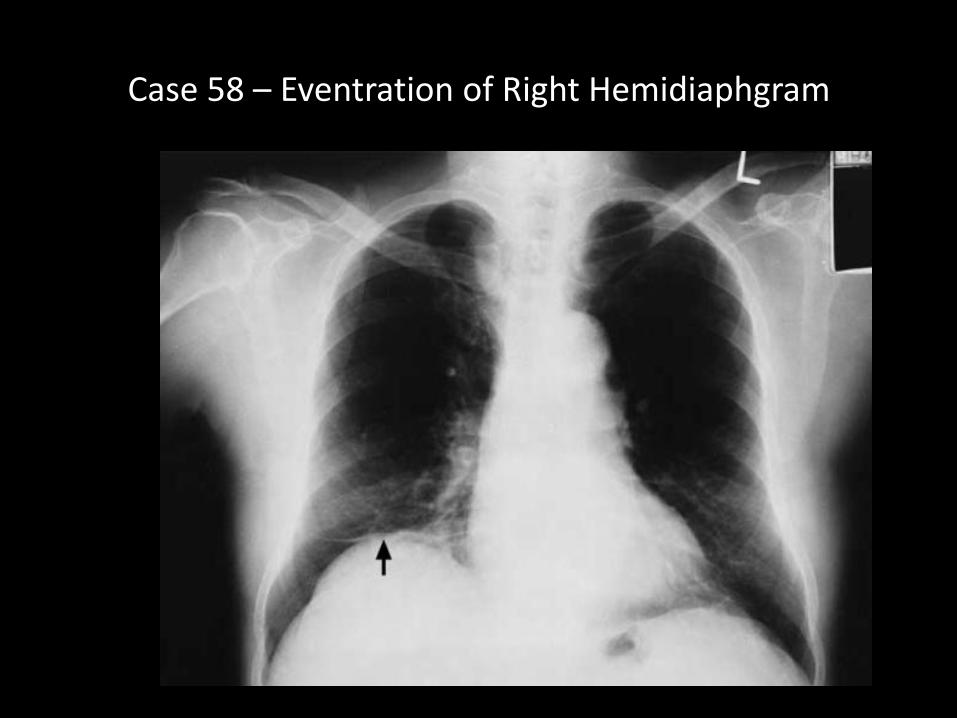
Case 58 – Eventration of Right Hemidiaphgram

Case 59 - Traumatic Aortic Disruption
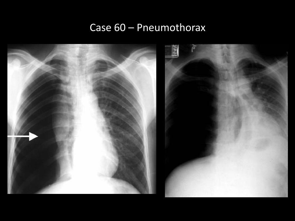
Case 60 – Pneumothorax
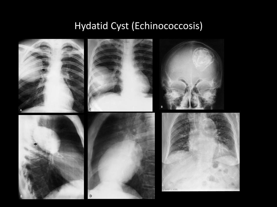
Hydatid Cyst (Echinococcosis)
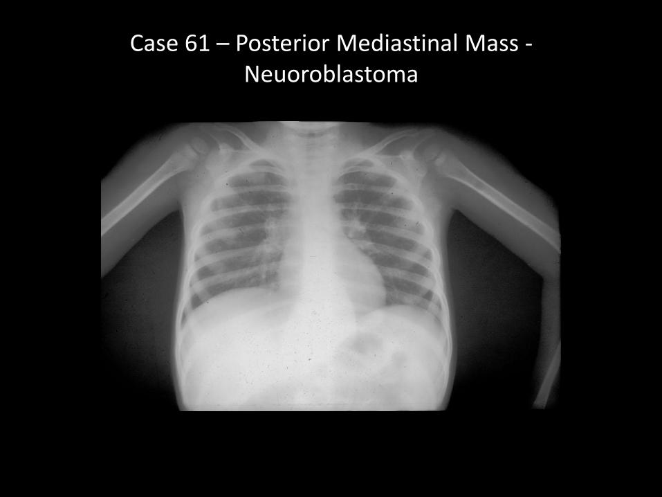
Case 61 – Posterior Mediastinal Mass -Neuoroblastoma
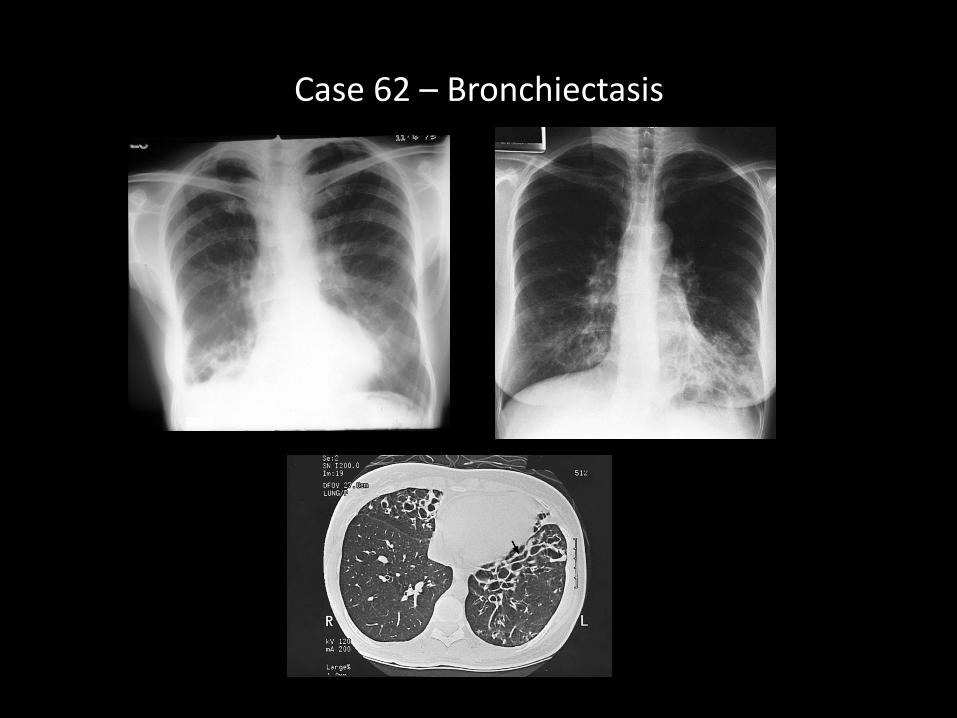
Case 62 – Bronchiectasis
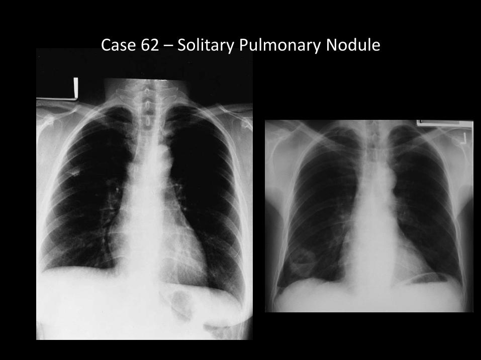
Case 62 – Solitary Pulmonary Nodule
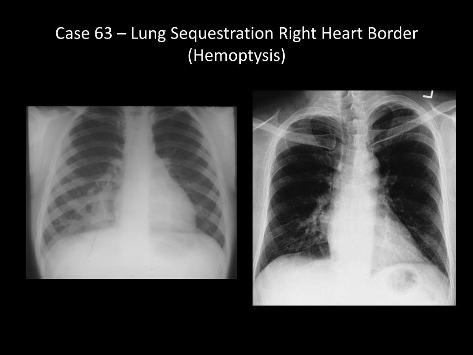
Case 63 – Lung Sequestration Right Heart Border (Hemoptysis)
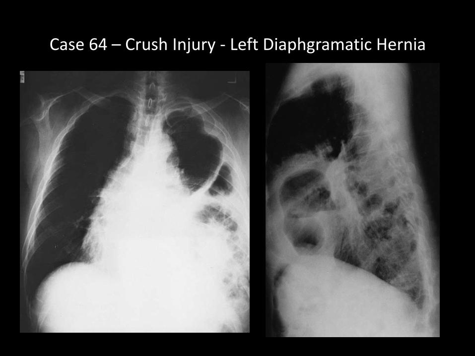
Case 64 – Crush Injury - Left Diaphgramatic Hernia
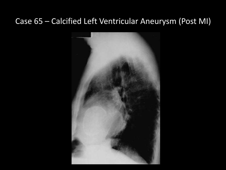
Case 65 – Calcified Left Ventricular Aneurysm (Post MI)
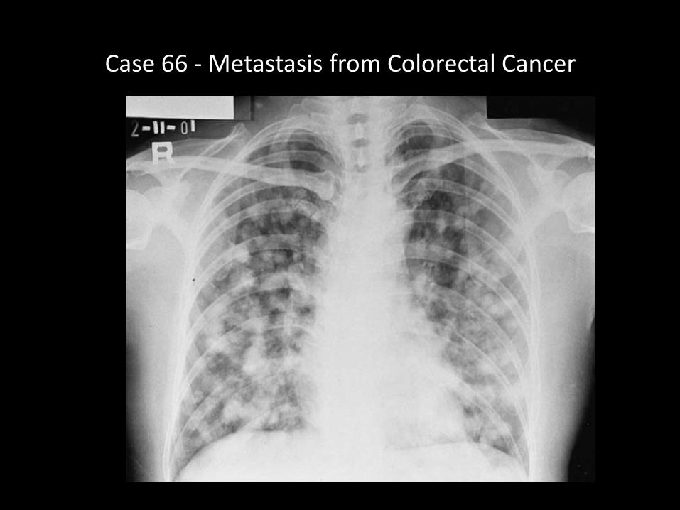
Case 66 - Metastasis from Colorectal Cancer
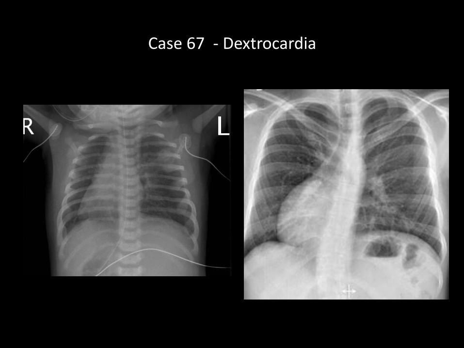
Case 67 - Dextrocardia
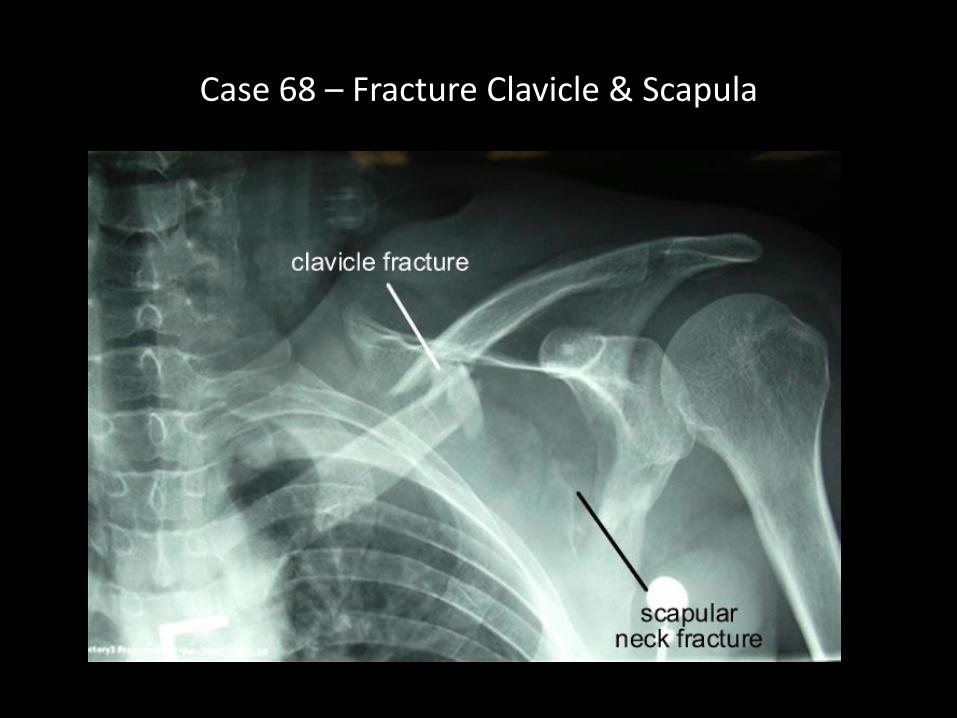
Case 68 – Fracture Clavicle & Scapula
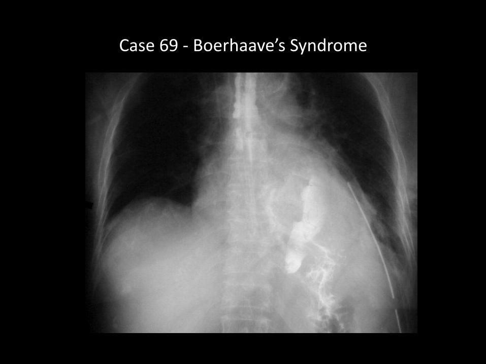
Case 69 - Boerhaave’s Syndrome
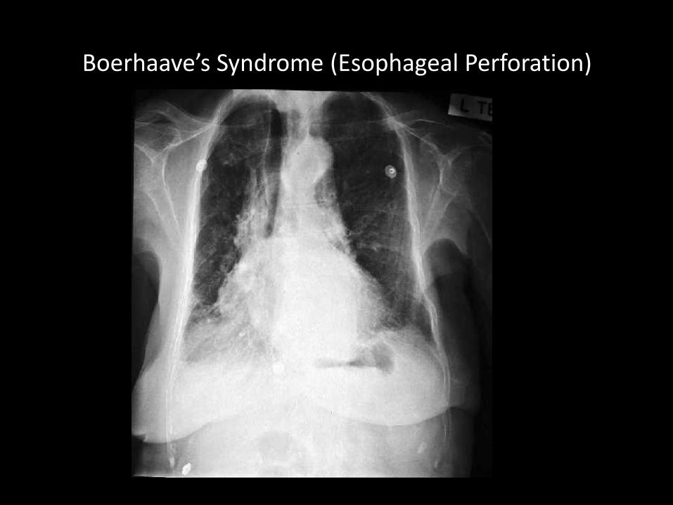
Boerhaave’s Syndrome (Esophageal Perforation)
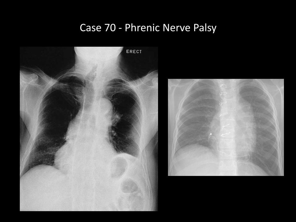
Case 70 - Phrenic Nerve Palsy
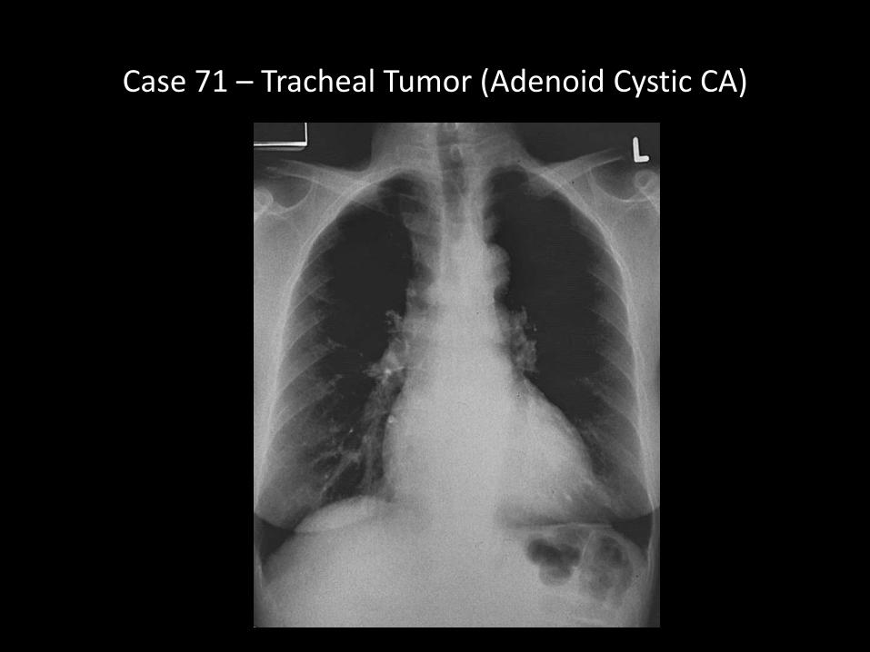
Case 71 – Tracheal Tumor (Adenoid Cystic CA)
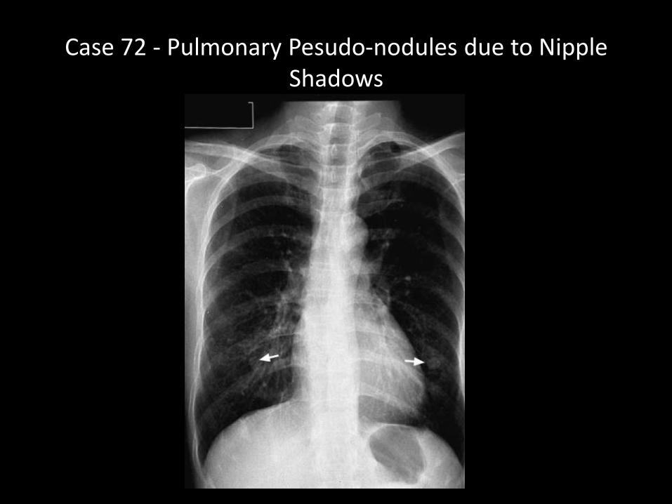
Case 72 - Pulmonary Pesudo-nodules due to Nipple Shadows
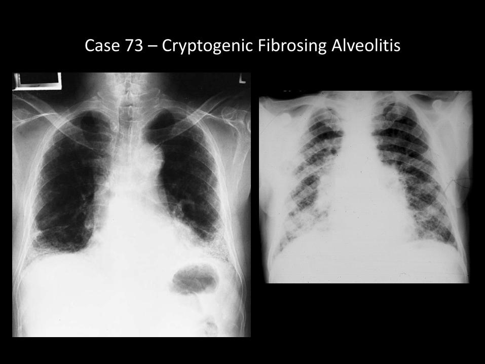
Case 73 – Cryptogenic Fibrosing Alveolitis
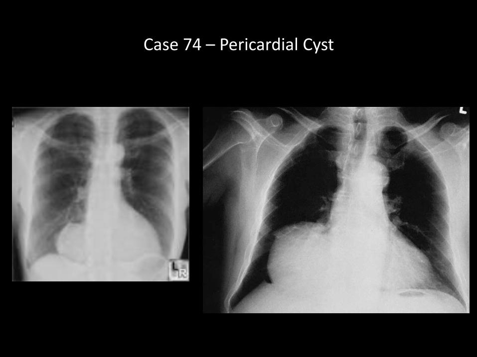
Case 74 – Pericardial Cyst
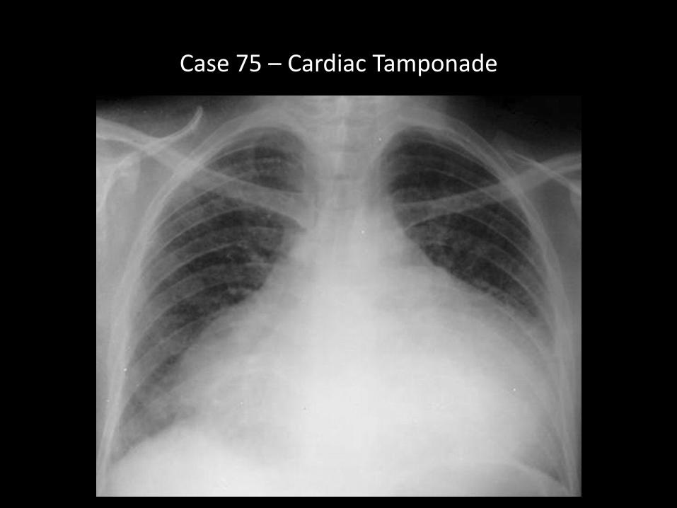
Case 75 – Cardiac Tamponade
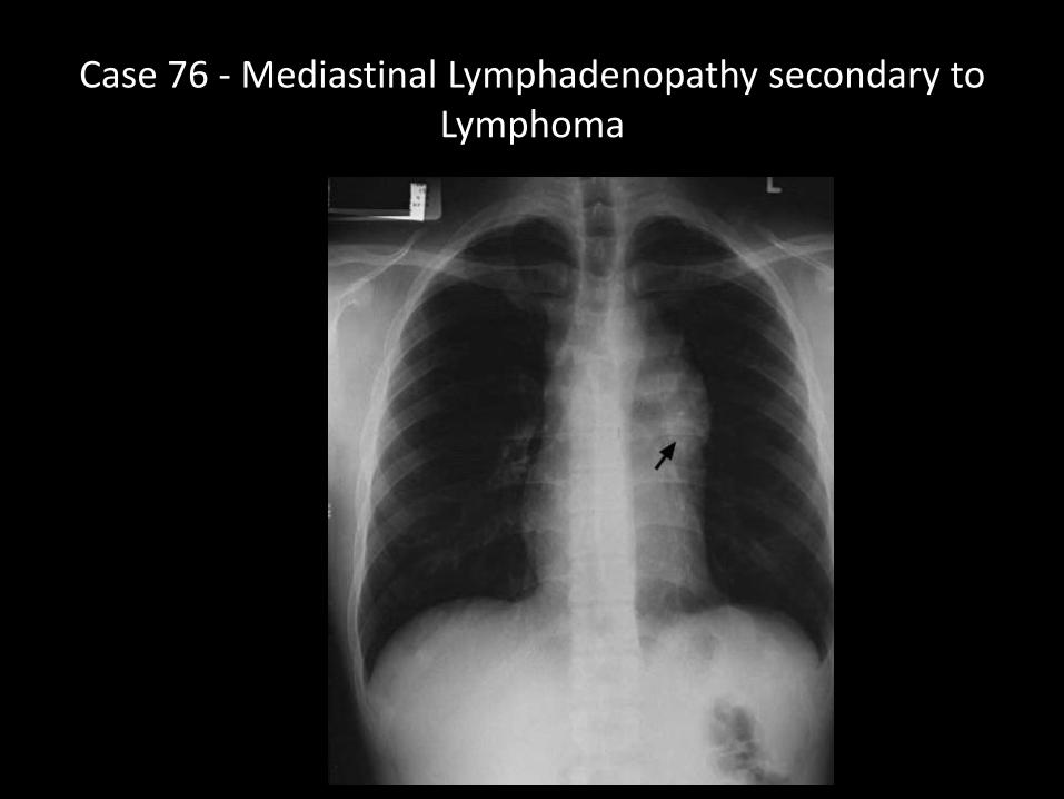
Case 76 - Mediastinal Lymphadenopathy secondary to Lymphoma
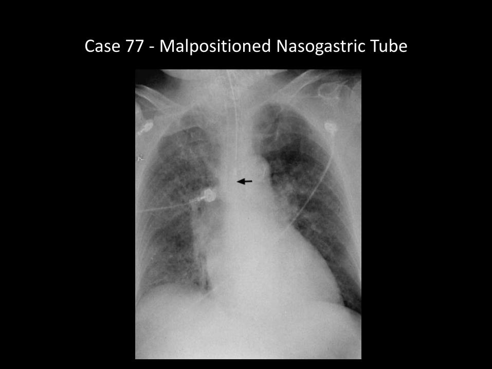
Case 77 - Malpositioned Nasogastric Tube
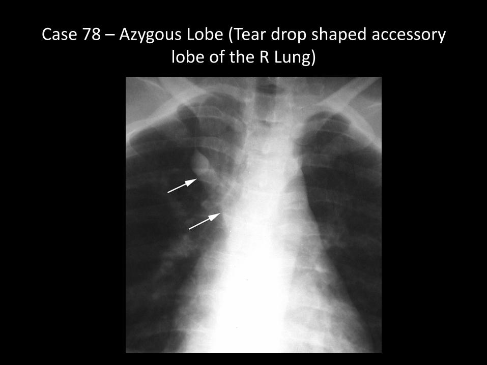
Case 78 – Azygous Lobe (Tear drop shaped accessory lobe of the R Lung)
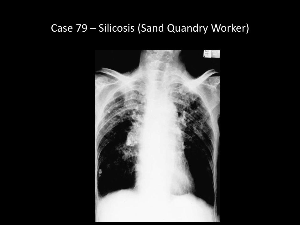
Case 79 – Silicosis (Sand Quandry Worker)
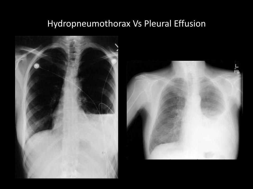
Hydropneumothorax Vs Pleural Effusion
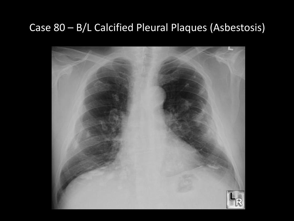
Case 80 – B/L Calcified Pleural Plaques (Asbestosis)

Case 81 – Lytic Lesion Right Humerus

Case 82 - Loculated Pleural Effusion R Lung
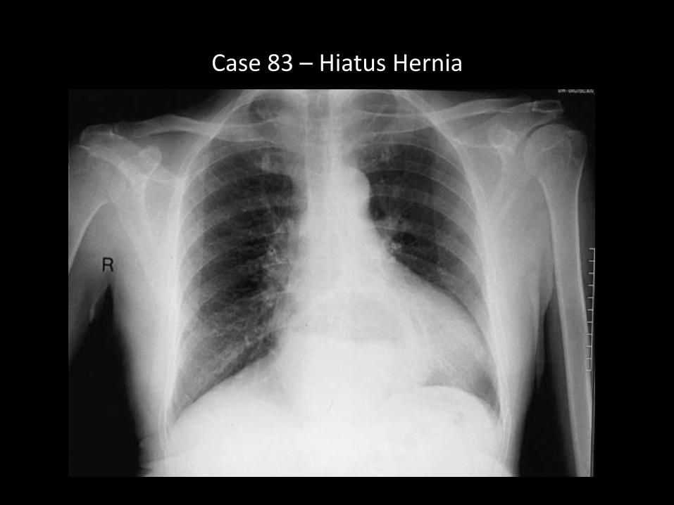
Case 83 – Hiatus Hernia
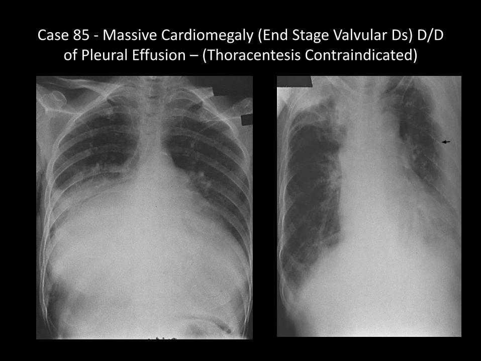
Case 85 - Massive Cardiomegaly (End Stage Valvular Ds) D/D of Pleural Effusion – (Thoracentesis Contraindicated)
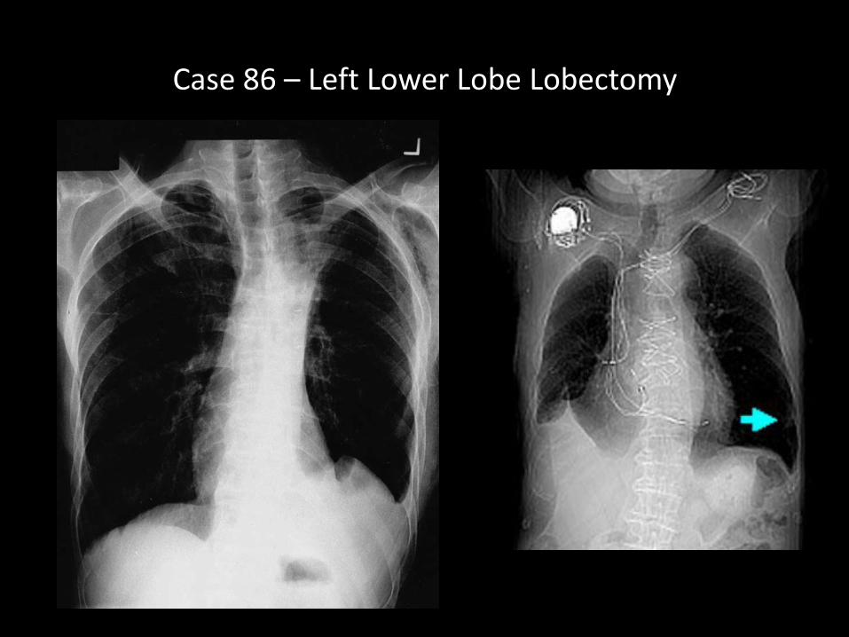
Case 86 – Left Lower Lobe Lobectomy
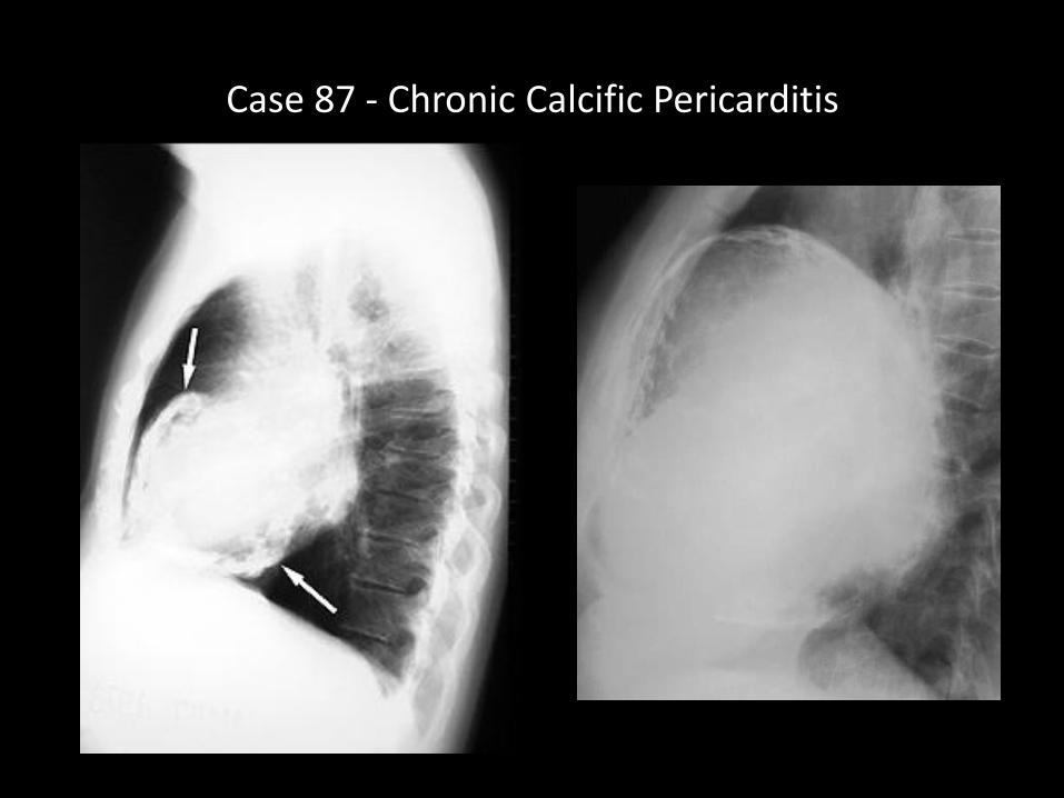
Case 87 - Chronic Calcific Pericarditis
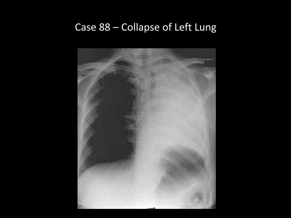
Case 88 – Collapse of Left Lung
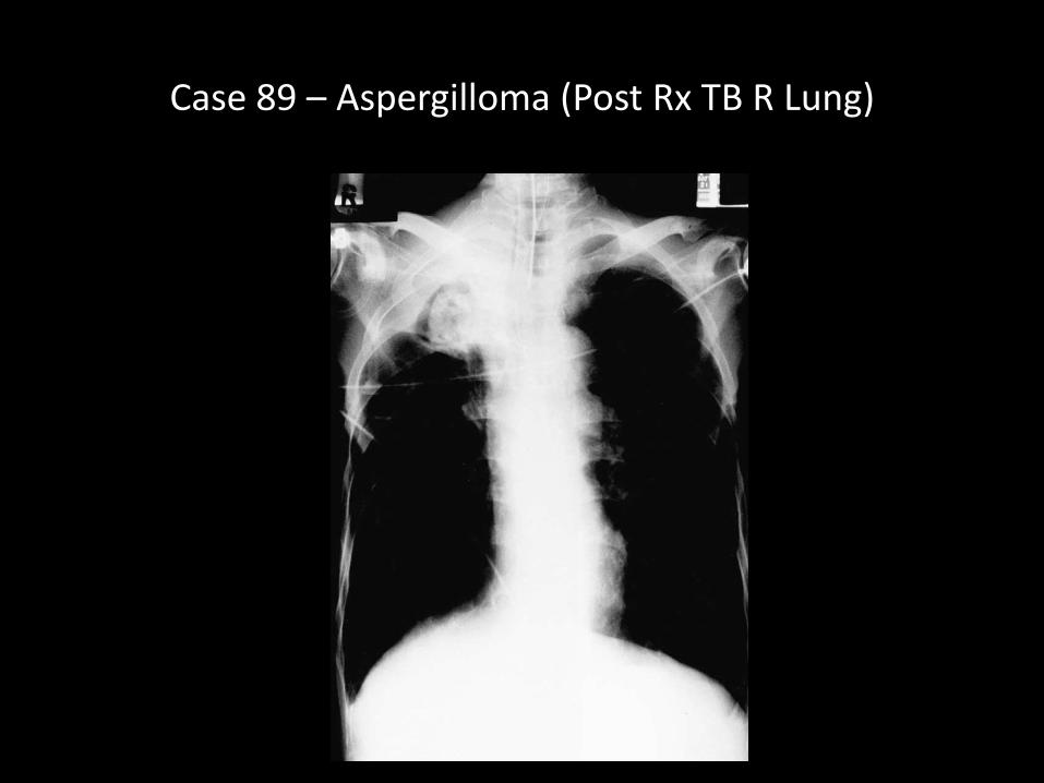
Case 89 – Aspergilloma (Post Rx TB R Lung)
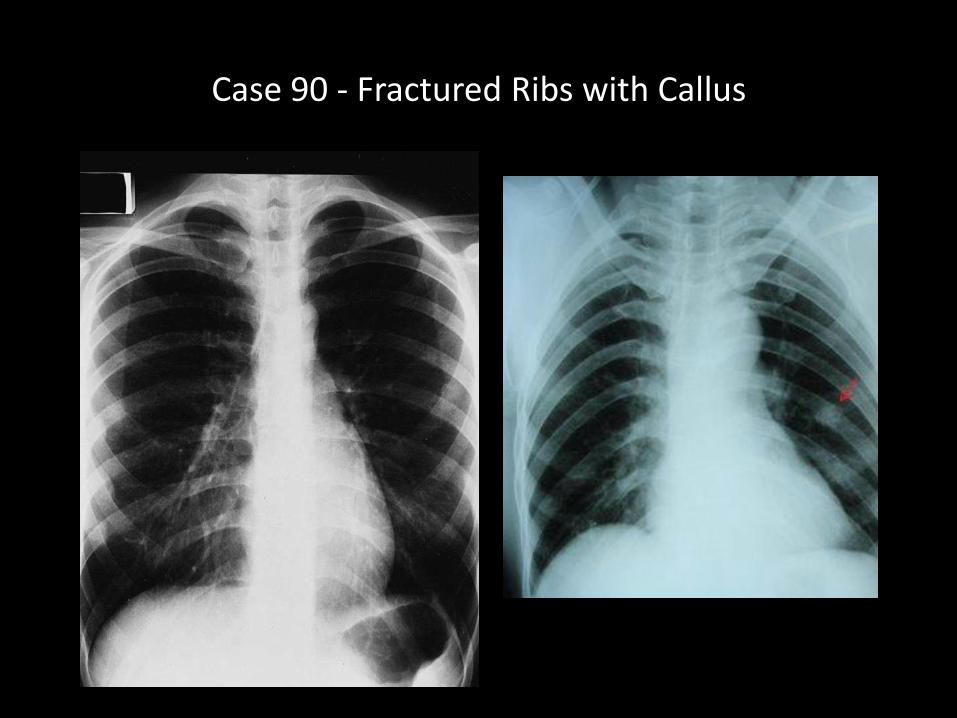
Case 90 - Fractured Ribs with Callus
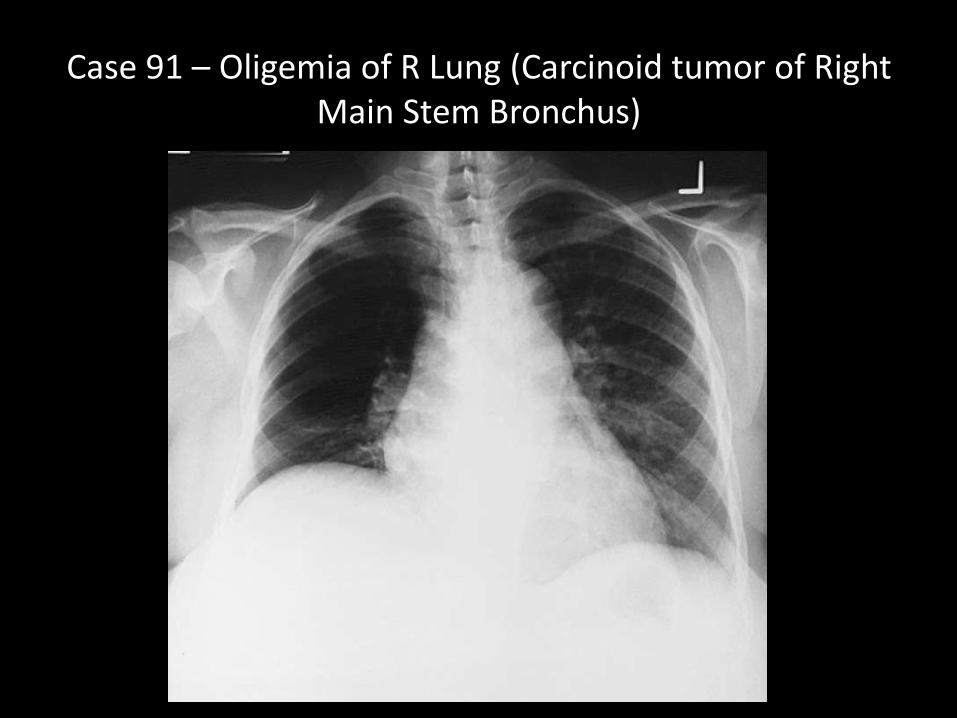
Case 91 – Oligemia of R Lung (Carcinoid tumor of Right Main Stem Bronchus)
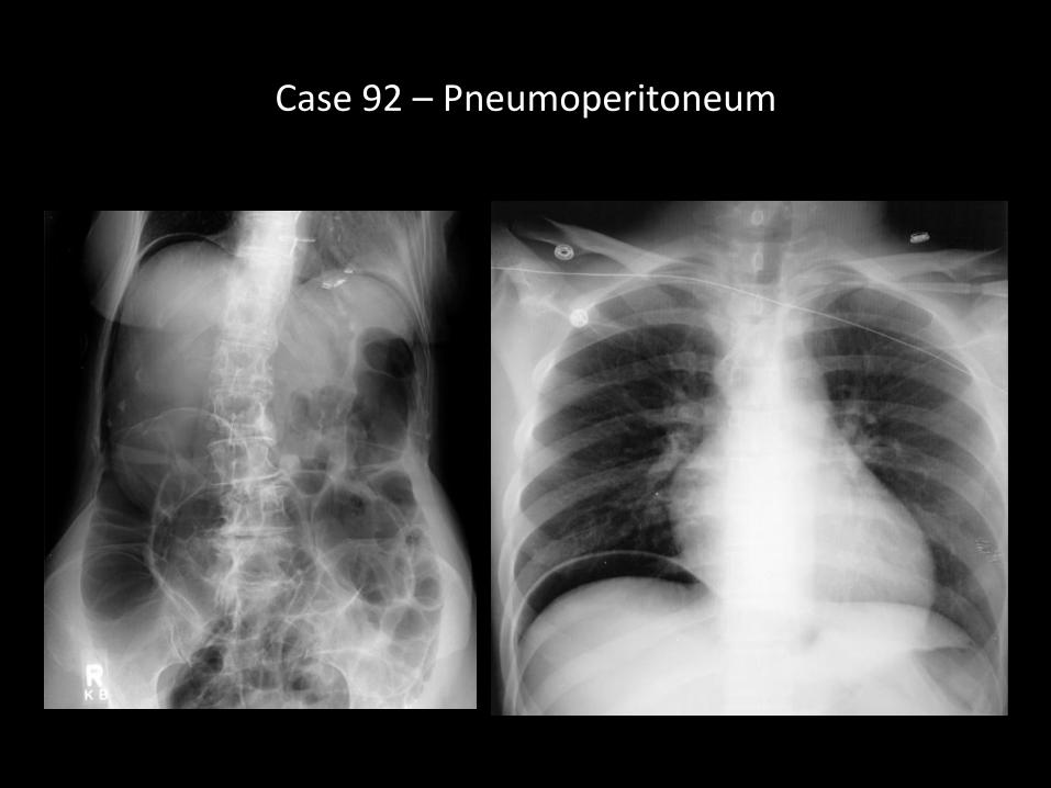
Case 92 – Pneumoperitoneum
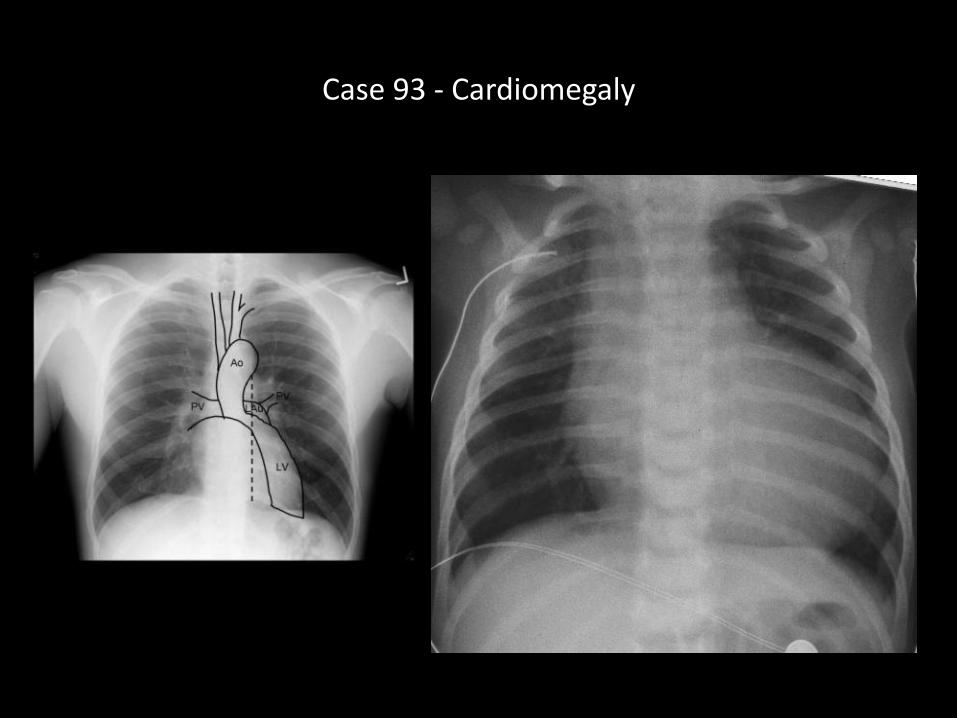
Case 93 - Cardiomegaly
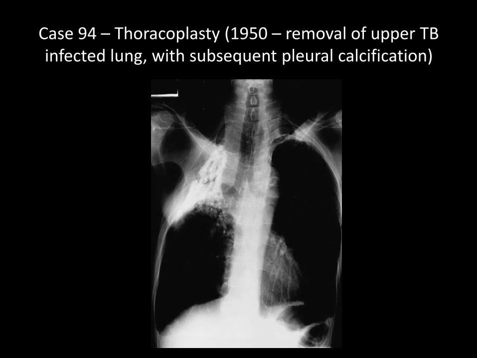
Case 94 – Thoracoplasty (1950 – removal of upper TB infected lung, with subsequent pleural calcification)
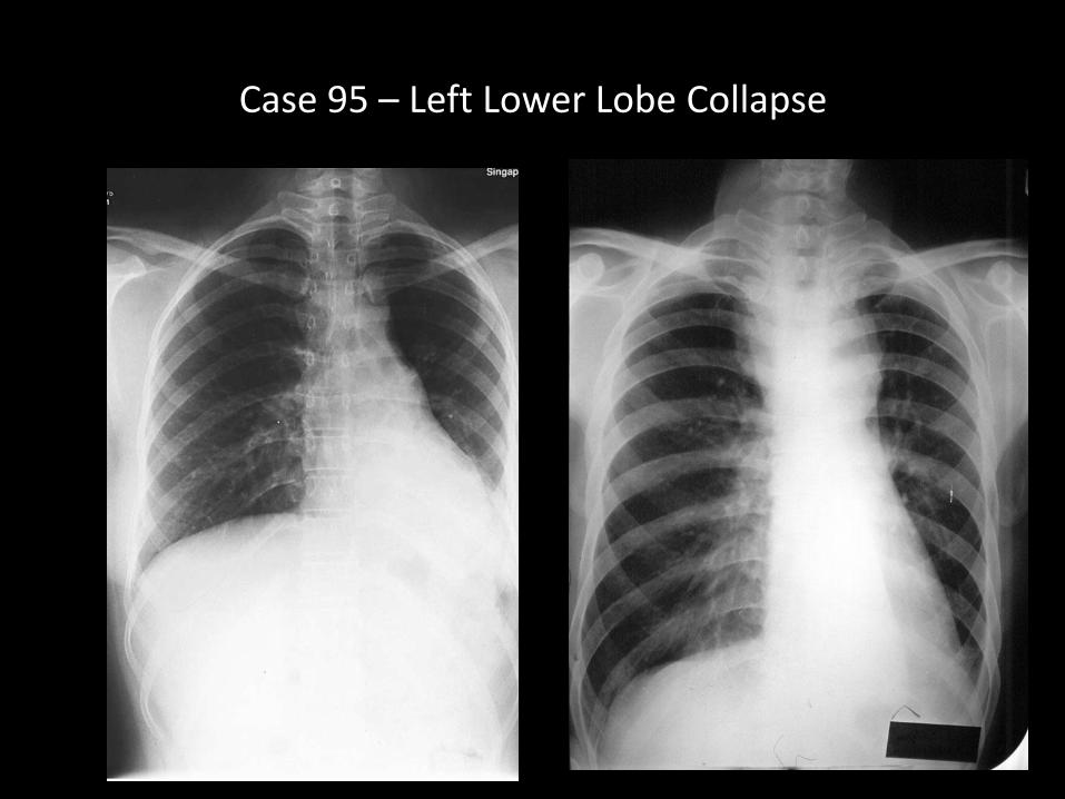
Case 95 – Left Lower Lobe Collapse
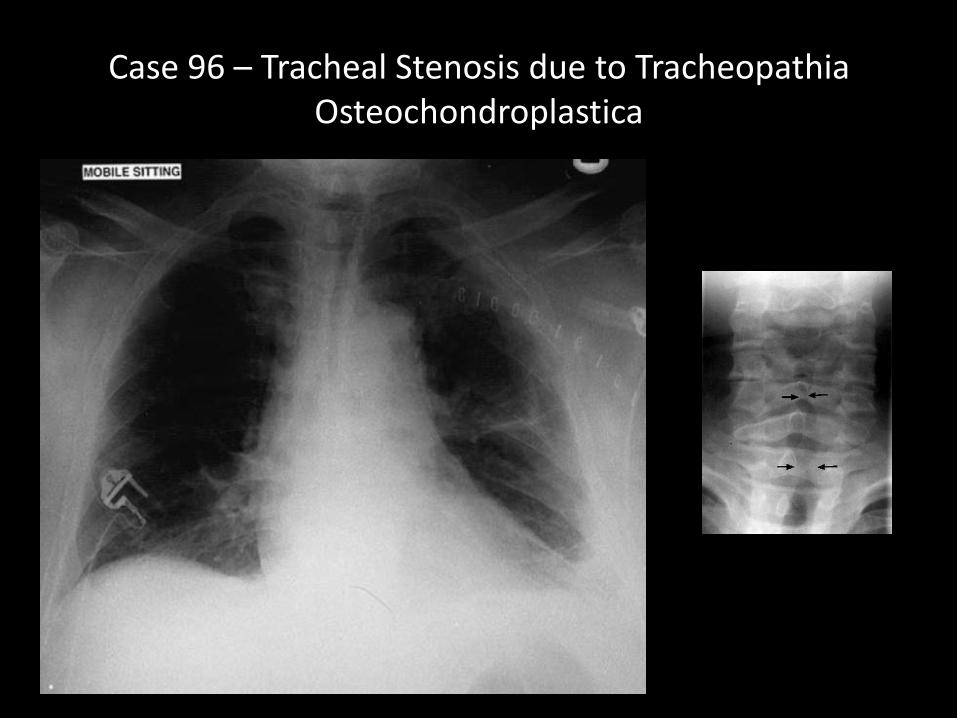
Case 96 – Tracheal Stenosis due to Tracheopathia Osteochondroplastica
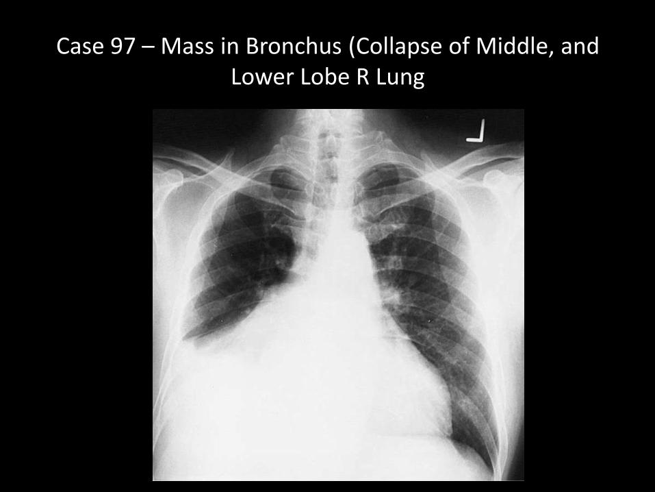
Case 97 – Mass in Bronchus (Collapse of Middle, and Lower Lobe R Lung
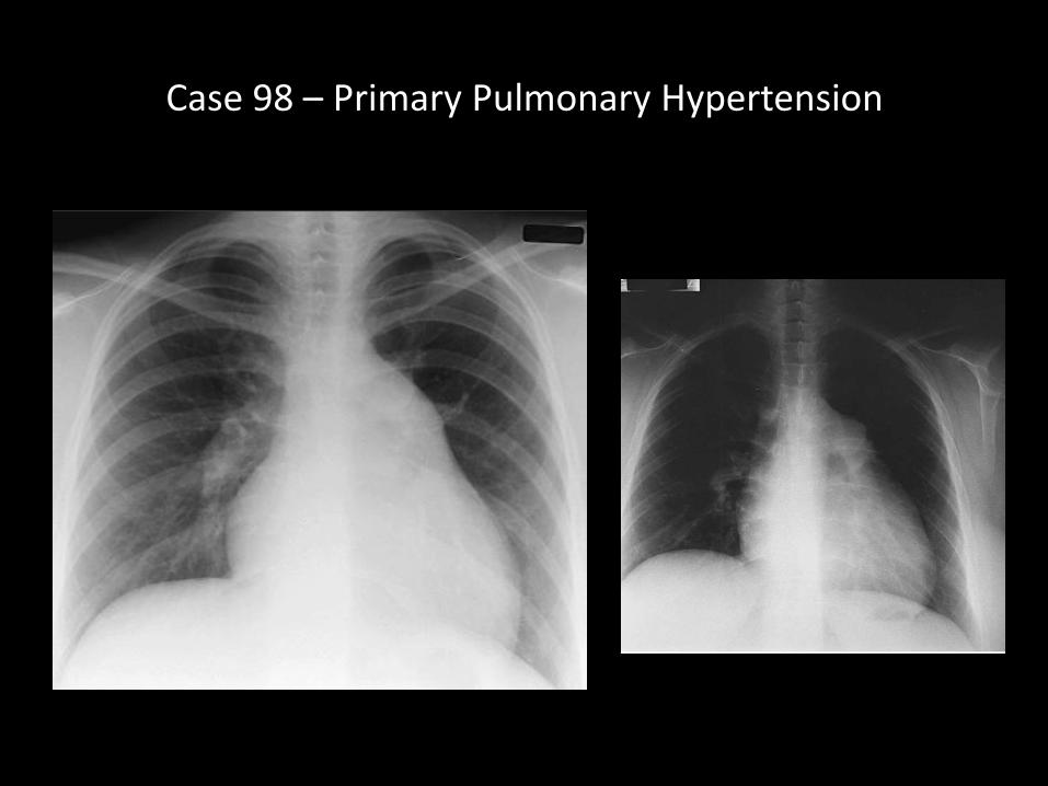
Case 98 – Primary Pulmonary Hypertension
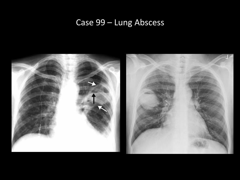
Case 99 – Lung Abscess
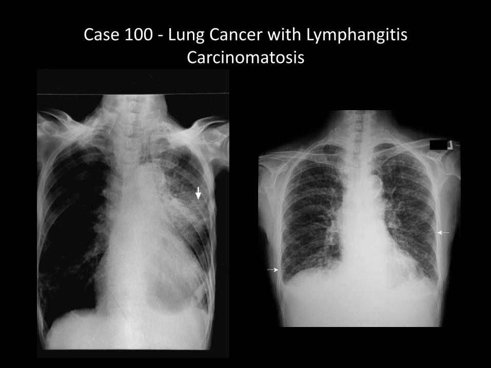
Case 100 - Lung Cancer with Lymphangitis Carcinomatosis
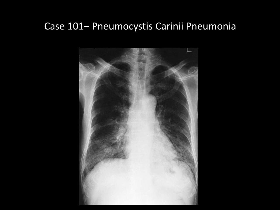
Case 101– Pneumocystis Carinii Pneumonia
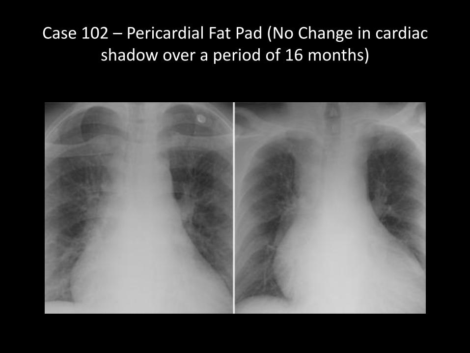
Case 102 – Pericardial Fat Pad (No Change in cardiac shadow over a period of 16 months)
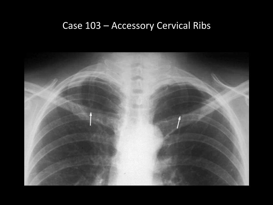
Case 103 – Accessory Cervical Ribs
Thank You