chemotaxis-based endosulfan biotransformation: enrichment and isolation of endosulfan-degrading...
TRANSCRIPT

This article was downloaded by: [Cukurova Universitesi]On: 04 November 2014, At: 07:20Publisher: Taylor & FrancisInforma Ltd Registered in England and Wales Registered Number: 1072954 Registered office: Mortimer House,37-41 Mortimer Street, London W1T 3JH, UK
Environmental TechnologyPublication details, including instructions for authors and subscription information:http://www.tandfonline.com/loi/tent20
Chemotaxis-based endosulfan biotransformation:enrichment and isolation of endosulfan-degradingbacteriaMuhil Vannan Seralathana, Saravanadevi Sivanesana, Srinivasan Nargunanathana, AmitBafanaa, Krishnamurthi Kannana & Tapan Chakrabartiaa Environmental Health Division, National Environmental Engineering Research Institute,Nagpur 440022, IndiaAccepted author version posted online: 20 Jun 2014.Published online: 21 Jul 2014.
To cite this article: Muhil Vannan Seralathan, Saravanadevi Sivanesan, Srinivasan Nargunanathan, Amit Bafana, KrishnamurthiKannan & Tapan Chakrabarti (2014): Chemotaxis-based endosulfan biotransformation: enrichment and isolation of endosulfan-degrading bacteria, Environmental Technology, DOI: 10.1080/09593330.2014.937464
To link to this article: http://dx.doi.org/10.1080/09593330.2014.937464
PLEASE SCROLL DOWN FOR ARTICLE
Taylor & Francis makes every effort to ensure the accuracy of all the information (the “Content”) containedin the publications on our platform. However, Taylor & Francis, our agents, and our licensors make norepresentations or warranties whatsoever as to the accuracy, completeness, or suitability for any purpose of theContent. Any opinions and views expressed in this publication are the opinions and views of the authors, andare not the views of or endorsed by Taylor & Francis. The accuracy of the Content should not be relied upon andshould be independently verified with primary sources of information. Taylor and Francis shall not be liable forany losses, actions, claims, proceedings, demands, costs, expenses, damages, and other liabilities whatsoeveror howsoever caused arising directly or indirectly in connection with, in relation to or arising out of the use ofthe Content.
This article may be used for research, teaching, and private study purposes. Any substantial or systematicreproduction, redistribution, reselling, loan, sub-licensing, systematic supply, or distribution in anyform to anyone is expressly forbidden. Terms & Conditions of access and use can be found at http://www.tandfonline.com/page/terms-and-conditions

Environmental Technology, 2014http://dx.doi.org/10.1080/09593330.2014.937464
Chemotaxis-based endosulfan biotransformation: enrichment and isolation ofendosulfan-degrading bacteria
Muhil Vannan Seralathan, Saravanadevi Sivanesan, Srinivasan Nargunanathan, Amit Bafana, Krishnamurthi Kannan∗ andTapan Chakrabarti
Environmental Health Division, National Environmental Engineering Research Institute, Nagpur 440022, India
(Received 22 January 2014; final version received 17 June 2014 )
The study was conducted to isolate endosulfan biotransforming or biodegrading microbes based on chemotaxis. Pseudomonasaeruginosa strain KKc3, Ochrobactrum sp. strain KKc4, Achromobacter xylosoxidans strain KKc6 and Bacillus megateriumKKc7 were isolated based on their migration towards endosulfan in a soil column. Out of the four bacteria, B. megateriumconverted endosulfan into toxic metabolite endosulfan sulphate, while the other three bacteria followed the non-toxic endo-sulfan diol pathway. The mixed culture system consisting of P. aeruginosa, Ochrobactrum sp and A. xylosoxidans couldremove 94% of total endosulfan by using endosulfan as the sole source of sulphur.
Keywords: chemotaxis; endosulfan; biotransformation; DGGE; kinetics
1. IntroductionBioremediation is a robust and proven technology for envi-ronmental cleanup. The success of bioremediation primar-ily depends on microbial strain selection. The batch scaleenrichment culturing method is extensively used for cultureselection during bioremediation. This concept has been fol-lowed from the time it was conceptualized by Beijerinckand Winogradsky.[1] However, culture enrichment bias wasobserved in due course of time. Besides, many studieshave suggested that it may distort the catabolic pathway’sdiversity.[2] Hence, different approaches such as in-rhizodirected selection and priming were developed. Simulta-neously, interdisciplinary studies were initiated to under-stand biodegradation process.[3] However, the well-knownchemotactic property of microbes was not utilized in micro-bial selection for bioremediation processes. In this regard,different studies have already proved that environmentalpollutants can act as chemo-attractants and the attractedmicrobes can metabolize the respective pollutants.[4]
In general, bacterial chemotaxis functions to keep thecells at their optimum environmental niche for growthand survival. Both positive and negative movements havebeen reported to achieve the same.[5,6] Bacteria suchas Pseudomonas sp, Kurthia sp, Bacillus circulans,[7]Agrobacterium radiobacter J14a [8] and Ralstonia sp.SJ98 [9] can chemotactically respond to pollutants. Envi-ronmental pollutants such as naphthalene,[7,10], atrazine[11] and aromatic acids [12] are known to attract certain
∗Corresponding author. Email: [email protected]
bacterial species. Recently, Sangwan et al. [13] observedthe expression of chemotaxis/motility-regulating genes atHCH (hexachlorocyclohexane) dump sites by metagenomicanalysis. It has been proved by mutational studies thatchemotaxis can bring pollutants and microbes in close prox-imity for enhanced biodegradation process.[12] Chemo-taxis may also promote the formation of microbial consortiawhich may restore the catabolic diversity of the niche.[14]
The current study used microbial chemotaxis to isolateendosulfan biotransforming bacterial species. Endosulfanis a well-known non-systemic insecticide cum acaricide. Itwas used in agricultural practice for the last few decades,especially for crops such as cotton, soya, coffee, tea andvegetables. Due to its adverse effects on environment andother health hazards, it was enlisted as POP (persistentorganic pollutant) in 2012 by Stockholm Convention. How-ever, it was exempted for use on few specific crop–pestcomplexes in India. Besides, a five-year phase-out periodwas permitted during which endosulfan could be used.[15]Although endosulfan is banned in many countries, its pres-ence in environment due to its recalcitrance is a seriousconcern. Hence, endosulfan remediation is still an impor-tant research problem. Isolation of bacterial species forendosulfan bioremediation and their characterization areequally important. Hence, the current investigation wasinitiated to isolate endosulfan biotransforming microbesby using a unique and novel strategy based on bacterialchemotaxis.
© 2014 Taylor & Francis
Dow
nloa
ded
by [
Cuk
urov
a U
nive
rsite
si]
at 0
7:20
04
Nov
embe
r 20
14

2 M.V. Seralathan et al.
2. Materials and methods2.1. Experimental set-upA prototype vertical soil matrix set-up was designed forthis study (Figure 1). The set-up was fabricated with thehelp of 250 ml capacity laboratory filtration unit (Tar-sons, India). The reservoir and collection tanks of the filterassembly were considered as two compartments and bothwere separated with autoclaved filter paper to avoid mix-ing of soils in the two compartments (Tarsons, India).Briefly, 10 kg of fertile, near-acidic or neutral pH topsoil samples were collected from National Environmen-tal Engineering Research Institute (NEERI) garden. Thesoil was thoroughly mixed; stones, roots and debris wereremoved. The samples were tested for the absence of pes-ticide residues by gas chromatograph-mass spectrometer(GC-MS) as described earlier elsewhere.[16] The soil wassubjected to routine autoclave cycles on five successivedays, and its sterility was confirmed by plating on nutrientagar plates. The autoclaved soil was spiked with 500 mg/kg(w/w) of technical grade endosulfan (>90% purity) andmixed thoroughly under aseptic condition. It was thenpacked in the reservoir compartment, while non-autoclavedand non-spiked soil was filled in the collection tank of thefiltration apparatus. Simultaneously, a control set-up wasdesigned with autoclaved soil in both the compartments.Soils in the two compartments were separated with the helpof autoclaved Whatman 1005 analytical filter paper (parti-cle filtration 2.5 μm) with filter holder. Water and air forthe total system were supplied through the collection tank(non-autoclaved soil). Aeration was done by a heavy dutyaquarium pump. Both water and air were sterilized through0.2 μm nylon membrane filter before entering the system.Moisture content was maintained between 20% and 30%with proper ventilation by siphoning tubes (Figure 1). Theset-up was maintained for 30 days study period.
2.2. Isolation and identification of bacterial culturesAfter 30 days of incubation, ∼50 g of soil from closeproximity to the filter paper of the reservoir compartment(autoclaved soil) was collected aseptically and stored at4◦C and −80◦C for further analysis. Endosulfan and itsmetabolites were extracted and analysed as per an earlierreport.[16] Presence of chemotactically attracted microbeswas screened with the help of non-sulphur medium (NSM)containing endosulfan as the sole source of sulphur asreported earlier elsewhere.[17] The isolates were identifiedby 16S rRNA gene sequencing as per an earlier study [18]and the nucleotide sequences were deposited in GenBank.
2.3. Flask studyBiodegradation efficacies of the selected cultures were stud-ied individually and in the mixed culture system in twodifferent media, i.e. NSM and non-carbon medium (NCM),with 100 mg/l initial endosulfan as per earlier reports.[17,19,20] The control flasks devoid of either culture or energysource were maintained under similar conditions to esti-mate the abiotic endosulfan transformation and bacterialgrowth pattern, respectively, under energy starved condi-tion. Samples were collected periodically and analysed forbacterial growth, residual concentration of endosulfan andits metabolites.
2.4. Growth curve analysisAt regular intervals, 1 ml of culture was collected andbacterial growth was recorded in terms of optical densityat 600 nm using UV–Visible spectrophotometer (Cintra,USA) as per the standard procedure.
Figure 1. Vertical soil column experimental set-up for chemotaxis. The 250 ml lab filtration unit was fabricated as prototype studymaterial. The reservoir compartment contained autoclaved soil, and non-autoclaved soil was filled in the collection tank.
Dow
nloa
ded
by [
Cuk
urov
a U
nive
rsite
si]
at 0
7:20
04
Nov
embe
r 20
14

Environmental Technology 3
2.5. Biodegradation and metabolite identificationTo study the degradation potential of the cultures, 5 ml ofsample at each time point was centrifuged at 10,000 g for5 min. The supernatant was extracted thrice with n-hexane:acetone (3:1 v/v), and the solvent phase was cleaned bypassing it through anhydrous sodium sulphate and flashevaporated at 45◦C. The residues were dissolved in 2 mlof GC-grade acetone as described earlier.[17] To estimatethe adsorption of endosulfan on cell surface, the cell pelletswere washed with autoclaved distilled water and then sus-pended in n-hexane: acetone (3:1). The solvent phase wasevaporated by flash evaporator, and the residues were dis-solved in 1 ml of acetone and stored at 4◦C until analysis.In case of soil, approximately 10 g of soil was subjectedto n-hexane: acetone extraction as described in an earlierreport.[16] The residues were dissolved in 2 ml of acetoneand stored at 4◦C until further analysis.
Metabolites were identified on Varian 3800 GC coupledwith Varian 2200 ion trap MS (mass spectrometer, Varian,USA) using electron ionization. Splitless injections of 1 μlvolume were carried out with 1079 split programmable tem-perature injector. The ion trap, manifold and transfer linewere kept at 200◦C, 80◦C and 280◦C, respectively. The GCoven was programmed from 80◦C to 180◦C at 25◦C permin with a 5-min hold at 300◦C. Separation was performedon Varian Chrompack MS grade WCOT fused-silica cap-illary column (30 m long, 0.25 mm I.D) CP-SIL 8CB, withhelium (ultra pure 99.99%) as the carrier gas at a flow rate of1.0 ml/min. The detector was programmed to scan a massrange of 40–450 amu. Metabolites were identified on thebasis of mass spectra and retention time using the NISTlibrary, and by comparison with the analytical standards.
2.6. Quantification and kinetics of biotransformationThe analytes were quantified as per USEPA method 525.The residual endosulfan was quantified by GC equippedwith ECD (electron capture detector, Perkin Elmer Clarus500, USA). Splitless injections of 1 μl volume of sampleswere resolved on DB5 column. Ultra-pure nitrogen gas wasused as the carrier gas at a flow rate of 0.8 ml/min with GCoven, injector and detector temperatures of 250◦C, 200◦Cand 300◦C, respectively. Different concentrations of ana-lytical standards were prepared in GC-grade acetone andused for quantification. The calibration curve was preparedby plotting peak area against concentration for all availablestandards. The kinetics of degradation was derived basedon the earlier reports.[18,21]
2.7. DGGE (denaturing gradient gel electrophoresis)analysis
DGGE was performed to confirm that the isolates werederived from the soil sample. 16S rRNA V3 variable region
specific amplification method was used as described ear-lier elsewhere. Briefly, the soil metagenome was extractedfrom 2.50 g of each sample by using ZR Soil Microbe DNAMiniPrep kit (Zymo Research, USA) as per the manufac-turer’s instructions. The DNA samples were quantified bynanodrop (Thermo Scientific, USA) and stored at −20◦Cuntil analysis. Hundred nanogram of soil DNA was used astemplate in 50 μl PCR as reported earlier elsewhere. Theamplification conditions were initial denaturation at 95◦Cfor 5 min, followed by 35 cycles of denaturation at 94◦C for30 s, annealing at 55◦C for 45 s and extension at 74◦C for1 min, and the final extension at 74◦C for 5 min. Amplifica-tion was carried out in BioRad S100 thermocycler (BioRad,UK). The amplicons were resolved in 2.0% agarose gel, andbands of ∼215 bp were excised and purified by using theGel DNA recovery kit (Zymo Research, USA). The purifiedproducts were quantified and equal amounts of ampliconswere run in DCode Universal Mutation System (BioRad,UK). DGGE was carried out on 8% acrylamide gel with adenaturant gradient of 40–70% in 1× TAE buffer at 60◦Cand a constant voltage of 120 V for 18 h. The gels werestained in running buffer containing 0.5 mg/l of ethidiumbromide, and images were acquired with Verasadoc system(BioRad, UK).[22]
The appearance of isolates in autoclaved soil was con-firmed by cDNA-based DGGE analysis. Briefly, total soilRNA was isolated on the initial and 30th days fromboth autoclaved and non-autoclaved soil samples by usingPowerSoil total RNA isolation kit (MOBio, USA). The iso-lated RNA was checked for its quality and quantity usingnanodrop UV spectrophotometer and 1% agarose gel elec-trophoresis. Clean-up was carried out by desalting columnsas per the manufacturer’s instruction (Pall Life Sciences,USA). DNA contamination was removed by DNase treat-ment and the same was confirmed by PCR using universalprimers. Hundred microgram of RNA was converted tocDNA using Invitrogen Power cDNA conversion kit as perthe manufacturer’s instructions.[23] The resulting cDNAwas used to detect the isolates with 16S rRNA V3 vari-able region universal primers as described earlier in thispaper.[22]
2.8. Soft Agar Swarm plate assayThe isolated species were checked for chemotaxis towardsendosulfan by using the soft agar swarm assay as describedearlier.[24] Briefly, soft agar plates of NSM and NCMcontaining 0.5% agar were prepared, 100 mg/l of endo-sulfan (>90% purity) was dropped at the centre of thepetri dish and excess solvent was allowed to evaporate.Mid-log-phase cultures were harvested from the nutrientbroth and washed twice with the respective media. The cellsuspensions containing 107 cells/ml were seeded on fourcorners (sub-centric) of the petri dish. After incubation at37◦C for 48 h, the plates were scored for colonies and zoneformation.
Dow
nloa
ded
by [
Cuk
urov
a U
nive
rsite
si]
at 0
7:20
04
Nov
embe
r 20
14

4 M.V. Seralathan et al.
3. Results and discussionTo the best of our knowledge, this is the first report aboutisolation of microbes based on chemotaxis for bioremedi-ation purpose. The study was conducted with a fabricatedprototype vertical soil matrix column. In a similar study byHanzel et al.,[25] vapour phase naphthalene was used aschemo-attractant to demonstrate the chemotactic responseof Pseudomonas putida PpG7 strain. In another study byPaul et al.,[26] open tray soil system was used to demon-strate bacterial chemotaxis towards p-nitrophenol. Apartfrom these reports, no other studies are available in the lit-erature about chemotaxis of microbes towards pollutants ina soil system.
The initial GC-MS analysis of garden soil samplesconfirmed that they were free from organochlorine andorganophosphate pesticides. Soil pH was 6.5–6.7 in 5%aqueous solution. After incubation for 30 days, the exper-imental set-up showed 20% reduction in initial spikedendosulfan concentration with the appearance of bioticmetabolite endosulfan sulphate, and other metabolites suchas endosulfan diol, endosulfan ether and endosulfan lactone(Figure 2). On the contrary, the control set showed only 2%
reduction in endosulfan with no known metabolites. Theidentification of endosulfan metabolites was confirmed bymass spectrometry (Supplementary Figure 1).
Four bacterial cultures were isolated from autoclavedsoil in the experimental set-up after the incubation period,while the control set-up remained sterile on all testedmedia. The 16S rRNA gene sequencing followed byBLAST analysis revealed that the isolated bacterial cultureswere Pseudomonas aeruginosa strain KKc3, Ochrobac-trum sp strain KKc4, Achromobacter xylosoxidans strainKKc6 and Bacillus megaterium KKc7 (GenBank accessionnumbers JF758468–JF758471). Gram staining revealedthat the strains KKc3, KKc4 and KKc6 were gram-negative rod-shaped bacteria, while KKc7 was grampositive. Hanging drop study showed that all the iso-lates were motile, which is a pre-requisite for chemo-taxis.
Batch scale studies confirmed that all the isolates couldmetabolize endosulfan with different efficiencies. Studieswere carried out in NSM and NCM with endosulfan asthe sole sulphur or carbon source, and all the isolateswere found to grow in both the media. However, the
(b) (a)
Figure 2. GC profiles of (a) 0th day and (b) 30th day autoclaved soil samples. Peak 1, endo lactone; Peak 2, endo ether; Peak 3, alphaendosulfan; Peak 4, endo diol; Peak 5, beta endosulfan and Peak 6, endosulfan sulphate. The mass spectra of these metabolites are givenin Supplementary Figure 1.
Dow
nloa
ded
by [
Cuk
urov
a U
nive
rsite
si]
at 0
7:20
04
Nov
embe
r 20
14

Environmental Technology 5
growth was more limited in NCM as compared withNSM. Among all the isolates, B. megaterium is not yetreported for endosulfan metabolism, while strains of theother three species have been well documented for endosul-fan biotransformation earlier.[27–30] Interestingly, thesethree organisms were also reported as chemotacticallyresponding species.[31–35] In this study, P. aeruginosa andA. xylosoxidans metabolized endosulfan into endosulfanlactone via the diol pathway, whereas Ochrobactrum bio-transformed endosulfan into endosulfan diol. Stain KKc7metabolized endosulfan into toxic metabolite endosulfansulphate, irrespective of the medium. B. megaterium hasearlier been reported for its potential against various pol-lutants such as PAH (polycyclic aromatic hydrocarbons),dyes and chromium.[36,37] Its bifunctional CytochromeP450 enzyme has been highlighted by various catabolicstudies.[38] Thus, strains KKc3, KKc4 and KKc6 detoxifiedendosulfan, whereas KKc7 followed the toxic endosulfan
sulphate pathway. The possibility of abiotic conversion wasruled out by control set-up in both the media, where nometabolic intermediate could be detected.
Of the four isolates, P. aeruginosa strain KKc3 wasrecorded as the fastest degrading bacterium under both non-sulphur and non-carbon conditions. The biotransformationrates were 0.098 and 0.094 mg/l/d, whereas the half-lifeperiods were 5.5 and 7.7 days in NSM and NCM, respec-tively. The endosulfan removal percentages were 80% and73% for NSM and NCM, respectively. P. aeruginosa hasbeen reported to degrade a range of pollutants and utilizethem as energy source,[39,40] including endosulfan. Strainssuch as TJ1.B1 and MN2B12 were reported for endosulfanbiotransformation with 40–93% degradation. Interestingly,P. aeruginosa is used as a model organism for understandingchemotactic character in ecological studies.[32,33,41,42]A. xylosoxidans strain KKc6 degraded endosulfan at therate of 0.0972 and 0.0979 mg/l/d with the half-life period
(a)
(b)
0
1
2
3
4
5
6
7
0 2 4 6 8 10 12 14
Time (Days)
OD
@ 6
00
KKc3 NSM control KKc3 NSM endosulfan KKc4 NSM control
KKc4 NSM endosulfan KKc6 NSM control KKc6 NSM endosulfan
KKc7 NSM control KKc7 NSM endosulfan
0
1
2
3
4
5
6
0 2 4 6 8 10 12 14
Time (Days)
OD
@ 6
00
KKc3 NCM control KKc3 NCM endosulfan KKc4 NCM control
KKc4 NCM endosulfan KKc6 NCM control KKc6 NCM endosulfan
KKC7 NCM control KKc7 NCM endosulfan
Figure 3. Growth curve patterns of isolates in (a) non-carbon condition with endosulfan as the carbon source, and (b) non-sulphurcondition with endosulfan as the sulphur source.
Dow
nloa
ded
by [
Cuk
urov
a U
nive
rsite
si]
at 0
7:20
04
Nov
embe
r 20
14

6 M.V. Seralathan et al.
0
20
40
60
80
100
120
0 2 4 6 8 10 12 14Time (Days)
Res
idu
al c
on
cen
trat
ion
of
end
osu
lfan
(%
)
KKc3 NSM KKc4 NSM KK6 NSM
KKc7 NSM Abiotic control NSM
0
20
40
60
80
100
120
0 2 4 6 8 10 12 14Time (Days)
Res
idu
al c
on
cen
trat
ion
of
end
osu
lfan
(%
)
KKc3 NCM KKc4 NCM KKc6 NCM
KKc7 NCM Abiotic control NCM
(a)
(b)
Figure 4. Degradation potential of isolates under (a) non-carboncondition with endosulfan as the carbon source, and (b)non-sulphur condition with endosulfan as the sulphur source.
of 7.13 and 7.07 days in NSM and NCM, respectively.Singh and Singh [30] reported that A. xylosoxidans C8Bcould biotransform endosulfan and endosulfan sulphate.Another strain A8 isolated from polychlorinated biphenyl(PCB) contaminated soil could use PCBs as carbon source,which supports the current observation on growth of thestrain.[34,43] It has also been studied for chemotactic char-acter. The strain Ochrobactrum KKc4 grew slowly in NSMas well as in NCM (Figure 3). However, it could stillmetabolize endosulfan to endodiol within the observationperiod with the half-life of 10.2 and 11.9 days, and degra-dation constant of 0.0697 and 0.058 mg/l/d for NSM andNCM, respectively. B. megaterium strain KKc7 biotrans-formed endosulfan to toxic metabolite endosulfan sulphatein both the media. The removal constant was 0.11 and0.0689 mg/l/d, whereas the endosulfan half-life period was6.09 and 10.02 days for NSM and NCM, respectively(Figure 4). Since strain KKc7 formed toxic metabolite, itwas omitted from mixed culture studies. The mixed cul-ture, consisting of KKc3, KKc4 and KKc6, showed rapidremoval of endosulfan between 2 and 6 days of incubationwith endosulfan as the sole source of sulphur, and achievedup to 94% endosulfan removal (Figure 5). Psuedomonasgrew faster than the other two strains in mixed culture
0
20
40
60
80
100
120
0 2 4 6 8 10 12 14Time (Days)
Res
idu
al C
on
cen
trat
ion
so
f E
nd
osu
lfan
(%
)
K = 0.2242 mg/l day–1
t1/2 = 3.09 days
R2 = 0.8981
00.5
11.5
22.5
33.5
44.5
5
0 5 10 15Time (Days)
Ln
Res
idu
al c
on
c o
fE
nd
osu
flan
(a)
(b)
Figure 5. Biodegradation potential of KKc3, KKc4 and KKc6in mixed culture system and its kinetics.
condition. First-order kinetics revealed that the mixed cul-ture system had the degradation potential of 0.22 mg/l/d(Figure 5) and the half-life period of endosulfan was 3.09days (R2 = 8.91).
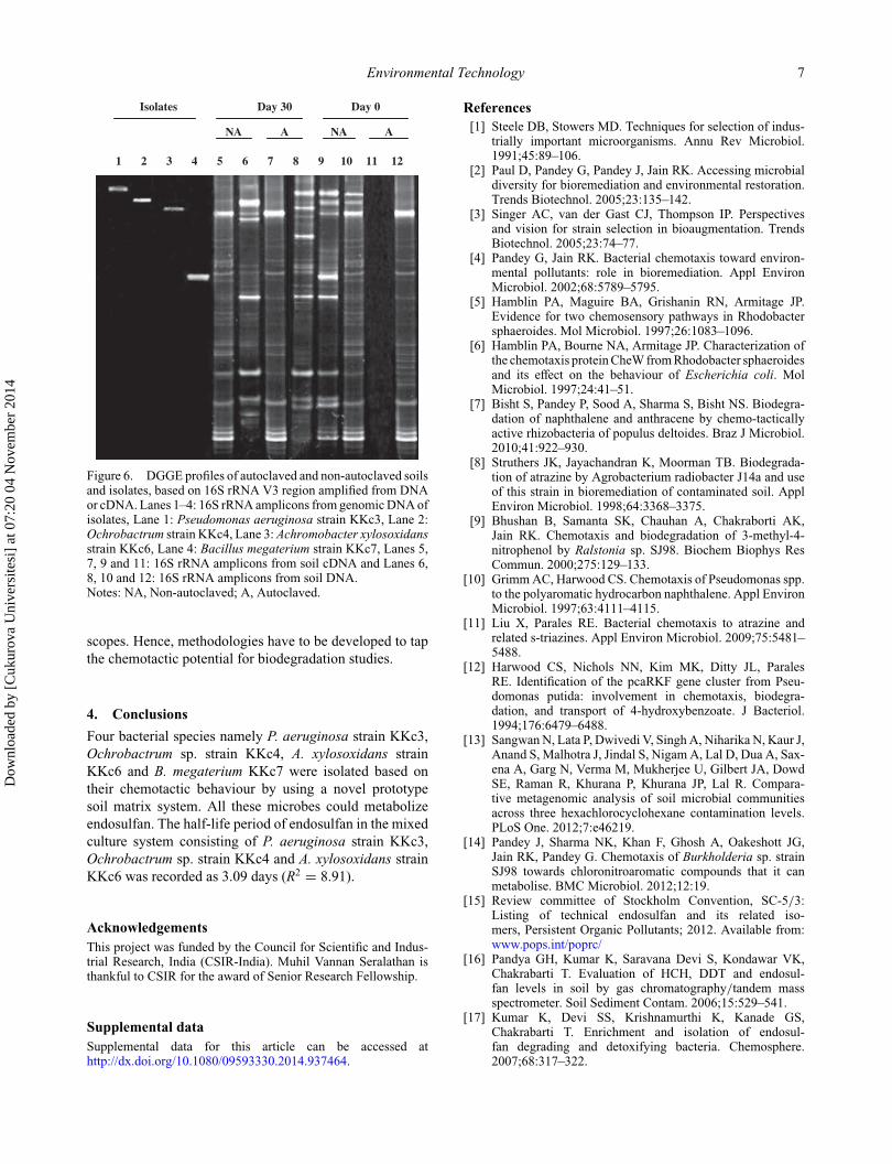
Both DNA- and cDNA-based DGGE analysis provedthat the isolated strains originated from the study soil sam-ples. It is well known that autoclaving destroys RNA, whileDNA is relatively resistant and can be amplified by PCReven after autoclaving.[44] In agreement with this, theDGGE profile of 16S rRNA V3 amplicon prepared fromautoclaved soil cDNA on day 0 did not show any bands. Itconfirmed complete absence of bacteria after autoclaving.Consequent reappearance of cDNA bands in autoclaved soilon day 30 clearly demonstrated migration of microbes fromnon-autoclaved to autoclaved soil (Figure 6). Further, themicrobial activity in autoclaved soil was confirmed by thedetection of biotic metabolite endosulfan sulphate. ThoughDGGE and the literature support the current observation onchemotaxis, the possibility of non-chemotactic migrationcannot be omitted, since soil is an open dynamic system andbacterial migration is controlled by many factors. The softagar plates did not show zone of attraction towards endo-sulfan for all the four organisms. We strongly believe thatthis might be due to the lack of bioavailability or problemin gradient formation on agar plates because of lower solu-bility of endosulfan. In the soil system endosulfan gradientmight be achieved by various possibilities such as mois-ture, biofilm formation or free flow of compounds, whereasthe same condition may not be created during the shorterincubation time on agar plates. The available techniquesfor chemotaxis studies such as microbial tracking, nanoflu-idic capillary system and capillary systems are limited in
Dow
nloa
ded
by [
Cuk
urov
a U
nive
rsite
si]
at 0
7:20
04
Nov
embe
r 20
14
Environmental Technology 7
Isolates Day 30 Day 0
NA A NA A
1 2 3 4 5 6 7 8 9 10 11 12
Figure 6. DGGE profiles of autoclaved and non-autoclaved soilsand isolates, based on 16S rRNA V3 region amplified from DNAor cDNA. Lanes 1–4: 16S rRNA amplicons from genomic DNA ofisolates, Lane 1: Pseudomonas aeruginosa strain KKc3, Lane 2:Ochrobactrum strain KKc4, Lane 3: Achromobacter xylosoxidansstrain KKc6, Lane 4: Bacillus megaterium strain KKc7, Lanes 5,7, 9 and 11: 16S rRNA amplicons from soil cDNA and Lanes 6,8, 10 and 12: 16S rRNA amplicons from soil DNA.Notes: NA, Non-autoclaved; A, Autoclaved.
scopes. Hence, methodologies have to be developed to tapthe chemotactic potential for biodegradation studies.
4. ConclusionsFour bacterial species namely P. aeruginosa strain KKc3,Ochrobactrum sp. strain KKc4, A. xylosoxidans strainKKc6 and B. megaterium KKc7 were isolated based ontheir chemotactic behaviour by using a novel prototypesoil matrix system. All these microbes could metabolizeendosulfan. The half-life period of endosulfan in the mixedculture system consisting of P. aeruginosa strain KKc3,Ochrobactrum sp. strain KKc4 and A. xylosoxidans strainKKc6 was recorded as 3.09 days (R2 = 8.91).
AcknowledgementsThis project was funded by the Council for Scientific and Indus-trial Research, India (CSIR-India). Muhil Vannan Seralathan isthankful to CSIR for the award of Senior Research Fellowship.
Supplemental dataSupplemental data for this article can be accessed athttp://dx.doi.org/10.1080/09593330.2014.937464.
References[1] Steele DB, Stowers MD. Techniques for selection of indus-
trially important microorganisms. Annu Rev Microbiol.1991;45:89–106.
[2] Paul D, Pandey G, Pandey J, Jain RK. Accessing microbialdiversity for bioremediation and environmental restoration.Trends Biotechnol. 2005;23:135–142.
[3] Singer AC, van der Gast CJ, Thompson IP. Perspectivesand vision for strain selection in bioaugmentation. TrendsBiotechnol. 2005;23:74–77.
[4] Pandey G, Jain RK. Bacterial chemotaxis toward environ-mental pollutants: role in bioremediation. Appl EnvironMicrobiol. 2002;68:5789–5795.
[5] Hamblin PA, Maguire BA, Grishanin RN, Armitage JP.Evidence for two chemosensory pathways in Rhodobactersphaeroides. Mol Microbiol. 1997;26:1083–1096.
[6] Hamblin PA, Bourne NA, Armitage JP. Characterization ofthe chemotaxis protein CheW from Rhodobacter sphaeroidesand its effect on the behaviour of Escherichia coli. MolMicrobiol. 1997;24:41–51.
[7] Bisht S, Pandey P, Sood A, Sharma S, Bisht NS. Biodegra-dation of naphthalene and anthracene by chemo-tacticallyactive rhizobacteria of populus deltoides. Braz J Microbiol.2010;41:922–930.
[8] Struthers JK, Jayachandran K, Moorman TB. Biodegrada-tion of atrazine by Agrobacterium radiobacter J14a and useof this strain in bioremediation of contaminated soil. ApplEnviron Microbiol. 1998;64:3368–3375.
[9] Bhushan B, Samanta SK, Chauhan A, Chakraborti AK,Jain RK. Chemotaxis and biodegradation of 3-methyl-4-nitrophenol by Ralstonia sp. SJ98. Biochem Biophys ResCommun. 2000;275:129–133.
[10] Grimm AC, Harwood CS. Chemotaxis of Pseudomonas spp.to the polyaromatic hydrocarbon naphthalene. Appl EnvironMicrobiol. 1997;63:4111–4115.
[11] Liu X, Parales RE. Bacterial chemotaxis to atrazine andrelated s-triazines. Appl Environ Microbiol. 2009;75:5481–5488.
[12] Harwood CS, Nichols NN, Kim MK, Ditty JL, ParalesRE. Identification of the pcaRKF gene cluster from Pseu-domonas putida: involvement in chemotaxis, biodegra-dation, and transport of 4-hydroxybenzoate. J Bacteriol.1994;176:6479–6488.
[13] Sangwan N, Lata P, Dwivedi V, Singh A, Niharika N, Kaur J,Anand S, Malhotra J, Jindal S, Nigam A, Lal D, Dua A, Sax-ena A, Garg N, Verma M, Mukherjee U, Gilbert JA, DowdSE, Raman R, Khurana P, Khurana JP, Lal R. Compara-tive metagenomic analysis of soil microbial communitiesacross three hexachlorocyclohexane contamination levels.PLoS One. 2012;7:e46219.
[14] Pandey J, Sharma NK, Khan F, Ghosh A, Oakeshott JG,Jain RK, Pandey G. Chemotaxis of Burkholderia sp. strainSJ98 towards chloronitroaromatic compounds that it canmetabolise. BMC Microbiol. 2012;12:19.
[15] Review committee of Stockholm Convention, SC-5/3:Listing of technical endosulfan and its related iso-mers, Persistent Organic Pollutants; 2012. Available from:www.pops.int/poprc/
[16] Pandya GH, Kumar K, Saravana Devi S, Kondawar VK,Chakrabarti T. Evaluation of HCH, DDT and endosul-fan levels in soil by gas chromatography/tandem massspectrometer. Soil Sediment Contam. 2006;15:529–541.
[17] Kumar K, Devi SS, Krishnamurthi K, Kanade GS,Chakrabarti T. Enrichment and isolation of endosul-fan degrading and detoxifying bacteria. Chemosphere.2007;68:317–322.
Dow
nloa
ded
by [
Cuk
urov
a U
nive
rsite
si]
at 0
7:20
04
Nov
embe
r 20
14

8 M.V. Seralathan et al.
[18] Bafana A, Chakrabarti T, Devi SS. Azoreductase and dyedetoxification activities of Bacillus velezensis strain AB.Appl Microbiol Biotechnol. 2008;77:1139–1144.
[19] Sutherland TD, Horne I, Lacey MJ, Harcourt RL, RussellRJ, Oakeshott JG. Enrichment of an endosulfan-degradingmixed bacterial culture. Appl Environ Microbiol. 2000;66:2822–2828.
[20] Sutherland TD, Weir KM, Lacey MJ, Horne I, Russell RJ,Oakeshott JG. Enrichment of a microbial culture capable ofdegrading endosulphate, the toxic metabolite of endosulfan.J Appl Microbiol. 2002;92:541–548.
[21] Bafana A, Devi SS, Krishnamurthi K, Chakrabarti T. Kinet-ics of decolourisation and biotransformation of direct black38 by C. hominis and P. stutzeri. Appl Microbiol Biotechnol.2007;74:1145–1152.
[22] Muyzer G, de Waal EC, Uitterlinden AG. Profiling of com-plex microbial populations by denaturing gradient gel elec-trophoresis analysis of polymerase chain reaction-amplifiedgenes coding for 16S rRNA. Appl Environ Microbiol.1993;59:695–700.
[23] Suresh A, Vannan M, Kumaran D, Gumus ZH, Sivadas P,Murugaian EE, Kekatpure V, Iyer S, Thangaraj K, Kuri-akose MA. Resistance/response molecular signature for oraltongue squamous cell carcinoma. Dis Markers. 2012;32:51–64.
[24] Parales RE. Nitrobenzoates and aminobenzoates arechemoattractants for Pseudomonas strains. Appl EnvironMicrobiol. 2004;70:285–292.
[25] Hanzel J, Harms H, Wick LY. Bacterial chemotaxis alongvapor-phase gradients of naphthalene. Environ Sci Technol.2010;44:9304–9310.
[26] Paul D, Singh R, Jain RK. Chemotaxis of Ralstonia sp.SJ98 towards p-nitrophenol in soil. Environ Microbiol.2006;8:1797–1804.
[27] Sarfraz H, Muhammad A, Muhammad S, Azeem K.Biodegradation of α- and β-endosulfan by soil bacteria.Biodegradation. 2007;18:731–740.
[28] Arshad M, Hussain S, Saleem M. Optimization of envi-ronmental parameters for biodegradation of alpha and betaendosulfan in soil slurry by Pseudomonas aeruginosa. J ApplMicrobiol. 2008;104:364–370.
[29] Kumar M, Lakshmi CV, Khanna S. Biodegradation andbioremediation of endosulfan contaminated soil. BioresourTechnol. 2008;99:3116–3122.
[30] Singh NS, Singh DK. Biodegradation of endosulfanand endosulfan sulfate by Achromobacter xylosoxidansstrain C8B in broth medium. Biodegradation. 2010;22:845–857.
[31] Bohin JP. Osmoregulated periplasmic glucans in proteobac-teria. FEMS Microbiol Lett. 2000;186:11–19.
[32] Craven RC, Montie TC. Motility and chemotaxis of threestrains of Pseudomonas aeruginosa used for virulence stud-ies. Can J Microbiol. 1981;27:458–460.
[33] Craven RC, Montie TC. Chemotaxis of Pseudomonasaeruginosa: involvement of methylation. J Bacteriol.1983;154:780–786.
[34] Strnad H, Ridl J, Paces J, Kolar M, Vlcek C, Paces V. Com-plete genome sequence of the haloaromatic acid-degradingbacterium Achromobacter xylosoxidans A8. J Bacteriol.2010;193:791–792.
[35] Hlozkova K, Suman J, Strnad H, Ruml T, Paces V, Kotrba P.Characterization of pbt genes conferring increased Pb andCd tolerance upon Achromobacter xylosoxidans A8. ResMicrobiol. 2013.
[36] Cheung KH, Gu J-D. Chromate reduction by Bacillus mega-terium TKW3 isolated from marine sediments. World JMicrobiol Biotechnol. 2005;21:213–219.
[37] Yao XF, Khan F, Pandey R, Pandey J, Mourant RG, JainRK, Guo JH, Russell RJ, Oakeshott JG, Pandey G. Degrada-tion of dichloroaniline isomers by a newly isolated strain,Bacillus megaterium IMT21. Microbiology. 2010;157:721–726.
[38] Carmichael AB, Wong LL. Protein engineering of Bacil-lus megaterium CYP102. The oxidation of polycyclicaromatic hydrocarbons. Eur J Biochem. 2001;268:3117–3125.
[39] Senthilkumar S, Anthonisamy A, Arunkumar S, Sivaku-mari V. Biodegradation of methyl parathion and endosulfanusing Pseudomonas aeruginosa and Trichoderma viridae. JEnviron Sci Eng. 2012;53:115–122.
[40] Zhang GL, Wu YT, Qian XP, Meng Q. Biodegradationof crude oil by Pseudomonas aeruginosa in the pres-ence of rhamnolipids. J Zhejiang Univ Sci B. 2005;6:725–730.
[41] Kim HE, Shitashiro M, Kuroda A, Takiguchi N, OhtakeH, Kato J. Identification and characterization of thechemotactic transducer in Pseudomonas aeruginosa PAO1for positive chemotaxis to trichloroethylene. J Bacteriol.2006;188:6700–6702.
[42] Craven R, Montie TC. Regulation of Pseudomonas aerug-inosa chemotaxis by the nitrogen source. J Bacteriol.1985;164:544–549.
[43] Jencova V, Strnad H, Chodora Z, Ulbrich P, Vlcek C, HickeyWJ, Paces V. Nucleotide sequence, organization and char-acterization of the (halo)aromatic acid catabolic plasmidpA81 from Achromobacter xylosoxidans A8. Res Microbiol.2008;159:118–127.
[44] Choi WS, Rodríguez RA, Sobsey MD. Persistence of viralgenomes after autoclaving. J Virol Methods. 2014;198:37–40.
Dow
nloa
ded
by [
Cuk
urov
a U
nive
rsite
si]
at 0
7:20
04
Nov
embe
r 20
14