chemokine cxcl12 activates dual cxcr4 and cxcr7-mediated signaling
TRANSCRIPT

RESEARCH Open Access
Chemokine CXCL12 activates dual CXCR4 andCXCR7-mediated signaling pathways inpancreatic cancer cellsEileen L Heinrich1, Wendy Lee1, Jianming Lu1, Andrew M Lowy2 and Joseph Kim1*
Abstract
Background: Previously assumed to be a select ligand for chemokine receptor CXCR4, chemokine CXCL12 is nowknown to activate both CXCR4 and CXCR7. However, very little is known about the co-expression of thesereceptors in cancer cells.
Methods: We used immunohistochemistry to determine the extent of co-expression in pancreatic cancer tissuesamples and immunoblotting to verify expression in pancreatic cancer cell lines. In cell culture studies, siRNA wasused to knock down expression of CXCR4, CXCR7, K-Ras and b-arrestin -2 prior to stimulating the cells withCXCL12. Activation of the mitogen-activated protein kinase pathway (MAPK) was assessed using both a Raf-pulldown assay and western blotting. The involvement of the receptors in CXCL12-mediated increases in cellproliferation was examined via an ATP-based proliferation assay.
Results: First, we discovered frequent CXCR4/CXCR7 co-expression in human pancreatic cancer tissues and celllines. Next, we observed consistent increases in ERK1/2 phosphorylation after exposure to CXCL12 or CXCL11, aCXCR7 agonist, in pancreatic cancer cell lines co-expressing CXCR4/CXCR7. To better characterize the receptor-mediated pathway(s), we knocked down CXCR4 or CXCR7, exposed the cells to CXCL12 and examinedsubsequent effects on ERK1/2. We observed that CXCR7 mediates the CXCL12-driven increase in ERK1/2phosphorylation. Knockdown of CXCR4 expression however, decreased levels of K-Ras activity. Conversely, KRASknockdown greatly reduced CXCL12-mediated increases in ERK1/2 phosphorylation. We then evaluated the roleof b-arrestin-2, a protein directly recruited by chemokine receptors. We observed that b-arrestin-2 knockdownalso inhibited increases in ERK1/2 phosphorylation mediated by both CXCR4 and CXCR7. Finally, we investigatedthe mechanism for CXCL12-enhanced cell proliferation and found that either receptor can modulate cellproliferation.
Conclusions: In summary, our data demonstrate that CXCR4 and CXCR7 are frequently co-expressed in humanpancreatic cancer tissues and cell lines. We show that b-arrestin-2 and K-Ras dependent pathways coordinate thetransduction of CXCL12 signals. Our results suggest that the development of therapies based on inhibiting CXCL12signaling to halt the growth of pancreatic cancer should be focused at the ligand level in order to account for thecontributions of both receptors to this signaling pathway.
Keywords: CXCR4, CXCR7, CXCL12, MAPK, pancreatic cancer
* Correspondence: [email protected] of Surgery, City of Hope Comprehensive Cancer Center, 1500East Duarte Road, Duarte, CA 91010, USAFull list of author information is available at the end of the article
Heinrich et al. Journal of Translational Medicine 2012, 10:68http://www.translational-medicine.com/content/10/1/68
© 2012 Heinrich et al; licensee BioMed Central Ltd. This is an Open Access article distributed under the terms of the Creative CommonsAttribution License (http://creativecommons.org/licenses/by/2.0), which permits unrestricted use, distribution, and reproduction inany medium, provided the original work is properly cited.

BackgroundIt is well-established that chemokines interact with Gprotein-coupled receptors (GPCRs) to activate down-stream signaling pathways that enhance cancer cellgrowth, migratory behavior, and cell survival [1,2]. Pre-vious studies have characterized the effects of chemo-kine CXCL12 in many cancers [3-5] including its role inpromoting local invasion and distant metastasis of pan-creatic cancer [4,6-8]. Its corresponding receptorCXCR4 has been widely investigated initially becausereports showed it is a co-receptor for T-tropic HIV-1and HIV-2 entry into CD4+ cells [9,10]. Since then,CXCL12 was found to be the specific ligand for CXCR4[11,12]. As such, the CXCL12-CXCR4 axis has been thefocus of research into therapeutic strategies for pancrea-tic and other cancers [7,13-15]. Recent data, however,shows that CXCL12 also binds to and activates chemo-kine receptor CXCR7 [16-19]. Therefore, downstreamcell functions, which have been previously attributed toCXCR4, may also result from CXCR7-mediatedsignaling.CXCR7 is expressed in many different tissues, includ-
ing neurons, immune cells, and endothelial cells; recep-tor-mediated signaling can occur by binding one of itstwo known ligands, CXCL11 or CXCL12 [17,18,20,21].It has a dedicated role in fetal cardiac development andB-cell localization as elucidated in CXCR7-deficientmice [17,18,20,21]. As with many other chemokinereceptors, CXCR7 is known to induce oncogenic pheno-types apart from its innate role in organogenesis andimmunity [17,18,20,21]. Similar to what is known aboutCXCR4, recent reports have indicated that CXCR7 pro-motes cancer cell survival through anti-apoptoticmechanisms [17,22]. However, in contrast to the down-stream effects of CXCR4, chemotaxis has not beenreported to be induced by CXCR7-mediated signaling[17]. Although these data may suggest divergent func-tions for CXCR4 and CXCR7 in cancer cells, little isknown regarding the frequency of co-expression andtherein the mechanism for propagation of CXCL12signals.We previously investigated CXCL12 signaling in pan-
creatic cancer cells and observed enhanced cell prolif-eration mediated by the MAPK pathway [23,24]. Here,our objective was to investigate the roles of CXCR4 andCXCR7 in CXCL12-driven activation of the MAPKpathway in human pancreatic cancer cells. We examinedb-arrestins which are recruited by GPCRs [25], as wellas K-Ras, which is known to regulate the MAPK path-way [26]. Our results demonstrate that CXCR4 andCXCR7 are co-expressed with high frequency in humanpancreatic cancers and that either receptor can regulatethe MAPK pathway. Our results suggest that both
CXCR4 and CXCR7 are potential targets in the develop-ment of effective therapies to halt the growth of pan-creatic cancer.
Materials and methodsCell culture and reagentsHuman pancreatic cancer cell lines AsPC-1, MiaPaCa-2,PANC-1, SU.86.86, HS-766 T and BxPC-3,were obtainedfrom American Type Culture Collection (ATCC; Mana-ssas, VA) within the past 5 years. FG cells, which are ametastatic derivative of the pancreatic adenocarcinomacells COLO-357, were provided by Dr. A. Lowy. Allcells used for the experiments presented in this studywere immediately cryopreserved in liquid nitrogen afterthey were obtained. All cell lines were assessed by DNAextraction, polymerase chain reaction (PCR) amplifica-tion, and sequencing for KRAS and TP53 gene muta-tions to verify the genotype of cells (data not shown).Cells were maintained in ATCC-recommended media at37°C and 5% CO2. Serum-starvation lasted for 12-24 hunless otherwise noted.CXCL12 and epidermal growth factor (EGF) were pur-
chased from Peprotech (Rocky Hill, NJ); CXCL11andCXCL10 from R&D Systems (Minneapolis, MN). Thefollowing antibodies were used: rabbit polyclonal antibo-dies against phospho-ERK1/2 and total ERK1/2 (CellSignaling; Beverly, MA), rabbit polyclonal antibodiesagainst CXCR4 (Abcam, Cambridge, MA) and CXCR7(Abcam and R&D Systems), a goat polyclonal antibodyagainst b-arrestin-2 (Abcam), and a mouse monoclonalantibody against K-Ras (Calbiochem; San Diego, CA).
ImmunoblottingCell lysates were collected as previously described [27].Twenty micrograms of protein were separated on 12%SDS-polyacrylamide gels and transfered onto PVDFmembranes (Millipore; Bedford, MA). The membraneswere blocked for 1 h and probed overnight with primaryantibodies. After washing, membranes were labeled withhorseradish peroxidase (HRP)-conjugated secondaryantibodies (BioRad; Hercules, CA). Blots were developedwith a chemiluminescence substrate (Amersham Phar-macia; Piscataway, NJ) and imaged.
Tissue stainingWe assessed CXCR4 and CXCR7 expression in forma-lin-fixed paraffin embedded (FFPE) specimens as pre-viously described [27]. Pancreatic cancers wereobtained from patients who had undergone resectionfor pancreatic adenocarcinoma with InstitutionalReview Board approval. Tissue blocks were sectioned(5 μm) and deparaffinized with xylene. After antigenretrieval was performed, a section was incubated with
Heinrich et al. Journal of Translational Medicine 2012, 10:68http://www.translational-medicine.com/content/10/1/68
Page 2 of 9

the anti-CXCR4 antibody and the next consecutivesection from the tissue block was incubated with theanti-CXCR7 antibody. Then, they were labeled withsecondary antibody (EnVision Plus; Dako, Carpinteria,CA), developed, and examined under microscopy at200× magnification.
Short interfering RNA (SiRNA)Pancreatic cancer cells were transfected with siRNA(100 nM) using RNAiMAX (Invitrogen; Carlsbad, CA)according to the manufacturer’s instructions and incu-bated for 48 h prior to application of treatments. siR-NAs used were control, CXCR4, CXCR7, b-arrestin-2and KRAS (Dharmacon; Lafayette, CO).
K-ras activityK-Ras activity was measured by a Raf pull-down assay(Millipore). In this enzyme-linked immunosorbent assay,cells maintained in serum-free media, were exposed toCXCL12 (100 ng/ml) or CXCL11 (200 ng/ml) for 15min and then lysed. The cell lysate (100 μg) was incu-bated with Raf-1 Ras Binding Domain (RBD)-agarose.K-Ras proteins captured by Raf-1-RBD were detectedand measured by the addition of an anti-K-Ras antibody(Millipore). An HRP-conjugated secondary antibody wasthen added. After adding a chemiluminescent substrate,signals were measured by a luminometer (Perkin-Elmer;Shelton, CT). Baseline K-Ras activity prior to stimula-tion with CXCL12 and CXCL11 was placed at zero; thedata presented represents the relative increases in K-Rasactivity due to stimulation. At least three independentassays were performed for each cell line. The meanabsorbance ± one SD was plotted for each treatmentgroup.
Cell proliferationCell proliferation was assessed using a proliferationassay (CellTiter-Glo, Promega; Madison, WI) based onthe quantification of ATP as previously described [27].Cells were plated in 96-well plates at a density of 5 ×10³ cells per well and exposed to CXCL12 in serum-freemedia for 72 h. Plates were incubated with CellTiter-Glo reagent and luminescence was measured. At leastthree independent cell proliferation assays were per-formed. Baseline proliferation prior to stimulation withCXCL12 and CXCL11 was placed at zero and theresults show the relative increases due to stimulation.The mean absorbance ± one SD was plotted for eachtreatment group.StatisticsStatistical analysis of the data was performed usingunpaired Student’s t-test. P values were two-sided andvalues of < 0.05 were considered statistically significant.
ResultsCXCR4 and CXCR7 are co-expressed in human pancreaticcancersTo assess the clinical frequency of CXCR4 and CXCR7co-expression, we performed immunohistochemical(IHC) staining in 51 FFPE human pancreatic cancer spe-cimens. IHC demonstrated high frequency of CXCR4and CXCR7 co-expression in these samples: 37 showeddouble staining, 5 showed single staining, while 9 hadno staining. Representative IHC of CXCR4 and CXCR7expression for three different patient samples are pre-sented in Figure 1. For each patient sample, the sectionstained with the CXCR4 antibody and the correspondingCXCR7 section are immediately adjacent slices of theFFPE tissue. We have previously shown the absence ofreceptor staining in normal human pancreatic tissuesamples with an increase in staining intensity overtumor stage [24].CXCR4 and CXCR7 expression was assessed in 7 pan-
creatic cancer cell lines by immunoblotting. All celllines expressed both CXCR4 and CXCR7, except forBxPC and SU.86.86 which lack CXCR4 (Figure 2). Ourresults showing no CXCR7 expression in HT29 coloncancer cells, which were utilized as a negative control,are consistent with published reports [22,28]. Weselected PANC-1, MiaPaCa2, and FG cells for furtherinvestigation.
CXCR4 and CXCR7 mediate the activation of the MAPKpathwayWe exposed PANC-1 and MiaPaCa2 cells to CXCL12,which induced an increase in ERK phosphorylation inagreement with our previous results [26]. Then, throughthe use of CXCL11, we demonstrated that CXCR7-mediated signaling alone can also increase ERK phos-phorylation (Figure 3a). By using CXCL12 and CXCL11,we show that both CXCR4 and CXCR7 can mediateERK phosphorylation in cells co-expressing thesereceptors.To further elucidate which receptor(s) mediate
CXCL12-driven ERK phosphorylation, we performedCXCR4 or CXCR7 knockdown in PANC-1 cells (Figure3b). Following knockdown, cells were exposed toCXCL12 (Figure 3c). CXCR7 knockdown was requiredto attenuate CXCL12-driven ERK phosphorylation. Ourresults suggest that CXCR7-mediated signaling can reg-ulate CXCL12-driven ERK phosphorylation in cells co-expressing CXCR4/CXCR7.
CXCR4 signaling is required for the CXCL12-inducedincrease in K-ras activityWe next sought to determine whether CXCR4 orCXCR7 signaling targeted K-Ras, a known regulator of
Heinrich et al. Journal of Translational Medicine 2012, 10:68http://www.translational-medicine.com/content/10/1/68
Page 3 of 9

the MAPK pathway. Utilizing a Raf pull-down assay, weobserved increased K-Ras activity in PANC-1 cells fol-lowing exposure to CXCL12 but not to the CXCR7ligand CXCL11 (Figure 4a). To further validate CXCR4as the receptor involved in the CXCL12-driven changein K-Ras activity, we again knocked down CXCR4 andCXCR7 expression in the cells prior to CXCL12 stimu-lation. We observed that CXCR4 knockdown, but notCXCR7, blocked CXCL12-induced changes in K-Rasactivity (Figrue 4b). This supports our finding in Figure4a that stimulating CXCR7 alone does not activate K-ras activity. We concluded that CXCL12-drivenincreases in K-Ras activity are mediated by CXCR4.To directly assess the role of K-Ras in CXCL12 signal-
ing, we knocked down KRAS using siRNA and thenexposed pancreatic cancer cell lines to CXCL12. Weobserved that KRAS knockdown greatly reduced
CXCL12-driven ERK1/2 phosphorylation (Figure 5).These results support the involvement of K-Ras inCXCL12-driven ERK phosphorylation in pancreatic can-cer cells. Since the reduction of ERK1/2 phosphorylationafter KRAS knockdown could be attributed to oncogeneaddiction due to a dysregulated signaling pathway [29],we tested for this condition by exposing cells to EGFafter KRAS knockdown and found that our results areunique to CXCL12 when compared to EGF and suggestthe absence of an oncogene addiction phenotype in cellsharboring mutant KRAS (Figure 5).
Signaling through CXCR4 and CXCR7 is b-arrestin-2-dependentWe sought to determine whether b-arrestin-2 isrequired for CXCR4 and CXCR7 signaling to the MAPKpathway. We treated cells with both CXCL12 and
Figure 1 CXCR4 and CXCR7 expression in human formalin-fixed paraffin-embedded pancreatic cancer specimens. Pancreatic cancertissue was obtained from patients who had undergone resection for pancreatic adenocarcinoma with Institutional Review Board approval. Tissueblocks were sectioned (5 μm) and deparaffinized with xylene. Antigen retrieval was performed and one section was incubated with the anti-CXCR4 antibody and the next consecutive section from the same tissue block was incubated with the anti-CXCR7 antibody.
Heinrich et al. Journal of Translational Medicine 2012, 10:68http://www.translational-medicine.com/content/10/1/68
Page 4 of 9
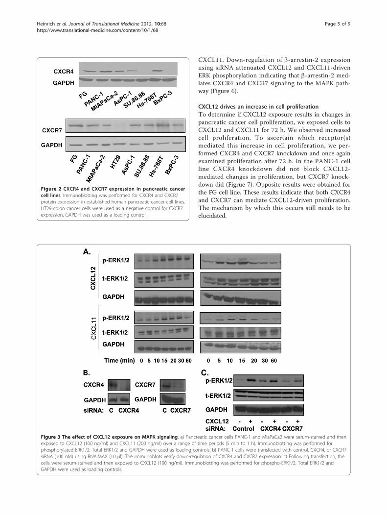
CXCL11. Down-regulation of b-arrestin-2 expressionusing siRNA attenuated CXCL12 and CXCL11-drivenERK phosphorylation indicating that b-arrestin-2 med-iates CXCR4 and CXCR7 signaling to the MAPK path-way (Figure 6).
CXCL12 drives an increase in cell proliferationTo determine if CXCL12 exposure results in changes inpancreatic cancer cell proliferation, we exposed cells toCXCL12 and CXCL11 for 72 h. We observed increasedcell proliferation. To ascertain which receptor(s)mediated this increase in cell proliferation, we per-formed CXCR4 and CXCR7 knockdown and once againexamined proliferation after 72 h. In the PANC-1 cellline CXCR4 knockdown did not block CXCL12-mediated changes in proliferation, but CXCR7 knock-down did (Figrue 7). Opposite results were obtained forthe FG cell line. These results indicate that both CXCR4and CXCR7 can mediate CXCL12-driven proliferation.The mechanism by which this occurs still needs to beelucidated.
Figure 2 CXCR4 and CXCR7 expression in pancreatic cancercell lines. Immunoblotting was performed for CXCR4 and CXCR7protein expression in established human pancreatic cancer cell lines.HT29 colon cancer cells were used as a negative control for CXCR7expression. GAPDH was used as a loading control.
Figure 3 The effect of CXCL12 exposure on MAPK signaling. a) Pancreatic cancer cells PANC-1 and MiaPaCa2 were serum-starved and thenexposed to CXCL12 (100 ng/ml) and CXCL11 (200 ng/ml) over a range of time periods (5 min to 1 h). Immunoblotting was performed forphosphorylated ERK1/2. Total ERK1/2 and GAPDH were used as loading controls. b) PANC-1 cells were transfected with control, CXCR4, or CXCR7siRNA (100 nM) using RNAiMAX (10 μl). The immunoblots verify down-regulation of CXCR4 and CXCR7 expression. c) Following transfection, thecells were serum-starved and then exposed to CXCL12 (100 ng/ml). Immunoblotting was performed for phospho-ERK1/2. Total ERK1/2 andGAPDH were used as loading controls.
Heinrich et al. Journal of Translational Medicine 2012, 10:68http://www.translational-medicine.com/content/10/1/68
Page 5 of 9

DiscussionChemokines regulate the chemotactic responses of cellsthat are essential for organogenesis and immunitythrough the orchestration of cell movement from onelocation to another [30-32]. Cancer cells have misappro-priated these regulatory mechanisms to stimulate theirown growth, invasion, and metastasis. Numerous studiesnow implicate chemokines and their correspondingreceptors in the invasive phenotype of many cancers [1].In particular, the CXCL12-CXCR4 axis has been wellstudied in gastrointestinal malignancies, but recentreports suggest that downstream effects once attributedto CXCR4 may also be secondary to CXCR7, an alter-nate or second receptor for CXCL12. As a result, weexamined CXCL12 activity in pancreatic cancer celllines. Among several novel discoveries in this investiga-tion, we identified high frequency of CXCR4/CXCR7
co-expression in human pancreatic cancer tissues andcell lines.Chemokine CXCL12 is characteristically expressed in
select tissues,[2] but may also be expressed via an autocrinefeedback loop mechanism in pancreatic cancer cells [7]. Assuch, the resources for CXCL12 to activate CXCR4 orCXCR7-mediated signaling pathways are present. Sinceour studies target pancreatic cancer, we examined CXCL12signaling within the framework of K-Ras. The gene encod-ing KRAS is frequently mutated in patients with pancreatic
Figure 4 K-Ras activity in response to CXCL12 exposure. a)Pancreatic cancer cells were serum-starved and then exposed toCXCL12 (100 ng/ml) and CXCL11 (200 ng/ml) for 15 min. Whole celllysates were assessed for K-Ras activity using a Raf pull-down assay.b) Cells were transfected with control, CXCR4, or CXCR7 siRNA (100nM). Following serum-starvation cells were treated with CXCL12(100 ng/ml) for 15 min. K-Ras activity was normalized to eachuntreated baseline level; and relative increases are depicted. Themean absorbance ± one SD was plotted for each treatment group.* designates p < 0.05.
Figure 5 The effects of KRAS knockdown on ERKphosphorylation following CXCL12 treatment. Pancreatic cancercells, FG and PANC-1 (left and right immunoblots respectively) weretransfected with control or KRAS siRNA (100 nM). Cells were serum-starved and then exposed to CXCL12 (100 ng/ml) or EGF (100 ng/ml) for 15 min. Immunoblotting was performed for K-Ras andphospho-ERK1/2. Total ERK1/2 and GAPDH were used as loadingcontrols.
Figure 6 b-arrestin-2’s role in CXCR4 and CXCR7-driven ERKphosphorylation. PANC-1 cells were transfected with control or b-arrestin-2 siRNA (100 nM) using RNAiMAX (10 μl). Cells were serum-starved and then exposed to CXCL12 (100 ng/ml) or CXCL11 (200ng/ml) for 15 min. The immunoblot shows b-arrestin-2 andphospho-ERK1/2 expression with total ERK1/2 and GAPDH servingas loading controls.
Heinrich et al. Journal of Translational Medicine 2012, 10:68http://www.translational-medicine.com/content/10/1/68
Page 6 of 9

cancer which results in a gain-of-function that may contri-bute to the pathogenesis and progression of this cancer[33,34]. Here, we made several novel observations regard-ing K-Ras. First, we discovered that pancreatic cancer cellsco-expressing CXCR4/CXCR7 had increased levels of ERKphosphorylation and K-Ras activity when exposed toCXCL12. These CXCL12-induced increases in K-Ras activ-ity were not observed with other ligands in endometrialand pancreatic cancer cells harboring mutant KRAS[35,36]. Our results, therefore, suggest that CXCL12 mayhyperactivate K-Ras activity levels even though there isbaseline mutant-derived K-Ras activity. Second, weobserved that CXCR4, rather than CXCR7, was the recep-tor that regulated this response. GPCRs can signal througha canonical or non-canonical pathway, activation of K-Rasmay occur via a CXCR4-mediated activation of the canoni-cal GPCR pathway [37-39]. CXCR7 on the other hand sig-nals through the non-canonical pathway [39], whichexplains why CXCR7 activation leads to ERK1/2 phosphor-ylation but not an increase in K-Ras activity. Β-arrestin-2 isan important member of either pathway, and we show thatits knockdown blocks ERK phosphorylation. Third, wedetermined that CXCL12-driven increases in cancer cellproliferation can occur through either receptor and henceeither signaling pathway.Specific inhibitors to CXCR4 and CXCR7 are cur-
rently unavailable for clinical use. AMD3100 wasbelieved to selectively bind and antagonize CXCR4activity [40,41]. Derivatives of AMD3100 are also underinvestigation for their effects on cancer cells [42]. Arecent study has demonstrated that AMD3100 specifi-cally binds to and activates CXCR7 [41]. Therefore, incontrast to its antagonism of the CXCL12-CXCR4 inter-action, AMD3100 positively modulates CXCL12 effectsand binding to CXCR7.
ConclusionIn summary, we report that CXCR4 and CXCR7 are co-expressed with high frequency in human pancreatic can-cer specimens and cell lines. CXCR4 and CXCR7 signal-ing is b-arrestin-2-dependent and controls CXCL12signals to the MAPK pathway. CXCL12 activates bothcanonical and non-canonical GPCR pathways in pan-creatic cancer cell lines. This has functional significancein that we show signaling through either pathway leadsto an increase in cell proliferation upon exposure toCXCL12. Hence our study suggests that efforts to thera-peutically target CXCL12 signaling in pancreatic cancershould be focused at the level of the ligand to accountfor both CXCR4 and CXCR7 activity.
AcknowledgementsWe appreciate the laboratory work of Xiaoming Shen, Ph.D. This workutilized City of Hope’s Pathology Core, which is supported in part by Cancer
Figure 7 The effect of CXCL12 on proliferation in pancreaticcancer cells. A quantitative ATP-based proliferation assay wasperformed on PANC-1 cells a) treated with CXCL12 and CXCL11 for72 h.. b) PANC-1 and c) FG cells were transfected with control,CXCR4, or CXCR7 siRNA (100 nM) prior to 72 h of exposure withCXCL12 (200 ng/ml). Proliferation levels presented were normalizedto each untreated baseline level, relative increases are depicted. Themean absorbance ± one SD was plotted for each treatment group.* designates p < 0.05.
Heinrich et al. Journal of Translational Medicine 2012, 10:68http://www.translational-medicine.com/content/10/1/68
Page 7 of 9

Center Support Grant NCI P30 CA33572. This study was also supported byNIH CA134637 (JK) and American Cancer Society RSG-11-070-01-TBE (JK). Thecontents are solely the responsibility of the authors and do not representthe official views of the NIH or ACS.
Author details1Department of Surgery, City of Hope Comprehensive Cancer Center, 1500East Duarte Road, Duarte, CA 91010, USA. 2Department of Surgery, MooresCancer Center, University of California San Diego, La Jolla, CA 92093, USA.
Authors’ contributionsJK: made substantial contributions to conception and design of thesestudies as well as analysis and interpretation of data, drafting the manuscriptand revising it critically; WL, JL: acquisition of data and drafting themanuscript; AL: analysis and interpretation of data and drafting themanuscript; ELH: analysis and interpretation of data and revision of the draft.All authors read and approved the final manuscript.
Competing interestsThe authors declare that they have no competing interests.
Received: 7 December 2011 Accepted: 2 April 2012Published: 2 April 2012
References1. Balkwill F: Cancer and the chemokine network. Nat Rev Cancer 2004,
4:540-550.2. Ratajczak M, Zuba-Surma E, Kucia M, Reca R, Wojakowski W, Ratajczak J: The
pleiotropic effects of the SDF-1-CXCR4 axis in organogenesis,regeneration and tumorigenesis. Leukemia 2006, 20:1915-1924.
3. Corcione A, Ottonello L, Tortolina G, Facchetti P, Airoldi I, Guglielmino R,Dadati P, Truini M, Sozzani S, Dallegri F, Pistola V: Stromal cell-derivedfactor-1 as a chemoattractant for follicular center lymphoma B cells. JNatl Cancer Inst 2000, 92:628-635.
4. Koshiba T, Hosotani R, Miyamoto Y, Ida J, Tsuji S, Nakajima S, Kawaguchi M,Kobayashi H, Doi R, Hori T, et al: Expression of stromal cell-derived factor1 and CXCR4 ligand receptor system in pancreatic cancer: a possiblerole for tumor progression. Clin Cancer Res 2000, 6:3530-3535.
5. Scotton C, Wilson J, Scott K, Stamp G, Wilbanks G, Fricker S, Bridger G,Balkwill F: Multiple actions of the chemokine CXCL12 on epithelial tumorcells in human ovarian cancer. Cancer Res 2002, 62:5930-5938.
6. Saur D, Seidler B, Schneider G, Algül H, Beck R, Senekowitsch-Schmidtke R,Schwaiger M, Schmid R: CXCR4 expression increases liver and lungmetastasis in a mouse model of pancreatic cancer. Gastroenterology 2005,129:1237-1250.
7. Marchesi F, Monti P, Leone B, Zerbi A, Vecchi A, Piemonti L, Mantovani A,Allavena P: Increased survival, proliferation, and migration in metastatichuman pancreatic tumor cells expressing functional CXCR4. Cancer Res2004, 64:8420-8427.
8. Mori T, Doi R, Koizumi M, Toyoda E, Ito D, Kami K, Masui T, Fujimoto K,Tamamura H, Hiramatsu K, et al: CXCR4 antagonist inhibits stromal cell-derived factor 1-induced migration and invasion of human pancreaticcancer. Mol Cancer Ther 2004, 3:29-37.
9. Feng Y, Broder C, Kennedy P, Berger E: HIV-1 entry cofactor: functionalcDNA cloning of a seven-transmembrane, G protein-coupled receptor.Science 1996, 272:872-877.
10. Endres M, Clapham P, Marsh M, Ahuja M, Turner J, McKnight A, Thomas J,Stoebenau-Haggarty B, Choe S, Vance P, et al: CD4-independent infectionby HIV-2 is mediated by fusin/CXCR4. Cell 1996, 87:745-756.
11. Bleul C, Farzan M, Choe H, Parolin C, Clark-Lewis I, Sodroski J, Springer T:The lymphocyte chemoattractant SDF-1 is a ligand for LESTR/fusin andblocks HIV-1 entry. Nature 1996, 382:829-833.
12. Oberlin E, Amara A, Bachelerie F, Bessia C, Virelizier J, Arenzana-Seisdedos F,Schwartz O, Heard J, Clark-Lewis I, Legler D, et al: The CXC chemokineSDF-1 is the ligand for LESTR/fusin and prevents infection by T-cell-line-adapted HIV-1. Nature 1996, 385:833-835.
13. Rubin J, Kung A, Klein R, Chan J, Sun Y, Schmidt K, Kieran M, Luster A, S RA:A small-molecule antagonist of CXCR4 inhibits intracranial growth ofprimary brain tumors. Proc Natl Acad Sci USA 2003, 100:13513-13518.
14. Zeelenberg I, Ruuls-Van Stalle L, Roos E: The chemokine receptor CXCR4 isrequired for outgrowth of colon carcinoma micrometastases. Cancer Res2003, 63:3833-3839.
15. Müller A, Homey B, Soto H, Ge N, Catron D, Buchanan M, McClanahan T,Murphy E, Yuan W, Wagner S, et al: Involvement of chemokine receptorsin breast cancer metastasis. Nature 2001, 410:50-56.
16. Balabanian K, Lagane B, Infantino S, Chow K, Harriague J, Moepps B,Arenzana-Seisdedos F, Thelen M, Bachelerie F: The chemokine SDF-1/CXCL12 binds to and signals through the orphan receptor RDC1 in Tlymphocytes. J Biol Chem 2005, 280:35760-35766.
17. Burns J, Summers B, Wang Y, Melikian A, Berahovich R, Miao Z, Penfold M,Sunshine M, Littman D, Kuo C, et al: A novel chemokine receptor for SDF-1 and I-TAC involved in cell survival, cell adhesion, and tumordevelopment. J Exp Med 2006, 203:2201-2213.
18. Miao Z, Luker K, Summers B, Berahovich R, Bhojani M, Rehemtulla A,Kleer C, Essner J, Nasevicius A, Luker G, et al: CXCR7 (RDC1) promotesbreast and lung tumor growth in vivo and is expressed on tumor-associated vasculature. Proc Natl Acad Sci USA 2007, 104:15735-15740.
19. Sierro F, Biben C, Martinez-Munoz L, Mellado M, Ransohoff R, Li M, Woehl B,Leung H, Groom J, Batten M, et al: Disrupted cardiac development butnormal hematopoiesis in mice deficient in the second CXCL12/SDF-1receptor, CXCR7. Proc Natl Acad Sci USA 2007, 104:14759-14764.
20. Wang Y, Li G, Stanco A, Long J, Crawford D, Potter G, Pleasure S, Behrens T:JL R: CXCR4 and CXCR7 have distinct functions in regulating interneuronmigration. Neuron 2011, 69:61-76.
21. Infantino S, Moepps B, Thelen M: Expression and regulation of the orphanreceptor RDC1 and its putative ligand in human dendritic and B cells. JImmunol 2006, 176:2197-2207.
22. Hattermann K, Held-Feindt J, Lucius R, Müerköster S, Penfold M, Schall T,Mentlein R: The chemokine receptor CXCR7 is highly expressed inhuman glioma cells and mediates antiapoptotic effects. Cancer Res 2010,70:3299-3308.
23. Shen X, Jackson D, Artinyan A, Thomas R, Lowy A, Kim J: Chemokinereceptor CXCR4 enhances proliferation in pancreatic cancer cellsthrough AKT and ERK dependent pathways. Pancreas 2010, 39:81-87.
24. Thomas R, Kim J, Revelo-Penafiel M, Angel R, Lowy A: The chemokinereceptor CXCR4 is expressed in pancreatic intraepithelial neoplasia. Gut2008, 57:1555-1560.
25. Rajagopal S, Rajagopal K, Lefkowitz R: Teaching old receptors new tricks:biasing seven-transmembrane receptors. Nat Rev Drug Discov 2010,9:373-386.
26. Pohl G, Ho C, Kurman R, Bristow R, Wang T, Shih I: Inactivation of themitogen-activated protein kinase pathway as a potential target-basedtherapy in ovarian serous tumors with KRAS or BRAF mutations. CancerRes 2005, 65:1994-2000.
27. Shen X, Mailey B, Ellenhorn J, Chu P, Lowy A, Kim J: CC chemokinereceptor 9 enhances proliferation in pancreatic intraepithelial neoplasiaand pancreatic cancer cells. J Gastrointest Surg 2009, 13:1955-1962.
28. Xu H, Wu Q, Dang S, Jin M, Xu J, Cheng Y, Pan M, Wu Y, Zhang C, Zhang Y:Alteration of CXCR7 expression mediated by TLR4 promotes tumor cellproliferation and migration in human colorectal carcinoma. PLoS One2011, 6:e27399.
29. Weinstein I, Joe A: Oncogene addiction. Cancer Res 2008, 68:3077-3080.30. Rossi D, Zlotnik A: The biology of chemokines and their receptors. Annu
Rev Immunol 2000, 18:217-242.31. Zlotnik A, Yoshie O: Chemokines: a new classification system and their
role in immunity. Immunity 2000, 12:121-127.32. Balkwill F: Chemokine biology in cancer. Semin Immunol 2003, 15:49-55.33. Deramaudt T, Rustgi A: Mutant KRAS in the initiation of pancreatic
cancer. Biochim Biophys Acta 2005, 1756:97-101.34. Hruban R, Wilentz R, Kern S: Genetic progression in the pancreatic ducts.
Am J Pathol 2000, 156:1821-1825.35. Kato K, Ueoka Y, Kato K, Tamura T, Nishida J, Wake N: Oncogenic Ras
modulates epidermal growth factor responsiveness in endometrialcarcinomas. Eur J Cancer 1998, 34:737-744.
36. Watanabe M, Nobuta A, Tanaka J, Asaka M: An effect of K-ras genemutation on epidermal growth factor receptor signal transduction inPANC-1 pancreatic carcinoma cells. Int J Cancer 1996, 67:264-268.
37. DeFea KA: Beta-arrestins as regulators of signal termination andtransduction: how do they determine what to scaffold? Cell Signal 2011,23:621-629.
Heinrich et al. Journal of Translational Medicine 2012, 10:68http://www.translational-medicine.com/content/10/1/68
Page 8 of 9

38. Rajagopal S, Rajagopal K, Lefkowitz RJ: Teaching old receptors new tricks:biasing seven-transmembrane receptors. Nat Rev Drug Discov 2010,9:373-386.
39. Rajagopal S, Kim J, Ahn S, Craig S, Lam CM, Gerard NP, Gerard C,Lefkowitz RJ: Beta-arrestin- but not G protein-mediated signaling by the“decoy” receptor CXCR7. Proc Natl Acad Sci USA 2010, 107:628-632.
40. Hatse S, Princen K, Bridger G, De Clercq E, Schols D: Chemokine receptorinhibition by AMD3100 is strictly confined to CXCR4. FEBS Lett 2002,527:255-262.
41. Kalatskaya I, Berchiche Y, Gravel S, Limberg B, Rosenbaum J, Heveker N:AMD3100 is a CXCR7 ligand with allosteric agonist properties. MolPharmacol 2009, 75:1240-1247.
42. De Clercq E: Potential clinical applications of the CXCR4 antagonistbicyclam AMD3100. Mini Rev Med Chem 2005, 5:805-824.
doi:10.1186/1479-5876-10-68Cite this article as: Heinrich et al.: Chemokine CXCL12 activates dualCXCR4 and CXCR7-mediated signaling pathways in pancreatic cancercells. Journal of Translational Medicine 2012 10:68.
Submit your next manuscript to BioMed Centraland take full advantage of:
• Convenient online submission
• Thorough peer review
• No space constraints or color figure charges
• Immediate publication on acceptance
• Inclusion in PubMed, CAS, Scopus and Google Scholar
• Research which is freely available for redistribution
Submit your manuscript at www.biomedcentral.com/submit
Heinrich et al. Journal of Translational Medicine 2012, 10:68http://www.translational-medicine.com/content/10/1/68
Page 9 of 9