cheek and inferior eyelid reconstruction after skin cancer...
TRANSCRIPT

Clin Plastic Surg 31 (2004) 49–67
Cheek and inferior eyelid reconstruction after skin
cancer ablation
William L. Murillo, MDa,b,*, William Fernandez, MDa,Diego J. Caycedo, MDa, Charles L. Dupin, MDb, Eileen S. Black, MDb
aDepartment of Surgery, Division of Plastic and Reconstructive Surgery, Universidad del Valle, Cali, ColombiabDepartment of Surgery, Division of Plastic and Reconstructive Surgery, Louisiana State University Medical Center,
1542 Tulane Avenue, New Orleans, LA 70112, USA
The cheek is the largest aesthetic unit of the face, susceptible to actinic damage and neoplastic changes.
with an underlying bony frame and an overlying soft
central part. Its outlines are different in each person,
due to changes with age, skeletal shape, and thickness
of adipose and muscular tissue. The perception of
the cheek varies depending on the angle from which
it is viewed. Menick [1] noted that it is impossible to
compare its symmetry to the contralateral cheek unit
from a single view.
The characteristics of the cheek, such as laxity
and low rigidity, allow for the use of a wide variety
of local and regional flaps for reconstruction. Its
close relation to the inferior eyelid constitutes a chal-
lenge, because alterations resulting from cheek recon-
struction may impact the function and symmetry of
the eyelid.
Etiology and epidemiology
A change from normal to neoplastic tissue is
caused by a variety of etiologic factors such as physical
agents, activation of cancer-promoting genes, viruses,
inhibition of cancer-suppression genes, and chemicals
[2]. Skin cancer has been linked to arsenic and ultra-
violet radiation exposure. Skin types I and II are most
0094-1298/04/$ – see front matter D 2004 Elsevier Inc. All right
doi:10.1016/S0094-1298(03)00122-6
* Corresponding author. Department of Surgery, Divi-
sion of Plastic and Reconstructive Surgery, Louisiana State
University Medical Center, 1542 Tulane Avenue, New
Orleans, LA 70112.
E-mail address: [email protected]
(W.L. Murillo).
White people living in countries close to the equator
or in subtropical zones are most susceptible to devel-
oping squamous cell and basal cell carcinomas [3].
Other factors associated with skin cancer are those
structural changes at the cutaneous sebaceous layer
where excessive free fatty acids and obstruction of the
pilosebaceous unit allow for chronic inflammatory
folliculitis. Alterations in the immune system, espe-
cially those in IgA, macrophages, white cells, and
Langerhans’ cells, play an important role in carcino-
genesis and may have a genetic component [4].
According to the Colombian National Cancer In-
stitute report for the year 2001 [5], skin cancer is the
third most common neoplastic lesion after cancer of
the cervix and breast. Nonmelanomas represent 8.7%
of skin cancers; among these, basal cell carcinomas
account for 53.4%, whereas squamous cell carcino-
mas represent 31.4% [5].
In tropical climates such as in Colombia, intense
sunlight can be felt during most of the day throughout
the year. Louisiana is a subtropical region, but it has a
very high percentage of sunny days. Like Colombia,
Louisiana has a large population that participates in
outdoor employment or recreation. In Colombia, the
proximity of areas of deep forests such as the Ama-
zon, the Pacific, the Darien, and the Orinoquia are
thought to be a protective factor against damage to
the ozone layer and therefore against skin cancer.
Other protective aspects are the wide range of ethnic
groups among which there is a high incidence of
people with dark skin, which is essentially an adaptive
resistance to ultraviolet rays. Diets rich in beta-caro-
s reserved.

W.L. Murillo et al / Clin Plastic Surg 31 (2004) 49–6750
tene [6], tomatoes [7], fruits, vegetables [8], and fish
[9] also are believed to be protective factors against
skin cancer.
Despite the previous considerations, people older
than 50 years of age with a history of extensive
occupational sunlight exposure compose a high per-
centage of actinic lesions and skin cancer in our
patients. Most of these patients are agricultural, fish-
eries, open field, or offshore workers who have an
average of 10 hours of daily solar exposure. Smok-
ing also has been implicated in about 5% of the skin
cancer cases and usually is associated with perioral
squamous cell carcinoma.
Fig. 2. Defects are closed along the minimal skin tension
lines (MSTL) or borders.
Anatomic considerationsThe peripheral boundaries of the cheek are formed
by hard and soft tissues. Superiorly, the lower eyelid
and the superior margin of the malar and zygomatic
arch form the limit. Medially, the border is formed
by the nasolabial and mesolabial fold and is supported
by the maxillary bone. Inferiorly, the mandibular
contour line and jowls form the frame. The lateral
edge is the preauricular crease and the posterior rim
of the mandible.
The anatomic layers of the cheek as described by
Gonzalez-Ulloa [10] include the skin, a homogeneous
layer of fascio-adipose tissue, and the superficial
musculo-aponeurotic system [11]. These layers are
interconnected by a system of ligaments. Between the
periosteum and dermis are four layers of mimetic
muscles: one superficial, two intermediate, and one
deep layer [12]. The masseter muscle, the buccal fat
pad, the facial nerve, the parotid gland, and Warthin’s
duct lie deep to the mimetic muscles. The osseous
Fig. 1. Anatomical subunits of the cheek.
structures that anchor the ligaments are the antero-
inferior border of the zygomatic arch, the inferior
orbital rim, and the inferior border of the mandible.
Branches of the external carotid artery perfuse the
cheek. The venous system drains to the jugular veins.
The terminal branches of the facial nerve supply the
motor innervation. The branches of the trigeminal
nerve provide the sensory innervation.
The elasticity of the cheek results from its soft
structure supported by facial attachments such as the
great zygomatic ligament, the anterior platysmal-
cutaneous ligament, the platysma, the auricular liga-
ments, and the mandibular ligaments [13]. The skin of
the medial and buccal subunit of the cheek is thick and
mobile. The skin of the zygomatic subunit is attached
firmly to the deep layer of soft tissue. The lateral
subunit is related closely to the fascia of the parotid
gland (Fig. 1).
Fig. 3. Advancement flap for medial lesion.

W.L. Murillo et al / Clin Plastic Surg 31 (2004) 49–67 51
The following layers compose the inferior eyelid:
skin, areolar tissue, muscle, tarsal plate, septum orbi-
talis, fat, and conjunctiva. The skin is thin and elastic.
The orbicular muscle—which is divided into pretar-
sal, preseptal, and orbital portions—is part of the
transition zone between the eyelid and the cheek.
For the purpose of planning cheek reconstruction,
Larrabee [14] divided the cheek into four subunits:
the medial, zygomatic, buccal, and lateral cheek sub-
units (see Fig. 1).
Fig. 4. Case 1. (A–E) Advancement fla
Principles of treatment
Many patients diagnosed with nonmelanoma skin
cancer have noticed the presence of the lesion within
the year before presentation. Patients frequently delay
seeking medical help and often treat the lesion with
antibiotic ointment or home remedies. Additionally, as
in Louisiana, social or economic issues may prevent
patients from seeking treatment. Advanced lesions
demand special attention and require meticulous his-
p closure of medial cheek defect.

Fig. 5. Flap based anterior for medial cheek defect.
W.L. Murillo et al / Clin Plastic Surg 31 (2004) 49–6752
topathologic mapping to ensure clear margins. If
complex reconstruction is required, closure should
be delayed until margins are confirmed. Once a com-
plete resection of the lesion in both radial extension
and depth is confirmed, a plan for an anatomic re-
construction is formulated. The outcome of each alter-
native is considered and the best option is determined
for each case.
Fig. 6. Case 2. (A–C) Paramedian foreh
Successful treatment depends on adequate plan-
ning and a multidisciplinary approach. Whenever pos-
sible, the reconstructive surgeon should participate
in the whole surgical process, including the ablation
of the tumor and the repair of the defect. The abla-
tion and reconstruction should be considered together
to gain every advantage, because each case presents
different challenges. When formulating a plan in
cheek and related inferior eyelid surgery, options for
reconstruction should be formulated before ablation.
Dividing the cheek into anatomic subunits is useful
to determine the type of closure to be used. Crossing
over the anatomic subunits of the cheek does not
constitute a limitation for an aesthetic repair, as it does
for the more central units of the face such as the
nose, lips, and eyelids. The plan should take into con-
sideration the size, depth, and location of the defect
as well as the timing and possible complications of
the surgery.
Medial area
In the medial area of the cheek the subcutaneous
fat allows for easy mobility of the attached skin, and
ead flap for medial canthus defect.

Fig. 7. Posterior-based flap for medial defect.
W.L. Murillo et al / Clin Plastic Surg 31 (2004) 49–67 53
defects that constitute up to 25% of the subunit may be
closed directly at the cheek–nose junction and meso-
labial fold (Fig. 2). Because the medial cheek subunit
becomes increasingly narrow as it abuts the nose and
there are inalterable boundaries (lower eyelid, nose,
and nasolabial fold), local flaps such as rhomboid,
bipedicle, or transposition flaps are difficult to
use. Alternatively, advancement flaps may be used
Fig. 8. Case 3. (A–F) Cervicofa
(Fig. 3). Deep permanent or long-lasting absorbable
sutures from the dermis of the flap should be fixed to
the periosteum of the nasal bone or pyriform aperture
to avoid distortion of the cheek–nose junction.
Defects that involve between 25% and 50% of the
medial cheek are closed with advancement flaps
elevated in the plane deep to the subdermal plexus.
Deep sutures must be placed to the periosteum
of the pyriform aperture to prevent distortion of the
nasofacial groove or the overlying lower lid unit. We
have used anchor fixation devices to ensure flap
support [Case 1 (Fig. 4A–E)]. Defects that involve
50% to 75% of the subunit are closed with cheek
rotation-advancement flaps (Fig. 5). If the defect
involves the medial canthus, a paramedian forehead
flap is useful [Case 2 (Fig. 6A–C)].
Defects that involve the entire medial subunit,
border the inferior eyelid, and involve the medial or
lateral canthus are best treated with cervicofacial flaps
(Fig. 7) [Case 3 (Fig. 8A–F)]. These large flaps sup-
ply enough tissue and allow for a reasonably tension-
free closure of the donor site. Jackson and Webster
[15] suggest de-epethelializing and concealing the
resulting dog-ears, which will increase the projec-
tion of the flap. Anchoring the flap to the periosteum
cial flap for medial defect.
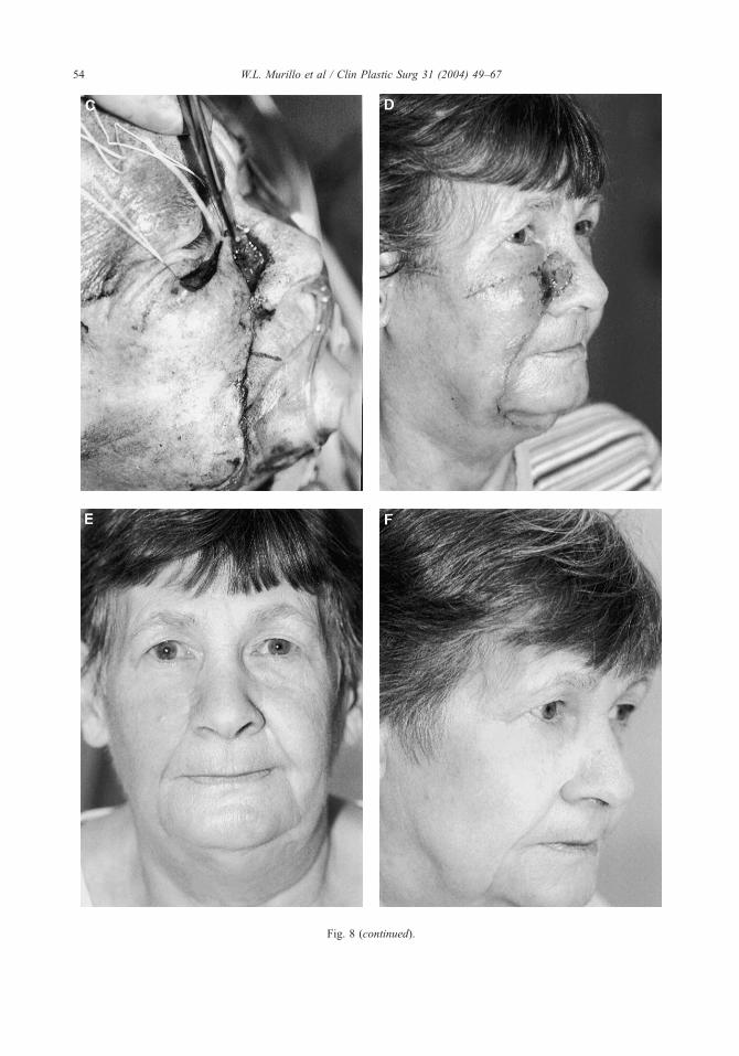
Fig. 8 (continued).
W.L. Murillo et al / Clin Plastic Surg 31 (2004) 49–6754

Fig. 9. V-Y and rhomboid flaps for cheek repair.
W.L. Murillo et al / Clin Plastic Surg 31 (2004) 49–67 55
of the zygomatic arch or malar bone and the use of
drains is recommended.
Lateral area
Repairing the lateral and adjacent areas of the
cheek follows principles similar to those described
Fig. 10. Case 4. (A, B) Rhombo
above for the medial area. Defects located in the
preauricular region that involve up to 50% of the
lateral subunit may be repaired with direct closure
(see Fig. 2). If a smaller defect is located inferiorly or
medially, especially if it encroaches on the buccal or
zygomatic area, options include a rhomboid flap or a
V-Y advancement flap (Fig. 9) [Cases 4 (Fig. 10A, B)
and 5 (Fig. 11A, B)]. When using a rhomboid flap,
the flap should be planned so that the closure of the
donor site is along the axis of maximum tissue laxity.
It also is important to choose the design so that the
donor closure does not leave a scar anterior to the
defect, encroaching on the central face. This scar will
be visible from the frontal view and should be
avoided. V-Y advancement flaps should be closed
along subunit borders or along minimal skin tension
lines [16–19].
Defects involving greater than 50% of the lateral
subunit are treated with extended direct closure, retro-
auricular transposition flaps [Case 6 (Fig. 12A–C)],
or cervical rotation flaps (Fig. 13). Total lateral de-
fects that also involve the buccal or zygomatic sub-
units typically require a cervicofacial flap [Case 7
(Fig. 14A–C)]. Whenever possible, the final incisions
id flap for lateral defect.

Fig. 11. Case 5. (A, B) V-Y advancement flap for lateral defect.
W.L. Murillo et al / Clin Plastic Surg 31 (2004) 49–6756
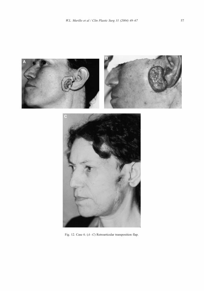
Fig. 12. Case 6. (A–C) Retroarticular transposition flap.
W.L. Murillo et al / Clin Plastic Surg 31 (2004) 49–67 57

Fig. 13. Larger lesions require more inferior flap development.
Fig. 14. Case 7. (A–C) Cervicofacial rotation flap for extensive defect.
W.L. Murillo et al / Clin Plastic Surg 31 (2004) 49–6758

Fig. 14 (continued).
W.L. Murillo et al / Clin Plastic Surg 31 (2004) 49–67 59
should be placed along the unit borders or should
follow Langer’s lines of minimal skin tension.
Zygomatic area
Defects that are smaller than 30% of the zygomatic
subunit can be repaired by direct closure (see Fig. 2).
Transposition flaps can be used when the tissue deficit
is between 30% and 70% of the subunit. Defects that
involve more than 70% of the zygomatic subunit may
require cheek rotation advancement flaps (Fig. 15)
[Case 8 (Fig. 16A–D)]. To prevent inferior flap dis-
placement and ectropion, anchoring sutures should be
placed lateral to the orbit and the flap should be
designed with a high lateral arch above the canthal
plane. Special attention must be given to the temporal
branch of the facial nerve, which runs at the subdermal
layer in this area.
Fig. 15. Cheek rotation flap for zygomatic defect.
Buccal area
Depending on the skin laxity, defects of up to 30%
are repaired by direct closure or a transposition flap.
Defects that involve 30% to 70% of the buccal sub-
unit will require transposition or cheek advancement
flaps [Cases 9 (Fig. 17A–C) and 10 (Fig. 18A–D)].
Defects of more than 70% of the subunit will require a
cervicofacial flap for repair [20,21]. This area is
unique because the central area does not have any
underlying bony support. Deeply invasive lesions in
this area may require oral lining as well as soft tissue
bulk. When this occurs, free flaps are a good recon-
structive option [Case 11 (Fig. 19A–E)].
Inferior eyelid
Eyelid reconstruction depends on the size and
position of the defect as well as the quality of sur-
rounding tissue. The main purpose of eyelid restora-
tion is to supply anatomic and functional protection of
the ocular globe with good support, mucosal lining,
and an appropriate skin cover. Whenever possible, it is
important to safeguard the structures providing lacry-
mal drainage. Reconstruction of the cheek must not
distort the anatomy of the lower eyelid.
Partial defects of any of the lamellas may be
closed directly or by using local flaps or grafts.
Full-thickness defects are more complex. Full-thick-
ness defects that are smaller than 30% of the eye-
lid may be repaired by direct closure [Case 12
(Fig. 20A–D)]. Defects of the anterior lamella that
involve greater than 30% of the eyelid may be re-
paired with regional flaps such as Tripier’s or Fricke’s
flaps. Resection of the total lid should be repaired
with a facial rotation flap such as the Mustarde flap
[Case 13 (Fig. 21A–F)] [22]. Despite its random
blood supply, this flap is reliable and provides good

Fig. 16. Case 8. (A–D) Cheek rotation flap for defect resulting from ablation of malignant tumor involving facial nerve.
W.L. Murillo et al / Clin Plastic Surg 31 (2004) 49–6760

Fig. 17. Case 9. (A–C) Transposition flap for buccal subunit defect.
W.L. Murillo et al / Clin Plastic Surg 31 (2004) 49–67 61

Fig. 18. Case 10. (A–D) Cheek advancement flap for buccal subunit.
W.L. Murillo et al / Clin Plastic Surg 31 (2004) 49–6762

Fig. 19. Case 11. (A–E) Free scapular flap, folded and de-epithialized to form lining and cheek resurfacing.
W.L. Murillo et al / Clin Plastic Surg 31 (2004) 49–67 63

Fig. 19 (continued).
W.L. Murillo et al / Clin Plastic Surg 31 (2004) 49–6764

Fig. 20. Case 12. (A–D) Twenty-five percent full-thickness lid resection closed.
W.L. Murillo et al / Clin Plastic Surg 31 (2004) 49–67 65

Fig. 21. Case 13. (A–F) Subtotal lower lid reconstruction with nasal septal chondro-mucosal graft and Mustarde flap.
W.L. Murillo et al / Clin Plastic Surg 31 (2004) 49–6766

W.L. Murillo et al / Clin Plastic Surg 31 (2004) 49–67 67
color match and easy donor site closure, especially in
the elderly.
Small defects of the posterior lamella can be closed
directly. Larger defects must be repaired using con-
junctival, oral, or nasal mucosal grafts supported with
either septal or conchal cartilage with the muco-
pericondrium. A tarsal-conjunctival flap of the supe-
rior eyelid also may be used to repair the posterior
lamella. At least 4 mm of the superior eyelid tar-
sal plate must be preserved to maintain its integrity
and function.
Summary
Most patients with actinic lesions and skin cancer
are skin type I or II, older than 50 years of age, and
have a history of extensive sunlight exposure. These
patients have been treated in our units according to
universal principles. A multidisciplinary team ap-
proach can produce encouraging long-term results.
The size and depth of the lesion are assessed in
planning the ablation. The residual defect after the
tumor resection is anticipated in the preoperative plan.
Adequate resection is mandatory, even if the recon-
struction must be delayed to ensure clear margins.
Attention to unit and subunit anatomy facilitates
adequate reconstruction with acceptable deformity.
Placing scars in borders or along the lines of minimal
skin tension reduces deformity. Planning the flap so
that the donor site is in tissue areas with maximum
laxity guards against donor site deformity. Flaps must
be planned to avoid excess tension on the lower lid
and central face. Attempts should be made to reduce
scarring in the central face as seen in the frontal view.
Respecting these principles will allow for reconstruc-
tion of the largest facial unit in a manner acceptable
to the patient.
References
[1] Menick FJ. Plast Reconstr Surg 2001;108(2):496–504.
[2] Schneider AS, Szanto PA. Neoplasia. In: Pathology/
Board Review Series. Harval Publishing; 1993.
p. 83–95.
[3] Wong VA, Marshal JA, Whitehead KJ, et al. Manage-
ment of periocular basal cell carcinoma with modified
en face frozen section controlled excision. Ophthal
Plast Reconstr Surg 18(6):430–3.
[4] Knudson Jr AG. Hereditary cancer, oncogenes and
antioncogenes. Cancer Res 1985;45:1437–43.
[5] Colombian National Cancer Institute Report. Available
at: www.incancerologia.gov.co. Accessed.
[6] Mathews-Roth MM, Pathack MA, Fitzpatrick TB. Be-
ta-carotene as a protective agent in erythropoietic
photoporphyria. N Engl J Med 1970;282:1231–4.
[7] Levy J, Bosin E, Feldman B, et al. Lycopene is a more
potent inhibitor of human cancer cell proliferation than
either alpha-carotene or beta-carotene. Nutr Cancer
1995;24:257–66.
[8] Michaud DS, Spiegelman D, Clinton SK, et al. Fruits
and vegetables intake and incidence of bladder cancer
in a male prospective cohort. J Natl Cancer Inst 1999;
91:605–13.
[9] Rose DP, Connolly JM. Omega-3 fatty acids as cancer
chemo preventive agents. Pharmacol Ther 1999;83:
217–44.
[10] Gonzalez-Ulloa M. Facial wrinkles, integral elimina-
tion. Plast Reconstr Surg 1962;29:658.
[11] Mitz V, Peyronie M. The superficial musculo-apo-
neirotic system (SMAS) in the parotid and cheek area.
Plast Reconstr Surg 1976;58:80.
[12] Freilinger G, Gruber H, Happak W, et al. Surgical
anatomy of the mimetic muscle system and the facial
nerve. Importance for reconstructive and aesthetic sur-
gery. Plast Reconstr Surg 1987;80:686.
[13] Furnas DW. The retaining ligaments of the cheek. Plast
Reconstr Surg 1989;83:11–6.
[14] Larrabee Jr W. Principles of facial reconstruction. Lip-
pincott-Raven; 1995. p. 120–49.
[15] Jackson IT, Webster HR. Craneofacial tumors. Clin
Plast Surg 1994;21(4):633–48.
[16] Grabb WC, Smith JW, editors. Plastic surgery, a con-
cise guide to clinical practice. 2nd edition. Boston:
Little Brown; 1973.
[17] Lamberty BGH, Healy C. Flaps: physiology, principles
of design and pitfalls. In: Cohen M, editor. Mastery
of plastic and reconstructive surgery. Boston: Little
Brown & Co.; 1995.
[18] Jackson IT. Local flaps in head and neck reconstruc-
tion. St. Louis: Mosby; 1985.
[19] Quaba AA, Sommerlad BC. ‘‘A square peg into a
round hole’’: a modified rhomboid flap and its clinical
application. Br J Plast Surg 1987;40:163.
[20] Feldman J. Facial burns. In: McCarthy, editor. Plastic
surgery, vol. 3. WB Saunders; 1990.
[21] Juri J, Juri C. Advancement and rotation of a large
cervicofacial flap for cheek repairs. Plast Reconstr
Surg 1979;64:692.
[22] Mustarde JC. Repair and reconstruction in the orbital
region. Edinburgh: Churchill Livingstone; 1980.