characterizing the function of the n-terminal domain of
TRANSCRIPT

University of Central Florida University of Central Florida
STARS STARS
Honors Undergraduate Theses UCF Theses and Dissertations
2017
Characterizing the Function of the N-Terminal Domain of Omi/Characterizing the Function of the N-Terminal Domain of Omi/
HtrA2 HtrA2
Christine Nguyen University of Central Florida
Part of the Molecular Biology Commons
Find similar works at: https://stars.library.ucf.edu/honorstheses
University of Central Florida Libraries http://library.ucf.edu
This Open Access is brought to you for free and open access by the UCF Theses and Dissertations at STARS. It has
been accepted for inclusion in Honors Undergraduate Theses by an authorized administrator of STARS. For more
information, please contact [email protected].
Recommended Citation Recommended Citation Nguyen, Christine, "Characterizing the Function of the N-Terminal Domain of Omi/HtrA2" (2017). Honors Undergraduate Theses. 242. https://stars.library.ucf.edu/honorstheses/242

CHARACTERIZING THE FUNCTION OF THE N-TERMINAL DOMAIN OF
OMI/HTRA2
by
CHRISTINE NGUYEN
A thesis submitted in partial fulfillment of the requirements
for the Honors in the Major Program in Biomedical Sciences
in the College of Medicine
and in the Burnett Honors College
at the University of Central Florida
Orlando, Florida
Fall Term, 2017
Thesis Chair: Dr. Antonis S. Zervos

ii
ABSTRACT
The yeast two-hybrid system was used to isolate and characterize protein interactors of the
N-terminal domain of the serine protease Omi/HtrA2 (high temperature requirement protein
A2) encompassing amino acids 31-133. This large domain of Omi/HtrA2 is usually cleaved and
removed through autoproteolysis to produce the mature form of the protein. Whether the N-
terminal domain has any function after its removal is unknown. Omi/HtrA2 is involved in a variety
of diseases including cancers, neurodegenerative disorders, and metabolic disorders, but thus far,
it is assumed that its normal function is the degradation of specific substrates. To characterize
any potential function of Omi/HtrA2’s unique amino terminus, specific interactors were isolated.
One such interactor was the small GTPase Rab2A protein. We discuss the implications of this
interaction and its biological significance.

iii
ACKNOWLEDGMENTS
I am indebted to my thesis committee members, who have been integral to the success of my
project. First and foremost, I am grateful for the mentorship provided by my thesis chair, Dr.
Antonis Zervos, who has allowed me to work in his lab for the past two years and encouraged
me to undertake this project. The opportunity to engage in research under his guidance has
allowed me to gain a deeper understanding of molecular biology and develop my critical
thinking skills as a scientist. I would also like to thank my other committee members including
Dr. Suren Tatulian and Dr. Robert Borgon, whose constructive criticism has allowed me to
improve my thesis. I am also extremely thankful for Dr. Lucia Cilenti; her expertise, patient
explanations, and advice have been indispensable throughout my project. Finally, I want to thank
my fellow lab members, who have provided constant encouragement and support.
iv
TABLE OF CONTENTS
LIST OF FIGURES ........................................................................................................................ v
LIST OF TABLES ......................................................................................................................... vi
CHAPTER 1: INTRODUCTION AND REVIEW OF LITERATURE ......................................... 1
Structure ...................................................................................................................................... 1
Function ...................................................................................................................................... 7
Implications for Disease ........................................................................................................... 10
CHAPTER 2: MATERIALS AND METHODS .......................................................................... 14
Yeast Two-Hybrid Overview .................................................................................................... 14
Polymerase Chain Reaction ...................................................................................................... 17
DNA Precipitation .................................................................................................................... 19
Restriction Enzyme Digestion .................................................................................................. 19
DNA Gel Electrophoresis ......................................................................................................... 19
DNA Ligation ........................................................................................................................... 20
Bacterial Transformation .......................................................................................................... 20
Isolation of Plasmid DNA from Bacteria (Boiling Method) .................................................... 21
Isolation of Plasmid DNA from Bacteria (Clean Miniprep - QIAPrep) ................................... 21
Yeast Transformation................................................................................................................ 22
Yeast SDS-PAGE and Western Blot ........................................................................................ 23
High Efficiency Yeast Transformation ..................................................................................... 23
Screening of Library ................................................................................................................. 24
Release of Plasmid from Yeast ................................................................................................. 24
KC8 Transformation ................................................................................................................. 25
Retransformation....................................................................................................................... 25
CHAPTER 3: RESULTS .............................................................................................................. 26
Previous data on the N-terminal Domain of Omi ..................................................................... 26
Expression and Stability of the Bait.......................................................................................... 30
Screen of HeLa cDNA library .................................................................................................. 32
CHAPTER 4: DISCUSSION ........................................................................................................ 36
REFERENCES ............................................................................................................................. 39
v
LIST OF FIGURES
Figure 1. Structure of Omi .............................................................................................................. 4 Figure 2. Alignment of the full length amino acid sequences of Omi/HtrA2 in Homo sapiens and
Mus musculus .......................................................................................................................... 5
Figure 3. Alignment of the N-terminal amino acid sequence of Omi/HtrA2 across numerous
species ..................................................................................................................................... 6 Figure 4. Programmed cell death via mitochondria ........................................................................ 8 Figure 5. Phenotype of Mnd2 mice............................................................................................... 13 Figure 6. The Yeast Two-Hybrid System ..................................................................................... 16
Figure 7. Omi constructs ............................................................................................................... 28 Figure 8. Flow cytometry to monitor apoptosis with different Omi constructs............................ 29
Figure 9. Expression and stability of LexA-Omi31-133 in yeast ................................................. 31
Figure 10. cDNA clone representing the Rab2A polypeptide is a specific interactor of LexA-
Omi31-133. ............................................................................................................................... 34 Figure 11. Full length Rab2A interacts specifically with LexA-Omi31-133 in yeast ...................... 35

vi
LIST OF TABLES
Table 1. Primer Sequences for Omi31-133. ..................................................................................... 18 Table 2. Primer Sequences for Full Length Rab2A ...................................................................... 33
1
CHAPTER 1: INTRODUCTION AND REVIEW OF LITERATURE
Structure
Omi/HtrA2 (high temperature requirement protein A2) is an ATP-independent serine
protease primarily localized in the mitochondrial intermembrane space, although it has also been
identified in both the endoplasmic reticulum and the nucleus [1-4]. Within mitochondria, Omi
plays key roles in maintenance of mitochondrial homeostasis and thus has been described as
performing a pro-survival function [1, 5, 6]. However, in response to stress, Omi translocates to
the cytosol, where it induces cellular apoptosis [2]. Omi was originally identified through a yeast
two-hybrid screen for protein interactors of Mxi2, an alternatively spliced form of the p38 stress
kinase [2]. It was later independently discovered through a yeast two-hybrid screen for protein
interactors of presenilin-1, a protein linked to Alzheimer’s disease [1].
Encoded by a gene found on human chromosome 2p12, Omi is a 458 amino acid protein
of molecular weight 49 kDa that is expressed ubiquitously [2, 3]. Omi belongs to a family of
proteins known as high temperature requirement A proteins (HtrA) and shows homology to
bacterial HtrAs, a class of proteins with dual chaperone and peptidase function that varies
according to the temperature [1]. Among mammals, the sequence for Omi has been found to be
highly conserved and constitutes one of four HtrA proteases that have been identified, along with
HtrA1, HtrA3, and HtrA4. Omi is unique within the mammalian HtrAs because it is mitochondrial
while its paralogs are secreted [1].
The sequence of Omi consists of a mitochondrial targeting signal, transmembrane domain,
a proteolytic trypsin like domain (including the catalytic triad of amino acids essential for its serine
protease function), and a PDZ domain (Figure 1) [16]. X-ray crystallography has revealed that

2
Omi consists of seven -helices and five -sheets [16]. In its active form, Omi exhibits a
homotrimeric structure with a pyramid shape: the IAP domain is located at the top of the pyramid
while the PDZ domain forms the base of the pyramid. This PDZ domain restricts access to the
active site and therefore regulates the activity Omi’s serine protease domain [16]. Trimerization is
crucial for Omi to function as mutation of a highly conserved phenylalanine 149 residue necessary
for trimerization has been shown to abolish Omi’s serine protease function [16, 29]. In its inactive
form in the IMS of the mitochondria, Omi’s transmembrane domain has been postulated to anchor
the protein to the inner mitochondrial membrane. Omi is also found in its soluble active form
within the IMS [18].
Alignment between the full length amino acid sequence of mouse and human Omi revealed
that the mature form of the protein (amino acids 134-458) shows a high degree of similarity with
about 95% of residues identical. However, the N-terminal domain has lower levels of similarity
with only 61% of residues conserved (Figure 2). Within the N-terminal domain, the sequence for
the transmembrane domain and mitochondrial targeting signal are similar, but the majority of the
differences are located in the linker region between the targeting signal and transmembrane
domain. Human Omi possesses a thrice repeated PRAXXTXXTP motif within the linker region
that is completely absent from mice. Alignment performed between the first 133 amino acids of
Omi across various species including humans, mice, rats, cows, chimpanzees, and guinea pigs
suggested that this PRAXXTXXTP motif is only found in primates (Figure 3). The function of
this PRAXXTXXTP motif is unknown, but when Omi is expressed in Escherichia coli cells, the
PRAXXTXXTP motif was shown to cause human Omi to be much more susceptible to proteolytic
degradation compared to mouse Omi [43].

3
Even though mitochondria contain their own genome, the proteins encoded by these genes
are limited and mainly include subunits of the respiratory chain as well as ATP synthase.
Therefore, the vast majority of mitochondrial proteins are nuclear encoded and after being
synthesized on cytosolic ribosomes, must be targeted to mitochondria. This post-translational
import usually occurs through an N-terminal signal sequence [32]. While the specific sequence of
these targeting signals is not highly conserved among mitochondrial proteins, certain
characteristics have been shown to be conserved including length (20-60 AA), charge (net
positive), and the presence of stabilizing N-terminal residues in the mature protein [42].
Comparative analysis of the mitochondrial N-termini of various proteins from mice, humans, and
yeast has revealed that Omi is unique in that its N-terminal pre-sequence is abnormally long at 133
amino acids [33]. This deviation serves as further impetus for investigating the potential fate of
this N-terminal domain after its cleavage to form mature Omi.

4
Figure 1. Structure of Omi
Omi is a 458 amino acid serine protease that consists of a large mitochondrial targeting signal, transmembrane domain,
proteolytic trypsin-like domain, and a PDZ domain (including a conserved AVPS motif crucial for interactions with
IAPs as well as the catalytic triad of amino acids). In its active form, Omi forms a homotrimeric pyramid structure
with its IAP-binding domain (IBD) at the top and its PDZ domain at the base. The PDZ domain controls substrate
access to the binding site [39].

5
Figure 2. Alignment of the full length amino acid sequences of Omi/HtrA2 in Homo sapiens and Mus
musculus
This alignment was performed using Clustal Omega with the numbers referring to the position of the amino acid
residues. The functional domains present in both species include: a mitochondrial targeting signal (aa 1-40) indicated
in blue font, a transmembrane domain (aa 105-125) boxed in purple, the PDZ domain (aa 364-445) in orange font,
and the catalytic triad (histidine 198, aspartate 228, and serine 306) in blue underlined font that is boxed in green. The
PRAAXXTXXP motif is indicated in red. An asterisk (*) indicates identical residues, a colon (:) indicates residues
with highly similar properties and a period (.) indicates residues with weakly similar properties.

6
Figure 3. Alignment of the N-terminal amino acid sequence of Omi/HtrA2 across numerous species
This alignment was performed using Clustal Omega and compares the first 133 amino acids of HtrA2 in Homo sapiens
(human), Mus musculus (mouse), Rattus norvegicus (rat), Bos taurus (cow), Cavia procellus (guinea pig), and Pan
troglodytes (chimpanzee). The numbers refer to the position of the amino acid residues. The functional domains
present include: a mitochondrial targeting signal (aa 1-40) indicated in blue font and the transmembrane domain (aa
105-125) boxed in purple. The PRAAXXTXXP motif is indicated in red. An asterisk (*) indicates identical residues,
a colon (:) indicates residues with highly similar properties and a period (.) indicates residues with weakly similar
properties.
7
Function
Omi plays dual roles in maintaining mitochondria homeostasis under normal conditions
and promoting cell death under conditions of stress. Omi triggers cell death through two different
pathways: caspase dependent cell death through interactions with inhibitors of apoptosis proteins
(IAP) or caspase independent cell death through usage of its serine protease domain [17]. To
induce caspase dependent cell death, Omi functions in a similar manner to proteins such as
Smac/DIABLO, Grim, Reaper, and HID. These proteins are released from mitochondria to the
cytosol following stress, and once in the cytosol, are able to bind to the BIR (baculovirus IAP
repeat) domain of IAPs to deter their function and trigger activation of caspases that bring about
cell death [19, 20]. In response to an apoptotic signal, Omi is processed into its active form through
cleavage of its first 133 amino acids to produce the processed protein, which reveals the AVPS
motif on its N-terminal domain essential for recognition and binding of the BIR or IAPs [19]. The
importance of this motif has been verified in studies that observed that Omi’s ability to induce cell
death is drastically reduced by mutations within its AVPS motif or its serine protease domain, and
its apoptotic function is altogether halted by mutations to both the AVPS motif and proteolytic
domain [19]. Omi has also been observed to cause apoptosis through interactions with proteins
outside of IAPs, such as ped/pea15 and HAX-1 [21, 22]. The serine protease activity of Omi, and
hence its apoptotic function, has been shown to be effectively inhibited by UCF-101 [23].

8
Figure 4. Programmed cell death via mitochondria
The release of pro-apoptotic proteins from the mitochondria into the cytosol induces cell death. One pathway through
which this occurs involves the release of Smac/DIABLO and Omi/HtrA2, two mitochondrial intermembrane space
proteins that antagonize IAPs. This leads to activation of caspase-3 and apoptosis. Omi/HtrA2 has also been postulated
to induce caspase-independent cell death through its serine protease activity [40].
9
As mentioned previously, Omi also plays a key pro-survival function within the
mitochondria. Increased mitophagy is observed for cells lacking functional Omi, due to Omi’s role
in regulating levels of the mitochondrial E3 ubiquitin ligase Mulan. Accumulation of the Mulan
causes a concomitant decrease in mitofusion 2 (a protein which promotes mitochondrial fusion),
leading to the observed mitophagy [19]. Furthermore, Omi has been suggested to play a key role
in mitochondrial protein quality control and maintenance of mitochondrial homeostasis. Omi aids
in the elimination of excessive or misfolded proteins within the mitochondrial intermembrane
space [24]. The absence of Omi results in elevated levels of the mitochondrial proteins Lon
protease 1 and prohibitin, leading to decreased mitochondrial membrane potential, the generation
of reactive oxygen species, and a decrease in ATP production [28].
Omi’s protective function is also clear through the Parkinsonian phenotype exhibited by
mnd2 mice that possess nonfunctional Omi through the homozygous mutation Ser276Cys. This
mutation in Omi was postulated to limit substrate access to the active site of Omi, as deletion of
the PDZ restores partial function [7]. Introduction of a human Omi transgene into the neuronal
tissues of mnd2 mice prevented development of the Parkinsonian phenotype, but the mice instead
exhibited hallmark signs of premature aging including weight loss, hair loss, curved spine, and
enlarged hearts [6]. Furthermore, overexpression of Omi in the neuronal cells of transgenic mice
revealed a normal phenotype rather than increased signs of apoptosis, leading to the conclusion
that Omi plays a neuroprotective role [25].

10
Implications for Disease
Non-functional Omi has been implicated in a variety of neurodegenerative disorders
including Parkinson’s disease (PD), Alzheimer’s disease (AD), and Huntington’s disease (HD).
Mnd2 (motor neuron degeneration 2) mice possessing a homozygous mutation for Ser276Cys
within Omi’s proteolytic domain showed hallmark symptoms generally observed in PD including
loss of neurons in the striatum of the basal ganglia, reduced body weight, reduced organ size, and
muscle wasting before an early death (Figure 5) [7]. These mice exhibited increased vulnerability
to stressed-induced permeabilization of the mitochondrial membrane that leads to cellular
apoptosis [17]. Phosphorylation of Omi has also been suggested to be important to the functioning
of its protease domain as mutations of the mitochondrial enzyme PTEN-induced putative kinase-
1 (PINK1), which has been implicated in early onset AD, resulted in lower levels of
phosphorylation of Omi near a site of mutation in human patients with PD [8].
This link between mutations affecting Omi’s proteolytic activity and PD has been
corroborated by screenings performed on German PD patients, which revealed high proportions
of patients with the G399S (located in the PDZ domain) or A141S (located in the N-terminus of
mature Omi) missense mutation compared to the general population [9]. Another study performed
with Belgian PD patients showed that a R404W mutation (located in the PDZ domain) as well as
variations in 5’ and 3’ regulatory regions that affect Omi expression increase susceptibility to PD
[27]. However, the veracity of these findings is yet to be confirmed, as another group examining
a cohort of North American patients found equal proportions of these mutations within a control
group compared to a group afflicted with young and late onset PD [10].

11
Omi has also been suggested to be involved in various types of cancers. Gastric mucosal
cells were found to have reduced expression of Omi compared to non-cancerous stomach cells,
leading to the conclusion that perhaps a drop in Omi’s apoptotic activity resulted in cancer
development [13]. Researchers have also identified potential links between Omi expression and
resistance to treatment with the alkylating agent cisplatin in ovarian cancer since cisplatin’s
mechanism of action involves increasing levels of Omi to promote apoptosis in cancerous cells
and cisplatin resistant cancer cells had markedly lower levels of cytosolic Omi [14]. Conversely,
Omi was found to be highly expressed in prostate cancer cells compared to both normal patients
and those with benign prostate hyperplasia, leading researchers to conclude Omi contributes to
prostate cancer development [15]. Because of the variability of gene expression observed among
the various types of cancers, it is difficult to draw a definitive conclusion on the role of Omi in the
development and progression of cancer. Nonetheless, Omi’s vital role in balancing cell death and
homeostasis suggests a high likelihood that aberrant expression of Omi could disturb this balance
and trigger cancer.
Whole exome sequencing (WES) has led to identification of recessive mutations in Omi
that cause 3-methylglutaconic aciduria (3-MGA-uria), a marker of metabolic dysfunction in which
excessive amounts of 3-methylglutaconic acid are excreted. A study of four human patients lacking
Omi due to a splicing mutation and a five base pair deletion observed increased susceptibility to
apoptosis, abnormal cristae structure, and neurodegeneration in addition to 3-MGA-uria [30].
Similarly, another study using WES on five patients that presented with 3-MGA-uria noted
recessive variants of Omi that caused seizures, hypotonia, neutropenia, and cardio-respiratory
problems [31]. Both studies noted no abnormalities in mitochondrial network morphology or

12
deviations in the mitochondrial respiratory chain, although abnormal levels OPA1, a protein
important in balancing fusion and fission in mitochondria, were observed [30, 31].
Thus, Omi has been implicated in a variety of diseases including cancers,
neurodegenerative disorders, and metabolic disorders. Additional insight into Omi’s mechanism
of function through identification of protein interactors of its N-terminal domain could provide
information about the pathways that result in disease phenotypes as well as possible methods to
prevent or mitigate symptoms.

13
Figure 5. Phenotype of Mnd2 mice
Mnd2 mice with a homozygous mutation within Omi’s catalytic domain exhibit neurological symptoms at 20 days
postnatal that include involuntary movement and posture, akinesis, and an early death within 30-40 days postnatal
[41].

14
CHAPTER 2: MATERIALS AND METHODS
Yeast Two-Hybrid Overview
The yeast two-hybrid screen was performed using the EGY48 yeast strain containing the
pSH 18-34 reporter construct, which includes the LacZ gene for β-galactosidase under the control
of LexA operators. These yeast also contain an endogenous leucine gene under the control of LexA
operators. The protein of interest (Omi31-133), termed the “bait,” was fused to the LexA DNA
binding protein through the use of the pGilda vector. The pGilda vector contained a HIS3
selectable marker as well as a GAL1 promoter, which is repressed by glucose and induced by
galactose. The potential interactors from the HeLa cDNA library, termed the “prey,” were fused
to the B42 transcriptional activator through the use of the pJG4-5 vector, which contains a GAL1
promoter as well as a TRP1 selectable marker. Interaction between the bait and prey resulted in an
active transcription factor that allowed for expression of two reporter genes. A positive interaction
was indicated by yeast that was able to survive in media lacking leucine as well as produced a
characteristic blue color in the presence of X-Gal.
Before performing the screen, a color and growth test were used to confirm that the two
reporter genes could not be activated without interaction between the bait and prey. For the growth
test, yeast that had been transformed with the pGilda vector containing Omi31-133 was grown
overnight in U-H- glucose media and then transformed with the empty pJG4-5 vector. Varying
dilutions were grown on U-H-W-L- Galactose/Raffinose plates. The inability of the yeast to grow
on medium lacking leucine confirmed that without interaction of the bait and prey, the gene for
leucine was not activated. For the color test, yeast was streaked on U-H-W- glucose plates and
incubated for a few days. Colonies from this transformation were plated on U-H-W-

15
Galactose/Raffinose/X-Gal plates. The absence of blue colonies indicated that the bait protein did
not self-activate to produce β-galactosidase.

16
Figure 6. The Yeast Two-Hybrid System
Interaction between the LexA-Omi31-133 bait (fused to the binding domain) and the unknown prey protein from the
HeLa cDNA library (fused to the activating domain) results in formation of an active transcription factor that binds to
the LexA promoter. This results in the expression of the endogenous LEU2 gene as well as the LacZ gene to form β-
galactosidase and leucine. On a plate with X-Gal, positive interactors form blue colonies. DNA sequencing allows for
identification of the specific interactor.

17
Polymerase Chain Reaction
The target DNA sequence corresponding to amino acids 31-133 of the N-terminal domain
of Omi was amplified through PCR. The primers used in the PCR reaction and included restriction
sites for EcoRI and BamHI (Table 1). For the PCR reaction, the following mixture was used: 20
ng of template DNA, 10 nM in 1 µL of each of the primers, 5 µL of 10X PCR Buffer containing
Mg2+, 1.5 units in 1 µL of Taq polymerase, 10 µM of dNTPs, and sterile water to bring the solution
to a final volume of 50 µL. The PCR program used was: denaturation for 2 minutes at 96oC
followed by 25 cycles of denaturation for 30 seconds at 96oC, annealing for 30 seconds at 62oC,
and elongation for 30 seconds at 72oC. The samples were then subjected to a final elongation for
7 minutes at 72oC and then kept at 4oC.

18
Table 1. Primer Sequences for Omi31-133.
Primer Sequence (5 3) Restriction Site
Forward GGCGAATTCTTGACCCCTGACCTCCGGG EcoRI
Reverse CGCGGATCCTTAGGCGAGGACGGCCGGAG BamHI
These forward and reverse primers were used to amplify the cDNA sequence for Omi31-133 with an EcoRI restriction
site included the forward primer and a BamHI restriction site included in the reverse primer.

19
DNA Precipitation
1/10 sample volume of 3M sodium acetate pH 5.5 and 2 sample volumes of 100% ethanol
kept at-20 oC were added to the DNA solution. This sample was mixed and then stored at -20 oC
for at least 20 minutes. The sample were then centrifuged at 13,000RPM at 4 oC for 20 minutes.
The supernatant was discarded. 250 µL of 70% ethanol was added to the pellet to wash away
residual salt. The sample was centrifuged again at 13,000RPM for 5 minutes at 4 oC. The
supernatant was discarded. The pellet was then dried for five minutes in a speed vacuum. The dried
pellet was re-suspended in 50 µL of sterile water.
Restriction Enzyme Digestion
Both the vector and DNA insert were digested with BamHI and EcoRI in preparation for
ligation. The restriction enzyme mixture included: 50 µL of DNA, 37 µL of sterile water, 10 µL
of 10X Restriction Enzyme Buffer, 1.5 µL BSA, and 1.5 µL of the restriction enzyme EcoRI at 10
units/µL. This mixture was incubated at 37oC. At the end of an hour, an additional 0.5 µL of EcoRI
was added. The mixture was left in the water bath for an additional hour for a total of two hours.
Following the two hours, the DNA was precipitated from the solution and the digestion was
repeated with the restriction enzyme BamHI. DNA gel electrophoresis was performed to determine
whether the digestion was successful and to estimate the DNA concentration in the vector and
insert solutions.
DNA Gel Electrophoresis
A 1.5% agarose gel was used to determine the size and concentration of the DNA present
in each sample. This gel was created by mixing 100 µL of 1X TAE (0.04 M Tris-acetate, 0.001 M
EDTA) with 1.5 g of agarose powder. This solution was microwaved for approximately 90-120
seconds until the agarose was observed to have completely dissolved. The solution was then cooled

20
under running cold water and 0.5 µg/µL ethidium bromide was added and mixed by swirling. The
solution was poured into a gel tray with well combs and left to solidify at room temperature. 2-5
µL of each sample of DNA was mixed with 5 µL of 10X Loading Buffer (30% glycerol, 2%
Orange-G in 1X TAE) and loaded into the wells of the gel. A GeneRuler 1 kb DNA ladder was
run along with the samples. The gel was covered with 1X TAE Buffer and run at 150V until the
dye had migrated an appropriate distance in the gel. The gel was visualized using ultraviolet light
in a Biorad Gel-Doc machine.
DNA Ligation
To determine the amount of vector and insert to use, gel electrophoresis was used to
visualize and estimate the concentration of DNA in the insert and vector solutions. The amount of
DNA used in the ligation should be roughly in a 3:1 insert to vector ratio. The reaction mixture
included 1.5 µL of Ligation Buffer, 1.5 µL of ATP, 1 µL DNA ligase, 1.5 µL of 10X buffer, and
varying amounts of insert and vector. A final volume of 15 µL was achieved by addition of sterile
water to bring up the volume of the reaction mixture. The samples were incubated at room
temperature for 20 minutes and at 72oC for 15 minutes in a heating block. The samples were stored
at 4oC.
Bacterial Transformation
Approximately 3-4 µL of the DNA ligation was added into a 100 µL aliquot of competent
DH5α bacterial cells and transferred to an electroporation cuvette cooled to 4 oC. The cuvette was
placed in the Bio-Rad Gene Pulser to deliver a single exponential decay pulse of 2.5 kV at 400Ω.
900 µl of room temperature LB medium was immediately added to the cuvette, which was gently
mixed and transferred to a microcentrifuge tube. This tube was incubated at 37 oC for one hour
21
while spinning. Various volumes of the cells were plated on LB agar plates containing ampicillin
and incubated overnight at 37 oC.
Isolation of Plasmid DNA from Bacteria (Boiling Method)
Individual colonies were isolated from the LB AMP plates and grown in 1.5 mL of LB
AMP media overnight while shaking at 37 oC. The following day, the suspensions were poured
into 1.5 mL microcentrifuge tubes and spun at 13,000 RPM for 2 minutes. The supernatant was
aspirated and the pellet was resuspended in 300 µL of a STET/Lysozyme solution (8% sucrose,
5% 100X Triton, 50 mM of Tris-HCl pH 8, 50 mM of EDTA, 10 mg/mL of lysozyme). These
samples were placed in a boiling water bath for 1 minute and centrifuged at 13,000RPM for 10
minutes. The pellet with cellular debris was removed with a toothpick and 200 µL isopropanol was
added to the supernatant. The samples were inverted and mixed before centrifuging at 13000 RPM
for 10 minutes. The supernatant was discarded and 300 µL of 70% ethanol was added to wash the
pellet. This solution was centrifuged for 5 minutes at 13,000 RPM. The supernatant was discarded
and the DNA pellet was dried in a speed vacuum for 10 minutes. The resulting dried pellet was
resuspended in 50 µL of TE.
Isolation of Plasmid DNA from Bacteria (Clean Miniprep - QIAPrep)
The QIAprep Spin Miniprep Kit was used to isolate plasmid DNA from bacteria that had
been incubated with shaking overnight at 37oC in 1.5 mL of LB AMP media. These suspensions
were transferred to microcentrifuge tubes and centrifuged at 13,000 RPM for 2 minutes at room
temperature. The supernatant was aspirated and the pellet was resuspended in 200 µL of
Resuspension Buffer P1 with RNase A (100 µg/mL). 200 µL of Lysis Buffer P2 was added, and
the tubes were gently inverted several times until a homogeneously colored suspension was
obtained. Next, 300 μL of Neutralization Buffer N3 was added and the tubes were once again

22
inverted several times until the solution appeared cloudy. The suspensions were centrifuged at
13,000 RPM for 10 minutes and the supernatant was transferred by pipette to a QIAprep spin
column with a 2 mL collection tube. These columns were centrifuged for 1 minute at 13,000 RPM
and the flow-through in the collection tube was discarded. The columns were then washed with
500 µL of PB Buffer and centrifuged for an additional 1 minute at 13,000 RPM to remove any
remaining wash buffer that might inhibit enzymatic reactions performed later on. The flow through
was discarded once again followed by another centrifugation step for 1 minute at 1,3000 RPM.
The spin columns were then transferred to a 1.5 mL clean microcentrifuge tube and 50 µL of EB
Buffer (10 mM Tris-HCl, pH 8.5) was added to the center of the column. This was allowed to sit
for a minute before centrifuging for 1 minute at 13,000 RPM to elute the DNA.
Yeast Transformation
A single colony was inoculated in 50 mL of medium and grown overnight shaking at 30°C.
The following day, the suspensions were centrifuged at 3800 RPM for 5 minutes and the
supernatant was discarded. The cells were then washed with 10 ml TE pH 7.5 (10mM Tris pH 5,
1 mM EDTA pH 8) before being centrifuged once again at 3800 RPM for 5 minutes. The cells
were resuspended in 5 mL LA (0.1 M LiAC in TE) and incubated in a shaker for 2 hours at 30°C.
The cells were centrifuged at 3800 RPM for 5 minutes and the supernatant was discarded. The
cells were once again resuspended in 5 mL of LA. 300 µL of this solution was aliquoted into
microcentrifuge tubes to which 1 µL of denatured salmon sperm DNA and 1 µg of DNA was
added. 700 µL of PEG (50% Peg 4000 in LA) was added and mixed thoroughly by micropipetting.
The solution was then incubated in a water bath at 30°C for 30 minutes and then 42°C for 15
minutes. These cells were centrifuged at 13200 RPM for 3 minutes and the supernatant was

23
discarded. The pellet was resuspended in 300 µL TE pH 7.5 and different volumes of the solution
was plated on U⁻H⁻ Glucose plates. These plates were incubated for 2-3 days at 30°C.
Yeast SDS-PAGE and Western Blot
Single colonies of yeast were grown shaking overnight at 30°C in 2 mL of U⁻H⁻W glucose
media. The following day, this suspension was centrifuged at 13200 RPM for 3 minutes at room
temperature and the resulting supernatant was discarded. These cells were then washed with 1 mL
of U⁻H⁻W⁻ galactose/raffinose media, and centrifuged at 13200 RPM for 3 minutes once again
with the supernatant discarded. This pellet was resuspended in 500 µL of U⁻H⁻W⁻
galactose/raffinose media. This solution was transferred to test tubes to which 500 µl U⁻H⁻
galactose/raffinose media had already been added. These cells were incubated while rotating for 4
hours at 30°C. At the end of the incubation period, the cells were transferred to microcentrifuge
tubes and boiled for 3 minutes. 0.1 g of 0.2 mm glass beads were added to the cells, which were
vortexed for 2 minutes. 70 µl of EBS (2% SDS, 80 mM Tris pH 6.8, 10% glycerol, 1.5% DTT,
and 0.1 mg/ml bromophenol blue) was added to the cells and the samples were boiled for 1 minute
and centrifuged for 1 minute at 13200 RPM. The supernatant was analyzed using SDS-PAGE with
a 12% resolving gel and 5% stacking gel, and a Western transfer was performed with a PVDF
membrane. α-LexA-HRP was used as the primary antibody for the bait fusion protein. This
allowed for confirmation that the fusion protein was expressed and stable.
High Efficiency Yeast Transformation
An individual colony of EGY48 yeast containing pSH18-34 as well as LexA-Omi31-133 in
pGilda was grown overnight at 30 °C while shaking in 50 ml of U-H- glucose medium. This
culture was diluted the following day to 300 mL with U-H- glucose medium and grown to an

24
OD600nm of 0.9. The cells were then harvested by centrifugation at 3800 RPM for 5 minutes, and
the resulting supernatant was discarded. The cells were washed with 20 mL of sterile water and
centrifuged for another 5 minutes at 3800 RPM with the supernatant discarded. The solutions
were resuspended in 20 mL LA (0.1M LiOAc in TE) and spun again for 5 minutes at 3800 RPM.
This pellet was resuspended in 5 mL of LA. 100 µl of competent yeast was aliquoted into 1.5 mL
microcentrifuge tubes.10 μg DNA (HeLa cDNA library in pJG4-5), 100 μg of denatured salmon
sperm DNA, and 600 μl of 40% PEG was added to the aliquots. The mixtures were resuspended
until homogeneous. The transformation reaction was then incubated at 30°C for 30 minutes and
then heat shocked at 42°C for 15 minutes. The samples were centrifuged and the supernatant was
aspirated. The pellet was resuspended in 500 μL of sterile water. 300 μL of this solution of yeast
was plated on 24 x 24 cm U⁻H⁻W⁻ galactose/raffinose plates and incubated at 30°C for 2-3 days.
Screening of Library
The yeast were grown on U⁻H⁻W⁻L⁻ galactose/raffinose plates for growth selection, and
positive colonies were plated onto a U⁻H⁻W⁻ glucose plate. This plate was incubated at 30°C for
2-3 days. These colonies were then streaked onto U⁻H⁻W⁻ glucose/X-Gal and U⁻H⁻W⁻
galactose/raffinose/X-Gal plates. Blue colonies on these plates indicated a positive interaction.
Release of Plasmid from Yeast
Positive colonies were selected to perform a release the pJG4-5 plasmid with the HeLa
cDNA library insert. These colonies were grown rotating at 30°C overnight in 1 mL of W⁻ glucose
media. The following day, 800 µL of yeast was transferred to in a microcentrifuge screw-lid tubes
and spun for 1 minute at 13200 RPM. The supernatant was aspirated and the pellet was
resuspended in 200 µL of yeast lysis solution (2% TritonX-100, 1% SDS, 100 mM NaCl, 10 mM

25
Tris pH 8.0, 1 mM EDTA). 0.3 g of 150 µm glass beads and 200 µL of phenol-cloroform-ioamyl
alcohol was added to the solution. The tubes were vortexed vigorously using a mini-beadbeater
for 2 minutes. They were then centrifuged for 5 minutes at 13,200 RPM until three distinct layers
were observed within the tube. The top layer contained the DNA of interest while the middle layer
contained protein and the bottom layer contained cellular debris. Hence, the top layer was kept and
a DNA precipitation procedure was performed.
KC8 Transformation
The plasmids were transformed into bacterial KC8 cells using electroporation. This
solution was plated onto WM9 AMP plates and incubated overnight at 37oC. A single colony was
selected from each plate and grown in LB AMP media overnight.
Retransformation
The boiling method was used to obtain the plasmid DNA from the KC8 cells. This was
transformed back into yeast cells containing the pSH18-34 plasmid and the pGilda vector carrying
the bait. These samples were plated onto U⁻H⁻W⁻ glucose plates. Colonies growing on these plates
were subsequently grown on U-H-W- Glucose/XGal plates and U-H-W- Galactose/Raffinose/X-Gal
plates for color selection. A colony that was blue on the U-H-W- Galactose/Raffinose/X-Gal plates
and white on the U-H-W- Glucose/XGal was deemed a true positive interactor.

26
CHAPTER 3: RESULTS
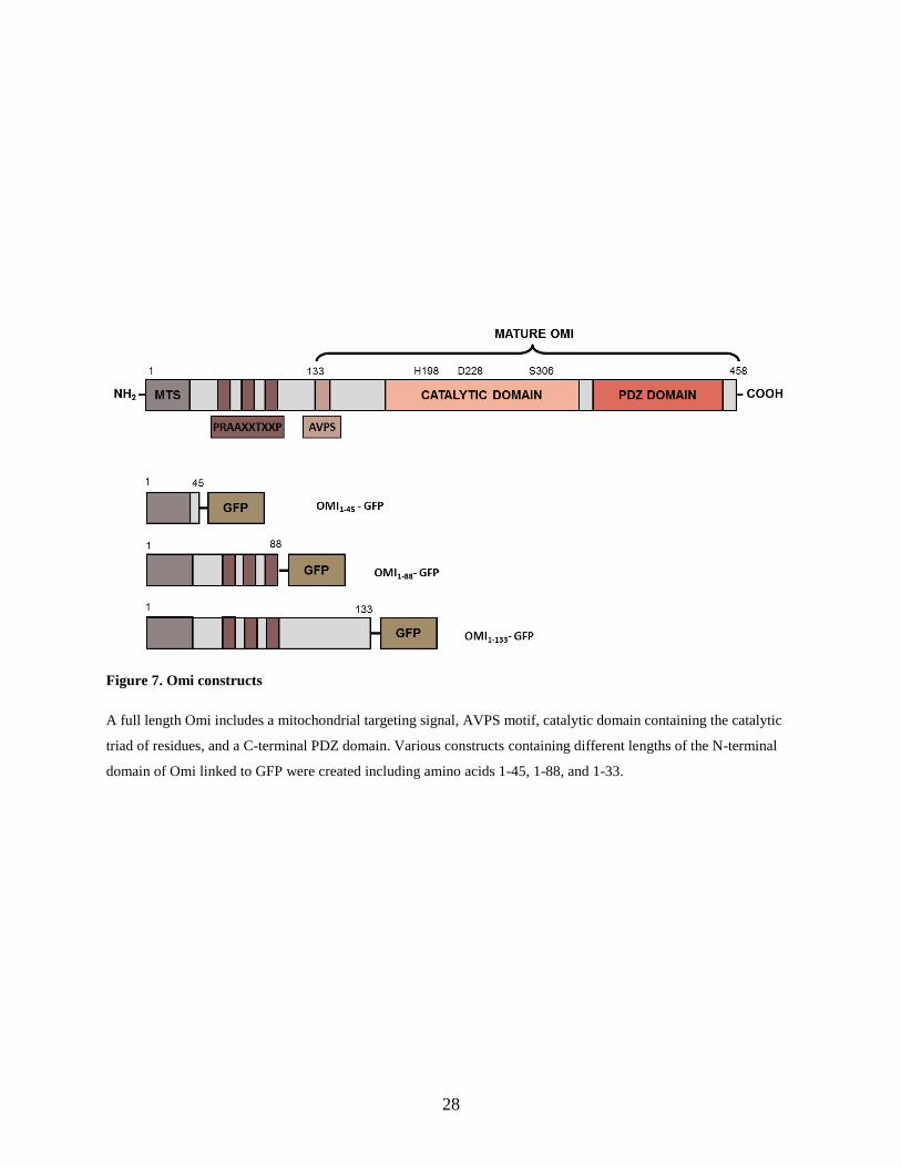
Previous data on the N-terminal Domain of Omi
Previous experiments performed in the Zervos lab indicated the potential function of the
N-terminal domain of Omi in promoting apoptosis under conditions of mitochondrial stress.
Various constructs were created including a full length Omi and different portions of the N-
terminal domain with amino acids 1-45 (includes the mitochondrial targeting signal and a few
additional amino acids), amino acids 1-88 (includes the mitochondrial targeting signal and all three
PRAAXXTXXP motifs), and amino acids 1-133 (includes the entire N-terminal domain that is
cleaved off to form the mature protein, stopping just short of the AVPS motif) (Figure 7). The
constructs were expressed as fusions to the green fluorescent protein (GFP).
These constructs were transfected into LLC-PK1 cells (pig proximal tubule epithelial cells)
and treated with antimycin (1 M) for 24 hours. Antimycin acts as an inhibitor of cellular
respiration by disrupting the electron transport chain and therefore prevents the production of ATP
through oxidative phosphorylation. In addition, it causes the creation of toxic free radicals harmful
to the cell. As a result, antimycin was used in this experiment to induce mitochondrial stress.
Apoptosis was monitored using flow cytometry with AnnexinV staining. AnnexinV binds
phosphatidylserine, and the presence of this molecule on the outer membrane of cells serves as an
indicator of apoptosis. Apoptosis data was acquired for a population of 10,000 GFP transfected
cells.
For all constructs, cells showed greater levels of apoptosis under conditions of stress
compared to untreated cells. Cells possessing full length Omi showed the greatest level of
apoptosis followed by the cells containing the full N-terminal domain. Cells containing Omi1-133

27
showed significantly higher levels of apoptosis than cells transfected with a truncated N-terminal
domain, suggesting that the N-terminal domain of Omi most likely plays a role in promoting
apoptosis, although the mechanism through which this occurs is unclear (Figure 8).
These preliminary results served as the impetus for this project. It suggested that the N-
terminal domain has a function within the cell, possibly related to apoptosis. Identifying interactors
of this N-terminal domain could potentially give clues as to the specific function of the N-terminal
domain of Omi.
28
Figure 7. Omi constructs
A full length Omi includes a mitochondrial targeting signal, AVPS motif, catalytic domain containing the catalytic
triad of residues, and a C-terminal PDZ domain. Various constructs containing different lengths of the N-terminal
domain of Omi linked to GFP were created including amino acids 1-45, 1-88, and 1-33.

29
Figure 8. Flow cytometry to monitor apoptosis with different Omi constructs
LLC-PK1 cells (pig proximal tubule epithelial cells) were transfected with the various Omi constructs and treated with
antimycin to induce mitochondrial stress (1 micromole for 24 hours). Flow cytometry with AnnexinV staining was
used to measure apoptosis in the population of transfected cells. 10,000 GFP positive cells were acquired.

30
Expression and Stability of the Bait
The DNA fragment corresponding to Omi’s N-terminal domain aa 31-133 was cloned into
the pGilda vector, and the expression and stability of the bait in yeast was tested. Yeast were
transformed with the LexA-Omi31-133 plasmid and plated on U-H- plates. These plates were allowed
to incubate for several days at 30oC. Single colonies were picked from these plates and grown
overnight in U-H- media. These yeast were induced the following day with U-H- galactose/raffinose
media and lysed. Uninduced (control) yeast were grown in U-H- glucose media. SDS PAGE and a
Western blot analysis with LexA specific antibodies was used to verify that the LexA-Omi31-133
bait was both expressed and stable within the yeast (Figure 9). Omi31-133 is roughly 10.5 kDa and
LexA is 24 kDa. Thus, it was expected that the LexA-Omi31-133 would be 34.5 kDa. The Western
blot shows no bands for the uninduced samples, since glucose represses the GAL1 promoter in
pGilda. Conversely, galactose/raffinose promotes expression with a GAL1 promoter, so yeast in
the induced condition expressed LexA or LexA-Omi31-133, showing bands at 24 kDa and 34.5 kDa,
respectively (Figure 9).

31
Figure 9. Expression and stability of LexA-Omi31-133 in yeast
The LexA-Omi31-133 protein is 34.5 kDa, and the LexA protein alone is 24 kDa. Yeast transfected with the empty
pGilda vector or the fusion bait LexA-Omi1-133 were grown in U-H- galactose/raffinose media (induced) or U-H-
glucose media (uninduced). Yeast extracts were separated by SDS-PAGE and analyzed by Western blotting using
LexA specific antibodies.

32
Screen of HeLa cDNA library
The H2Y screen was used to isolate interactors against the LexA-Omi31-133 bait as described
in the Methods section. Several yeast colonies appeared that could potentially contain a specific
interactor. The plasmid from these colonies was isolated and sequenced. DNA sequencing revealed
the cDNA represents a partial length sequence for Rab2A, encoding amino acids 7-212 (Figure
10). PCR and specific primers were used to amplify a full length cDNA clone for the human Rab2A
protein (Table 2). This was then cloned back to the pJG-4-5 vector and used to transform yeast.
The full length Rab2A protein was also able to interact with the LexA-Omi31-133 bait (Figure 11).

33
Table 2. Primer Sequences for Full Length Rab2A
Primer Sequence (5 3) Restriction Site
Forward GGCGAATTCATGGCGTACGCCTATCTCTTC EcoRI
Reverse CCGCTCGAGTCAACAGCAGCCGCCCCC XhoI
These forward and reverse primers were used to create a full length clone for Rab2A with an EcoRI restriction site
included the forward primer and an XhoI restriction site included in the reverse primer.

34
Figure 10. cDNA clone representing the Rab2A polypeptide is a specific interactor of LexA-Omi31-133.
DNA and corresponding amino acid sequence of the partial clone representing part of the Rab2A
protein that was isolated as a specific interactor in our yeast two-hybrid screen (blue). The DNA
and amino acid sequence for the full length protein also includes the portion in black.

35
Figure 11. Full length Rab2A interacts specifically with LexA-Omi31-133 in yeast
The cDNA library screen originally identified a partial length sequence of Rab2A as an interactor of Omi21-133. A full
length clone of Rab2A was produced, cloned back to the pJG-4-5 vector, and used to transform yeast. The
characteristic blue color produced in the presence of X-Gal indicating expression of the reporter system confirms that
full length Rab2A also interacts with Omi21-133.

36
CHAPTER 4: DISCUSSION
Omi/HtrA2 was first identified as a mammalian homolog to the bacterial heat shock protein
HtrA, and since its discovery, multiple studies have implicated this mitochondrial protease in the
quality control of mitochondrial proteins, mitochondrial dynamics, autophagy, as well as apoptosis
[2, 17, 24, 28, 38]. It has been assumed that all properties assigned to Omi are entirely mediated
by its ability to function as a serine protease and degrade specific substrates. Some of these
substrates have already been identified and represent a diverse group of both mitochondrial and
cytoplasmic proteins. A paradox in the biological function of Omi is the presence of a very large
N-terminal domain in the precursor Omi protein that is cleaved and remove to create the functional
active protease. The normal function of this large N-terminal domain is unknown. It is assumed to
be a mitochondrial targeting sequence, which does not help to explain its size or the various
structural characteristics that it contains. We hypothesized that the amino terminal of Omi after it
is removed from the precursor protein is stable and has a distinct function. In this work, we tried
to identify the potential function of Omi’s N-terminus by isolating and characterizing its specific
interactors. This method has been used successfully in the past to define the function of a protein
through its interactions. In addition, it provides information as to the potential stability of the N-
terminus when expressed in yeast.
We performed a yeast two-hybrid screen, using the N-terminal domain of Omi (amino
acids 31-133). Omi 1-30 amino acids represent the mitochondrial targeting sequence and were not
included, as the success of the yeast two-hybrid relies on an interaction that occurs within the
nucleus. The expression and stability of the LexA-Omi31-133 suggested the Omi31-133 polypeptide
can be expressed and is stable in yeast cells. The yeast two-hybrid screen of a HeLa cDNA library

37
revealed a novel interactor, the Rab2A protein. Rab2A was able to specifically and strongly
interact with the LexA-Omi31-133.
The Rab (Ras-related in the brain) GTPases comprise a family of proteins within the Ras
superfamily that are found within all eukaryotic organisms. Localized on the cytosolic side of
intracellular membranes, Rab GTPases are essential for coordinating vesicle traffic within the cell
through interactions with specific effector molecules such as tethering factors, kinases, and
phosphatases [34, 36]. Rab has been shown to influence numerous aspects of trafficking including
vesicle uncoating, budding, motility, fusion, and receptor signaling as well as play key roles in
cellular signaling [34].
Rab GTPase activation is controlled by the antagonistic effects of GDP/GTP exchange
factors (GEFs) and RAB GTPase activating proteins (GAPs). Rab alternates between two states:
active Rab is bound to GTP while inactive Rab is bound to GDP. GEFs catalyze the conversion of
GDP for GTP to form the active protein, and GAPs produce the opposite effect by promoting GTP
hydrolysis to GDP [35]. With over 60 members of Rab proteins in the human genome, many Rab
proteins have been shown to be the result of gene duplication events and have high sequence
identity to one another. Even between eukaryotes, Rab function is highly conserved; yeast lacking
a Rab enzyme can effectively use a mammalian homolog [34].
The Rab GTPase identified as an interactor of Omi, Rab2A, is suggested to be a key player
in the process of autophagy and endocytosis. Rab2A was discovered to be a binding partner of a
tethering complex known as HOPS (homotypic fusion and vacuole protein sorting), which is
responsible for degradation of autophagosomes and endosomes. Combined with previous
knowledge of Rab7’s interaction with HOPS, a model was proposed through which Rab2A works

38
in conjunction with Rab7 in order to promote fusion of autophagosomes and endosomes with
lysosomes. Rab2A knockdown or siRNA treatment resulted in accumulation of autophagic
vesicles within the cell [37]. Previous research has revealed that Omi activates autophagy (more
specifically mitophagy) by regulating the E3 ubiquitin ligase Mulan [38]. Thus, Rab2A’s role in
autophagy is of interest because it could describe another pathway through which Omi regulates
autophagy within the cell. The potential link between Omi and Rab2A in autophagy has clinical
significance since autophagy degrades pathogenic protein aggregates whose build up has been
implicated in neurodegenerative diseases [38].
Further experimentation needs to be completed to confirm that this interaction between
Rab2A and Omi occurs in mammalian cells under physiological conditions. If a specific antibody
against the N-terminus of Omi can be created, experiments can be done to determine the
subcellular localization of this domain (mitochondrial versus cytoplasmic). In addition, a specific
antibody could be used to monitor the expression and stability of the amino terminal domain and
its potential trafficking to the lysosomes during autophagy. Finally, whether the amino terminal
domain of Omi is involved in the pathological conditions where the protease has been implicated
needs further investigation. It is expected that the amino terminal domain of Omi is created through
auto-degradation. As such, mutations that affect the protease activity of Omi will block the release
of the amino terminal domain from the precursor protein. This could prove detrimental to cells by
affecting several biological processes including autophagy. Thus, elucidation of the role of the N-
terminal domain can result in better understanding of human disease and pathology.

39
REFERENCES
1. Gray, C. W., Ward, R. V., Karran, E., Turconi, S., Rowles, A., Viglienghi, D., ... &
Savopoulos, J. (2000). Characterization of human HtrA2, a novel serine protease involved in
the mammalian cellular stress response. European Journal of Biochemistry, 267(18), 5699-
5710. http://dx.doi.org/10.1046/j.1432-1327.2000.01589.x
2. Faccio, L., Fusco, C., Chen, A., Martinotti, S., Bonventre, J. V., & Zervos, A.S. (2000).
Characterization of a novel human serine protease that has extensive homology to bacterial
heat shock endoprotease HtrA and is regulated by kidney ischmia. Journal of Biological
Chemistry, 275(4), 2581-2588. http://dx.doi.org/10.1074/jbc.275.4.2581
3. Hegde, R., Srinivasula, S. M., Zhang, Z., Wassell, R., Mukattash, R., Cilenti, L., ... &
Alnemri, E. S. (2002). Identification of Omi/HtrA2 as a mitochondrial apoptotic serine
protease that disrupts inhibitor of apoptosis protein-caspase interaction. Journal of Biological
Chemistry, 277(1), 432-438. http://dx.doi.org/10.1074/jbc.M109721200
4. Kadomatsu, T., Mori, M., & Terada, K. (2007). Mitochondrial import of Omi: the definitive
role of the putative transmembrane region and multiple processing sites in the amino-
terminal segment. Biochemical and biophysical research communications, 361(2), 516-521.
http://dx.doi.org/10.1016/j.bbrc.2007.07.053
5. Cilenti, L., Ambivero, C. T., Ward, N., Alnemri, E. S., Germain, D., & Zervos, A. S. (2014).
Inactivation of Omi/Htra2 protease leads to deregulation of mitochondrial Mulan E3
ubiquitin ligase and increased mitophagy. Biochimica et Biophysica Acta: Molecular Cell
Research, 1843(7), 1295-1307. http://dx.doi.org/10.1016/j.bbamcr.2014.03.027
6. Kang, S., Fernandes-Alnemri, T., & Alnemri, E. S. (2013). A novel role for the
mitochondrial HTRA2/OMI protease in aging. Autophagy, 9(3), 420-421.
http://dx.doi.org/10.4161/auto.22920
7. Jones, J. M., Datta, P., Srinivasula, S. M., Ji, W., Gupta, S., Zhang, Z., . . . Meisler, M. H.
(2003). Loss of Omi mitochondrial protease activity causes the neuromuscular disorder of
mnd2 mutant mice. Nature, 425, 721-727. http://dx.doi.org/10.1038/nature02052
8. Plun-Favreau, H., Klupsch, K., Moisoi, N., Gandhi, S., Kjaer, S., Frith, D., ... & Wood, N.
W. (2007). The mitochondrial protease HtrA2 is regulated by Parkinson's disease-associated
kinase PINK1. Nature cell biology, 9(11), 1243-1252. http://dx.doi.org/10.1038/ncb1644
9. Strauss, K. M., Martins, L. M., Plun-Favreau, H., Marx, F. P., Kautzmann, S., Berg, D., ... &
Wolburg, H. (2005). Loss of function mutations in the gene encoding Omi/HtrA2 in
Parkinson's disease. Human molecular genetics, 14(15), 2099-2111.
http://dx.doi.org/10.1093/hmg/ddi215

40
10. Simón-Sánchez, J., & Singleton, A. B. (2008). Sequencing analysis of OMI/HTRA2 shows
previously reported pathogenic mutations in neurologically normal controls. Human
molecular genetics, 17(13), 1988-1993. http://dx.doi.org/10.1093/hmg/ddn096
11. Park, H.-J., Kim, S.-S., Seong, Y.-M., Kim, K.-H., Goo, H. G., Yoon, E. J., . . . Rhim, H.
(2006). β-amyloid precursor protein is a direct cleavage target of HtrA2 serine protease. The
Journal of Biological Chemistry, 281, 34277-34287.
http://dx.doi.org/0.1074/jbc.M603443200
12. Inagaki, R., Tagawa, K., Qi, M.-L., Enokido, Y., Ito, H., Tamura, T., . . . Okazawa, H.
(2008). Omi/HtrA2 is relevant to the selective vulnerability of striatal neurons in
Huntington’s disease. European Journal of Neuroscience, 28(1), 30-40.
http://dx.doi.org/0.1111/j.1460-9568.2008.06323.x
13. Lee, S., Lee, J., Kim, H., Kim, S., & Yoo, N. (2003). Immunohistochemical analysis of
Omi/HtrA2 expression in stomach cancer. APMIS, 111(5), 586-590.
http://dx.doi.org/10.1034/j.1600-0463.2003.1110508.x
14. Yang, X., Xing, H., Gao, Q., Chen, G., Lu, Y., Wang, S., & Ma, D. (2005). Regulation of
HtrA2/Omi by X-linked inhibitor of apoptosis protein in chemoresistance in human ovarian
cancer cells. Gynecologic Oncology, 97(2), 413-421.
http://dx.doi.org/10.1016/j.ygyno.2004.12.055
15. Hu, X., Xu, Y., Chen, X., Ping, H., Chen, Z., & Zeng, F. (2006). Immunohistochemical
analysis of Omi/HtrA2 expression in prostate cancer and benign prostatic hyperplasia.
APMIS, 114(12), 893-898. http://dx.doi.org/10.1111/j.1600-0463.2006.apm_271.x
16. Li, W., Srinivasula, S. M., Chai, J., Li, P., Wu, J. W., Zhang, Z., ... & Shi, Y. (2002).
Structural insights into the pro-apoptotic function of mitochondrial serine protease
HtrA2/Omi. Nature Structural & Molecular Biology, 9(6), 436-441.
http://dx.doi.org/10.1038/nsb795
17. Suzuki, Y., Imai, Y., Nakayama, H., Takahashi, K., Takio, K., & Takahashi, R. (2001). A
serine protease, HtrA2, is released from the mitochondria and interacts with XIAP, inducing
cell death. Molecular cell, 8(3), 613-621. http://dx.doi.org/10.1016/S1097-2765(01)00341-0
18. Kadomatsu, T., Mori, M., & Terada, K. (2007). Mitochondrial import of Omi: the definitive
role of the putative transmembrane region and multiple processing sites in the amino-
terminal segment. Biochemical and biophysical research communications, 361(2), 516-521.
http://dx.doi.org/10.1016/j.bbrc.2007.07.053
19. Verhagen, A. M., Silke, J., Ekert, P. G., Pakusch, M., Kaufmann, H., Connolly, L. M., ... &
Moritz, R. L. (2002). HtrA2 promotes cell death through its serine protease activity and its
ability to antagonize inhibitor of apoptosis proteins. Journal of Biological Chemistry, 277(1),

41
445-454. http://dx.doi.org/10.1074/jbc.M109891200
20. Hegde, R., Srinivasula, S. M., Zhang, Z., Wassell, R., Mukattash, R., Cilenti, L., ... &
Alnemri, E. S. (2002). Identification of Omi/HtrA2 as a mitochondrial apoptotic serine
protease that disrupts inhibitor of apoptosis protein-caspase interaction. Journal of Biological
Chemistry, 277(1), 432-438. http://dx.doi.org/10.1074/jbc.M109721200
21. Cilenti, L., Soundarapandian, M. M., Kyriazis, G. A., Stratico, V., Singh, S., Gupta, S., ... &
Zervos, A. S. (2004). Regulation of HAX-1 anti-apoptotic protein by Omi/HtrA2 protease
during cell death. Journal of Biological Chemistry, 279(48), 50295-50301.
http://dx.doi.org/10.1074/jbc.M406006200
22. Trencia, A., Fiory, F., Maitan, M. A., Vito, P., Barbagallo, A. P. M., Perfetti, A., ... &
Formisano, P. (2004). Omi/HtrA2 promotes cell death by binding and degrading the anti-
apoptotic protein ped/pea-15. Journal of Biological Chemistry, 279(45), 46566-46572.
http://dx.doi.org/ 10.1074/jbc.M406317200
23. Cilenti, L., Lee, Y., Hess, S., Srinivasula, S., Park, K. M., Junqueira, D., . .. Zervos, A. S.
(2003). Characterization of a novel and specific inhibitor for the pro-apoptotic protease
Omi/HtrA2. The Journal of Biological Chemistry, 278(13), 11489-11494.
http://dx.doi.org/10.1074/jbc.M212819200
24. Radke, S., Chander, H., Schäfer, P., Meiss, G., Krüger, R., Schulz, J. B., & Germain, D.
(2008). Mitochondrial protein quality control by the proteasome involves ubiquitination and
the protease Omi. Journal of Biological Chemistry, 283(19), 12681-12685.
http://dx.doi.org/10.1074/jbc.C800036200
25. Liu, M. J., Liu, M. L., Shen, Y. F., Kim, J. M., Lee, B. H., Lee, Y. S., & Hong, S. T. (2007).
Transgenic mice with neuron-specific overexpression of HtrA2/Omi suggest a
neuroprotective role for HtrA2/Omi. Biochemical and biophysical research communications,
362(2), 295-300. http://dx.doi.org/10.1016/j.bbrc.2007.07.118
26. Tang, A., Downward, J., & Iaccarino, I. (n.d). The serine protease Omi/HtrA2 regulates
apoptosis by binding XIAP through a reaper-like motif. Journal Of Biological
Chemistry, 277(1), 439-444.
27. Nuytemans, K., Rousseau, F., Theuns, J., Schymkowitz, J., Van Broeckhoven, C., Bogaerts,
V., & ... Pickut, B. (n.d). Genetic variability in the mitochondrial serine protease HTRA2
contributes to risk for Parkinson disease. Human Mutation, 29(6), 832-840.
28. Goo, H., Rhim, H., & Kang, S. (2014). Research Article: HtrA2/Omi influences the stability
of LON protease 1 and prohibitin, proteins involved in mitochondrial
homeostasis. Experimental Cell Research, 328(Cell polarity), 456-465.
doi:10.1016/j.yexcr.2014.07.032

42
29. Nam, M., Seong, Y., Park, H., Choi, J., Kang, S., & Rhim, H. (2006). The homotrimeric
structure of HtrA2 is indispensable for executing its serine protease activity. Experimental
And Molecular Medicine, 38(1), 36.
30. Mandel, H., Saita, S., Edvardson, S., Jalas, C., Shaag, A., Goldsher, D., & ... Elpeleg, O.
(2016). Deficiency of HTRA2/Omi is associated with infantile neurodegeneration and 3-
methylglutaconic aciduria. Journal of Medical Genetics, (10). 690.
31. Olahova, M., Thompson, K., Hardy, S. A., Barbosa, I. A., Besse, A., Anagnostou, M., & ...
Taylor, R. W. (2017). Pathogenic variants in HTRA2 cause an early-onset mitochondrial
syndrome associated with 3-methylglutaconic aciduria. Journal Of Inherited Metabolic
Disease, (1), 121. doi:10.1007/s10545-016-9977-2
32. Dudek, J., Rehling, P., & van der Laan, M. (2013). Review: Mitochondrial protein import:
Common principles and physiological networks. BBA - Molecular Cell
Research, 1833(Protein Import and Quality Control in Mitochondria and Plastids), 274-285.
doi:10.1016/j.bbamcr.2012.05.028
33. Calvo, S. E., Julien, O., Clauser, K. R., Shen, H., Kamer, K. J., Wells, J. A., & Mootha, V. K.
(2017). Comparative Analysis of Mitochondrial N-Termini from Mouse, Human, and
Yeast. Molecular & Cellular Proteomics: MCP, 16(4), 512-523.
doi:10.1074/mcp.M116.063818
34. Stenmark, H. (2009). Rab GTPases as coordinators of vesicle traffic. Nature Reviews
Molecular Cell Biology, (8), 513.
35. Stenmark, H., & Olkkonen, V. M. (2001). The Rab GTPase family. Genome Biology, 2(5),
3007.1–3007.7. https://doi.org/10.1186/gb-2001-2-5-reviews3007
36. Dikic, I., Kern, A., Behl, C., & Dikic, I. (n.d). The integration of autophagy and cellular
trafficking pathways via RAB GAPs. Autophagy, 11(12), 2393-2397.
37. Lorincz, P., Toth, S., Benko, P., Lakatos, Z., Boda, A., Glatz, G., & ... Juhasz, G. (2017).
Rab2 promotes autophagic and endocytic lysosomal degradation. The Journal Of Cell
Biology, (7), 1937. doi:10.1083/jcb.201611027
38. Li, B., Hu, Q., Wang, H., Man, N., Ren, H., Wen, L., & ... Wang, G. (2010). Omi/HtrA2 is a
positive regulator of autophagy that facilitates the degradation of mutant proteins involved in
neurodegenerative diseases. Cell Death & Differentiation, 17(11), 1773-1784.
doi:10.1038/cdd.2010.55
39. Goo, H., Rhim, H., & Kang, S. (2017). Pathogenic Role of Serine Protease HtrA2/Omi in
Neurodegenerative Diseases. Current Protein & Peptide Science, 18(7), 746-757.
doi:10.2174/1389203717666160311115750

43
40. Bras, M., Queenan, B., & Susin, S. (2005). Programmed cell death via mitochondria:
Different modes of dying. Biochemistry (00062979), 70(2), 231-239. doi:10.1007/s10541-
005-0105-4
41. Rathke-Hartlieb, S., Schlomann, U., Heimann, P., Meisler, M. H., Jockusch, H., & Bartsch, J.
W. (2002). Regular Article: Progressive Loss of Striatal Neurons Causes Motor Dysfunction
in MND2 Mutant Mice and Is Not Prevented by Bcl-2. Experimental Neurology, 17587-97.
doi:10.1006/exnr.2002.7868
42. Dudek, J., Rehling, P., & van der Laan, M. (2013). Review: Mitochondrial protein import:
Common principles and physiological networks. BBA - Molecular Cell
Research, 1833(Protein Import and Quality Control in Mitochondria and Plastids), 274-285.
doi:10.1016/j.bbamcr.2012.05.028
43. Seong, Y., Park, H., Seong, G., Choi, J., Yoon, S. K., Min, B., & ... Rhim, H. (2004). N-
terminal truncation circumvents proteolytic degradation of the human HtrA2/Omi serine
protease in Escherichia coli: rapid purification of a proteolytically active HtrA2/Omi. Protein
Expression And Purification, 33200-208. doi:10.1016/j.pep.2003.10.002