characterization of the transport mechanism and permeant binding
TRANSCRIPT
Characterization of the Transport Mechanism andPermeant Binding Profile of the Uridine PermeaseFui1p of Saccharomyces cerevisiae*
Received for publication, May 30, 2006, and in revised form, July 17, 2006 Published, JBC Papers in Press, July 19, 2006, DOI 10.1074/jbc.M605129200
Jing Zhang‡§¶1, Kyla M. Smith‡�, Tracey Tackaberry‡§¶, Xuejun Sun§¶, Pat Carpenter‡§¶, Melissa D. Slugoski‡�,Morris J. Robins**2, Lars P. C. Nielsen**3, Ireneusz Nowak**, Stephen A. Baldwin‡‡, James D. Young‡�4,and Carol E. Cass‡§¶5
From the ‡Membrane Protein Research Group and the Departments of §Oncology and �Physiology, University of Alberta, and the¶Cross Cancer Institute, Edmonton, Alberta T6H 1Z2, Canada, the **Department of Chemistry and Biochemistry, Brigham YoungUniversity, Provo, Utah 84602-5700, and the ‡‡Astbury Centre for Structural Molecular Biology, Institute for Membrane andSystems Biology, University of Leeds, Leeds LS2 9JT, United Kingdom
The uptake of Urd into the yeast Saccharomyces cerevisiaeis mediated by Fui1p, a Urd-specific nucleoside transporterencodedby theFUI1 gene andamemberof the yeast Furpermeasefamily, which also includes the uracil, allantoin, and thiamine per-meases. When Fui1p was produced in a double-permease knock-out strain (fur4�fui1�) of yeast, Urd uptake was stimulated atacidic pH and sensitive to the protonophore carbonyl cyanidem-chlorophenylhydrazone. Electrophysiological analysis ofrecombinant Fui1p produced in Xenopus oocytes demonstratedthat Fui1p-mediated Urd uptake was dependent on protoncotransportwitha1:1 stoichiometry.Mutagenesis analysis of threecharged amino acids (Glu259, Lys288, and Asp474 in putative trans-membrane segments 3, 4, and 7, respectively) revealed that onlyLys288 was required formaintaining highUrd transport efficiency.Analysis of binding energies betweenFui1p anddifferentUrd ana-logs indicated that Fuip1 interacted with C(3�)-OH, C(2�)-OH,C(5)-H, and N(3)-H of Urd. Fui1p-mediated transport of Urd wasinhibited by analogswithmodifications at C-5�, but was not inhib-ited significantly by analogs with modifications at C-3�, C-5, andN-3or inversionsof configurationatC-2�andC-3�.This character-ization of Fui1p contributes to the emerging knowledge of thestructure and function of the Fur family of permeases, includingthe Fui1p orthologs of pathogenic fungi.
Nucleoside transporters are integralmembrane proteins thatmediate the uptake and release of naturally occurring nucleo-
sides and cytotoxic nucleoside analogs (1–4). Mammaliannucleoside transporters are classified into two structurallyunrelated protein families, the concentrative (CNTs)6 andequilibrative (ENTs) nucleoside transporters (1, 2, 5). Nucleo-side permeation into Saccharomyces cerevisiae is mediated byFui1p, a permease with high specificity for Urd and with nosequence similarities to any of the mammalian nucleosidetransporters (6, 7). S. cerevisiae cells also salvage nucleobasesthrough Fur4p (uracil permease) and Fyc2p (purine-cytosinepermease), but they appear to lack the capacity to transportthymidine and purine nucleosides across plasma membranes(8). Although considerable information is available for theFur4p and Fyc2p nucleobase transporters of S. cerevisiae(9–17), relatively little is known about Fui1p.Fui1p belongs to the uracil/allantoin permease family (Fur
family) of yeast, which also includes Fur4p, Thi10p (thiaminepermease), and Dal4p (allantoin permease). Fui1p (629 aminoacids, 72 kDa) shares high amino acid identity (50–60%) withthe other family members. The predicated topology of Fur4pconsists of 10 transmembrane (TM) segments with longN- andC-terminal tails, which have been shown to be intracellular(17). It is believed that the two-dimensional Fur4p structuralmodel could be extended to all members of the yeast uracil/allantoin permease family (14). The similarity of amino acidsequences is greatest in the putative TM segments of the fourproteins. Based on the high sequence identity of Fur4p andFui1p, we hypothesized that these two transporters might havesimilar transport mechanisms and that Fui1p might operate asan electrogenic proton/permeant symporter. Charged aminoacid residues in the membrane-spanning regions of transport-ers are known to play important roles in permeant binding (18,19), proton coupling (20), transporter stability and activity (21),and plasmamembrane targeting (22). Although Fur4p containsthree charged amino acid residues in TM regions, only the onelocated in TM segment 4 (Lys272), which is highly conserved in
* This work was supported in part by Canadian Cancer Society grants fromthe National Cancer Institute of Canada (to C. E. C. and J. D. Y.), the Cana-dian Institutes of Health Research (to C. E. C. and J. D. Y.), and pharmaceu-tical company unrestricted gift funds (to M. J. R.). The costs of publicationof this article were defrayed in part by the payment of page charges. Thisarticle must therefore be hereby marked “advertisement” in accordancewith 18 U.S.C. Section 1734 solely to indicate this fact.
1 Supported by studentships from the Canadian Institutes of Health Researchand the Alberta Heritage Foundation for Medical Research and Depart-ment of Oncology Endowed Studentship.
2 J. Rex Goates Professor of Chemistry.3 Supported by a Brigham Young University undergraduate research
fellowship.4 Heritage Scientist of the Alberta Heritage Foundation for Medical Research.5 Canada Research Chair in Oncology. To whom correspondence should be
addressed: Dept. of Oncology, Cross Cancer Inst., 11540 University Ave.,Edmonton, Alberta T6H 1Z2, Canada. Tel.: 780-432-8320; Fax: 780-432-8425; E-mail: [email protected].
6 The abbreviations used are: CNTs, concentrative nucleoside transporters;ENTs, equilibrative nucleoside transporters; TM, transmembrane; h,human; ORF, open reading frame; GFP, green fluorescent protein; CMM,complete minimal medium; FUrd, 5-fluorouridine; ChCl, choline chloride;MES, 4-morpholineethanesulfonic acid; CCCP, carbonyl cyanide m-chloro-phenylhydrazone; MeUrd, methyluridine; ddUrd, dideoxyuridine.
THE JOURNAL OF BIOLOGICAL CHEMISTRY VOL. 281, NO. 38, pp. 28210 –28221, September 22, 2006© 2006 by The American Society for Biochemistry and Molecular Biology, Inc. Printed in the U.S.A.
28210 JOURNAL OF BIOLOGICAL CHEMISTRY VOLUME 281 • NUMBER 38 • SEPTEMBER 22, 2006
by guest on February 15, 2018http://w
ww
.jbc.org/D
ownloaded from

the uracil/allantoin permease family, was identified as a criticalresidue involved in uracil binding and translocation (14). Thefunctional importance of the corresponding lysine residue ofFui1p (Lys288) in TM segment 4 and of the only two othercharged TM residues (in TM segments 3 and 7) was investi-gated in this study.In vivo labeling of DNA using nucleosides and nucleoside
analogs such as thymidine and 5-bromo-2�-dUrd has long beena cornerstone of replication studies. S. cerevisiae has been usedextensively as a model organism in defining the genetic ele-ments required for DNA replication. In the absence of theintroduction of heterogeneous nucleoside transporters (e.g.human (h) ENT1) (23), Fui1p is the dominant route that allowsentry of nucleosides and nucleoside analogs into S. cerevisiae.Among the nucleoside transporters identified so far from bac-teria to higher eukaryotes, only S. cerevisiae Fui1p mediatestransport of Urd but not that of other naturally occurring py-rimidine and purine nucleosides, implying a specialized func-tion for Urd in S. cerevisiae. The abundance of Fur4p is deter-mined by extracellular uracil availability by regulation of theefficiency of its ubiquitylation (24). Fui1p has also been shownto be sorted for early vacuolar degradation in cells exposed totoxic levels of Urd, indicating that extracellular Urd controlsFui1p trafficking and prevents harmful Urd uptake that resultsin a decrease in growth rate (24). Knowledge of the transportmechanism and permeant selectivities of Fui1p will contributeto an understanding of its physiological significance in the bud-ding yeast S. cerevisiae, one of the most important model orga-nisms for DNA replication and repair studies.Fui1p orthologs of Candida albicans and Candida glabrata
with high sequence identities to Fui1p of S. cerevisiae (�70%)were revealed from contigs (groups of overlapping clones) ofthe Stanford C. albicans genome sequence data bank and theassembled open reading frame (ORF) data bank of the C. gla-brata genome (GenBankTM GI:50287475) (25), respectively.One of the most commonly encountered human pathogens, C.albicans causes awide variety of infections, ranging from super-ficial disorders in generally healthy individuals to invasive, rap-idly fatal systemic infections in individuals with impairedimmunity. C. glabrata has emerged as the second causativeagent of human candidiasis worldwide and is more resistant todrug therapy thanC. albicans. Few classes of drugs are effectiveagainst these fungal infections, and drug efficacy is limited bytoxicity and side effects. Nucleoside antibiotics such as thenikkomycins and neopolyoxins have been considered as candi-date inhibitors of opportunistic candidal infections in AIDSand organ transplant patients (26). Efforts to develop moreeffective nucleoside analogs are under way (27).We report here the functional characterization of Fui1p in a
double-permease knock-out yeast strain (fur4�fui1�) thatenabled us to analyze Fui1p-mediated Urd uptake in an other-wise nucleoside transport-free background. Fui1p transportedUrd into yeast with high affinity and high capacity in a proton-dependent manner. The roles of three charged amino acid res-idues (Glu259, Lys288, andAsp474) in putative TM segments 3, 4,and 7, as well as the cellular location of themutant transporters,were investigated using site-directed mutagenesis and greenfluorescent protein (GFP) and c-Myc tags. Of the three charged
residues, only Lys288 was important for the transport capacityof Fui1p. A quantitative inhibitor sensitivity assay was used togain an understanding of the structural regions of Urd thatinteract with Fui1p. Because transportability is a potentialdeterminant of the cytotoxic efficacy of nucleoside analogdrugs, knowledge of the Urd binding profile of Fui1p will guidethe design of novel antifungal nucleoside analogs that mayselectively target Fui1p orthologs in pathogenic fungi.
MATERIALS AND METHODS
Strains and Media—BY4742-YBR021W (MAT�, his3, leu2,lys2, ura3, fur4�), which contains a disruption in FUR4, thegene encoding the endogenous uracil permease, was purchasedfrom the American Type Culture Collection (Manassas, VA)and used as the parental yeast strain to generate the double-permease knock-out strain fur4�fui1� (previously namedfui1::HIS3) by deleting FUI1 using the PCR-mediated one-stepgene disruption method as described previously (28). Otherstrains were generated by transformation of the yeast-Esche-richia coli shuttle vector pYPGE15 (29) into fur4�fui1� using astandard lithium acetate method (30).Yeast strains were maintained in complete minimal medium
(CMM) containing 0.67% yeast nitrogen base (Difco), aminoacids (as required to maintain auxotrophic selection), and 2%glucose (CMM/Glc). Agar plates contained CMMwith varioussupplements and 2% agar (Difco). Plasmids were propagated inE. coli strain TOP10F� (Invitrogen) and maintained in Luriabroth with ampicillin (100 �g/ml).Plasmid Construction—All oligonucleotide primers were
synthesized by Invitrogen. For S. cerevisiae expression, theFUI1ORFswere amplified from vector pYSE2-FUI1 (6) by PCRmethodology using primers 5�-XbaI-FUI1 (5�-CTG TCT AGAATG CCG GTA TCT GAT TCT GGA TTC-3�, with therestriction site underlined) and 3�-XhoI-FUI1 (5�-CGA CTCGAG TTA GAT ATA TCG TAT CTT TTC ATA GC-3�). Forconstruction of c-Myc-tagged Fui1p, the c-Myc tag (CAG ATCCTCTTCTGAGATGAGTTTTTGTTC) was introduced intoprimer 3�-XhoI-FUI1. For Xenopus oocyte expression, primers5�-BamHI-FUI1 (5�-GTC GGA TCC ATG CCG GTA TCTGAT TCT GGA TTC-3�) and 3�-XbaI-FUI1 (5�-CGA TCTAGA TTA GAT ATA TCG TAT CTT TTC ATA G-3�) wereused. To constructGFP-tagged Fui1p, theORF of FUI1withouta stop codon was first amplified using forward primer 5�-XbaI-FUI1 and a reverse primer containing (3� to 5�) 21 bases withhomology to FUI1 and a unique tag sequence complementaryto the first 50 nucleotides of the ORF of GFP. C-terminallyGFP-tagged Fui1p was obtained by overlapping PCR using theproduct of the first run PCR and the pGFPuv vector (Promega,Madison, WI) as templates and 5�-XbaI-FUI1 and 3�-KpnI-GFP (5�-CTGGGTACCCTATTTGTAGAGCTCATCCATGCC) as primers. GFP-ORF was also amplified by PCR usingforward primer 5�-XbaI-GFP (5�-CGT TCT AGA ATG GCCAGC AAA GGA GAA CTT-3�) and reverse primer 3�-EcoRI-GFP (5�-CGTGAATTCCTATTTGTAGAGCTCATCCATGCC). The amplifiedORFswere inserted into pYPGE15 (a highcopy number episomal yeast vector that expresses the insertedDNA constitutively under the transcriptional control of thephosphoglycerate kinase promoter) to generate pYPFUI1, pYP-
Uridine Permease of Yeast
SEPTEMBER 22, 2006 • VOLUME 281 • NUMBER 38 JOURNAL OF BIOLOGICAL CHEMISTRY 28211
by guest on February 15, 2018http://w
ww
.jbc.org/D
ownloaded from

FUI1-GFP, and pYPGFP or into the Xenopus expression vectorpGEM-HE to generate pGEFUI1. pYPFUI1-K288A, pYPFUI1-K288E, pYPFUI1-K288R, pYPFUI1-E259A, pYPFUI1-D474A,and pYPFUI1-E259A,D474A and the corresponding GFP- andc-Myc-tagged versions were generated using the QuikChangeXL site-directed mutagenesis kit (Stratagene, La Jolla, CA).The PCRs were performed using Pwo polymerase (RocheApplied Science), and all constructs were verified by DNAsequencing using an ABI PRISM 310 sequence detection sys-tem (PerkinElmer Life Sciences).Nucleoside Transport in S. cerevisiae—The uptake of
[3H]Urd or 5-[3H]fluorouridine (FUrd; Moravek BiochemicalsInc., Brea, CA) into logarithmically proliferating yeast cells wasmeasured using a cell harvester as described previously (31, 32).Yeast cells containing pYPFUI1, pYPFUI1-GFP, or individualmutant transporters were grown in CMM/Glc to A600 � 0.7–0.9, washed twice with fresh medium, and resuspended toA600 � 4.0. Uptake assays were performed at room temperatureat pH 4.5 unless specified otherwise by adding 50-�l portions ofyeast suspensions to 50-�l portions of twice concentrated3H-labeled nucleoside in CMM/Glc in 96-well microtiterplates. Yeast cells were collected on filter mats using aMicro96cell harvester (Skatron Instruments, Lier, Norway) and rapidlywashed with deionized water. The individual filter circles cor-responding to wells of themicrotiter plates were removed fromfilter mats and transferred to vials for scintillation counting.The binding ofUrd and its analogs to recombinant Fui1pwas
assessed bymeasuring their abilities to inhibit inward transportof 1 �M [3H]Urd in an “inhibitor sensitivity” assay as follows.Yeast cells producing Fui1p were incubated with 1 �M [3H]Urdfor 30 s in the absence or presence of graded concentrations ofUrd orUrd analogs. The 30-s exposures to [3H]Urdwere shownin time course experiments to be sufficient to provide true ini-tial rates of uptake into yeast cells, thereby providing rates oftransport across plasma membranes rather than rates of intra-cellular metabolism (see Fig. 1, upper panel). Each experimentwas repeated at least three times. Nonspecific radioactivity wasdetermined in the presence of 10 mM nonradioactive Urd, andthese values were subtracted from total uptake values. Datawere subjected to nonlinear regression analysis using Graph-Pad Prism software (Version 3.0; GraphPad Software, Inc., SanDiego, CA) to obtain IC50 values (concentrations that inhibitedreactions by 50%) for Urd and Urd analogs. Ki (inhibitory con-stant) values were determined from the Cheng-Prusoff equa-tion (33) and theKm values forUrd.Gibbs free energy (�G0)wascalculated from �G0 � �RT ln(Ki), where R is the gas constantand T is the absolute temperature. The thermodynamic stabil-ity of transporter-inhibitor complexes was quantitatively esti-mated from �G0 as described (34).Measurements of Fui1p-induced H� Currents and H�/Urd
Coupling Ratios—pGEFUI1 was linearized with NheI and tran-scribed with T7 polymerase using the mMESSAGEmMACHI-NETM transcription system (Ambion, Inc., Austin, TX). In vitrosynthesized transcripts were injected into isolatedmature stageVI oocytes from Xenopus laevis as described previously (35).Mock-injected oocytes were injected with water alone. Electro-physiological studies used transport medium in which cholinewas substituted for sodium, i.e. 100 mM choline chloride
(ChCl), 2mMKCl, 1mMCaCl2, 1mMMgCl2, and 10mMHEPES(for pH values �6.5) or 10 mM MES (for pH values �6.5). Pro-ton dependence was tested in ChCl-containing transportmedium at pH 4.5–8.5.Urd-induced membrane currents were measured in Fui1p-
producing oocytes at room temperature (20 °C) using aGeneClamp 500B oocyte clamp (Axon Instruments, FosterCity, CA) in the two-electrode voltage-clamp mode asdescribed previously (35) and interfaced to an IBM-compatiblePC via a Digidata 1200A/D converter and controlled bypCLAMP software (Version 8.0; Axon Instruments). Themicroelectrodes were filled with 3 M KCl and had resistancesthat ranged from 0.5 to 2.5 M� megaohms. Oocytes were pen-etrated with the microelectrodes, and their membrane poten-tials were monitored for periods of 10–15 min. Oocytes werediscarded when membrane potentials were unstable or morepositive than �30 mV. The oocyte membrane potential wasclamped at a holding potential of �50 mV, and Urd was addedin the appropriate transport medium. Current signals were fil-tered at 20 Hz (four-pole Bessel filter) and sampled at intervalsof 20ms. For data presentation, the signals were further filteredat 0.5 Hz by the pCLAMP program suite.The H�/Urd coupling ratio for Fui1p was determined by
simultaneously measuring H� currents and uptake of [14C]Urd(200 �M, 1 �Ci/ml; Amersham Biosciences) under voltage-clamp conditions. Individual Fui1p-producing oocytes wereplaced in a perfusion chamber and voltage-clamped at a holdingpotential of �50mV in sodium-free (100 mMChCl) and nucle-oside-free medium (pH 5.5) for a 10-min period to monitorbase-line currents. When the base line was stable, the nucleo-side-free medium was exchanged with medium of the samecomposition containing [14C]Urd. Current was measured for 2min, and uptake was terminated by washing the oocyte withnucleoside-free medium until the current returned to the baseline. The oocyte was then transferred to a scintillation vial andsolubilized with 1% (w/v) SDS for quantitation of oocyte-asso-ciated radioactivity. Urd-induced current was calculated as thedifference between base-line current and total inward current.The total charge translocated into the oocyte during the uptakeperiod was calculated from the current-time integral and cor-related with the measured radiolabeled flux for each oocyte todetermine the charge/uptake ratio. Basal [14C]Urd uptake wasdetermined in control water-injected oocytes (from the samedonor frog) under equivalent conditions and used to correct forendogenous non-mediated nucleoside uptake over the sameincubation period. Coupling ratios (�S.E.) were calculatedfrom slopes of least-squares fits of Urd-dependent charge ver-sus Urd accumulation in oocytes.Confocal Microscopy of Yeast—Logarithmically growing yeast
cells transformed with a GFP-tagged vector (10 �M, A600 � 0.7–0.9) were mixed with 30 �l of anti-fading mounting medium,smeared on a glass slide, and checked for green fluorescenceusing an excitation wavelength of 488 nm. Confocal imageswere collected using a Zeiss LSM510 confocal laser scanningmicroscope with a 63 1.4 objective (Plan-Apochromat) usinga frame size of 512 512 pixels with a pixel resolution of 0.08�m and a pixel depth of 8 bits.
Uridine Permease of Yeast
28212 JOURNAL OF BIOLOGICAL CHEMISTRY VOLUME 281 • NUMBER 38 • SEPTEMBER 22, 2006
by guest on February 15, 2018http://w
ww
.jbc.org/D
ownloaded from

Isolation of PlasmaMembranes and Immunoblotting—Yeastmembranes were fractionated on sucrose gradients asdescribed (36). Briefly, 1 liter of yeast cells at A600 � 1 wascollected, washedwith sucrose breaking buffer (0.4 M sucrose, 1mM EDTA, and 10 mM Tris (pH 7.4)) containing additionalprotease inhibitors (Complete protease inhibitor mixture,Roche Applied Science), and lysed by vortexing in the presenceof glass beads (425–600 �m; Sigma) for 15 min at 4 °C. Unbro-ken cells and glass beads were removed from lysates by centrif-ugation at 500 g for 20 min at 4 °C, and membrane fractionswere obtained by centrifugation of lysates at 21,000 g for 40min at 4 °C. The resulting crude membrane pellets were resus-pended in sucrose breaking buffer containing protease inhibi-tors. The crude membranes were layered onto a stepwisesucrose gradient (0.4, 1.1, 1.65, and 2.25 M sucrose) containing10 mM Tris, 1 mM EDTA (pH 7.4), and protein inhibitor mix-ture. After centrifugation at 80,000 g (Beckman SW 41 Tirotor) for 14 h at 4 °C, fractions of band 3 from the top, whichcontained enriched plasma membranes, were collected andresuspended with sucrose breaking buffer containing proteaseinhibitors. After centrifugation at 21,000 g for 90min at 4 °C,the pellets were dissolved with sucrose breaking buffer, and theproteins present in the membranes were separated electro-phoretically and analyzed by immunoblotting as described pre-viously (31).The primary antibodies used in immunoblottingweremono-
clonal antibodies against the c-Myc epitope tag (9E10; BAbCo,Richmond, CA), against Pma1 (plasma membrane marker;Abcam, Cambridge, MA), against Dpm1p (dolichol phosphatemannose synthase endoplasmic reticulum membrane marker;Invitrogen), and against the V-ATPase 100-kDa subunit (vacu-ole membrane marker; Invitrogen). The proteins were visual-ized by enhanced chemiluminescence (ECL, Amersham Bio-sciences) and autoradiography on a film. The filmwas scanned,and the quantities of the proteins were evaluated using Image-Quant software (Version 5.2; GE Healthcare).Urd Analogs—The structures of Urd and its analogs were
given previously (31). The Urd analogs used in this study wereeither obtained from R.I. Chemical, Inc. (Orange, CA) or syn-thesized as described previously (31). Stock solutions of testcompounds were prepared in water orMe2SO (Sigma), and thefinal concentration of Me2SO in transport reactions was 0.1%when Me2SO was used as a solvent.
RESULTS
Transport Characteristics of Fui1p
The fur4�fui1� yeast strain with or without pYPGE15 hasno active uptake of Urd as described previously (28). At pH 4.5,the uptake of 1 �M [3H]Urd into fur4�fui1� yeast containingpYPFUI1 was rapid and linear over 90 s, with a mean rate(�S.E.) of 285 � 5 pmol/mg of protein/s (Fig. 1, upper panel).This rate was reduced to 0.3 � 0.1 pmol/mg of protein/s in thepresence of 10mM nonradioactive Urd, indicating the presenceof functional Fui1p in yeast plasma membranes. Urd uptakerates were determined for all subsequent experiments usingincubation periods of 30 s for recombinant Fui1p produced inyeast, thereby providing large signal-to-noise ratios while
maintaining initial rates of uptake. FUrd is cytotoxic to S. cer-evisiae (6, 7), and the evidence that its transport into yeast ismediated by Fui1p is based on the association of resistance toFUrd with the disruption of the FUI1 gene (7). The time coursefor uptake of 1 �M [3H]FUrd into fur4�fui1� containing pYP-FUI1 was linear for at least 10 min (Fig. 1, lower panel), and itsrate (4.0 � 0.2 pmol/mg of protein/s) was the same as thatobserved during the first 120 s (inset), indicating that uptakeintervals of 10 min provided initial rates, evidently because thetransport step of the FUrd uptake process was rate-limiting.The uptake rate of FUrdwas only 1.5% of that of Urd, indicatingthat FUrd was a poor permeant for Fui1p. Consistent with theresults of a previous study (6), similar experiments with other3H-labeled nucleosides demonstrated that adenosine, inosine,guanosine, cytidine, and thymidine were not transported byFui1p (data not shown).Fui1p-mediated Urd uptake in yeast was strictly pH-depend-
ent, with an optimum at pH 4–5, and the protonophore car-bonyl cyanide m-chlorophenylhydrazone (CCCP; 400 �M)strongly inhibitedUrd uptake at every pH condition tested (Fig.2A). Fui1p-mediated Urd uptake (pH 5.0) was sensitive toCCCP in a concentration-dependentmanner, with amean IC50
FIGURE 1. Time courses of Urd and FUrd transport into yeast. The uptakeof 1 �M [3H]Urd (upper panel) or [3H]FUrd (lower panel) by fur4�fui1� yeasttransformed with pYPFUI1 was measured in CMM/Glc (pH 4.5) either alone (fand F) or with 10 mM nonradioactive Urd (� and E). The inset in the lowerpanel shows the time courses for the first 120 s of [3H]FUrd uptake. Each datapoint represents the means � S.E. of eight determinations; error bars are notshown where they are smaller than the symbol. Each curve represents one ofthree identical experiments that gave qualitatively similar uptake rates.
Uridine Permease of Yeast
SEPTEMBER 22, 2006 • VOLUME 281 • NUMBER 38 JOURNAL OF BIOLOGICAL CHEMISTRY 28213
by guest on February 15, 2018http://w
ww
.jbc.org/D
ownloaded from

value (�S.E.) of 50 � 4 �M (Fig. 2B), indicating that a protongradient was required for Fui1p function.The pH dependence of Fui1p was also studied in Xenopus
oocytes producing recombinant Fui1p. Fig. 2C illustrates theeffect of an imposed H� gradient on Urd-induced proton cur-rents mediated by Fui1p. Perfusion of Fui1p-produing oocyteswith medium containing 100 �M Urd induced net inward cur-rents, themagnitude of which increased as the extracellular pHwas lowered. Inward currents were not detected at pH 8.5,strongly supporting the conclusion that protons were cotrans-ported with Urd. No currents were observed in control water-injected oocytes under any of the conditions tested.The H�/nucleoside stoichiometry of FUI1 was directly
determined by simultaneously measuring Urd-induced cur-rents and [14C]Urd uptake under voltage-clamp conditions.The H�/nucleoside coupling ratio was determined in sodium-
free medium (pH 5.5) at a mem-brane potential of �50 mV. Theinset in Fig. 2D shows a representa-tive Urd-dependent current record-ing in a Fui1p-producing oocyte.Each data point in Fig. 2D repre-sents a single oocyte, and theH�/nucleoside coupling ratio, givenby the slope of the linear fit of charge(pmol) versus uptake (pmol), was0.86 � 0.09, indicating a proton/Urd stoichiometry of 1:1. Urd ismetabolized only slowly by oocytes,mostly by phosphorylation into lesspermeable forms (37). It is thereforeunlikely that the measured H�/nu-cleoside stoichiometry of Fui1p wasinfluenced by flow of acid or baseproduced by addition ofUrd.As evi-dence of this, the current recordingin the inset in Fig. 2D returned tozero as soon as externally appliedUrd was removed.
Mutational Analysis of ChargedAmino Acid Residues in Putative TMSegments 3, 4, and 7
There are only three charged res-idues in the 10 TM segments in thetopology model of Fui1p (38): gluta-mate, lysine, and aspartate at posi-tions 259, 288, and 474, respectively.The hydrophobic locations of thesecharged residues suggested thatthey could be important in trans-porter structure and function. Todetermine the effects of mutationsof these amino acids, site-directedmutagenesis was undertaken atGlu259, Lys288, andAsp474. All of theresulting mutants (Fui1p-K288A,Fui1p-K288E, Fui1p-K288R, Fui1p-
E259A, Fui1p-D474A, and Fui1p-E259A,D474A) and theirGFP- and c-Myc-tagged proteins remained functional,although some exhibited substantial changes in kinetic proper-ties. The apparent Km and Vmax values for Urd transport byyeast producingwild-type ormutant Fui1pwere determined bymeasuring initial uptake rates at increasing concentrations of[3H]Urd (Fig. 3). In all cases, Urd uptake conformed to simpleMichaelis-Menten kinetics (Fig. 3, upper panel). Wild-typeFui1p showed high apparent affinity and capacity for Urduptake (Km � 10.8� 0.9�M andVmax � 2500� 60 pmol/mg ofprotein/s, mean � S.E., n � 3). The apparent Km of Fui1preported here was lower than that reported previously (20 �M)(6), probably because the contribution to Urd uptake by theuracil permease Fur4p was eliminated in this study by using thedouble-permease knock-out strain fur4�fui1�.
Replacement of either or both of the acidic glutamate and
FIGURE 2. pH and proton dependence of Urd uptake mediated by Fui1p. A, effects of pH and CCCP on Urdtransport into yeast. The uptake of 1 �M [3H]Urd into fur4�fui1� yeast producing Fui1p was measured over 30 sin transport medium at different pH values in the presence (�) and absence (f) of 400 �M CCCP. Each datapoint represents the mean � S.E. of eight determinations; error bars are not shown where they are smaller thanthe symbol. B, inhibitory effects of CCCP on Urd transport mediated in yeast by Fui1p. fur4�fui1� yeast cellstransformed with pYPFUI1 were incubated with CCCP at graded concentrations for 5 min before initiation ofthe transport assay. The uptake of 1 �M [3H]Urd was measured over a 30-s incubation period in the presenceand absence of CCCP. Uptake values in the presence of CCCP are given as the percentage of uptake values inthe absence of CCCP. Each data point represents the mean � S.E. of quadruplicate determinations; error barsare not shown where they are smaller than the symbol. Three or four independent experiments gave similarresults; data from individual representative experiments are shown. C, proton currents induced in oocytes byexposure of recombinant Fui1p to Urd. Shown are averaged inward currents in Fui1p-producing oocytesperfused with 100 �M Urd in sodium-free (100 mM ChCl) transport medium at a membrane potential of �50mV. Currents are the means � S.E. of five different oocytes from the same batch of cells used on the same day.No currents were observed in control water-injected oocytes. D, stoichiometry of H�/Urd symport by recom-binant Fui1p. A plot of charge versus [14C]Urd uptake was generated with 10 different Fui1p-producing oocytesin transport medium containing 100 mM ChCl and acidified to pH 5.5 at a membrane holding potential of �50mV. Integration of the Urd-induced current was used to calculate the net cation influx (charge) and wascorrelated to the net [14C]Urd influx (flux). Each data point represents a single oocyte. Linear regression analysisof the data is indicated by the solid line. The dashed line indicates a theoretical 1:1 charge/uptake ratio. Linearfits passed through the origin. The inset is a representative example of the current generated during applica-tion of [14C]Urd in a Fui1p-producing oocyte.
Uridine Permease of Yeast
28214 JOURNAL OF BIOLOGICAL CHEMISTRY VOLUME 281 • NUMBER 38 • SEPTEMBER 22, 2006
by guest on February 15, 2018http://w
ww
.jbc.org/D
ownloaded from

aspartate residues at positions 259 and 474 of Fui1p, respectively,with a neutral alanine residue had relatively small effects on thekinetic parameters of uptake (Fig. 3 and Table 1), indicating thatthese residues in TM segments 3 and 7, respectively, were notcritical for Urd uptake. In contrast, the replacement of lysine atposition 288with alanine produced amutant (Fui1p-K288A)witha significantly reducedVmax valuecomparedwith thatofwild-typeFui1p, although the apparent Km values of wild-type and mutant
Fui1p were similar. The resulting transport efficiency (Vmax/Km)was only 12%of that ofwild-typeFui1p.This result also confirmedthat the uptake kinetics represented those of the transport processrather than metabolism because the only difference between thetwoyeast strainswas thealterationofa singleaminoacid residue inFui1p. To determine whether replacement of the positive chargeof the lysine residuewas responsible for the reduced transportabil-ity, further site-directed mutagenesis was carried out to analyzethe function of Lys288. Replacement of this residue with positivelycharged arginine or negatively charged aspartate produced Fui1p-K288R and Fui1p-K288E, respectively, both of which displayedkinetic properties similar to those of Fui1p-K288A (Fig. 3 andTable 1).This result suggested thatLys288was evidently critical formaintaining the high transport efficiency of Fui1p but not for pro-tonbinding and/or translocation, a conclusion thatwas supportedby the results obtained whenUrd uptake wasmeasured in buffersat different pH values in that none of the Lys288 mutants showedaltered pHdependence of uptake (data not shown).Urd uptake byFui1p-E259A, Fui1p-D474A, or the Fui1p-E259A,D474A doublemutant (with the negative charges removed from both residues259 and 474) also exhibited strong dependence on pH, with opti-mal uptake into yeast occurring at pH 5.0 (data not shown), indi-cating that Glu259 and Asp474 were also not required for protonbinding and/or translocation.
Cellular Resistance to FUrd by Yeast Producing Wild-typeFui1p or Its Mutants
Yeast cells are sensitive to FUrd, and disruption of the FUI1gene results in resistance to FUrd cytotoxicity (7). Todeterminewhether any of the FUI1mutations altered sensitivity to FUrd,fur4�fui1� yeast cells producing either wild-type Fui1p or oneof the Fui1pmutants were exposed to graded concentrations ofFUrd. Similar growth patterns were observed for all situationsin the absence of FUrd (Fig. 4). When fur4�fui1� yeast cellsthat contained the insert-free vector (pYPGE15) were plated onsolid medium containing graded concentrations of FUrd (Fig.4), growth was observed at the highest concentration tested(100 �M), whereas growth of fur4�fui1� yeast producing wild-type Fui1p, Fui1p-E259A, or Fui1p-D474A was inhibited at thelowest concentration tested (5 �M). No growth was observed infur4�fui1� yeast producing the Fui1p-E259A,D474A doublemutant at 5�M FUrd (data not shown).When fur4�fui1� yeastcells were transformedwith plasmids containing inserts encod-ing any of the three Lys288 mutants, resistance to FUrd was
FIGURE 3. Kinetic properties of Fui1p and Fui1p mutants. The mediatedcomponent of Urd transport (uptake rates of [3H]Urd at particular Urd con-centrations minus uptake rates at those concentrations in the presence of 10mM nonradioactive Urd) was plotted as a function of concentration (upperpanel) and subsequently converted to v versus v/[S] plots (lower panel) todetermine the kinetic constants for wild-type Fui1p (E), Fui1p-E259A (�),Fui1p-K288A (�), Fui1p-K288E (Œ), Fui1p-K288R (F), Fui1p-D474A (�), andFui1p-E259A,D474A (ƒ) using GraphPad Prism software. The lower panel isthe Eadie-Hofstee plot of the data presented in the upper panel. Each valueis the mean � S.E. of nine determinations, and error bars are not shown wherethey were smaller than the data points. Each curve represents one of threeidentical experiments that gave qualitatively similar results.
TABLE 1Kinetic properties of Urd transport by wild-type and mutant Fui1pThe Km and Vmax values were determined using GraphPad Prism software by nonlinear regression analysis. Representative plots for Urd transport by Fui1p, Fui1p-K288A,Fui1p-K288E, Fui1p-K288R, Fui1p-E259A, Fui1p-D474A, and Fui1p-E259A,D474A are shown in Fig. 3. The Km and Vmax values shown are the means � S.E. of threeseparate experiments. The normalized Vmax/Km values were obtained by dividing the Vmax/Km values by the corresponding relative abundance values as determined byimmunoblot analysis as illustrated in Fig. 5 (B and C).
Fui1p Apparent Km Apparent Vmax Vmax/KmVmax/Km
(normalized to protein abundance)�M pmol/mg/s
Wild-type 10.8 � 0.9 2500 � 60 232 232E259A 9.2 � 1.5 2100 � 94 228 204K288A 7.8 � 1.1 210 � 28 27 9.2K288R 12.2 � 1.5 380 � 41 31 22K288E 13.5 � 2.0 390 � 37 29 5.0D474A 9.1 � 1.3 2200 � 88 241 180E259A,D474A 7.9 � 1.8 2210 � 92 280 190
Uridine Permease of Yeast
SEPTEMBER 22, 2006 • VOLUME 281 • NUMBER 38 JOURNAL OF BIOLOGICAL CHEMISTRY 28215
by guest on February 15, 2018http://w
ww
.jbc.org/D
ownloaded from

observed at 5, 10, and 20 �M (Fig. 4), suggesting a role of Lys288in FUrd transport.
Detection of GFP- and c-Myc-tagged Fui1p and Fui1p Mutants
Because some of the Fui1p mutants exhibited reduced Vmaxvalues for Urd in comparison with wild-type Fui1p, the GFP tagwas used to determine whether impaired transporter capacitywas due to changes in localization. Wild-type and mutantFui1p-GFP proteins displayed kinetic parameters that werewithin 98–104% of those obtained for the correspondinguntagged versions (data not shown), indicating that addition ofthe GFP tag to the C-terminal end of the wild-type and mutantFui1p proteins did not change their transport properties. Asshown in Fig. 5A, yeast producing Fui1p mutants exhibited flu-orescence patterns similar to those of wild-type Fui1p, whichshowed plasma membrane localization and intracellular accu-mulations. The intracellular patterns varied from bright fluo-rescent areas, which were likely due to retention of overex-pressed proteins in vacuoles for degradation (24), to fluorescentcircles, which were likely due to the presence of the proteins inintracellular membranes. Diffuse green fluorescence wasobserved in yeast transformed with pYPGFP, and no fluores-cence was observed in yeast transformed with the insert-freevector (pYPGE15) (Fig. 5A, lower panels).Considering that GFP is relatively large and therefore may
itself contribute to the retention of the tagged protein in intra-cellular compartments, Fui1p and its mutants were also C-ter-minally tagged with c-Myc and quantified by immunoblotting.c-Myc-tagged Fui1p and its mutants exhibited kinetic proper-ties that were within 96–103% of those obtained for the corre-sponding untagged versions (data not shown), indicating thatthe c-Myc tag did not change the transport properties of Fui1por its mutants and could therefore be used for the quantifica-tion studies described below.Yeast plasma membranes were isolated, fractionated, and
analyzed by immunoblotting (Fig. 5, B and C). Four majorbands were collected after equilibrium density centrifugationon sucrose gradients and subjected to immunoblotting (Fig.5B). When the abundance of Pma1 (a plasma membranemarker) was compared with that of Dpm1p and V-ATPase(endoplasmic reticulum and vacuolar membrane markers,respectively), it was evident that band 3 contained enrichedfractions of plasma membranes (Fig. 5B) with relatively smallquantities of intracellular membranes (Fig. 5C). The band den-
sities of c-Myc-tagged Fui1p and its mutants were digitized bydensitometry using ImageQuant software and normalized tothe band densities of the corresponding Pma1 (loading con-trols). The value of the ratio of the density of the c-Myc-taggedtransporter to that of the corresponding Pma1 represented theabundance of c-Myc-tagged Fui1p or its mutants. The “relativeabundance” of each of the Fui1p mutants was expressed as theratio of its abundance to that of c-Myc-tagged wild-type Fui1p,which was taken as 1 (Fig. 5D). With the exception of a largeincrease in Fui1p-K288A, the Fui1p mutants exhibited smalldifferences in abundance relative to that of c-Myc-tagged wild-type Fui1p. When the Vmax/Km values of Fui1p and its mutantswere normalized to their corresponding relative abundancelevels (shown in Fig. 5D), all three Lys288 mutants exhibited10% of the transport efficiency of wild-type Fui1p, whereasthe other mutants exhibited transport efficiencies that weresimilar to that ofwild-type Fui1p (Table 1), confirming a criticalrole of Lys288 in Fui1p function.
Interaction of Urd Analogs with Fui1p
To gain an understanding of the structural regions of Urdthat interact with Fui1p, inhibitor sensitivity assays using Urdanalogs with modifications of the base and/or sugar moietieswere used as described previously in studies of recombinanthuman transporters produced in yeast (28, 31, 39). The inhibi-tion of initial rates of Urd uptake (i.e. the transport step in theuptake process) was assumed to be competitive because (i) theinhibitors tested were close structural analogs of Urd, and (ii)Fui1p was most likely to be the only plasma membrane proteinthat interacted with the potential inhibitors. Representativeconcentration-effect curves of some of the analogs for inhibi-tion of Fui1p-mediated Urd transport are shown in Fig. 6. In allcases, the Hill coefficients of the concentration-effect curveswere close to�1 (mean� S.E. of�0.9� 0.2), indicating a singlebinding site for the inhibitors. The mean Ki values (�S.E.) andthe correspondingGibbs free energy values are listed inTable 2.Base Modifications—There appeared to be strong interac-
tions between Fui1p and C-5 of Urd because addition of a sub-stituent with different sizes at C-5 resulted in Ki values of �3mM and decreases of �12.9 kJ/mol in �G0 values. Because2�-dUrd is a low affinity inhibitor of Urd uptake, the inability of5-fluoro-2�-dUrd, 5-bromo-2�-dUrd, 5-iodo-2�-dUrd, 5-ethyl-2�-dUrd, and thymidine to inhibit Urd uptake was likely dueprimarily to modifications at C-5. Position 3 of the base moiety
FIGURE 4. Cellular resistance to FUrd. fur4�fui1� yeast cells transformed with plasmid pYPFUI1, pYPFUI1-E259A, pYPFUI1-K288A, pYPFUI1-K288R, pYPFUI1-K288E, pYPFUI1-D474A, or pYPGE15 and grown in CMM/Glc under logarithmic conditions were harvested and resuspended in CMM/Glc to A600 values of 1.0,0.1, 0.01, and 0.001. Ten-�l portions of the diluted cultures were plated on solid medium containing graded concentrations (0 –100 �M) of FUrd, and cell growthon the plates was assessed after 3 days at 30 °C.
Uridine Permease of Yeast
28216 JOURNAL OF BIOLOGICAL CHEMISTRY VOLUME 281 • NUMBER 38 • SEPTEMBER 22, 2006
by guest on February 15, 2018http://w
ww
.jbc.org/D
ownloaded from

FIGURE 5. Localization and protein abundance of GFP- or c-Myc-tagged wild-type Fui1p and its mutants produced in yeast. A, plasma membrane andintracellular locations of GFP-tagged wild-type Fui1p and its mutants. Representative fur4�fui1� yeast cells producing GFP alone (control) or GFP-fused Fui1p,Fui1p-E259A, Fui1p-K288A, Fui1p-K288R, Fui1p-K288E, Fui1p-D474A, or Fui1p-E259A,D474A (not shown) were immobilized on glass slides using polylysineand viewed under a confocal microscope. EV, empty vector (pYPGE15). B, immunoblotting to detect enrichment of plasma membranes. Membranes fromfur4�fui1� yeast producing c-Myc-fused Fui1p-E259A or Fui1p-E259A,D474A were subjected to fractionation by continuous sucrose gradient centrifugationas described under “Materials and Methods,” and four major fractions were isolated and subjected to immunoblotting using monoclonal antibodies againstPma1, Dpm1p, and the V-ATPase 100-kDa subunit. C, immunoblotting to detect c-Myc-tagged Fui1p and its mutants in yeast. Plasma membranes wereisolated as described under “Materials and Methods” from fur4�fui1� yeast producing Fui1p or c-Myc-fused Fui1p, Fui1p-E259A, Fui1p-K288A, Fui1p-K288R, Fui1p-K288E, Fui1p-D474A, or Fui1p-E259A,D474A. Plasma membrane preparations (10 �g of protein/sample) were subjected to SDS-PAGE,after which the proteins were transferred to polyvinylidene fluoride membranes that were subjected to immunoblotting with monoclonal antibodiesagainst c-Myc, Pma1, Dpm1p, and the V-ATPase 100-kDa subunit. Results were similar for three independent experiments, only one of which is shown.The molecular mass of each protein is indicated. D, relative abundance of c-Myc-tagged Fui1p and its mutants in isolated yeast plasma membranes. Thebands detected by immunoblotting as illustrated in B were quantified using ImageQuant software (Version 5.2). The band densities of c-Myc-taggedFui1p mutants were normalized to those of the corresponding Pma1 proteins, and the resulting values (representing the abundance of each of thec-Myc-tagged mutants) are presented as a ratio relative to the abundance of c-Myc-tagged wild-type Fui1p. The means � S.E. of average proteinabundance (n � 3) were determined with GraphPad Prism software.
Uridine Permease of Yeast
SEPTEMBER 22, 2006 • VOLUME 281 • NUMBER 38 JOURNAL OF BIOLOGICAL CHEMISTRY 28217
by guest on February 15, 2018http://w
ww
.jbc.org/D
ownloaded from

(N(3)-H) also contributed a recognition determinant for Fui1p/Urd interactions because 3-methyluridne (MeUrd) did notinhibit Urd uptake (Ki � 3 mM). There was a difference of�12.9 kJ/mol in binding energy relative to the �G0 of Urd,suggesting loss of hydrogen bonding. Hydrogen bondingbetween Fui1p and position 3 of the base was further sup-ported by the effect of the change of N(3)-H to C(3)-H, whichis not a donor for hydrogen bonding; 3-deaza-Urd yielded aKi of �3 mM.Sugar Modifications—The C(2�)-OH group appeared to be a
critical determinant for high affinity binding of Urd by Fui1pbecause its removal (2�-dUrd) produced a difference of 8.9kJ/mol in �G0, with a 51-fold increase in Ki, suggesting thathydrogen bonding could be important. Any further modifica-tions at C-2�, e.g. substitution of an azido group for a hydrogenatom at C-2� of 2�-dUrd (2�-azido-2�-dUrd), addition of a
methyl group (2�-O-MeUrd), or inversion of the orientation ofthe hydroxyl group (1-(�-D-arabinofuranosyl)uracil), dramati-cally reduced interactions with Fui1p (apparent affinities for2�-azido-2�-dUrd, 2�-O-MeUrd, and 1-(�-D-arabinofuranosyl)-uracil of Ki � 3 mM).
The C(3�)-OH group also appeared to interact strongly withFui1p because its removal (3�-dUrd) yielded Ki values of �3mM, with losses of �12.9 kJ/mol in �G0. Although 2�-dUrdinhibited Fui1p-mediated Urd uptake, the additional removalof the C(3�)-OH group (2�,3�-dideoxyuridine (ddUrd)) abol-ished the inhibitory effects. The contribution of the C(3�)-OHgroup as a recognition determinant for binding to Fui1p wasalso apparent from the effects of substitution of an azido orO-methyl group at these positions; Fui1p-mediated Urd uptakeremained unchanged in the presence of high concentrations of3�-azido-3�-dUrd or 3�-O-MeUrd. 3�-Azido-3�-deoxythymi-dine failed to inhibit Fui1p-mediated Urd uptake. AlthoughFui1p strongly bound Urd with the C(3�)-OH group below the
FIGURE 6. Inhibition of recombinant Fui1p-mediated Urd uptake by someUrd analogs. The uptake of 1 �M [3H]Urd into fur4�fui1� yeast producingFui1p was measured over a period of 30 s in the presence of graded concen-trations of test compounds. The test compounds were Urd (F), 2�-dUrd (f),5�-dUrd (ƒ), 2�,5�-ddUrd (*), 3�-dUrd (�), 5�-O-MeUrd (�), 1-(�-D-xylofurano-syl)uracil (�), 5�-chloro-5�-dUrd (‚), 5�-azido-5�-dUrd (E), 5-fluoro-5�-dUrd(Œ), 3-MeUrd (�), and FUrd (). Uptake values in the presence of Urd com-pounds are given as a percentage of uptake values in their absence. Each datapoint represents the mean � S.E. of quadruplicate determinations; error barsare not shown where they are smaller than the symbol. Three or four inde-pendent experiments gave similar results, and results from representativeexperiments are shown.
TABLE 2Ki and Gibbs free energy values for inhibition of Fui1p-mediated Urduptake in yeast by Urd analogsThe uptake of 1 �M �3H�Urd into fur4� fui1� yeast expressing pYPFUI1 was meas-ured over 30 s in the presence of graded concentrations of nonradioactive Urd orUrd analogs. IC50 values (mean � S.E., n � 3–4) were determined using GraphPadPrism software (Version 3.0) and were converted to Ki values (33) using a Km(mean � S.E., n � 3) of 10.8 � 0.9 �M for recombinant Fui1p. Gibbs free energy(�G0) was calculated from �G0 � �RT ln(Ki).
Urd compounds Ki �G0 �(�G0)�M
Urd 10.2 � 0.4 26.1 0Base modifications5-Bromo-Urda �3000bFUrda �3000b5-Iodo-Urda �3000b5-MeUrd �3000b3-MeUrd �3000c3-Deaza-Urd �3000c
Sugar modifications2�-dUrd 520 � 57 17.2 8.93�-dUrd �3000b 13.2 �12.95�-dUrd 2.7 � 0.7 29.1 �3.01-(�-D-Arabinofuranosyl)uracil �3000c1-(�-D-Xylofuranosyl)uracil �3000b2�,3�-ddUrd �3000b2�,5�-ddUrd 144 � 36 20.1 6.03�,5�-ddUrd �3000b2�-O-MeUrd �3000b 15.8 �8.03�-O-MeUrd �3000c5�-O-MeUrd 13.6 � 3.5 25.4 0.72�-Azido-2�-dUrd �2000b 14.1 �123�-Azido-3�-dUrd �2000c5�-Azido-5�-dUrd 14.4 � 1.9 25.3 0.85�-Chloro-5�-dUrd 2.3 � 0.3 29.4 �3.32�,3�-O-Isopropylidene-Urd �3000c
Base and sugar modifications3�-Azido-3�-deoxythymidine �3000c5-Bromo-2�-dUrd �3000b5-Ethyl-2�-dUrd �3000b5-Fluoro-2�-dUrda �3000b5-Iodo-2�-dUrd �3000bThymidinea �3000b5-Fluoro-5�-dUrda 438 � 47 17.6 8.5
a The inhibitory effects of these Urd analogs were tested at 1 or 10 mM in ourprevious study (6). In the previous study (6), yeast cells were preincubatedwith theUrd analog (1 or 10mM) for 20min before the addition of �3H�Urd, whereas in thisstudy, the Urd analogs and �3H�Urd were added simultaneously as describedunder “Materials and Methods.” This difference in the experimental conditionsexplains why greater inhibition of some Urd analogs was observed in the previousstudy than in this study.
b Inhibition of 50% was observed.c No inhibition was observed.
Uridine Permease of Yeast
28218 JOURNAL OF BIOLOGICAL CHEMISTRY VOLUME 281 • NUMBER 38 • SEPTEMBER 22, 2006
by guest on February 15, 2018http://w
ww
.jbc.org/D
ownloaded from

sugar ring plane, its affinity for 1-(�-D-xylofuranosyl)uracil, anepimer of Urd with the C(3�)-OH group oriented above theplane of the sugar ring, wasmarkedly reduced (Ki � 3mM). Theinverted orientation of the hydroxyl group evidently producedan analog that could no longer interact with Fui1p. Similarly,2�,3�-O-isopropylidene-Urd failed to inhibit Urd uptake, as wasalso observed with 2�,3�-ddUrd.C-5� of Urd was not required for binding to Fui1p because
5�-dUrd and 5�-chloro-5�-dUrd were both potent inhibitors,with somewhat lowerKi values than Urd itself (t test, p 0.05).Fui1p displayed relatively high affinities for 5�-azido-5�-dUrdand 5�-O-MeUrd (Fig. 5), supplying further evidence that theC(5�)-OH group was not essential for Fui1p/Urd interactions.Although Fui1p exhibited a low apparent affinity for 2�-dUrd(Ki � 520 � 57 �M), the additional removal of the C(40)-OHgroup partially restored the affinity for Fui1p (2�,5�-ddUrd,Ki � 144 � 36 �M). The slightly higher binding energy of 2�,5�-ddUrd compared with 2�-dUrd (�(�G0) � �2.9 kJ/mol relativeto the �G0 of 2�-dUrd) was evidently due to energy gained byremoval of the hydroxyl group at C-5�, resulting in an analogthat has a better fit in the binding pocket of Fui1p. Also, inhibi-tion of Fui1p-mediated Urd transport with FUrd was barelydetectable, whereas relatively high concentrations of 5-fluoro-5�-dUrd (Ki � 438 � 47 �M) inhibited Urd uptake.
DISCUSSION
Fui1p is theprimarypermease involved in theuptakeofnucleo-sides into the yeast S. cerevisiae (39). The uracil permease Fur4pexhibits weak transportability for Urd.7 Compared with othernucleoside transporters identified so far, Fui1pexhibitshighly spe-cific permeant selectivity. As a high affinity, high capacity trans-porter for Urd, Fui1p has also been suggested to mediate theuptake of the cytotoxic nucleoside analog FUrd and was initiallydiscovered because a mutant selected for resistance to FUrd hadlost the capacity to import Urd as well as FUrd (7).We undertookthis study to characterize the transportmechanismand to identifystructural determinants of Urd that are important for binding toFui1p.Results from experiments with both yeast and oocytes produc-
ing wild-type Fui1p suggested a simple proton/Urd cotransportmechanism.The optimalUrd transportability under acidic condi-tions and the sensitivity to proton uncoupling byCCCP suggestedthat Fui1p functions througha secondary active transport process.Two-electrode voltage-clamp analysis of oocytes producingrecombinant Fui1p showed that proton currents induced by Urdwere stimulated and inhibited by externally acidic and alkaline pHenvironments, respectively, and that the H�/Urd coupling ratiowas 1:1. In yeast and plants, protons are likely to be the preferredcoupling ions for nutrient transport, and the proton electrochem-ical gradient ismaintained by plasmamembraneH�-ATPase (41,42). Utilization of the transmembrane proton gradient to energizeactive transport has been demonstrated for other members of theFur family of transporters (15, 43), as well as for ENTs of parasiticprotozoa (3) and CNTs of C. albicans and C. elegans (44, 45). Incontrast, mammalian CNTs function predominantly as Na�/nucleoside symporters, althoughhCNT3 is also able toutilize pro-
tons (46).TheH�-ATPase fromS. cerevisiae is known tobe essen-tial for intracellular pH regulation, nutrient uptake, and cellgrowth (42). H�-ATPase activity determines cellular H� extru-sion, which might affect extracellular proton concentrations andthus regulate the rate ofUrduptakemediatedbyFui1p.Oneof themechanisms of growth control by H�-ATPase in yeast may bemodulating the uptake of extracellular nutrients, including Urd.Highly conserved charged residues within TM segments of
transporter proteins are likely candidates for involvement inbinding and translocation of ions. According to the topologymodel for the Fur family (17), the predicted TM segments ofFur4p contain three charged amino acid residues (Glu243,Lys272, and Glu539 in putative TM segments 3, 4, and 9, respec-tively) that are highly conserved in other family members, ofwhich only the lysine residue was shown to be important foractivity (14). In this study, the kinetic properties of the threeconserved charged amino acid residues in putative TM seg-ments 3, 4, and 7 of Fui1p were characterized in mutagenesisand transport assays. The two negatively charged residues(Glu259 and Asp474 in TM segments 3 and 7, respectively) werenot essential for Fui1p function because mutation to alaninehad no effect on transport activity. In addition, neither Glu259nor Asp474 appeared to be involved in the sensing or binding ofprotons because removal of the negative charge separately or intandem did not change the proton dependence of Urd trans-port. However, although mutation of the conserved lysine(Lys288) in TM segment 4 of Fui1p to positively charged, nega-tively charged, or neutral residues resulted in similar affinitiesfor Urd, it resulted in greatly decreased Vmax values for Urdtransport. In contrast, mutation of the corresponding lysine(Lys272) in TM segment 4 of Fur4p changed both the bindingand translocation of uracil (14).The results of the GFP and c-Myc tagging studies established
that the decreases in the transport capacity of the Fui1p Lys288mutants were not due to decreased abundance of themutant pro-teins in yeast plasma membranes relative to wild-type Fui1p. TheGFP-taggedmutant proteins also had similar cellular distributionpatterns. Thus, Lys288 per se was important for Urd transportcapacity. Kcat/Km is a measure of catalytic efficiency, and Kcat �Vmax/[E]T, where [E]T is the amount of transporter. [E]T of theFui1pLys288mutantswasnot reducedcomparedwith thatofwild-type Fui1p. Replacement of lysine at position 288 with arginine,glutamate, or alanine affected Urd transport efficiency similarly,suggesting that this lysine, which lies toward the extracellularaspect of putative TM segment 4 of Fui1p, plays a key role in Urdtranslocation.The effects of these substitutionswere further dem-onstrated by the FUrd cytotoxicity profiles of the Lys288 mutants.Yeast producing the Lys288 mutants survived inmedium contain-ing low FUrd concentrations, which killed yeast producing eitherFui1p or its Glu259 and/or Asp474 mutants. In the presence ofhigher concentrations of FUrd (e.g. 100 �M), the Lys288 mutantswere alsokilled, indicating that theLys288mutantswere functionalbut had reduced transport capacities.Structural regions of theUrdmolecule involved in binding to
Fui1pwere probed by analysis of inhibition profiles and bindingenergies as described previously (31, 47). These regions wereidentified as C(2�)-OH, C(3�)-OH, C-5, andN(3)-H. The loss of�12.9 kJ/mol of Gibbs free energy when the C(3�)-OH and7 J. Zhang and C. E. Cass, unpublished data.
Uridine Permease of Yeast
SEPTEMBER 22, 2006 • VOLUME 281 • NUMBER 38 JOURNAL OF BIOLOGICAL CHEMISTRY 28219
by guest on February 15, 2018http://w
ww
.jbc.org/D
ownloaded from
N(3)-H groups weremodified suggested that these groups wereinvolved in hydrogen bonding with Fui1p. Fui1p also exhibitedlow affinities for Urd analogs with C-5 modifications. Removalof theC(2�)-OHgroup resulted in a loss of 8.9 kJ/mol of bindingenergy. TheC-5� region in the sugarmoiety did not appear to berequired for Urd binding becausemodifications at this positiondid not cause substantial losses in binding energy. Theunchanged (or even increased with both C-2� and C-5� modifi-cations) affinities for Urd analogs with C-5� substituents indi-cated that this position could be modified without seriouslyaffecting binding to Fui1p.The Urd binding profiles of Fui1p have both similarities and
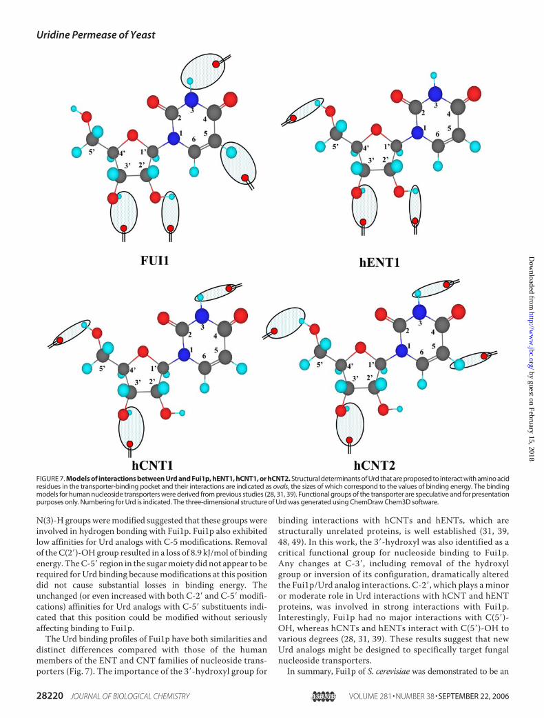
distinct differences compared with those of the humanmembers of the ENT and CNT families of nucleoside trans-porters (Fig. 7). The importance of the 3�-hydroxyl group for
binding interactions with hCNTs and hENTs, which arestructurally unrelated proteins, is well established (31, 39,48, 49). In this work, the 3�-hydroxyl was also identified as acritical functional group for nucleoside binding to Fui1p.Any changes at C-3�, including removal of the hydroxylgroup or inversion of its configuration, dramatically alteredthe Fui1p/Urd analog interactions. C-2�, which plays a minoror moderate role in Urd interactions with hCNT and hENTproteins, was involved in strong interactions with Fui1p.Interestingly, Fui1p had no major interactions with C(5�)-OH, whereas hCNTs and hENTs interact with C(5�)-OH tovarious degrees (28, 31, 39). These results suggest that newUrd analogs might be designed to specifically target fungalnucleoside transporters.In summary, Fui1p of S. cerevisiae was demonstrated to be an
FIGURE 7. Models of interactions between Urd and Fui1p, hENT1, hCNT1, or hCNT2. Structural determinants of Urd that are proposed to interact with amino acidresidues in the transporter-binding pocket and their interactions are indicated as ovals, the sizes of which correspond to the values of binding energy. The bindingmodels for human nucleoside transporters were derived from previous studies (28, 31, 39). Functional groups of the transporter are speculative and for presentationpurposes only. Numbering for Urd is indicated. The three-dimensional structure of Urd was generated using ChemDraw Chem3D software.
Uridine Permease of Yeast
28220 JOURNAL OF BIOLOGICAL CHEMISTRY VOLUME 281 • NUMBER 38 • SEPTEMBER 22, 2006
by guest on February 15, 2018http://w
ww
.jbc.org/D
ownloaded from

electrogenic transporterwith aH�/Urd coupling stoichiometry of1:1. Fui1p mediates the high affinity transport of Urd and the lowbut significant transport of the cytotoxic nucleoside analog FUrd.Studies of the Urd analog binding profiles of Fui1p revealed sub-stantial differences fromthoseof thehumannucleoside transport-ers (hCNTsandhENTs).This raisespossibilities for thedesignandapplication of Urd analog drugs with cytotoxic substituent(s) thatwould be transported differentially by fungal and human nucleo-side transporters.Of the three charged residues in themembrane-spanning regions of Fui1p, only the lysine at position 288 wasfound to be important for transport of Urd. Because all of theLys288 mutants were found to be targeted to yeast plasma mem-branes with minimal changes in abundance, the severe impair-mentof transport capacity likely resulted fromdefects in the trans-location process. Additional studies are needed to fully elucidatethe structural features of Fui1p that are critical for permeant bind-ing and translocation.
REFERENCES1. Baldwin, S. A., Mackey, J. R., Cass, C. E., and Young, J. D. (1999)Mol.Med.
Today 5, 216–2242. Cass, C. E., Young, J. D., Baldwin, S. A., Cabrita, M. A., Graham, K. A.,
Griffiths, M., Jennings, L. L., Mackey, J. R., Ng, A. M. L., Ritzel, M. W. L.,Vickers,M. F., andYao, S. Y.M. (1999) inMembrane Transporters asDrugTargets (Amidon, G. L., and Sadee, W., eds) 1st Ed., Kluwer Academic/Plenum Publishers, New York
3. Landfear, S. M., Ullman, B., Carter, N. S., and Sanchez, M. A. (2004)Eukaryot. Cell 3, 245–254
4. Pastor-Anglada, M., Molina-Arcas, M., Casado, F. J., Bellosillo, B., Co-lomer, D., and Gil, J. (2004) Leukemia (Basingstoke) 18, 385–393
5. Vickers, M. F., Young, J. D., Baldwin, S. A., and Cass, C. E. (2000) Emerg.Ther. Targets 4, 515–539
6. Vickers, M. F., Yao, S. Y. M., Baldwin, S. A., Young, J. D., and Cass, C. E.(2000) J. Biol. Chem. 275, 25931–25938
7. Wagner, R., de Montigny, J., deWergifosse, P., Souciet, J. L., and Potier, S.(1998) FEMS Microbiol. Lett. 159, 69–75
8. Visser, F., Zhang, J., Raborn, R. T., Baldwin, S. A., Young, J. D., and Cass,C. E. (2005)Mol. Pharmacol. 67, 1291–1298
9. Volland, C., Garnier, C., and Haguenauer-Tsapis, R. (1992) J. Biol. Chem.267, 23767–23771
10. Seron, K., Blondel,M.O., Haguenauer-Tsapis, R., andVolland, C. (1999) J.Bacteriol. 181, 1793–1800
11. Volland, C., Urban-Grimal, D., Geraud, G., and Haguenauer-Tsapis, R.(1994) J. Biol. Chem. 269, 9833–9841
12. Pinson, B., Pillois, X., Brethes, D., Chevallier, J., and Napias, C. (1996) Eur.J. Biochem. 239, 439–444
13. Pinson, B., Napias, C., Chevallier, J., Van den Broek, P. J., and Brethes, D.(1997) J. Biol. Chem. 272, 28918–28924
14. Pinson, B., Chevallier, J., and Urban-Grimal, D. (1999) Biochem. J. 339,37–42
15. Eddy, A. A., and Hopkins, P. (1998) Biochem. J. 336, 125–13016. Marchal, C., Haguenauer-Tsapis, R., and Urban-Grimal, D. (2000) J. Biol.
Chem. 275, 23608–2361417. Garnier, C., Blondel, M. O., and Haguenauer-Tsapis, R. (1996) Mol. Mi-
crobiol. 21, 1061–107318. Unkles, S. E., Rouch, D. A., Wang, Y., Siddiqi, M. Y., Glass, A. D., and
Kinghorn, J. R. (2004) Proc. Natl. Acad. Sci. U. S. A. 101, 17549–1755419. Muth, T. R., and Schuldiner, S. (2000) EMBO J. 19, 234–24020. Grewer, C.,Watzke, N., Rauen, T., and Bicho, A. (2003) J. Biol. Chem. 278,
2585–259221. Kaback, H. R. (1997) Proc. Natl. Acad. Sci. U. S. A. 94, 5539–554322. Arastu-Kapur, S., Ford, E., Ullman, B., and Carter, N. S. (2003) J. Biol.
Chem. 278, 33327–33333
23. Vernis, L., Piskur, J., and Diffley, J. F. (2003) Nucleic Acids Res. 31, e12024. Blondel, M. O., Morvan, J., Dupre, S., Urban-Grimal, D., Haguenauer-
Tsapis, R., and Volland, C. (2004)Mol. Biol. Cell 15, 883–89525. Dujon, B., Sherman,D., Fischer,G., Durrens, P., Casaregola, S., Lafontaine,
I., de Montigny, J., Marck, C., Neuveglise, C., Talla, E., Goffard, N., Fran-geul, L., Aigle, M., Anthouard, V., Babour, A., Barbe, V., Barnay, S.,Blanchin, S., Beckerich, J. M., Beyne, E., Bleykasten, C., Boisrame, A.,Boyer, J., Cattolico, L., Confanioleri, F., DeDaruvar, A., Despons, L., Fabre,E., Fairhead, C., Ferry-Dumazet, H., Groppi, A., Hantraye, F., Hennequin,C., Jauniaux,N., Joyet, P., Kachouri, R., Kerrest, A., Koszul, R., Lemaire,M.,Lesur, I., Ma, L., Muller, H., Nicaud, J. M., Nikolski, M., Oztas, S., Ozier-Kalogeropoulos, O., Pellenz, S., Potier, S., Richard, G. F., Straub, M. L.,Suleau, A., Swennen, D., Tekaia, F., Wesolowski-Louvel, M., Westhof, E.,Wirth, B., Zeniou-Meyer, M., Zivanovic, I., Bolotin-Fukuhara, M., Thi-erry, A., Bouchier, C., Caudron, B., Scarpelli, C., Gaillardin, C., Weissen-bach, J., Wincker, P., and Souciet, J. L. (2004) Nature 430, 35–44
26. Kimura, K., and Bugg, T. D. (2003) Nat. Prod. Rep. 20, 252–27327. Rapp, R. P. (2004) Pharmacotherapy 24, 4S–28S; quiz 29S–32S28. Zhang, J., Smith, K. M., Tackaberry, T., Visser, F., Robins, M. J., Nielsen,
L. P., Nowak, I., Karpinski, E., Baldwin, S. A., Young, J. D., and Cass, C. E.(2005)Mol. Pharmacol. 68, 830–839
29. Brunelli, J. P., and Pall, M. L. (1993) Yeast 9, 1309–131830. Ito, H., Fukuda, Y., Murata, K., and Kimura, A. (1983) J. Bacteriol. 153,
163–16831. Zhang, J., Visser, F., Vickers, M. F., Lang, T., Robins, M. J., Nielsen, L. P.,
Nowak, I., Baldwin, S. A., Young, J. D., and Cass, C. E. (2003) Mol. Phar-macol. 64, 1512–1520
32. Visser, F., Baldwin, S. A., Isaac, R. E., Young, J. D., and Cass, C. E. (2005)J. Biol. Chem. 280, 11025–11034
33. Cheng, Y., and Prusoff,W. H. (1973) Biochem. Pharmacol. 22, 3099–310834. De Koning, H. P., and Jarvis, S. M. (2001) Acta Trop. 80, 245–25035. Smith, K.M., Ng, A.M. L., Yao, S. Y.M., Labedz, K. A., Knaus, E. E.,Wiebe,
L. I., Cass, C. E., Baldwin, S. A., Chen, X.-Z., Karpinski, E., and Young, J. D.(2004) J. Physiol. 558, 807–823
36. Ausubel, F. M., Brent, R., Kingston, R. E., Moore, D. D., Seidman, J. G.,Smith, J. A., and Struhl, K. (eds) (1997) Current Protocols in MolecularBiology, John Wiley & Sons, New York
37. Huang, Q. Q., Harvey, C. M., Paterson, A. R., Cass, C. E., and Young, J. D.(1993) J. Biol. Chem. 268, 20613–20619
38. de Montigny, J., Straub, M. L., Wagner, R., Bach, M. L., and Chevallier,M. R. (1998) Yeast 14, 1051–1059
39. Vickers, M. F., Zhang, J., Visser, F., Tackaberry, T., Robins, M. J., Nielsen,L. P., Nowak, I., Baldwin, S. A., Young, J. D., and Cass, C. E. (2004)Nucleo-sides Nucleotides Nucleic Acids 23, 361–373
40. Acton, E. M., Goerner, R. N., Uh, H. S., Ryan, K. J., Henry, D. W., Cass,C. E., and LePage, G. A. (1979) J. Med. Chem. 22, 518–525
41. Serrano, R. (1984) Curr. Top. Cell. Regul. 23, 87–12642. Serrano, R., Kielland-Brandt, M. C., and Fink, G. R. (1986) Nature 319,
689–69343. Hopkins, P., Chevallier, C., Jund, R., and Eddy, A. A. (1988) FEMSMicro-
biol. Lett. 49, 173–17744. Slugoski, M. D., Loewen, S. K., Ng, A.M. L., Baldwin, S. A., Cass, C. E., and
Young, J. D. (2004) Yeast 21, 1269–127745. Xiao, G.,Wang, J., Tangen, T., and Giacomini, K.M. (2001)Mol. Pharma-
col. 59, 339–34846. Ritzel, M. W. L., Ng, A. M. L., Yao, S. Y. M., Graham, K., Loewen, S. K.,
Smith, K. M., Hyde, R. J., Karpinski, E., Cass, C. E., Baldwin, S. A., andYoung, J. D. (2001)Mol. Membr. Biol. 18, 65–72
47. Wallace, L. J., Candlish, D., andDe Koning, H. P. (2002) J. Biol. Chem. 277,26149–26156
48. Vickers, M. F., Kumar, R., Visser, F., Zhang, J., Charania, J., Raborn, R. T.,Baldwin, S. A., Young, J. D., and Cass, C. E. (2002) Biochem. Cell Biol. 80,639–644
49. Patil, S. D., Ngo, L. Y., and Unadkat, J. D. (2000) Cancer Chemother. Phar-macol. 46, 394–402
Uridine Permease of Yeast
SEPTEMBER 22, 2006 • VOLUME 281 • NUMBER 38 JOURNAL OF BIOLOGICAL CHEMISTRY 28221
by guest on February 15, 2018http://w
ww
.jbc.org/D
ownloaded from

James D. Young and Carol E. CassSlugoski, Morris J. Robins, Lars P. C. Nielsen, Ireneusz Nowak, Stephen A. Baldwin,
Jing Zhang, Kyla M. Smith, Tracey Tackaberry, Xuejun Sun, Pat Carpenter, Melissa D.Saccharomyces cerevisiaeUridine Permease Fui1p of
Characterization of the Transport Mechanism and Permeant Binding Profile of the
doi: 10.1074/jbc.M605129200 originally published online July 19, 20062006, 281:28210-28221.J. Biol. Chem.
10.1074/jbc.M605129200Access the most updated version of this article at doi:
Alerts:
When a correction for this article is posted•
When this article is cited•
to choose from all of JBC's e-mail alertsClick here
http://www.jbc.org/content/281/38/28210.full.html#ref-list-1
This article cites 47 references, 23 of which can be accessed free at
by guest on February 15, 2018http://w
ww
.jbc.org/D
ownloaded from