characterization of the pilin ortholog of the helicobacter pylori type
TRANSCRIPT

JOURNAL OF BACTERIOLOGY, Aug. 2006, p. 5865–5877 Vol. 188, No. 160021-9193/06/$08.00�0 doi:10.1128/JB.00060-06Copyright © 2006, American Society for Microbiology. All Rights Reserved.
Characterization of the Pilin Ortholog of the Helicobacter pylori TypeIV cag Pathogenicity Apparatus, a Surface-Associated Protein
Expressed during Infection†Joanna Andrzejewska,1‡§ Sae Kyung Lee,1 Patrick Olbermann,2 Nina Lotzing,2 Elena Katzowitsch,1
Bodo Linz,3 Mark Achtman,3 Clarence I. Kado,4 Sebastian Suerbaum,1,2
and Christine Josenhans1,2*‡Institute for Hygiene and Microbiology, University of Wuerzburg, Josef Schneider Strasse 2, D-97080 Wuerzburg, Germany1;
Institute for Medical Microbiology and Hospital Epidemiology, Hannover Medical School, Hannover, Germany2;Max Planck Institute for Infection Biology, Berlin, Germany3; and Davis Crown Gall Group,
University of California, One Shields Avenue, Davis, California 956164
Received 13 January 2006/Accepted 25 May 2006
The Helicobacter pylori cag pathogenicity island (cag PAI) encodes components of a type IV secretion system(T4SS) involved in host interaction and pathogenicity. Previously, seven cag PAI proteins were identified ashomologs of Agrobacterium tumefaciens Vir proteins, which form a paradigm T4SS. The T pilus composed of theprocessed VirB2 pilin is an external structural part of the A. tumefaciens T4SS. In H. pylori, cag-dependentassembly of pili has not been observed so far, nor has a pilin (VirB2) ortholog been characterized. We have hereidentified, using a motif-based search, an H. pylori cag island protein (HP0546) that possesses sequence andpredicted structural similarities to VirB2-like pilins of other T4SSs. The HP0546 protein displays interstrainvariability in its terminal domains. HP0546 was expressed as a FLAG-tagged fusion protein in Escherichia coli,A. tumefaciens, and H. pylori and was detected as either two or three bands of different molecular masses in theinsoluble fraction, indicating protein processing. As reported previously, isogenic H. pylori mutants in theputative cag pilin gene had reduced abilities to induce cag PAI-dependent interleukin-8 secretion in gastricepithelial cells. Fractionation analysis of H. pylori, using a specific antiserum raised against an N-terminalHP0546 peptide, showed that the protein is partially surface exposed and that its surface localization dependedupon an intact cag system. By immunoelectron microscopy, HP0546 was localized in surface appendages, withsurface exposure of an N-terminal epitope. Pronounced strain-to-strain variability of this predicted surface-exposed part of HP0546 indicates a strong selective pressure for variation in vivo.
The cag pathogenicity island (cag PAI) of Helicobacter pyloriencodes proteins with homologies to structural and functionalcomponents of type IV secretion systems (T4SS) of other bac-teria (1, 6, 15). These systems are multicomponent membrane-spanning transport systems dedicated to the secretion or trans-location of high-molecular-mass biomolecules, such as protein-coupled DNA or proteins, into the environment or intorecipient cells (8, 25, 27). The cag island is involved in thepathogenesis of gastric inflammation and gastric cancer in thehuman host (42). It enables the bacterium to translocate theCagA effector protein into host cells, which, as a consequence,causes a growth factor-like phenotype in infected epithelialcells (30, 31, 37). CagA is crucial for the development of cellmorphology changes and the disruption of cell-cell contacts(tight junctions), effects which likely play a role in the devel-opment of ulcers and cancer during chronic H. pylori infectionof the stomach. It is so far the only H. pylori macromolecule
effector known to be translocated by the cag T4SS (3, 10, 17,31, 43). In addition, the apparatus encoded by the cag PAI isinstrumental in the induction of proinflammatory cytokines,such as interleukin-8 (IL-8), in human epithelial cells (39),which is a marker for host interaction and a hallmark of dis-ease. This effect may be caused by muramyl tripeptide trans-location and thereafter signal induction via the host patternrecognition protein NOD1 (45) and may be enhanced by thetranslocated CagA protein (4, 15, 38). Several H. pylori pro-teins encoded on the cag PAI were identified as homologs ofVir proteins from Agrobacterium tumefaciens, which constitutea model of T4SS (5, 6, 33). In this paradigmatic T4SS of A.tumefaciens, in addition to a complex transmembrane transportsystem, the membrane-associated and surface-exposed T pilusis an essential structural and functional component of type IVsecreton (9, 27). The major subunit of the multimeric T pilus isthe processed and cyclized pilin protein VirB2, which wasfound to participate in the translocation of the T-DNA-proteincomplex (25, 28). Up to now, neither a pilin ortholog in thecomplete genome sequences (2, 44) nor cag island-depen-dent or -independent assembly of typical pili has been de-tected in H. pylori. Recently, sheathed surface structuresdependent on a functional cag pathogenicity island, notclosely resembling classical T pili, but with a central pilus-like structure, have been detected in H. pylori (33, 43). Inthese studies, surface-exposed components of the pilus-like
* Corresponding author. Mailing address: Hannover Medical School,Institute for Medical Microbiology, Carl-Neuberg-Strasse 1, Hannover30625, Germany. Phone: 49 511 5324346. Fax: 49 511 5324355. E-mail:[email protected].
† Supplemental material for this article may be found at http://jb.asm.org/.
‡ J.A. and C.J. contributed equally.§ Present address: ICON Clinical Research GmbH, Langen, Ger-
many.
5865
on February 11, 2018 by guest
http://jb.asm.org/
Dow
nloaded from

structures were identified as domains of cag-encoded pro-teins HP0527, HP0528, and HP0532 using immunogold la-beling and electron microscopy (EM) (33, 43).
In our present study, we identified an H. pylori cag islandprotein (encoded by the HP0546 gene) that has significantsimilarity to VirB2-like pilins from other T4SS. The HP0546protein was characterized to be important for the completefunction of the cag T4SS in host interaction and to provide astructural component on the surfaces of the bacteria. Theprotein was expressed and detected in Escherichia coli, A. tu-mefaciens, and H. pylori. A strain-specific antiserum against anN-terminal peptide of HP0546 was generated and revealedthat HP0546 was surface exposed in wild-type bacteria but notin a cag virB4 (HP0544) mutant. In electron microscopy,HP0546 (N-terminal epitope) was detected to be exposed atdifferent sites on bacterial surfaces, within larger amorphousappendages. Taken together, the results suggest that the pro-tein is a surface-associated VirB2-like pilin subunit that isfunctionally linked to the H. pylori cag apparatus.
MATERIALS AND METHODS
Bacterial strains and growth conditions. H. pylori N6 and KE 26695 wild-typestrains, 88-3887 (the motile derivative of 26695), and a flagellumless fliP-negativederivative of N6 (14, 20, 44), which all harbor a functional cag secretion system,were used for protein analysis and for eukaryotic cell infections. NCTC11637,SS1, J99 (2), LSU2003, LSU1062-3, LSU1014, NQ315, RE10001, RE10002, andBO265 (13, 41), additional H. pylori strains of different geographical origins, eachpossessing a functionally intact cag island, were used for the preparation ofproteins and extrabacterial appendages and for amplification and sequencing ofthe strain-specific HP0546 genes. H. pylori strains were cultured on blood agarplates (Columbia agar base II; Oxoid, Wesel, Germany) containing 10% horseblood and the following antibiotics: vancomycin (10 mg/liter), polymyxin B (2,500U/liter), trimethoprim (5 mg/liter), and amphotericin B (4 mg/liter). H. pyloristrains were preincubated on plates for 24 to 48 h at 37°C under microaerobicconditions for the infection assays. Mutant strains were propagated on bloodagar plates with the addition of chloramphenicol (10 mg/liter) and/or kanamycin(100 mg/liter).
E. coli strains MC1061 and DH5� were used for the DNA cloning experimentsand were propagated in Luria broth or on Luria-Bertani plates supplementedwith kanamycin (100 mg/liter), chloramphenicol (20 mg/liter), ampicillin (100mg/liter), and tetracycline (10 mg/liter) antibiotics as required.
The Agrobacterium tumefaciens bald strain NT1REB (without flagella) (24)was used for heterologous expression of HP0546.
Cell lines, growth conditions, and maintenance. For infection with H. pylori,the human gastric carcinoma cell lines AGS and HM02 (isolated from adeno-carcinoma of the stomach from Caucasian patients) and MKN28 (originallyisolated from a Japanese cancer patient) (47) were used. Cells were routinelycultured in RPMI 1640 medium (buffered with 25 mM HEPES; Invitrogen) andsupplemented with 10% heat-inactivated fetal bovine serum (FBS; Invitrogen) ina 6% CO2 incubator. Cells were subcultured every 3 to 4 days for maintenance.
Cell infections and determination of cytokine release. Cell infections andcoincubations with proteins were performed on subconfluent cell layers (epithe-lial cell lines, 70% to 90% confluent). Cells in 24-well tissue culture plates werewashed three times and preincubated prior to infection for 30 min in freshmedium with serum and without antibiotics. At the time point of infection,mid-log-phase bacteria, washed and resuspended in fresh cell culture medium(RPMI 1640, 25 mM HEPES, 10% heat-inactivated FBS), were added at amultiplicity of infection of 50 bacteria per cell, and the incubation plates werecentrifuged at 500 � g for 3 min to synchronize the infection. The coincubationwas carried out for 20 h. Mock-infected cells were prepared as negative controls.For cytokine measurements, supernatants were harvested, cleared by centrifu-gation, and stored at �80°C until use. IL-8 release into the cell supernatants wasquantitated using an OptEIA IL-8 enzyme-linked immunosorbent assay kit byBD Pharmingen (San Diego, CA) according to the manufacturer’s instructions.Cell supernatants were used at appropriate dilutions for measurement. Eachassay was performed in triplicate. Means and standard deviations for at leastthree independently performed coincubations were calculated. The level of con-fidence was set at 95%.
DNA methods. DNA purification and cloning procedures were performed asdescribed elsewhere (35). Large-scale plasmid purifications were performed us-ing QIAGEN column purification protocols (QIAGEN, Hilden, Germany).DNA fragments were extracted from agarose gels using a QiaEX DNA purifi-cation kit (QIAGEN). DNA restriction and modification enzymes were obtainedfrom Invitrogen Life Technologies or Roche Biochemicals and were used ac-cording to the manufacturer’s protocols. Plasmids and oligonucleotide primersare listed in Table 1 and Table 2, respectively.
Protein methods and Western blotting. Protein concentrations were deter-mined by Bradford (Bio-Rad protein assay dye reagent; Hercules, CA) or bicin-choninic acid (Pierce, Rockford, IL) assay. Equal amounts of protein per samplewere separated on 16.5% denaturing Tris-Tricine sodium dodecyl sulfate (SDS)-polyacrylamide gels or on 14% Tris-glycine SDS-polyacrylamide gel electro-phoresis (PAGE) gels and, for Western blotting, transferred to nitrocellulosemembranes of 0.2-�m pore size (Schleicher & Schuell, Germany). The mem-branes were blocked in Tris-buffered saline-0.1% Tween 20 or in Tris-bufferedsaline-0.1% Tween 20 with 5% nonfat dried milk for 1 h and afterwards incu-bated sequentially with anti-FLAG tag mouse monoclonal antibody M2 (dilu-tion, 1:500; SIGMA) or affinity-purified anti-HP0546 polyclonal antiserum (di-lution, 1:1,000) overnight at 4°C and then peroxidase-conjugated goat anti-rabbitimmunoglobulin G (IgG) or alkaline phosphatase-conjugated goat anti-mouseIgG (dilution, 1:50,000 or 1:5,000; Jackson ImmunoResearch Laboratories) for2 h. Immunolabeled proteins were revealed using Nitro Blue Tetrazolium-BCIP(5-bromo-4-chloro-3-indolyphosphate) color detection or chemiluminescencesubstrate Enhanced SuperSignal West (Pierce).
Isolation of H. pylori surface appendages. H. pylori surface appendages, con-taining flagella and other surface-associated material, were isolated as previouslydescribed (20). Briefly, cells were grown to late log phase, harvested from bloodagar plates, and subjected to mild shearing forces by repeated (20 times) pushingof the suspension through 26-gauge syringe needles. Intact bacterial cells and thesheared-off material were separated by differential centrifugation steps (first at8,000 � g for 20 min and then at 40,000 � g for 1 h in an Optima CU-TL100Beckman ultracentrifuge), and the protein content of the preparations was de-termined. The preparations were also evaluated by electron microscopy andcontained predominantly flagella in flagellated strains (confirmed by Westernimmunoblotting using flagellin-specific antisera), which indicated that the pro-tocol successfully isolates surface-associated material.
Production of a HP0546-specific antiserum. A peptide of HP0546 (strain26695) was chosen, according to published material on various VirB2-like pro-teins (27), for the production of an antiserum. We selected the peptide TSPAEGVTETKTLVIQ (16 amino acids [aa]) at the N terminus of the predictedmature HP0546 protein, immediately following the putative leader peptide cleav-age site (amino acids 31 to 46 of the predicted HP0546 protein), since it has beensuggested that in other bacteria (E. coli [11, 29, 34] and A. tumefaciens [25]), theN termini of the processed VirB2-like pilin proteins may be surface exposed inthe correctly folded mature pilin subunits and in the pilus polymers. The peptidewas synthesized (Biosyntan/Biogenes, Berlin, Germany), and two rabbits weresubcutaneously injected with the peptide conjugated to a carrier to increase itsimmunogenicity. After four booster injections, the serum was obtained andaffinity purified using the specific peptide. Western blots of whole cells andmembrane preparations of H. pylori were used as controls for the specificity ofthe peptide antibody and recognized only a single protein species (see Results).
Deletion and insertion mutageneses of HP0546 and HP0544 genes. For thefurther functional characterization of HP0546, mutations in the HP0546 genewere constructed by gene deletion and replacement and subsequent allelic ex-change in different H. pylori strains. The HP0546 gene with its own promoterregion (1,048 bp) was amplified from strain 26695 using primers HP0546_10 andHP0546_4 (BamH1 sites) and cloned into pUC18 (plasmid pCJ202-0). InversePCR of the whole pCJ202-0 plasmid was performed using primers HP0546inv_1and HP0546_2 (ClaI sites). Thereby, an almost complete deletion of the HP0546coding sequence was engineered, in order to exclude the generation of a partialprotein with residual function, while preserving possible regulatory sequences fordownstream genes at the 3� end of the coding sequence. A kanamycin resistancecassette (aphA3�-III, derived from pILL600) (23) was inserted into the inverselyamplified plasmid in the same transcriptional orientation as the gene, resulting inpCJ202-1. Subsequently, plasmid pCJ202-1 was introduced into H. pylori strainsN6, 26695, and 88-3887 (the motile derivative of 26695) by natural transforma-tion. The HP0544 (cag island virB4) gene was inactivated in strain 88-3887 by arapid method without cloning steps, using three subsequent PCRs and trans-forming final PCR products into H. pylori wild-type strains for marker exchangemutagenesis (details of mutant construction available on request). Correct re-placement of the target genes in the chromosome by double crossover and
5866 ANDRZEJEWSKA ET AL. J. BACTERIOL.
on February 11, 2018 by guest
http://jb.asm.org/
Dow
nloaded from

marker exchange by directional integration of the kanamycin resistance cassettewere determined by PCR using different primer combinations.
Expression of the H. pylori HP0546 protein in H. pylori, A. tumefaciens, and E.coli and engineering of HP0546 internal FLAG tag fusion constructs. For ex-pression of HP0546 in H. pylori, the gene containing its own promoter (PCRproduct generated using 26695 genomic DNA as a template and primersHP0546_3 and HP0546_4 [BamHI sites]) was cloned as a 586-bp fragment intothe H. pylori/E. coli shuttle plasmid pHel2 (18). The resulting plasmid waspCJ204. An inducible plasmid used for expression of the HP0546 protein in A.tumefaciens was constructed as follows. A 2,025-bp fragment of pTrc99A with theinducible trc promoter was amplified (primers pTrc99A_1s and pTrc99A_2s), gelpurified, and cut using BglII. It was ligated with a 4,980-bp BglII fragment fromthe binary plasmid pCAMBIA-0380 for E. coli and A. tumefaciens (16), whichwas generated by PCR amplification (primers pCAM380_1 and pCAM380_3[Table 2]). The resulting plasmid, pCJ203-0, contains replication origins for A.tumefaciens and for E. coli, a kanamycin resistance gene (aadA) for selection, thelacIq gene encoding the Lac repressor, the inducible trc promoter of pTrc99Awith a multiple cloning site, and strong transcriptional terminators (rrnB) down-stream of the multiple cloning site. HP0546 was cloned into pCJ203-0 (cut withNco1) as a 567-bp fragment, exclusive of its own promoter, generated by primersHP0546Nco_1 and HP0546Nco_2 (NcoI sites). Genomic DNA of H. pylori 26695was used as a template. The resulting plasmids were pCJ203-1r and pCJ203-1w(inverse orientation of insert, used as a negative control). The insertion site andthe orientation of the HP0546 gene relative to the trc promoter in pCJ203-1r andpCJ203-1w were confirmed by sequencing. Plasmid pCJ205, containing the
HP0546 gene downstream of an inducible Ptrc promoter in pHel2, was con-structed by insertion of the Nco1-amplified HP0546 fragment as described aboveinto the pCJ201 plasmid (unpublished), to yield pCJ201-1. In addition, plasmidspCJ204, pCJ203-1, and pCJ201-1 were all engineered by PCR mutagenesis tocontain an in-frame fusion with a FLAG tag sequence (MDYKDDDD) withinthe coding region of the HP0546 amino acid sequence (between aa 44 and aa 45)(Fig. 1B), to permit detection of the HP0546 gene in Western blots (resultingplasmids were pCJ205, pCJ207, and pCJ206 [Table 1] [details of constructionavailable on request]). The correct FLAG tag fusion was confirmed by sequenceanalysis of all plasmids. For expression analyses, all plasmids were transformedinto the appropriate host bacteria (H. pylori N6, H. pylori 26695, E. coli MC1061,and A. tumefaciens NT1REB). All pTrc-derivative plasmids for inducible expres-sion in E. coli and A. tumefaciens were constructed such that they should allowthe expression of the HP0546 protein from its own start codon without anyadditional N- and C-terminal amino acids.
Complementation of the HP0546 gene in H. pylori. For complementation,HP0546 plasmid pCJ204 (pHel2 derivative), including its own 5� untranslatedregion, containing putative promoter sequences, was transformed into the H.pylori N6 wild-type strain and the H. pylori 88-3887 isogenic HP0546 mutant. Incases where transformants were obtained on chloramphenicol plates, productionof the HP546 protein was determined by Western blotting.
Fluorescent labeling of cells and microscopy. Immunofluorescent labeling ofsurface-exposed HP0546 on H. pylori in the presence and in the absence ofhuman epithelial cells grown on gelatin-coated coverslips was performed usingaffinity-purified HP0546 antiserum (dilution, 1:1,000). Binding of primary anti-
TABLE 1. Plasmids used in this study
Plasmid/vector Size (bp) Commentsa Source or reference
pHel2 5,000 Cmr, RepEc, RepHp, multicopy shuttle vector for E. coli and H. pylori 18
pILL600 5,700 Ampr, Kmr, RepEc, source of Km resistance cassette 23
pUC18 2,690 Ampr, RepEc, high-copy-number cloning vector
pTrc99A 4,584 Ampr, RepEc, source of inducible Ptrc
pCAMBIA-0,380 6,812 Kmr, RepEc, RepAt, binary vector for A. tumefaciens 16
pCJ201-1 7,593 Cmr, RepEc, RepHp, inducible Ptrc/promoterless HP0546 gene in sametranscriptional orientation as Ptrc in pHel2
This work
pCJ201-0 7,025 Cmr, RepEc, RepHp, Ptrc in pHel2, control plasmid for pCJ201-1r and pCJ206 This work
pCJ202-1 5,074 Ampr, Kmr, RepEc, HP0546 disrupted by Km resistance cassette in pUC18 This work
pCJ203-0At Kmr, RepEc, RepAt, fusion of partial sequence of pCAMBIA-0380 with induciblepromoter of vector pTrc99a to serve as inducible expression plasmid forA. tumefaciens
This work
pCJ203-1r 7,573 Kmr, RepEc, RepAt, inducible Ptrc/promoterless HP0546 gene are in the sametranscriptional orientation in pCJ203-0At
This work
pCJ203-1w 7,573 Kmr, RepEc, RepAt, inducible Ptrc/promoterless HP0546 gene are in transcriptionalorientation opposite to that of promoter in pCJ203-0At (negative control forexpression experiments)
This work
pCJ204 6,048 Cmr, RepEc, RepHp, HP0546 in pHel2, control plasmid for pCJ205 (withoutFLAG tag)
This work
pCJ205 6,072 Cmr, RepEc, RepHp, HP0546 with FLAG tag integrated into HP0546, cloned inpHel2, source of immunodetectable HP0546 fusion protein
This work
pCJ206 7,617 Cmr, RepEc, RepHp, derivative of pCJ201-1 containing FLAG-tagged HP0546fusion protein
This work
pCJ207 7,597 Kmr, RepEc, RepAt, derivative of pCJ203-1r containing FLAG-tagged HP0546fusion protein
This work
a Ampr, ampicillin resistant; Cmr, chloramphenicol resistant; Kmr, kanamycin resistant; RepEc, plasmid which can replicate in E. coli; RepHp, plasmid which canreplicate in H. pylori.
VOL. 188, 2006 H. PYLORI cag PILIN 5867
on February 11, 2018 by guest
http://jb.asm.org/
Dow
nloaded from

body was detected using Alexa Fluor 488-coupled goat anti-rabbit secondaryantibody (dilution, 1:5,000; Molecular Probes). The samples were viewed in aLeica TCS confocal microscope and the TIFF image files processed with AdobePhotoshop.
Electron microscopy. H. pylori and infected cells were prepared for electronmicroscopy as follows. Cells were seeded in drops of cell medium on EM goldgrids (300 mesh, Formvar-carbon coated; Plano, Germany), which had beencleansed and sterilized by being rinsed for 1 h in 70% ethanol of the purestgrade in bidistilled H2O. Cells were grown overnight in RPMI 1640-25 mMHEPES-10% FBS in a humid chamber (using a process similar to that de-scribed in reference 19) and then washed once and overlaid carefully withdrops of fresh medium supplemented with 10% FBS. Cells were infected witha droplet of H. pylori bacteria resuspended in cell culture medium to anoptical density at 600 nm of approximately 1.0. Cells were coincubated withthe bacteria for 2 or 3 h. Then, the medium was removed gently, and fixingagent (2% paraformaldehyde in 100 mM phosphate buffer, pH 7.0) waslayered on the grids for 20 min. Subsequently, the grids were treated forimmunolabeling with the affinity-purified anti-HP0546 antiserum (1:100) in2% goat serum-2% fish skin gelatin-2% bovine serum albumin in phosphate-buffered saline, pH 7.4, for 30 min. After appropriate washing steps in 0.1%bovine serum albumin in phosphate-buffered saline, the primary antibody wasdetected by incubation in goat anti-rabbit IgG secondary antibody coupled to10 nm colloidal gold (1:80). After final washing steps and a secondary fixationin 2.5% glutaraldehyde for 10 min, the grids were negatively stained using 1%phosphotungstate, pH 7.0. The grids were viewed using a Zeiss EM-10 trans-mission electron microscope, and pictures were taken on Kodak EM film anddigitized by high-resolution scanning.
Bioinformatic analyses. Identification of a putative pilin of the H. pylori cagPAI T4SS was performed by in silico analyses; pilins of nine different T4SS werealigned using CLUSTAL W software (GCG Wisconsin Package) and subse-quently used as an input file for the program ProfileMake (GCG), which definesspecific amino acid motifs of a homologous protein family. Pilins from thefollowing systems were compared: (i) plasmid pKM101, E. coli (TraM); (ii)plasmid RP4, E. coli multiresistance plasmid (TrbC); (iii) E. coli plasmid R388(TrwM); (iv) plasmid IncF1 R386, E. coli (TraA); (v) Agrobacterium tumefaciens(VirB2); (vi) Brucella suis (VirB2); (vii) Bartonella henselae (VirB2); (viii) Bor-detella pertussis (PtlA); and (ix) Legionella pneumophila (LvhB2). The output fileof the pilins created by the ProfileMake software was further analyzed by per-forming ProfileSearch (GCG) on the PIR and Swiss-Prot databases. In thesubsequently received output file, the derived HP0546 protein of the H. pylori cagPAI was identified as one single H. pylori protein containing amino acid motifsof a possible T4SS pilin. Subsequently, the HP0546 protein was automatically(MULTALIGN, CLUSTAL W) and further manually aligned with the nine
other pilins using the GeneDoc software (www.psc.edu/biomed/genedoc) (Fig.1A). The amino acid hydrophobicity profile in comparison to those for otherVirB2-like pilins was tested in silico using a membrane protein hydrophobicityalgorithm (12).
RESULTS
Identification of a virB2-like gene (HP0546) in the H. pyloricag pathogenicity island. The Vir system of A. tumefaciens,which transports Ti plasmid transfer DNA into plant cells, inassociation with specific Vir proteins, is a prototype of the typeIV apparatus (27), of which structural proteins VirB1 throughVirB11 and VirD4 are the essential constituents (22). The Tpilus, a multisubunit structure predominantly consisting ofVirB2 subunits, forms an important part of the VirB T4SS (9).In H. pylori, neither a T4SS-associated pilus nor a VirB2-likepilin subunit associated with the cag T4SS has been character-ized so far. We identified the cag PAI-encoded HP0546(Cag25, CagC) as a candidate VirB2-orthologous pilin proteinof the cag apparatus in H. pylori using a database search, withpeptide motifs compiled from amino acid sequences of nineknown orthologous pilins of T4SS from different bacteria (Fig.1A), as detailed in Materials and Methods. The 115-aa pre-dicted full-length HP0546 protein with its putative leader pep-tide (see below) has an overall amino acid identity of 19%(CLUSTAL W) to the A. tumefaciens VirB2 protein (Fig. 1).T4SS pilins of other bacteria show low overall amino acididentities to each other and to the prototype VirB2 protein ofA. tumefaciens, but all display a characteristic hydrophobicityprofile, with two hydrophobic peaks in the mature polypeptide,after cleavage of a leader peptide, indicating hydrophobic orputative transmembrane alpha helices. Hydrophobicity plot-ting (not shown) revealed that the HP0546-derived protein(when the putative leader peptide cleaved between aa 29 and30 is omitted) contained two hydrophobic domains (aa 35 to 47and aa 72 to 95), very similar to other structurally well-char-
TABLE 2. Oligonucleotides used in this study for PCR and nucleotide sequence determination
Primer Purpose Sequence (underlined site)a
HP546p1 (forward) PCR 5�-ATA GGA TCC TCT CAC TCT GAT CAG CTT GG-3� (BamHI)HP546p2 (reverse) PCR 5�-ATA GGA TCC TTC CTT TCA AAT TGA AAT CAA TCG-3� (BamHI)HP546_3 (forward) PCR 5�-ATA GGA TCC GAT GAA TGC AAG GTG AGT GC-3� (BamHI)HP546_4 (reverse) PCR 5�-AAT GGA TCC GAT AGT CGC CTT GAA ACT ATC-3� (BamHI)HP546_5 (forward) PCR 5�-AAT AGA TCT TGA TTT CAA TTT GAA AGG AAA CG-3� (BglII)HP546Nco_1 (forward) PCR 5�-TAT ACC ATG GTG ATT TCA ATT TGA AAG GAA ACG-3� (NcoI)HP546Nco_2 (reverse) PCR 5�-TAT ACC ATG GGA TAG TCG CCT TGA AAC TAT C-3� (NcoI)HP546inv_1 (forward) PCR 5�-ATA TCG ATC ATT TCT GTT CTA GCG ATC G-3� (ClaI)HP546inv_2 (reverse) PCR 5�-ATA TCG ATG ATT GGT TGT TAC CAC TAG C-3� (ClaI)HP546 FLAG1 PCR 5�-ATG GAT TAC AAG GAT GAC GAC GAT ATT CAG ATC ATT TCT
GTT CTA GC-3� (FLAG tag)HP546 FLAG2 PCR 5�-ATC GTC GTC ATC CTT GTA ATC CAT AAC CAA AGT TTT AGT
CTC AGT AAC-3� (FLAG tag)pCAM380_1 (forward) PCR 5�-ATA AGA TCT AGG ACG CAT TGA CCG AGG-3� (BglII)pCAM380_3 (reverse) PCR 5�-ATA AGA TCT GAA CAG TGA ATT GGA GTT CG-3� (BglII)pTrc991 (forward) PCR 5�-ATA AGA TCT TCA CCG TCA TCA CCG AAA CG-3� (BglII)pTrc992 (reverse) PCR 5�-ATA AGA TCT GAG TTT GTA GAA ACG CAA AAA GG-3� (BglII)lac_pHel Nucleotide sequencing 5�-ACT TCC AGT TCA ACA TCA GC-3�km1 Nucleotide sequencing 5�-CTG CTA AGG TAT ATA AGC TGG TGG G-3�km2 Nucleotide sequencing 5�-CAT ACT GTT CTT CCC CGA TAT CCT C-3�pTrc_MCS2 Nucleotide sequencing 5�-ACT TCT GAG TTC GGC ATG G-3�pUC/M13 (reverse) Nucleotide sequencing 5�-TCA CAC AGG AAA CAG CTA TGA C-3�
a The site in parentheses is represented by the underlined nucleotides.
5868 ANDRZEJEWSKA ET AL. J. BACTERIOL.
on February 11, 2018 by guest
http://jb.asm.org/
Dow
nloaded from

acterized VirB2-like pilins (Fig. 1). By means of alignment withother pilins (Fig. 1), the potential leader peptide cleavage siteof HP0546 was modeled. The A. tumefaciens VirB2 pilin andrelated proteins are expressed as propilins and are processed at
their amino and carboxy termini before integration into thepilus polymers (9, 25). In A. tumefaciens VirB2, a leader pep-tide is cleaved off between a stretch of three alanine residues atpositions 45 to 47 and the adjacent glutamine residue at posi-
FIG. 1. Alignment of H. pylori HP0546 protein (strain 26695) with various known type IV pilins of other bacteria. (A) The alignment shown wascreated with CLUSTAL W and revised manually (see Materials and Methods). Designations of protein origins, protein names, and bacterial species/plasmids are shown to the left of each alignment. Abbreviations for the plasmids/bacterial species: pKm10, pKM10 of E. coli; RP4, RP4 of E. coli; R388,R388 of E. coli; Eco, E. coli; atu, A. tumefaciens; bsui, Brucella suis; bhe, Bartonella henselae; borpe, Bordetella pertussis; Lepn, Legionella pneumoniae;Hpyl, H. pylori. Shading of the amino acids according to their physicochemical properties was performed with the GeneDoc software (www.psc.edu/biomed/genedoc). (B) The manual alignment of H. pylori HP0546 with the A. tumefaciens VirB2 type IV pilin protein shows 20% amino acid identity.The leader peptide cleavage site (putative for HP0546, at A29), the insertion site of an internal FLAG tag epitope (between V44 and I45) in HP0546,and the peptide (T31 to Q47; bold and underlined) which was used for generation of the specific antiserum are indicated in panel B.
VOL. 188, 2006 H. PYLORI cag PILIN 5869
on February 11, 2018 by guest
http://jb.asm.org/
Dow
nloaded from

tion 48. By analogy, we positioned the putative cleavage site inHP0546 between Ala (position 29) and Val (position 30),which represents a hydrophilic amino acid stretch between twohydrophobic domains. In Fig. 1B, the leader peptide cleavagesites in HP0546 (putative) and in A. tumefaciens VirB2 areindicated. The proposed leader peptide in HP0546 is shorterthan that in A. tumefaciens VirB2 but comparable to those inother pilins of the VirB2 family (Fig. 1A). The putative matureHP0546 protein after cleavage was calculated to have a mo-lecular mass of 9.7 kDa (as opposed to 13 kDa with the leaderpeptide).
To ascertain that a complete HP0546-like sequence is con-tained in cag PAIs of different H. pylori isolates and to gain anoverview of the possible diversity of this gene and its derivedprotein, the nucleotide sequences of HP0546 from variousstrains from different geographical origins were amplified byPCR and determined (see Materials and Methods; also datanot shown). The gene was present and complete in all (12)strains investigated and showed synonymous and nonsynony-mous strain-to-strain variation. The derived amino acid se-quences of the HP0546 proteins of these 12 H. pylori strainswere aligned and compared (see Fig. S1 in the supplementalmaterial). Amino acid variation was found within the putativeleader peptide (aa 1 to 29), at the proposed mature N terminusfollowing the putative leader sequence (aa 29 to 40), and alsowithin the last four amino acids at the C terminus (aa 110 to115). We then set out to characterize further the expressionand localization of the pilin ortholog HP0546 by expressingand characterizing the protein in three bacterial species whichcan assemble T4SS of different functions, namely, in E. coli,A. tumefaciens, and H. pylori.
HP0546 was expressed and processed in E. coli, A. tumefa-ciens, and H. pylori and localized to the insoluble (membrane-associated) fraction. The HP0546 protein was first expressed inE. coli MC1061 using the three plasmids pCJ206, pCJ207, andpCJ205, each containing an internally FLAG-tagged in-framegene fusion of HP0546 (Materials and Methods and Fig. 1B).We thereby tested protein expression from all of these plasmids inE. coli before these plasmids were used further as inducible plas-mids in E. coli(pCJ206) and A. tumefaciens(pCJ207) and as non-inducible expression plasmids in H. pylori(pCJ205). After induc-tion of the expression plasmids pCJ206 and pCJ207 by IPTG(isopropyl-�-D-thiogalactopyranoside) (see Materials and Meth-ods), harvested cells were fractionated into surface-exposedproteins (shearing) as well as insoluble and soluble fractions byultrasonication. The three fractions were analyzed by Tris-Tricine SDS-PAGE and subsequent Western blotting againstthe FLAG tag (Fig. 2A).
Expression levels and band patterns of the HP0546 proteinin E. coli were found to be similar in Western immunoblots(anti-FLAG) after induction of plasmids pCJ206 (Fig. 2A) andpCJ207 (not shown). In the immunoblots, two bands that werenot present in the negative controls were detected in the in-soluble fractions after ultrasonication (pCJ201-0 not contain-ing the HP0546 insert) (Table 1 and Fig. 2A). The two bands,including the FLAG tag (1 kDa), had apparent molecularmasses of between 7 kDa and 16.5 kDa, which corresponded tomasses calculated from the amino acid sequence with andwithout the putative leader peptide (13 kDa and 9.7 kDa [seeabove]). The two bands might be indicative of a processing of
the protein in E. coli (see Discussion). No HP0546 proteinbands were detected in the soluble fraction of E. coli cellsunder these conditions or in the fractions after mechanicalshearing (surface-associated material). When HP0546 was ex-pressed under the control of its own promoter in E. coli (plas-mid pCJ205), only a low expression level was detected in West-ern blots (not shown).
Our aim was also to express HP0546 in A. tumefaciens, sincethe Vir T4SS encoded on the Agrobacterium Ti plasmid is verywell characterized, and the conditions of the expression of itscomponents are well established (24), which may offer im-proved methods for further investigating the functions of theprotein (27). We also wanted to know whether processing ofHP0546 observed in E. coli MC1061, a K-12 E. coli strain lesswell characterized for T4SS functions, also occurs in A. tume-faciens. For heterologous expression of HP0546, we used theA. tumefaciens bald strain NT1REB, which does not containthe pTi and does not form flagella (7). In the presence of pTi,this strain is able to form T pili from fully processed VirB2(11), making this strain suitable for characterization of theorthologous HP0546 protein in the absence of flagella. A. tu-mefaciens strain NT1REB was transformed with plasmidspCJ203-1r (control for HP0546 without a FLAG tag) andpCJ207, which encodes FLAG-tagged HP0546. A. tumefaciensNT1REB(pCJ207) and controls were induced in liquid culture(0.5 mM IPTG, 200 mM acetosyringone, 19°C, 2 to 3 days),separated into soluble, insoluble, and sheared fractions by son-ication, and analyzed by immunodetection on Western blotsusing anti-FLAG tag antibody (Fig. 2B). HP0546 was detectedexclusively in the insoluble fraction of A. tumefaciens
FIG. 2. Expression of FLAG-tagged HP0546 in three different bac-terial hosts, E. coli, A. tumefaciens, and H. pylori, reveals that theprotein is localized in the insoluble bacterial fractions and is processed.(A) Expression of HP0546 (of strain 26695) in E. coli MC1061. (B) Ex-pression of HP0546 in A. tumefaciens NTREB1. (C) Expression ofHP0546 in H. pylori 26695. Proteins (5 �g per lane) were separated on16.5% Tris-Tricine SDS-PAGE gels, and Western blots were devel-oped with anti-FLAG tag M2 monoclonal antibody. Only proteinsfrom insoluble fractions after ultrasonication are shown, since no sig-nals were detected in the soluble fractions. Lanes 1, insoluble fractions,which show HP0546 protein expression (plasmid constructs used forHP0546 expression are pCJ206 [A], pCJ207 [B], and pCJ205 [C]);lanes 2, insoluble fractions of negative controls without expression ofspecific FLAG-tagged proteins (plasmids used as negative controls arepCJ201-0 [A], pCJ203-1w [B], and pCJ201-0 [C]). Molecular masses ofstandard proteins are indicated on the left; arrows indicate proteinsspecifically detected with the anti-FLAG tag antibody.
5870 ANDRZEJEWSKA ET AL. J. BACTERIOL.
on February 11, 2018 by guest
http://jb.asm.org/
Dow
nloaded from

NT1REB(pCJ207). In contrast to expression in E. coli, threeHP0546-specific bands, of which the two smaller bands ap-peared to have very similar molecular masses, were labeledusing the FLAG tag antibody. As in E. coli, HP0546 was notdetected in the soluble and surface-associated fractions. Theseresults confirmed the functionality of the expression plasmidpCJ203 for A. tumefaciens and showed that HP0546 is ex-pressed mostly as an insoluble protein (putatively membraneassociated) and is processed in A. tumefaciens. This bears thepotential for further functional and complementation studiesof the HP0546 protein in the heterologous system of VirB2-deficient A. tumefaciens strains.
One further goal was the overexpression and detection ofHP0546 in H. pylori, in order to study its localization, process-ing, and function in the native system. pCJ205 (HP0546 with aninternal FLAG tag under the control of its own promoter,which, due to an approximately fivefold gene copy effect,should permit a slight overexpression of the protein) andpCJ201-0 (negative control) were transformed into the H. py-lori N6 and N6 fliP (flagellumless mutant) strains. Soluble andinsoluble proteins and surface-associated materials were pre-pared as described above for E. coli and A. tumefaciens. Im-munoblot analyses performed with anti-FLAG tag antibodyrevealed two specific bands, which were not present in thenegative controls, in the insoluble fraction of H. pyloriN6(pCJ205) (Fig. 2C). The molecular masses of the two pro-tein bands appeared to be similar to those for E. coliMC1061(pCJ206) (Fig. 2). In the soluble fraction and in ma-terial sheared from the surfaces, again, no bands were detectedwith the anti-FLAG antibody (not shown).
To investigate the possibility of multimer formation byHP0546, nondenaturing PAGE was performed and furtheranalyzed by Western immunoblotting. No signal was obtainedafter immunoblotting with anti-FLAG tag antibody (notshown), which suggests that the internal FLAG tag is notaccessible in native HP0546 in monomeric or multimeric state.
Immunodetection after denaturing PAGE was also per-formed for samples of the H. pylori N6(pCJ205) fliP mutantafter introduction of the HP0546 expression plasmid, a flagel-lar basal body mutant not able to form flagellar appendages,which we used in order to facilitate detection in electron mi-croscopy of small surface appendages. One single specificband, which corresponded to the smaller band detected in theinsoluble fraction of H. pylori N6(pCJ205) (Fig. 2C), was de-tected in the insoluble fraction by anti-FLAG tag antibody inthis mutant (not shown).
In Coomassie blue-stained SDS-PAGE, no differences inband pattern were observed between HP0546-positive samplesof E. coli, H. pylori, and A. tumefaciens and their respectivenegative controls, indicating an overall low expression level ofHP0546 in these expression systems under in vitro growthconditions (not shown).
Identification of HP0546 in H. pylori by a specific antiserum.A specific antiserum against HP0546 was produced, since theanti-FLAG tag antibody proved to be of limited sensitivity andspecificity for the detection of HP0546 in its native from and inmicroscopy (data not shown). The HP0546-specific antiserumwas raised in rabbits against a specific N-terminal peptide ofHP0546 from H. pylori 26695 (Materials and Methods), which,as we hypothesized, may be surface associated in mature pilin
protein (see Discussion). To aid in specific immunodetectionby microscopy, the serum was affinity purified by column chro-matography using the same HP0546 peptide. In Western blotsof denaturing PAGE, the affinity-purified anti-HP0546 peptideantiserum recognized one single band (with a molecular massof approximately 10 kDa, corresponding to the faster-migrat-ing band in the FLAG-tagged HP0546 expression strains [seeabove]) in lysates of heterologous H. pylori strain N6 and in E.coli, both expressing the HP0546 of strain 26695 from a plas-mid (Fig. 3A). Otherwise, in the absence of plasmid expres-sion, the specific antiserum detected the HP0546 protein onlyin the wild-type H. pylori strain 26695 and its motile variant88-3887 (Fig. 3A and B), not in strain N6 or in the more than10 additional H. pylori strains tested (Fig. 3A and not shown).In some Western blots, in addition to the 10-kDa band, ahigh-molecular-mass band (�100 kDa) was weakly detected inthe insoluble fraction of HP0546-overexpressing H. pylori, us-ing the specific serum (Fig. 3A). The specific bands were alsonot detected in the HP0546 mutants of 26695 and 88-3887 (Fig.3A and B; also see below), which confirmed the specificity ofthe detection and the successful mutagenesis of the gene. Fur-ther analysis of the HP0546 nucleotide and derived amino acidsequences in several strains and repeated Western blotting ofvarious strains with different HP0546 sequences showed thatthe N-terminal peptide-specific antiserum was entirely strainspecific for strains 26695 and 88-3887. In an additional collec-tion of 30 different H. pylori strains from different geographicalorigins (B. Linz, unpublished), we could find only one singlestrain which had exactly the same N-terminal HP0546 se-quence adjacent to the putative leader peptide as strain 26695.However, this strain (M49) did not possess a functional cagPAI and apparently did not express the protein. When welooked again in more detail for localization of native HP0546with this high-affinity and high-specificity antiserum in frac-tionated bacteria of strain 88-3887, we could detect the proteinmost strongly in the insoluble fraction and in surface-associ-ated material sheared from intact bacteria of the specific strainbut also in smaller amounts in the soluble fraction (Fig. 3A andB). When bacteria were incubated for 1 h at 37°C in cell culturemedium in the presence of lysed AGS cells, the protein wasexpressed in amounts similar to those for plate-grown bacteria,but under the former conditions, shearing of surface materialyielded much less HP0546 protein than it did from plate-grownbacteria (not shown). Successful bacterial fractionation wasconfirmed with antisera against proteins specific for the solublebacterial cytoplasmic fraction (UreB urease subunit), themembrane fraction (antiserum against flagellar basal body pro-tein FlhA), and the surface-associated fraction (FlaB flagellin)of H. pylori (Fig. 3B). Another novel observation in the bacte-rial fractions analyzed using the strain-specific antiserum wasthat HP0546 protein in the surface-associated fraction mi-grated at a slightly higher molecular mass in SDS-PAGE thancytoplasmic or membrane-associated HP0546 (Fig. 3B). Fla-gellumless H. pylori 88-3887 fliP mutants also expressed theHP0546 protein, like the wild type (Fig. 3B). These resultsobtained with the specific antiserum extended our localizationstudies of overexpressed Flag-tagged HP0546 in a heterolo-gous H. pylori strain and in other bacterial species.
Functional characterization of HP0546 by mutagenesis. TheHP0546 protein was inactivated in three different H. pylori
VOL. 188, 2006 H. PYLORI cag PILIN 5871
on February 11, 2018 by guest
http://jb.asm.org/
Dow
nloaded from

strains (26695, 88-3887, and N6) by allelic exchange mutagen-esis (see Materials and Methods). In the 26695 and 88-3887HP0546 mutants, the lack of expression of the HP0546 proteinwas confirmed by Western blotting with the epitope-specificantiserum (Fig. 3A and B). Since the peptide antiserum pro-duced against the HP0546 protein was specific for strains
26695 and 88-3887 (but did not react with strain N6 [seeabove]), it was not possible to confirm by immunoblot analysisthe expected lack of the protein in the H. pylori N6 HP0546mutant. No obvious morphological or cell adherence differ-ences between the mutants and the respective wild-type strainswere observed with or without coincubation with human gas-
FIG. 3. Strain-specific detection of HP0546 protein in H. pylori using an antiserum raised against a peptide. Western blotting and immuno-labeling of HP0546 (with and without FLAG tag), expressed in different H. pylori wild-type and plasmid-transformed strains and detected by astrain- and epitope-specific antiserum raised against an N-terminal peptide of HP0546, and control samples are shown. (A) Expression andstrain-specific detection of HP0546 (from strain 26695) in E. coli MC1061 and in different H. pylori strains and mutants, including negative controlsamples which either do not express the protein (HP0546 mutant) or express a protein with a different N-terminal sequence (N6 and the N6 fliPmutant). Ten micrograms of protein (whole bacterial lysates) was loaded in each lane. (B) Western blot on which fractions of strain 88-3887 andits fliP (devoid of flagella), HP0546, and HP0544 mutants were analyzed, using the strain-specific anti-HP0546 antiserum. Insoluble (membrane)(I), soluble (cytoplasmic) (S), and surface-associated (extracellular) (E) bacterial fractions are depicted. HP0546 protein was found in the wild typein all three fractions, with predominance in the insoluble fraction. Note the slightly higher molecular mass of surface-associated HP0546. Fourmicrograms of protein was loaded in each lane. The upper two panels show fractionation controls for the same blot, developed using bothanti-HPFlhA antiserum (raised against the membrane marker protein FlhA of the flagellar basal body) and anti-HPFlaB antiserum (raised againstthe extracellular marker protein flagellin B [20]). Arrows indicate protein bands, which were specifically detected by the respective antisera. Bandsof lower molecular mass detected on FlhA-coincubated membranes and very weak bands detected by anti-HPFlaB are nonspecific bands.
5872 ANDRZEJEWSKA ET AL. J. BACTERIOL.
on February 11, 2018 by guest
http://jb.asm.org/
Dow
nloaded from
tric epithelial cells (electron microscopy and adherence assays[data not shown]). Mutants of N6 and 26695/88-3887 inHP0546 showed strongly diminished abilities to induce IL-8secretion in gastric epithelial cells in comparison to the wild-type strains (IL-8 secretion levels were approximately 15% ofthe wild-type level for the HP0546 mutants in both strains)(Fig. 4), as was previously described for different H. pyloristrains and their isogenic HP0546 mutants (15, 38). Functionalcomplementation of the loss of HP0546 by transformationof the HP0546 mutant of strain 88-3887 with HP0546-contain-ing shuttle plasmids (pCJ204 or pCJ205 [Table 1]) was notachieved, since no transformant clones were obtained after fiveattempts at natural transformation and electroporation instrain 88-3887, whereas transformants leading to heterologousprotein expression were obtained for H. pylori strain N6 (Fig.3A), which has a much higher transformation efficiency.
H. pylori HP0546 was localized by microscopy on the sur-faces of intact bacteria using the epitope-specific antiserum.For the next step, we used the purified specific antiserum toreveal the localization of HP0546 in intact wild-type bacteriausing specific immunolabeling and microscopy. Immunofluo-rescence microscopy of fixed, intact H. pylori bacteria labeledwith the HP0546-specific, affinity-purified antiserum confirmedthe assumed surface localization that had been suggested bythe previous fractionation experiments and showed a patchydistribution of HP0546-specific signal on the surfaces of H.pylori 26695 and 88-3887 bacteria (Fig. 5A). HP0546-specificlabeling on bacterial surfaces was observed regardless ofwhether the bacteria were cultured in the presence or absenceof eukaryotic cells, but visual inspection suggested higher sur-
face expression levels of the protein in bacteria adhering tocells (Fig. 5A). No signal on bacterial surfaces or elsewherewas observed with the HP0546-specific antibody in fluores-cence microscopy samples of 88-3887 HP0546 mutants (Fig.5B) or of human AGS cells alone. Immunogold labeling ofspecimens for electron microscopy confirmed this result andshowed that amorphous appendages on the surfaces of H.pylori bacteria were labeled by the specific antiserum (Fig. 6).Bacteria clearly carried flagella and HP0546-containing mate-rial on their surfaces simultaneously (Fig. 6). Specimens ofHP0546 mutants and control specimens of cells without bac-teria, which were incubated with secondary antibody only, hadbound less than one gold particle per 100 �m2, demonstratinga high specificity of detection with the specific antiserum.These results clearly demonstrate that HP0546 is a surface-associated protein of intact H. pylori and that the peptide-specific antiserum recognizes a surface-exposed domain of theprotein in a mature structure at least partially composed ofHP0546 protein. We next tested whether surface localizationof HP0546 is dependent on the presence of a functional cagT4SS. To this end, we constructed a cag island virB4 (HP0544,cagE) ATPase mutant deficient in cag system function (asindicated by its reduced ability to induce IL-8 secretion in AGScells). The mutant still expressed HP0546 in amounts similar tothose for the wild-type strain, but HP0546 protein was notdetected on bacterial surfaces by either fractionation (Fig. 3B)or immunofluorescence (not shown). In this mutant, higheramounts of cytoplasmic HP0546 were detected (Fig. 3B).
The affinity-purified antiserum raised against a surface-exposed epitope of the putative pilin HP0546 does not inhibitIL-8 release by infected epithelial cells. We next sought todetermine whether the anti-HP0546 peptide antiserum wasable to interfere with the functionality of the cag apparatus, ofwhich HP0546 is predicted to form an important external part.In a preincubation step, the antiserum and negative controlsera (dilution, 1:50 or 1:100) were added to HP88-3887 bacte-ria in cell culture medium supplemented with 10% FBS for 1 hand then supplemented at the same concentration during a 4-hor overnight coincubation of gastric epithelial cells with thebacteria. The affinity-purified antiserum under these condi-tions did not change the outcome of the bacterial coincubationwith AGS cells (IL-8 release, hummingbird phenotype [notshown]). Preincubation with the anti-HP0546 peptide anti-serum did not reduce the viability of the bacteria, which wasconfirmed by plating and counting of CFU after incubation for1 h in the presence or absence of the peptide antiserum (notshown).
DISCUSSION
We described and partially characterized the HP0546 pro-tein, encoded on the cag pathogenicity island of H. pylori,which has similarity to VirB2-like pilins of other T4SS (9, 26,27). HP0546 is surface exposed, strain variable, and predomi-nantly localized in the bacterial membrane fraction. The hy-pothesis that HP0546 may be a VirB2 pilin ortholog was alsoraised by a different group during the course of the presentexperiments in a review article (21), based on protein and genesynteny comparisons. The HP0546 gene was present in all H.pylori strains with an intact and functional cag PAI. The strain-
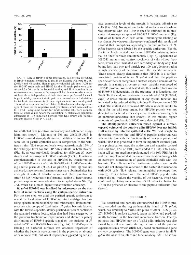
FIG. 4. Role of HP0546 in cell interaction. IL-8 release is reducedin HP0546 mutants compared to that in the isogenic wild-type 88-3887(26695) and N6 strains. Human gastric epithelial cell lines (AGS forthe 88-3887 strain pair and HM02 for the N6 strain pair) were coin-cubated for 20 h with the bacterial strains, and IL-8 secretion in thesupernatants was measured by enzyme-linked immunosorbent assay.At least three independent cell infections were performed for eachisogenic wild-type/mutant strain pair, and means/standard deviationsfor triplicate measurements of these triplicate infections are depicted.The results are summarized as relative IL-8 induction values (percent-ages of those for the respective wild-type strains, which were each setto 100%). Background values for mock-infected cells were deducedfrom all other values before the calculation. �, statistically significantdifferences in IL-8 induction between wild-type strains and isogenicmutants (paired t test; P 0.005).
VOL. 188, 2006 H. PYLORI cag PILIN 5873
on February 11, 2018 by guest
http://jb.asm.org/
Dow
nloaded from
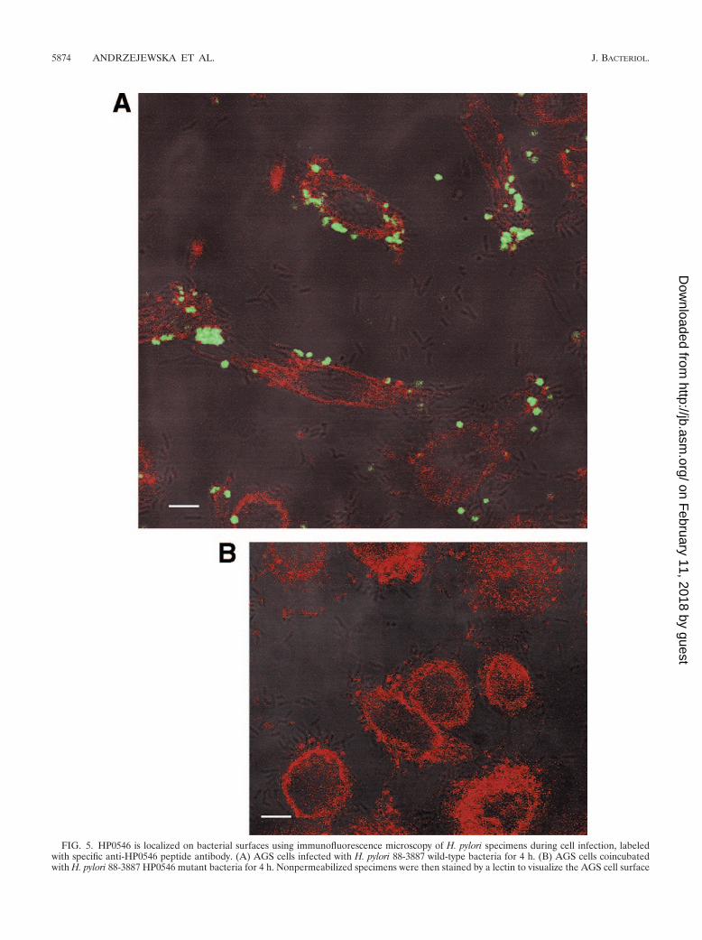
FIG. 5. HP0546 is localized on bacterial surfaces using immunofluorescence microscopy of H. pylori specimens during cell infection, labeledwith specific anti-HP0546 peptide antibody. (A) AGS cells infected with H. pylori 88-3887 wild-type bacteria for 4 h. (B) AGS cells coincubatedwith H. pylori 88-3887 HP0546 mutant bacteria for 4 h. Nonpermeabilized specimens were then stained by a lectin to visualize the AGS cell surface
5874 ANDRZEJEWSKA ET AL. J. BACTERIOL.
on February 11, 2018 by guest
http://jb.asm.org/
Dow
nloaded from

to-strain variability of the HP0546-derived proteins was signif-icant and localized (i) to the N terminus (putative leader pep-tide), (ii) to the domain adjacent to the postulated cleavagesite of the leader peptide, and (iii) to the extreme C terminus.An antiserum raised against the N-terminal domain of HP0546adjacent to the putative leader peptide cleavage site (TSPAEGVTETKTLVIQ) proved to be strain specific for the strain(26695) against which it was raised and recognized a small proteinof approximately 10 kDa, which was absent from the HP0546mutant. The protein was predominantly localized in the mem-brane fractions and to a lesser extent on bacterial surfaces. Im-munofluorescence and electron microscopy with intact nonper-
meabilized bacteria suggested that HP0546 subunits form or arepart of a multimeric structure on bacterial surfaces. Expressionand surface localization of native HP0546-containing material inindividual H. pylori bacteria were not uniform. In electron micros-copy specimens, the specific antiserum against HP0546 revealedsurface-exposed material with no fixed location on bacterial cellsand no defined morphology or length, which clearly exceeded theexpected dimensions of monomeric HP0546. The possibility thatthe lack of defined structure may be due to preparation artifactscannot be excluded. Structure will be studied further, with em-ployment of different preparation techniques. We identified onesecond strain (M49), harboring a HP0546 sequence identical to
using wheat germ agglutinin coupled to Texas Red and subsequently fixed and immunolabeled using anti-HP0546 peptide antiserum (1:1,000),followed by anti-rabbit IgG coupled to Alexa Fluor 488 (1:5,000). Specimens were analyzed using confocal laser scanning microscopy, andrepresentative focal planes are depicted as overlays between signals detected in the green (HP0546-specific signal) and red (cell surface staining)emission channels, combined with a differential interference contrast image of the same specimen. White bars are a size marker of 5 �m. No signalin the green channel (HP0546-specific) was detected in panel B.
FIG. 6. Electron microscopy using anti-HP0546 antibody for immunolabeling of cag-positive H. pylori 88-3887 bacteria reveals surface-associated material. H. pylori bacteria were coincubated with AGS cells on electron microscopy grids for 2 h and then fixed and immunolabeledusing gold-coupled secondary antibodies as described in Materials and Methods. Two representative bacterial specimens, which carry HP0546-specific material labeled with 10-nm gold grains at polar (top) and lateral (bottom) localizations, respectively, are shown. The bottom right paneldepicts an inset at a higher magnification. Arrows point to flagella. Black bars are size markers of 0.5 �m.
VOL. 188, 2006 H. PYLORI cag PILIN 5875
on February 11, 2018 by guest
http://jb.asm.org/
Dow
nloaded from

that for 26695, in a collection of diverse H. pylori isolates from allover the world (13) (B. Linz, unpublished data), but this straincarried a nonfunctional cag PAI and did not react with the specificserum, suggesting that the protein is not expressed in this strain.We additionally determined the expression and localization ofHP0546 in an isogenic cag virB4 (HP0544) mutant, which shows afunctional defect of the cag system (15), and whose VirB4 or-tholog in A. tumefaciens is required for T-pilus assembly (48).HP0546 was expressed in the HP0544 mutant but appeared tolose surface localization, indicating that its surface exposure isdependent on the cag T4SS. Secretion, surface expression, andprocessing of HP0546 in various cag mutants will be examined inmore detail in further experiments.
We were able to express HP0546 as an internal FLAG tagfusion protein in both E. coli and A. tumefaciens, both of whichcan form functional T4SS with roles in DNA and proteintransport. In both heterologous hosts, even in the absence of afunctional T4SS, the HP0546 protein localized mostly to theinsoluble fraction, presumably the membrane. These resultssupport the concept that the expression and membrane local-ization of this protein are quite likely not dependent on a fullyfunctional type IV apparatus and that membrane insertion maybe determined by the general secretory pathway (32, 36), sim-ilar to evidence obtained for A. tumefaciens VirB2 (9, 25, 48).The detection of at least two bands in all bacterial speciesanalyzed for the expression of the FLAG-tagged HP0546 usingan anti-FLAG tag antibody indicated that the protein is pro-cessed. The detection of only one band (the one of lower mass)in Western blots using the specific anti-HP0546 peptide anti-serum may suggest that the epitope (part of the synthesizedpeptide) against which the antiserum is directed is masked inthe unprocessed form of the protein by the leader peptide. TheHP0546 protein was detected as three bands in A. tumefaciensNT1REB. A. tumefaciens VirB2 and orthologous E. coli TrB-CRP4 are cyclized after the cleavage of the leader peptide, bycovalent linkage between the processed N and C termini (11,21, 26). Cyclized A. tumefaciens VirB2 migrates slightly fasterin SDS-PAGE than noncyclized VirB2 (25, 28). Partial cycli-zation of HP0546 in A. tumefaciens might explain the occur-rence of three protein bands in Western blots. Two HP0546-specific bands were also detected when HP0546-FLAG wasexpressed in H. pylori, indicating that processing of HP0546 isnot due to the heterologous expression systems but seems tooccur also in H. pylori. Attempts to perform N-terminal proteinsequencing of the HP0546 band detected by the specific anti-peptide antiserum were not successful (not shown). This maybe due to cyclization of the mature protein, similar to that of A.tumefaciens VirB2 or to other ways of N-terminal processing.The slight elevation of the molecular mass of the surface-associated native HP0546 in H. pylori 88-3887 over those of itsmembrane-bound and cytoplasmic forms was also indicative ofa further processing of the mature surface-associated HP0546in H. pylori. The nature of this processing or posttranslationalmodification could not be determined here and will be thesubject of further studies, including the analysis of various H.pylori cag mutants and the application of mass spectrometry.
The microscopic visualization of material on bacterial sur-faces using the strain-specific anti-H. pylori HP0546 peptideantiserum provided evidence that the peptide used for thegeneration of the antiserum likely comprises a surface-exposed
part of the mature pilin ortholog, probably within a multimericassembly, and that it is not included in the predicted leaderpeptide. The amino acid epitope reacting with the strain-spe-cific antibody could be narrowed down tentatively to the se-quence SPAEGVT (aa 32 to 38 of predicted HP0546), sincethis motif varied and was different from that in HP0546 ofstrain 26695 in all other (except one) of the 42 tested strains(see Fig. S1 in the supplemental material). This result indi-cated that the sequence SPAEGVT is surface exposed in themature structure formed by HP0546 subunits. A high selectivepressure for variation on this surface-localized epitope mayexplain the high strain-to-strain variation of its amino acidsequence, leading to its absolutely strain-dependent recogni-tion by the specific antiserum. The high variability of this sur-face-localized epitope of HP0546 in different strains may pro-vide host specificity for interaction with a receptor and/or ameans of antigenic variation.
Surface localization of the HP0546 protein was dependenton the cag system and appeared increased upon contact witheukaryotic cells, although the expression level of the proteinseemed largely independent of the presence of human hostcells. The surface appendage containing HP0546 subunits mayform a functional pilus of the cag T4SS, promoting host inter-action, although our structural characterization has not re-vealed a defined pilus structure so far. In recent publications,cag PAI-associated, pilus-like structures, containing three cagPAI-encoded proteins, HP0527, HP0528, and HP0532, whichbear only limited similarity to the HP0546-containing materialidentified in the present study, were visualized on bacterialsurfaces after cell contact (33, 43). Further studies will have toclarify whether and how both of these structural findings canbe reconciled. In confirmation of previous results, HP0546deletion mutants displayed diminished abilities to induce IL-8release in gastric epithelial cells in comparison to the wild-typestrain carrying an intact cag PAI (15, 38). The mutants also lostthe ability to translocate the effector protein CagA into hostcells (not shown; see also reference 31). The HP0546 proteinappeared not to be strongly involved in bacterial adhesion tohuman gastric epithelial cells, since the isogenic mutants didnot display deficiencies in adherence assays (our unpublishedresults). The specific antiserum directed against an apparentlysurface-exposed epitope of HP0546 did not inhibit the functionof the cag PAI T4SS in coculture experiments with AGS gastricepithelial cells in our assays. However, since HP0546 is surfaceassociated and very likely one of the proteins localized at theoutermost edge of the cag PAI apparatus, which may have theability to interact directly with epithelial cell receptors, the furthercharacterization of this protein and its potential human recep-tors bears potential for the development of a vaccine or aspecific therapeutic agent against cag-positive strains of H.pylori, which have been shown to be more pathogenic and havea higher association with ulcers and cancer in infected humans(1, 17, 40, 46).
ACKNOWLEDGMENTS
We thank Allison Stack, Verena Ryan, Daniela Fischer, and DanielaGoeppel for excellent technical assistance, all lab members for criticalcomments and helpful suggestions, and Matthias Frosch for continu-ous support. We are grateful to P. T. Hajdukiewicz and the CAMBIAIntellectual Property Resource (Canberra, Australia) for the kind giftof plasmid pCAMBIA-0380.
5876 ANDRZEJEWSKA ET AL. J. BACTERIOL.
on February 11, 2018 by guest
http://jb.asm.org/
Dow
nloaded from

This work was supported by the German Research Council (grant Jo344/2-1 and International Research Training Group grant IRTG 587/IIto S. K. Lee) and by the PathoGenoMik Research Center Wuerzburgof the German Ministry of Education and Research (BMBF).
REFERENCES
1. Akopyants, N. S., S. W. Clifton, D. Kersulyte, J. E. Crabtree, B. E. Youree,C. A. Reece, N. O. Bukanov, E. S. Drazek, B. A. Roe, and D. E. Berg. 1998.Analyses of the cag pathogenicity island of Helicobacter pylori. Mol. Micro-biol. 28:37–53.
2. Alm, R. A., L.-S. L. Ling, D. T. Moir, B. L. King, E. D. Brown, P. C. Doig,D. R. Smith, B. Noonan, B. C. Guild, B. L. deJonge, G. Carmel, P. J.Tummino, A. Caruso, M. Uria-Nickelsen, D. M. Mills, C. Ives, R. Gibson, D.Merberg, S. D. Mills, Q. Jiang, D. E. Taylor, G. F. Vovis, and T. J. Trust.1999. Genomic-sequence comparison of two unrelated isolates of the humangastric pathogen Helicobacter pylori. Nature 397:176–180.
3. Backert, S., E. Ziska, V. Brinkmann, U. Zimny-Arndt, A. Fauconnier, P. R.Jungblut, M. Naumann, and T. F. Meyer. 2000. Translocation of the Heli-cobacter pylori CagA protein in gastric epithelial cells by a type IV secretionapparatus. Cell. Microbiol. 2:155–164.
4. Brandt, S., T. Kwok, R. Hartig, W. Konig, and S. Backert. 2005. NF-kappaBactivation and potentiation of proinflammatory responses by the Helicobacterpylori CagA protein. Proc. Natl. Acad. Sci. USA 102:9300–9305.
5. Buhrdorf, R., C. Forster, R. Haas, and W. Fischer. 2003. Topological anal-ysis of a putative virB8 homologue essential for the cag type IV secretionsystem in Helicobacter pylori. Int. J. Med. Microbiol. 293:213–217.
6. Censini, S., C. Lange, Z. Xiang, J. E. Crabtree, P. Ghiara, M. Borodovsky,R. Rappuoli, and A. Covacci. 1996. cag, a pathogenicity island of Helicobacterpylori, encodes type I-specific and disease-associated virulence factors. Proc.Natl. Acad. Sci. USA 93:14648–14653.
7. Chesnokova, O., J. B. Coutinho, I. H. Khan, M. S. Mikhail, and C. I. Kado.1997. Characterization of flagella genes of Agrobacterium tumefaciens, andthe effect of a bald strain on virulence. Mol. Microbiol. 23:579–590.
8. Christie, P. J. 2001. Type IV secretion: intercellular transfer of macromol-ecules by systems ancestrally related to conjugation machines. Mol. Micro-biol. 40:294–305.
9. Christie, P. J., K. Atmakuri, V. Krishnamoorthy, S. Jakubowski, and E.Cascales. 2005. Biogenesis, architecture, and function of bacterial type IVsecretion systems. Annu. Rev. Microbiol. 59:451–485.
10. Churin, Y., L. Al Ghoul, O. Kepp, T. F. Meyer, W. Birchmeier, and M.Naumann. 2003. Helicobacter pylori CagA protein targets the c-Met receptorand enhances the motogenic response. J. Cell Biol. 161:249–255.
11. Eisenbrandt, R., M. Kalkum, E. M. Lai, R. Lurz, C. I. Kado, and E. Lanka.1999. Conjugative pili of IncP plasmids, and the Ti plasmid T pilus arecomposed of cyclic subunits. J. Biol. Chem. 274:22548–22555.
12. Engelman, D. M., T. A. Steitz, and A. Goldman. 1986. Identifying nonpolartransbilayer helices in amino acid sequences of membrane proteins. Annu.Rev. Biophys. Biophys. Chem. 15:321–353.
13. Falush, D., T. Wirth, B. Linz, J. K. Pritchard, M. Stephens, M. Kidd, M. J.Blaser, D. Y. Graham, S. Vacher, G. I. Perez-Perez, Y. Yamaoka, F. Meg-raud, K. Otto, U. Reichard, E. Katzowitsch, X. Wang, M. Achtman, and S.Suerbaum. 2003. Traces of human migrations in Helicobacter pylori popula-tions. Science 299:1582–1585.
14. Ferrero, R. L., V. Cussac, P. Courcoux, and A. Labigne. 1992. Constructionof isogenic urease-negative mutants of Helicobacter pylori by allelic exchange.J. Bacteriol. 174:4212–4217.
15. Fischer, W., J. Puls, R. Buhrdorf, B. Gebert, S. Odenbreit, and R. Haas.2001. Systematic mutagenesis of the Helicobacter pylori cag pathogenicityisland: essential genes for CagA translocation in host cells and induction ofinterleukin-8. Mol. Microbiol. 42:1337–1348.
16. Hajdukiewicz, P., Z. Svab, and P. Maliga. 1994. The small, versatile pPZPfamily of Agrobacterium binary vectors for plant transformation. Plant Mol.Biol. 25:989–994.
17. Hatakeyama, M. 2004. Oncogenic mechanisms of the Helicobacter pyloriCagA protein. Nat. Rev. Cancer 4:688–694.
18. Heuermann, D., and R. Haas. 1998. A stable shuttle vector system forefficient genetic complementation of Helicobacter pylori strains by transfor-mation and conjugation. Mol. Gen. Genet. 257:519–528.
19. Jin, Q., and S. Y. He. 2001. Role of the Hrp pilus in type III protein secretionin Pseudomonas syringae. Science 294:2556–2558.
20. Josenhans, C., K. A. Eaton, T. Thevenot, and S. Suerbaum. 2000. Switchingof flagellar motility in Helicobacter pylori by reversible length variation of ashort homopolymeric sequence repeat in fliP, a gene encoding a basal bodyprotein. Infect. Immun. 68:4598–4603.
21. Kalkum, M., R. Eisenbrandt, R. Lurz, and E. Lanka. 2002. Tying rings forsex. Trends Microbiol. 10:382–387.
22. Krall, L., U. Wiedemann, G. Unsin, S. Weiss, N. Domke, and C. Baron. 2002.Detergent extraction identifies different VirB protein subassemblies of thetype IV secretion machinery in the membranes of Agrobacterium tumefa-ciens. Proc. Natl. Acad. Sci. USA 99:11405–11410.
23. Labigne-Roussel, A., P. Courcoux, and L. Tompkins. 1988. Gene disruption
and replacement as a feasible approach for mutagenesis of Campylobacterjejuni. J. Bacteriol. 170:1704–1708.
24. Lai, E. M., O. Chesnokova, L. M. Banta, and C. I. Kado. 2000. Genetic andenvironmental factors affecting T-pilin export and T-pilus biogenesis in relationto flagellation of Agrobacterium tumefaciens. J. Bacteriol. 182:3705–3716.
25. Lai, E. M., R. Eisenbrandt, M. Kalkum, E. Lanka, and C. I. Kado. 2002.Biogenesis of T pili in Agrobacterium tumefaciens requires precise VirB2propilin cleavage and cyclization. J. Bacteriol. 184:327–330.
26. Lai, E. M., and C. I. Kado. 1998. Processed VirB2 is the major subunit of thepromiscuous pilus of Agrobacterium tumefaciens. J. Bacteriol. 180:2711–2717.
27. Lai, E. M., and C. I. Kado. 2000. The T-pilus of Agrobacterium tumefaciens.Trends Microbiol. 8:361–369.
28. Lai, E. M., and C. I. Kado. 2002. The Agrobacterium tumefaciens T piluscomposed of cyclic T pilin is highly resilient to extreme environments. FEMSMicrobiol. Lett. 210:111–114.
29. Manchak, J., K. G. Anthony, and L. S. Frost. 2002. Mutational analysis ofF-pilin reveals domains for pilus assembly, phage infection and DNA trans-fer. Mol. Microbiol. 43:195–205.
30. Moese, S., M. Selbach, T. Kwok, V. Brinkmann, W. Konig, T. F. Meyer, andS. Backert. 2004. Helicobacter pylori induces AGS cell motility and elonga-tion via independent signaling pathways. Infect. Immun. 72:3646–3649.
31. Odenbreit, S., J. Puls, B. Sedlmaier, E. Gerland, W. Fischer, and R. Haas.2000. Translocation of Helicobacter pylori CagA into gastric epithelial cells bytype IV secretion. Science 287:1497–1500.
32. Peabody, C. R., Y. J. Chung, M. R. Yen, D. Vidal-Ingigliardi, A. P. Pugsley,and M. H. Saier, Jr. 2003. Type II protein secretion and its relationship tobacterial type IV pili and archaeal flagella. Microbiology 149:3051–3072.
33. Rohde, M., J. Puls, R. Buhrdorf, W. Fischer, and R. Haas. 2003. A novelsheathed surface organelle of the Helicobacter pylori cag type IV secretionsystem. Mol. Microbiol. 49:219–234.
34. Sagulenko, E., V. Sagulenko, J. Chen, and P. J. Christie. 2001. Role ofAgrobacterium VirB11 ATPase in T-pilus assembly and substrate selection. J.Bacteriol. 183:5813–5825.
35. Sambrook, J., E. F. Fritsch, and T. Maniatis. 1989. Molecular cloning: alaboratory manual. Cold Spring Harbor Laboratory Press, Cold Spring Har-bor, N.Y.
36. Sauvonnet, N., G. Vignon, A. P. Pugsley, and P. Gounon. 2000. Pilus forma-tion and protein secretion by the same machinery in Escherichia coli. EMBOJ. 19:2221–2228.
37. Segal, E. D., J. Cha, J. Lo, S. Falkow, and L. S. Tompkins. 1999. Altered states:involvement of phosphorylated CagA in the induction of host cellular growthchanges by Helicobacter pylori. Proc. Natl. Acad. Sci. USA 96:14559–14564.
38. Selbach, M., S. Moese, T. F. Meyer, and S. Backert. 2002. Functional analysis ofthe Helicobacter pylori cag pathogenicity island reveals both VirD4-CagA-de-pendent and VirD4-CagA-independent mechanisms. Infect. Immun. 70:665–671.
39. Shimoyama, T., and J. E. Crabtree. 1997. Mucosal chemokines in Helico-bacter pylori infection. J. Physiol. Pharmacol. 48:315–323.
40. Shimoyama, T., and J. E. Crabtree. 1998. Bacterial factors and immunepathogenesis in Helicobacter pylori infection. Gut 43(Suppl. 1):S2–S5.
41. Suerbaum, S., J. Maynard Smith, K. Bapumia, G. Morelli, N. H. Smith, E.Kunstmann, I. Dyrek, and M. Achtman. 1998. Free recombination withinHelicobacter pylori. Proc. Natl. Acad. Sci. USA. 95:12619–12624.
42. Suerbaum, S., and P. Michetti. 2002. Helicobacter pylori infection. N. Engl.J. Med. 347:1175–1186.
43. Tanaka, J., T. Suzuki, H. Mimuro, and C. Sasakawa. 2003. Structural defi-nition on the surface of Helicobacter pylori type IV secretion apparatus. Cell.Microbiol. 5:395–404.
44. Tomb, J.-F., O. White, A. R. Kerlavage, R. A. Clayton, G. G. Sutton, R. D.Fleischmann, K. A. Ketchum, H. P. Klenk, S. Gill, B. A. Dougherty, K.Nelson, J. Quackenbush, L. Zhou, E. F. Kirkness, S. Peterson, B. Loftus, D.Richardson, R. Dodson, H. G. Khalak, A. Glodek, K. McKenney, L. M.Fitzegerald, N. Lee, M. D. Adams, E. K. Hickey, D. E. Berg, J. D. Gocayne,T. R. Utterback, J. D. Peterson, J. M. Kelley, M. D. Cotton, J. M. Weidman, C.Fujii, C. Bowman, L. Watthey, E. Wallin, W. S. Hayes, M. Borodovsky, P. D.Karp, H. O. Smith, C. M. Fraser, and J. C. Venter. 1997. The complete genomesequence of the gastric pathogen Helicobacter pylori. Nature 388:539–547.
45. Viala, J., C. Chaput, I. G. Boneca, A. Cardona, S. E. Girardin, A. P. Moran,R. Athman, S. Memet, M. R. Huerre, A. J. Coyle, P. S. DiStefano, P. J.Sansonetti, A. Labigne, J. Bertin, D. J. Philpott, and R. L. Ferrero. 2004.Nod1 responds to peptidoglycan delivered by the Helicobacter pylori cagpathogenicity island. Nat. Immunol. 5:1166–1174.
46. Webb, P. M., J. E. Crabtree, D. Forman, et al. 1999. Gastric cancer, cytotoxin-associated gene A-positive Helicobacter pylori, and serum pepsinogens: an inter-national study. Gastroenterology 116:269–276.
47. Yamada, H., T. Aihara, and S. Okabe. 2001. Mechanism for Helicobacterpylori stimulation of interleukin-8 production in a gastric epithelial cell line(MKN 28): roles of mitogen-activated protein kinase and interleukin-1beta.Biochem. Pharmacol. 61:1595–1604.
48. Yuan, Q., A. Carle, C. Gao, D. Sivanesan, K. A. Aly, C. Hoppner, L. Krall,N. Domke, and C. Baron. 2005. Identification of the VirB4-VirB8-VirB5-VirB2 pilus assembly sequence of type IV secretion systems. J. Biol. Chem.280:26349–26359.
VOL. 188, 2006 H. PYLORI cag PILIN 5877
on February 11, 2018 by guest
http://jb.asm.org/
Dow
nloaded from