characterization of the flavoenzyme xiak as an n
TRANSCRIPT

ChemicalScience
EDGE ARTICLE
Ope
n A
cces
s A
rtic
le. P
ublis
hed
on 0
4 M
ay 2
017.
Dow
nloa
ded
on 1
1/28
/202
1 11
:06:
00 P
M.
Thi
s ar
ticle
is li
cens
ed u
nder
a C
reat
ive
Com
mon
s A
ttrib
utio
n-N
onC
omm
erci
al 3
.0 U
npor
ted
Lic
ence
.
View Article OnlineView Journal | View Issue
Characterization
aCAS Key Laboratory of Tropical Marine Bi
Laboratory of Marine Materia Medica, So
Chinese Academy of Sciences, 164 West Xi
E-mail: [email protected]; czhang@sbInstitute of Marine Natural Products, Scho
Resource Exploitation and Protection Colla
University, 135 West Xingang Road, GuangzcHefei National Laboratory of Microscale P
University of Science and Technology of ChidHigh Magnetic Field Laboratory, Chinese A
ChinaeState Key Laboratory of Bioorganic and
Institute of Organic Chemistry, Chinese A
Shanghai 200032, ChinafInstitut fur Pharmazeutische Biologie u
Marburg, Deutschhausstrasse 17a, 35037 MgState Key Laboratory of Microbial Techno
University, Jinan 250100, ChinahDivision of Chemical Biology and Medi
Department of Chemistry, University of Te
E-mail: [email protected]
† Electronic supplementary informationprocedures, materials, and character10.1039/c7sc01182b
‡ Q. Zhang and H. Li contributed equally
Cite this: Chem. Sci., 2017, 8, 5067
Received 15th March 2017Accepted 27th April 2017
DOI: 10.1039/c7sc01182b
rsc.li/chemical-science
This journal is © The Royal Society of C
of the flavoenzyme XiaK as an N-hydroxylase and implications inindolosesquiterpene diversification†
Qingbo Zhang,‡a Huixian Li,‡ab Lu Yu,cd Yu Sun,e Yiguang Zhu,a Hanning Zhu,a
Liping Zhang,a Shu-Ming Li,f Yuemao Shen, g Changlin Tian, cd Ang Li,e
Hung-wen Liu*h and Changsheng Zhang *a
Flavoenzymes are ubiquitous in biological systems and catalyze a diverse range of chemical
transformations. The flavoenzyme XiaK from the biosynthetic pathway of the indolosesquiterpene
xiamycin A is demonstrated to mediate the in vivo biotransformation of xiamycin A into multiple
products, including a chlorinated adduct as well as dimers characterized by C–N and N–N linkages
that are hypothesized to form via radical-based mechanisms. Isolation and characterization of XiaK in
vitro shows that it acts as a flavin-dependent N-hydroxylase that catalyzes the hydroxylation of
xiamycin A at the carbazole nitrogen to form N-hydroxyxiamycin, a product which was overlooked in
earlier in vivo experiments because its chemical and chromatographic properties are similar to those
of oxiamycin. N-Hydroxyxiamycin is shown to be unstable under aerobic conditions, and
characterization by electron paramagnetic resonance spectroscopy demonstrates formation of an N-
hydroxycarbazole radical adduct. This radical species is proposed to serve as a key intermediate
leading to the formation of the multiple xiamycin A adducts. This study suggests that non-enzyme
catalyzed reactions may play a greater role in the biosynthesis of natural products than has been
previously recognized.
o-resources and Ecology, Guangdong Key
uth China Sea Institute of Oceanology,
ngang Road, Guangzhou 510301, China.
csio.ac.cn
ol of Marine Sciences, South China Sea
borative Innovation Center, Sun Yat-sen
hou 510006, China
hysical Sciences, School of Life Science,
na, Hefei, 230027, China
cademy of Sciences, Hefei, 230031, P. R.
Natural Products Chemistry, Shanghai
cademy of Sciences, 345 Lingling Road,
nd Biotechnologie, Philipps-Universitat
arburg, Germany
logy, School of Life Science, Shandong
cinal Chemistry, College of Pharmacy,
xas at Austin, Austin, TX, 78712, USA.
(ESI) available: The experimentalization of compounds. See DOI:
to this work.
hemistry 2017
Introduction
Recent technological advances have accelerated microbialnatural product discovery, leading to a renaissance of interest inthis eld throughout academia as well as in the pharmaceuticalindustry. The recognition of the isolation of avermectins andartemisin by the Nobel Prize in 2015 has initiated a new wave ofnatural product drug discovery.1 In recent years, a growingnumber of indolosesquiterpenes with antibacterial, antitumorand antiviral properties have been isolated from actinomy-cetes.2–7 The diverse structures and broad activity proles ofindolosesquiterpenes have spurred interest in the chemicalsynthesis of this class of natural products.8–12 To learn nature'sstrategy for synthesizing this class of compounds, the biosyn-thetic gene cluster of the indolosesquiterpene xiamycin A (XMA,1, Fig. 1) has been independently identied from marine-derived Streptomyces pactum SCSIO 02999 by us,13 anda mangrove-endophyte Streptomyces sp., HKI0576, has beenidentied by the Hertweck group.14 Further studies haveunveiled the enzymatic cyclization cascade leading to the pen-tacyclic ring structure of 1 (Fig. 1). The pathway is initiated byoxidoreductase XiaO-catalyzed epoxidation (2 / 3),15 followedby membrane protein XiaH-mediated terpene cyclization (3 /
4),15 P450 enzyme XiaM-catalyzed tri-hydroxylations (4 / 5),16
and indole oxygenase XiaI-catalyzed central ring closure (5 / 6
Chem. Sci., 2017, 8, 5067–5077 | 5067
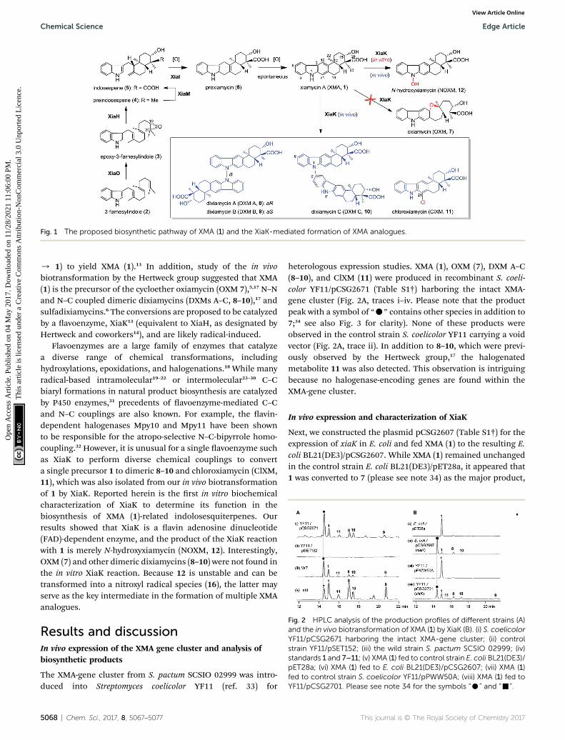
Fig. 1 The proposed biosynthetic pathway of XMA (1) and the XiaK-mediated formation of XMA analogues.
Fig. 2 HPLC analysis of the production profiles of different strains (A)and the in vivo biotransformation of XMA (1) by XiaK (B). (i) S. coelicolorYF11/pCSG2671 harboring the intact XMA-gene cluster; (ii) controlstrain YF11/pSET152; (iii) the wild strain S. pactum SCSIO 02999; (iv)standards 1 and 7–11; (v) XMA (1) fed to control strain E. coli BL21(DE3)/pET28a; (vi) XMA (1) fed to E. coli BL21(DE3)/pCSG2607; (vii) XMA (1)fed to control strain S. coelicolor YF11/pPWW50A; (viii) XMA (1) fed toYF11/pCSG2701. Please see note 34 for the symbols “C” and “-”.
Chemical Science Edge Article
Ope
n A
cces
s A
rtic
le. P
ublis
hed
on 0
4 M
ay 2
017.
Dow
nloa
ded
on 1
1/28
/202
1 11
:06:
00 P
M.
Thi
s ar
ticle
is li
cens
ed u
nder
a C
reat
ive
Com
mon
s A
ttrib
utio
n-N
onC
omm
erci
al 3
.0 U
npor
ted
Lic
ence
.View Article Online
/ 1) to yield XMA (1).13 In addition, study of the in vivobiotransformation by the Hertweck group suggested that XMA(1) is the precursor of the cycloether oxiamycin (OXM 7),5,17 N–Nand N–C coupled dimeric dixiamycins (DXMs A–C, 8–10),17 andsulfadixiamycins.6 The conversions are proposed to be catalyzedby a avoenzyme, XiaK13 (equivalent to XiaH, as designated byHertweck and coworkers14), and are likely radical-induced.
Flavoenzymes are a large family of enzymes that catalyzea diverse range of chemical transformations, includinghydroxylations, epoxidations, and halogenations.18 While manyradical-based intramolecular19–22 or intermolecular23–30 C–Cbiaryl formations in natural product biosynthesis are catalyzedby P450 enzymes,31 precedents of avoenzyme-mediated C–Cand N–C couplings are also known. For example, the avin-dependent halogenases Mpy10 and Mpy11 have been shownto be responsible for the atropo-selective N–C-bipyrrole homo-coupling.32 However, it is unusual for a single avoenzyme suchas XiaK to perform diverse chemical couplings to converta single precursor 1 to dimeric 8–10 and chloroxiamycin (ClXM,11), which was also isolated from our in vivo biotransformationof 1 by XiaK. Reported herein is the rst in vitro biochemicalcharacterization of XiaK to determine its function in thebiosynthesis of XMA (1)-related indolosesquiterpenes. Ourresults showed that XiaK is a avin adenosine dinucleotide(FAD)-dependent enzyme, and the product of the XiaK reactionwith 1 is merely N-hydroxyxiamycin (NOXM, 12). Interestingly,OXM (7) and other dimeric dixiamycins (8–10) were not found inthe in vitro XiaK reaction. Because 12 is unstable and can betransformed into a nitroxyl radical species (16), the latter mayserve as the key intermediate in the formation of multiple XMAanalogues.
Results and discussionIn vivo expression of the XMA gene cluster and analysis ofbiosynthetic products
The XMA-gene cluster from S. pactum SCSIO 02999 was intro-duced into Streptomyces coelicolor YF11 (ref. 33) for
5068 | Chem. Sci., 2017, 8, 5067–5077
heterologous expression studies. XMA (1), OXM (7), DXM A–C(8–10), and ClXM (11) were produced in recombinant S. coeli-color YF11/pCSG2671 (Table S1†) harboring the intact XMA-gene cluster (Fig. 2A, traces i–iv. Please note that the productpeak with a symbol of “C” contains other species in addition to7;34 see also Fig. 3 for clarity). None of these products wereobserved in the control strain S. coelicolor YF11 carrying a voidvector (Fig. 2A, trace ii). In addition to 8–10, which were previ-ously observed by the Hertweck group,17 the halogenatedmetabolite 11 was also detected. This observation is intriguingbecause no halogenase-encoding genes are found within theXMA-gene cluster.
In vivo expression and characterization of XiaK
Next, we constructed the plasmid pCSG2607 (Table S1†) for theexpression of xiaK in E. coli and fed XMA (1) to the resulting E.coli BL21(DE3)/pCSG2607. While XMA (1) remained unchangedin the control strain E. coli BL21(DE3)/pET28a, it appeared that1 was converted to 7 (please see note 34) as the major product,
This journal is © The Royal Society of Chemistry 2017
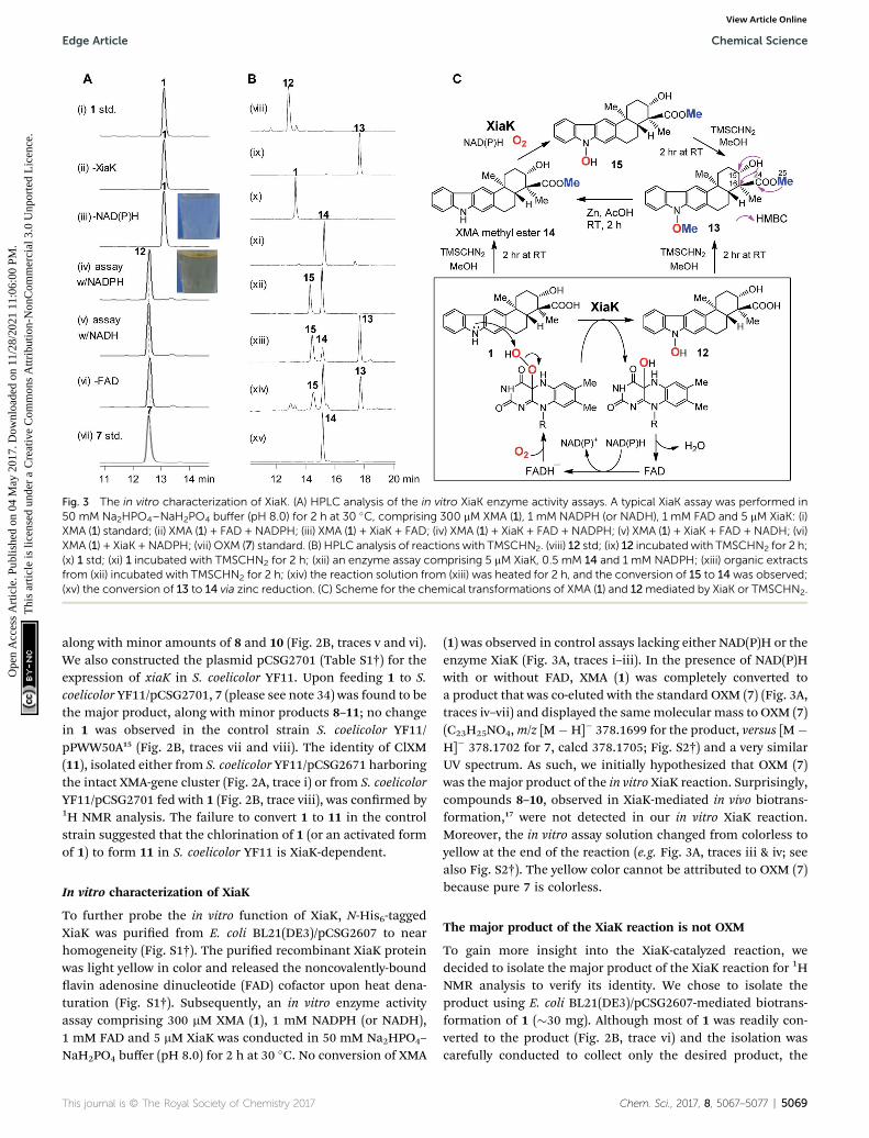
Fig. 3 The in vitro characterization of XiaK. (A) HPLC analysis of the in vitro XiaK enzyme activity assays. A typical XiaK assay was performed in50 mM Na2HPO4–NaH2PO4 buffer (pH 8.0) for 2 h at 30 �C, comprising 300 mM XMA (1), 1 mM NADPH (or NADH), 1 mM FAD and 5 mM XiaK: (i)XMA (1) standard; (ii) XMA (1) + FAD + NADPH; (iii) XMA (1) + XiaK + FAD; (iv) XMA (1) + XiaK + FAD + NADPH; (v) XMA (1) + XiaK + FAD + NADH; (vi)XMA (1) + XiaK +NADPH; (vii) OXM (7) standard. (B) HPLC analysis of reactions with TMSCHN2. (viii) 12 std; (ix) 12 incubatedwith TMSCHN2 for 2 h;(x) 1 std; (xi) 1 incubated with TMSCHN2 for 2 h; (xii) an enzyme assay comprising 5 mM XiaK, 0.5 mM 14 and 1 mM NADPH; (xiii) organic extractsfrom (xii) incubated with TMSCHN2 for 2 h; (xiv) the reaction solution from (xiii) was heated for 2 h, and the conversion of 15 to 14was observed;(xv) the conversion of 13 to 14 via zinc reduction. (C) Scheme for the chemical transformations of XMA (1) and 12mediated by XiaK or TMSCHN2.
Edge Article Chemical Science
Ope
n A
cces
s A
rtic
le. P
ublis
hed
on 0
4 M
ay 2
017.
Dow
nloa
ded
on 1
1/28
/202
1 11
:06:
00 P
M.
Thi
s ar
ticle
is li
cens
ed u
nder
a C
reat
ive
Com
mon
s A
ttrib
utio
n-N
onC
omm
erci
al 3
.0 U
npor
ted
Lic
ence
.View Article Online
along with minor amounts of 8 and 10 (Fig. 2B, traces v and vi).We also constructed the plasmid pCSG2701 (Table S1†) for theexpression of xiaK in S. coelicolor YF11. Upon feeding 1 to S.coelicolor YF11/pCSG2701, 7 (please see note 34) was found to bethe major product, along with minor products 8–11; no changein 1 was observed in the control strain S. coelicolor YF11/pPWW50A15 (Fig. 2B, traces vii and viii). The identity of ClXM(11), isolated either from S. coelicolor YF11/pCSG2671 harboringthe intact XMA-gene cluster (Fig. 2A, trace i) or from S. coelicolorYF11/pCSG2701 fed with 1 (Fig. 2B, trace viii), was conrmed by1H NMR analysis. The failure to convert 1 to 11 in the controlstrain suggested that the chlorination of 1 (or an activated formof 1) to form 11 in S. coelicolor YF11 is XiaK-dependent.
In vitro characterization of XiaK
To further probe the in vitro function of XiaK, N-His6-taggedXiaK was puried from E. coli BL21(DE3)/pCSG2607 to nearhomogeneity (Fig. S1†). The puried recombinant XiaK proteinwas light yellow in color and released the noncovalently-boundavin adenosine dinucleotide (FAD) cofactor upon heat dena-turation (Fig. S1†). Subsequently, an in vitro enzyme activityassay comprising 300 mM XMA (1), 1 mM NADPH (or NADH),1 mM FAD and 5 mM XiaK was conducted in 50 mM Na2HPO4–
NaH2PO4 buffer (pH 8.0) for 2 h at 30 �C. No conversion of XMA
This journal is © The Royal Society of Chemistry 2017
(1) was observed in control assays lacking either NAD(P)H or theenzyme XiaK (Fig. 3A, traces i–iii). In the presence of NAD(P)Hwith or without FAD, XMA (1) was completely converted toa product that was co-eluted with the standard OXM (7) (Fig. 3A,traces iv–vii) and displayed the samemolecular mass to OXM (7)(C23H25NO4,m/z [M�H]� 378.1699 for the product, versus [M�H]� 378.1702 for 7, calcd 378.1705; Fig. S2†) and a very similarUV spectrum. As such, we initially hypothesized that OXM (7)was the major product of the in vitro XiaK reaction. Surprisingly,compounds 8–10, observed in XiaK-mediated in vivo biotrans-formation,17 were not detected in our in vitro XiaK reaction.Moreover, the in vitro assay solution changed from colorless toyellow at the end of the reaction (e.g. Fig. 3A, traces iii & iv; seealso Fig. S2†). The yellow color cannot be attributed to OXM (7)because pure 7 is colorless.
The major product of the XiaK reaction is not OXM
To gain more insight into the XiaK-catalyzed reaction, wedecided to isolate the major product of the XiaK reaction for 1HNMR analysis to verify its identity. We chose to isolate theproduct using E. coli BL21(DE3)/pCSG2607-mediated biotrans-formation of 1 (�30 mg). Although most of 1 was readily con-verted to the product (Fig. 2B, trace vi) and the isolation wascarefully conducted to collect only the desired product, the
Chem. Sci., 2017, 8, 5067–5077 | 5069
Chemical Science Edge Article
Ope
n A
cces
s A
rtic
le. P
ublis
hed
on 0
4 M
ay 2
017.
Dow
nloa
ded
on 1
1/28
/202
1 11
:06:
00 P
M.
Thi
s ar
ticle
is li
cens
ed u
nder
a C
reat
ive
Com
mon
s A
ttrib
utio
n-N
onC
omm
erci
al 3
.0 U
npor
ted
Lic
ence
.View Article Online
major compound retrieved from the isolation process was 1.Only a very minor quantity (around 2mg) of the desired productwas obtained. Although the isolated product was pure andcolorless, no 1H NMR signals were discernible in MeOH-d3,which was used routinely in our NMR characterization ofauthentic OXM (7).5 Surprisingly, regeneration of 1 from thepuried product was noted during NMR characterization,indicating a reverse transformation of the isolated product to 1in free solution. However, the standard OXM (7) is a stablecompound, and the spontaneous conversion of 7 to 1 has neverbeen observed. These cumulative data suggested that whileOXM (7) is a metabolite of the producing strain, the majorproduct isolated from the XiaK reaction is not OXM (7) buta closely related compound (designated 12) having a nearlyidentical HPLC retention time (Fig. S2A†), the same molecularweight, and a similar UV spectrum (Fig. S2B†). The co-elution of7 and 12 under the HPLC conditions and the spontaneousconversion of 12 to 1 are consistent with an early observationthat much less OXM (7, 0.29 mg L�1) than XMA (1, 6.86 mg L�1)was isolated from the wild type S. pactum SCSIO 02999,5
although the production titer of 7 (actually a mixture of 7 and12, see also note 34) was much higher than that of 1 based onHPLC analysis (Fig. 2A, trace iii).13
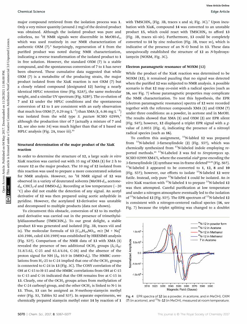
Fig. 4 EPR spectra of 12 (as a powder, in acetone, and in MeOH), OXM(7) (in acetone), and 15N-12 (in MeOH), measured at room temperature.
Structural determination of the major product of the XiaKreaction
In order to determine the structure of 12, a large scale in vitroXiaK reaction was carried out with 35 mg of XMA (1) for 2 h toensure 12 as the major product. The 10 mg of 12 isolated fromthis reaction was used to prepare a more concentrated solutionfor NMR analysis. However, no 1H NMR signal of 12 wasobserved in a variety of deuterated solvents (MeOH-d3, acetone-d6, CHCl3-d and DMSO-d6). Recording at low temperature (�20�C) also did not enable the detection of any signal. An acetylderivative of 12 was then prepared using acetic anhydride inpyridine. However, the acetylated 12-derivative was unstableand decomposed to multiple products (data not shown).
To circumvent this obstacle, conversion of 12 to its methyl-ated derivative was carried out in the presence of trimethylsi-lyldiazomethane (TMSCHN2). To our great delight, a stableproduct 13 was generated and isolated (Fig. 3B, traces viii andix). The molecular formula of 13 (C25H29NO4, m/z [M + Na]+
430.1986, calcd 430.1989) was established by HRESIMS analysis(Fig. S3†). Comparison of the NMR data of 13 with XMA (1)revealed the presence of two additional OCH3 groups (dC/dH:51.8/3.62, C-25 and 63.4/4.04, C-26) and the absence of theproton signal for NH (dH 10.9 in DMSO-d6). The HMBC corre-lations from H3-25 to C-24 implied that one of the OCH3 groupsis connected to C-24 in 13 (Fig. 3C). The COSY correlation of theOH at C-15 to H-15 and the HMBC correlations from OH at C-15to C-15 and C-16 indicated that the OH remains free at C-15 in13. Clearly, one of the OCH3 groups arises from methylation ofthe C-24 carboxyl group, and the other OCH3 is linked to N-1 in13. Thus, 13 can be assigned as N-methoxy-xiamycin methylester (Fig. S3, Tables S2 and S3†). In separate experiments, wechemically prepared xiamycin methyl ester 14 by reaction of 1
5070 | Chem. Sci., 2017, 8, 5067–5077
with TMSCHN2 (Fig. 3B, traces x and xi; Fig. 3C).7 Upon incu-bation with XiaK, compound 14 was converted to an unstableproduct 15, which could react with TMSCHN2 to afford 13(Fig. 3B, traces xii–xiv). Furthermore, 13 could be completelyconverted to 14 by zinc reduction (Fig. 3B, trace xv), which isindicative of the presence of an N–O bond in 13. These dataunequivocally established the structure of 12 as N-hydroxyx-iamycin (NOXM, Fig. 3C).
Electron paramagnetic resonance of NOXM (12)
While the product of the XiaK reaction was determined to beNOXM (12), it remained puzzling that no signal was detectedwhen the puried 12 was subjected to NMR analysis. A possiblescenario is that 12 may co-exist with a radical species (such as16, see Fig. 7) whose paramagnetic properties may complicatethe NMR characterization. To test this hypothesis, the EPR(electron paramagnetic resonance) spectra of 12 were recordedtogether with the reference compounds XMA (1) and OXM (7)under three conditions: as a powder, in acetone and in MeOH.The results showed that XMA (1) and OXM (2) are EPR silent(Fig. S4†); however, 12 displayed a triplet EPR signal with a g-value of 2.0051 (Fig. 4), indicating the presence of a nitroxylradical species (such as 16).
To conrm this assignment, 15N-labled 12 was preparedfrom 15N-labeled 3-farnesylindole (2) (Fig. S5†), which waschemically synthesized from 15N-labeled indole employing re-ported methods.15 15N-Labeled 2 was fed to Streptomyces sp.SCSIO 02999 XM47i, where the essential xiaP gene encoding the3-farnesylindole (2) synthase was in-frame deleted13,35 (Fig. S6†).15N-labeled 2 appeared to be converted to 1, 12, 8 and 10(Fig. S5†); however, our efforts to isolate 15N-labeled 12 werefutile. Instead, only pure 15N-labeled 1 could be isolated. An invitro XiaK reaction with 15N-labeled 1 to prepare 15N-labeled 12was then attempted. Careful purication at low temperatureand under a nitrogen atmosphere eventually led to the isolationof 15N-labeled 12 (Fig. S5†). The EPR spectrum of 15N-labeled 12is consistent with a nitrogen-centered radical species (16, seeFig. 7) because the triplet splitting was changed to a doublet
This journal is © The Royal Society of Chemistry 2017

Edge Article Chemical Science
Ope
n A
cces
s A
rtic
le. P
ublis
hed
on 0
4 M
ay 2
017.
Dow
nloa
ded
on 1
1/28
/202
1 11
:06:
00 P
M.
Thi
s ar
ticle
is li
cens
ed u
nder
a C
reat
ive
Com
mon
s A
ttrib
utio
n-N
onC
omm
erci
al 3
.0 U
npor
ted
Lic
ence
.View Article Online
signal due to the substitution of the 14N nucleus with 15N at theradical center (Fig. 4). A theoretical simulation also supportedthis conclusion (Fig. S7†). However, the spin concentration inthe 12 sample was estimated to be only around 3.6% by calcu-lation using MTSL (1-oxyl-2,2,5,5-tetramethyl-D3-pyrroline-3-methyl methanethiosulfonate) as a standard (Fig. S8†).Despite the low concentration of the nitroxyl radical in thesample, the unpaired electron spin associated with compound16 was sufficient to broaden the NMR signals of 12 through spinrelaxation. An attempt to quench the radical by the addition ofascorbic acid was ineffective, as the NMR spectrum of theresulting sample remained featureless. Compounds exhibitingsimilar “NMR silent” characteristics have been previously re-ported for intermediates (e.g. kinobscurinone) in kinamycinbiosynthesis.36 Treatment with ion-exchange resin and washingwith aqueous EDTA also failed to produce a visible NMR spec-trum. Likewise, the structure of kinobscurinone (naturally iso-lated36 or chemically synthesized37) could only be determined bychemical derivatizations.
Characterization of XiaK as an N-hydroxylase
The above results indicated that XiaK is an N-hydroxylase thatcatalyzes the hydroxylation of 1 at N-1 to generate an unstableproduct, NOXM (12). Phylogenetic analysis (Fig. S9†) and struc-tural modelling (Fig. S10†) indicate that XiaK is a member of theclass A avoprotein monooxygenase family.38,39 Sequencecomparison of XiaK with other known class A avoproteinmonooxygenases reveals that XiaK consists of a conserved motifof glycine residues GXGXXG and a bab-Rossmann fold secondarystructure, which are characteristic of FAD binding domains(Fig. S11†). To further understand the XiaK-catalyzed N-hydrox-ylation, the kinetic parameters were determined (Fig. 5). XiaKdisplayed a Km value of 16.2 mM and a kcat value of 260.4 min�1
toward 1, with a kcat/Km value of 16.1 mM�1 min�1. The efficiencyof the XiaK-catalyzed N-hydroxylation is much higher than thosecatalyzed by other avoenzymes40–44 and P450 enzymes.45–47 Thetypical kcat/Km values of a variety of N-hydroxylation reactionsrange from 0.0053 to 1.21 mM�1 min�1 (Table S4†).
The XiaK-catalyzed reaction likely involves nucleophilicattack at the peroxy oxygen of a avin-4a-hydroperoxy
Fig. 5 Determination of XiaK kinetic parameters for the substrate 1ranging from 5 to 200 mM.
This journal is © The Royal Society of Chemistry 2017
intermediate by the carbazole nitrogen of 1 to cleave the O–Obond and expel 4a-hydroxyavin. Subsequent elimination ofwater from avin to regenerate the oxidized coenzyme therebycompletes the catalytic cycle (Fig. 3C). Although the avin-4a-hydroperoxy species has been proposed as an intermediate inreactions catalysed by other avoprotein monooxygenases,38 itis only transiently formed during turnover. Because XiaK is anefficient catalyst of N-hydroxylation (Table S4†), it is difficult todirectly detect the avin-4a-hydroperoxy intermediate shown inFig. 3C. Further characterization of this intermediate willrequire stop-ow spectroscopic and/or rapid kinetic experi-ments, which will be pursued in the near future.
Time course of the XiaK reaction
To learn more about the XiaK catalysis, a time course analysis ofthe in vitro XiaK reaction was also performed. As shown inFig. 6A, XMA (1) was converted to 12 in a time-dependentmanner, achieving near completion in 2 h (Fig. 6A, traces i–viii). During the period from 5 min to 2 h, another product 16was also detected, albeit in minute quantity (Fig. 6A, traces ii–viii). The molecular weight of 16 was determined by LC-HRESIMS analysis as m/z 378 (379.1770 [M + H]+, 377.1607 [M� H]�, Fig. S12†), which is one mass unit less than that of 12.Given the fact that 16 absorbs in the visible region (Fig. S12†),the yellow color of the XiaK reaction may be attributed to thenitroxyl radical 16. Interestingly, the ratio of 16 to 12 steadilyincreased from 4 h to 8 h, during which time a small amount of1 was detected (Fig. 6A, traces ix and x). Aer 24 h, more 12 wasconverted to 1 and 16 (Fig. 6A, trace xi); however, prolongedincubation (5 d or 12 d) (Fig. 6A, traces xii and xiii) resulted indepletion of 16, with 1 as the predominant product. Theappearance of additional products, such as 17 and 18, was alsonoted; both exhibited similar UV spectra to 1 (Fig. S13†). Thus,the nascent product of the XiaK reaction is 12, which is theprecursor of 16–19. However, formation of 16–19 may not beXiaK-catalyzed.
To characterize the products generated during prolongedincubation with XiaK, a large scale XiaK reaction with 80 mg ofXMA (1) for 7 d was performed. From this reaction, 3.3 mg of 17(designated xiamycin F, XMF), 8.0 mg of 18 (designated xia-mycin G, XMG) and 1.0 mg of 19 (designated xiamycin H, XMH)were isolated (Fig. 6B). HRESIMS analyses established molec-ular formulas of C23H23NO4 for 17 (Fig. S14†), C23H23NO5 for 18(Fig. S15†), and C22H19NO for 19 (Fig. S16†). Comparison of theNMR data of 1 and 17 (Fig. S14, Tables S2 and S3†) revealed that17 contains a C-19 carbonyl group (dC 198.3) instead of a C-19methylene group (dC 31.8, dH 3.09) as in 1. This assignment issupported by HMBC correlations from H-21 and H-18 to C-19 in17 (Fig. S14†). Inspection of the NMR data of 18 indicated thatits structure closely resembles that of 17 (Fig. S15, Tables S2 andS3†). The signal for H-21 (dH 8.08) in 17 is absent in 18, and thechemical shi of C-21 is deshielded from dC 110.6 in 17 to dC
152.2 in 18, indicating a hydroxy substitution at C-21 in 18(Fig. 6B). Detailed NMR analysis also established the structureof 19 (Fig. 6B, S16, Tables S2 and S3†) as a highly oxidizedcongener of 1.
Chem. Sci., 2017, 8, 5067–5077 | 5071
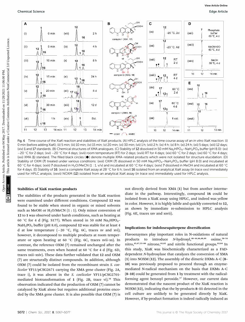
Fig. 6 Time course of the XiaK reaction and stabilities of XiaK products. (A) HPLC analysis of the time course assay of an in vitro XiaK reaction: (i)0min (before adding XiaK); (ii) 5 min; (iii) 10min; (iv) 15min; (v) 20min; (vi) 30min; (vii) 1 h; (viii) 2 h; (ix) 4 h; (x) 8 h; (xi) 24 h; (xii) 5 days; (xiii) 12 days;(xiv) 1 and 17 standards. (B) Chemical structures of XMA analogues. (C) Stability of 12 dissolved in 50mMNa2HPO4–NaH2PO4 buffer (pH 8.0): (xv)�20 �C for 2 days; (xvi) �20 �C for 4 days; (xvii) room temperature (RT) for 2 days; (xviii) RT for 4 days; (xix) 60 �C for 2 days; (xx) 60 �C for 4 days;(xxi) XMA (1) standard. The filled black circles (C) denote multiple XMA-related products which were not isolated for structure elucidation. (D)Stability of OXM (7) treated under various conditions: (xxii) OXM (7) dissolved in 50 mM Na2HPO4–NaH2PO4 buffer (pH 8.0) and incubated at60 �C for 4 days; (xxiii) 7 dissolved in H2O/MeCN (1 : 1, v/v) and incubated at 60 �C for 4 days; (xxiv) 7 dissolved in MeOH and incubated at 60 �Cfor 4 days. (E) Stability of 16: (xxv) a complete XiaK assay at 28 �C for 6 h; (xxvi) 16 isolated from an analytical XiaK assay (in trace xxv) immediatelyused for HPLC analysis; (xxvii) NOXM (12) isolated from an analytical XiaK assay (in trace xxv) immediately used for HPLC analysis.
Chemical Science Edge Article
Ope
n A
cces
s A
rtic
le. P
ublis
hed
on 0
4 M
ay 2
017.
Dow
nloa
ded
on 1
1/28
/202
1 11
:06:
00 P
M.
Thi
s ar
ticle
is li
cens
ed u
nder
a C
reat
ive
Com
mon
s A
ttrib
utio
n-N
onC
omm
erci
al 3
.0 U
npor
ted
Lic
ence
.View Article Online
Stabilities of XiaK reaction products
The stabilities of the products generated in the XiaK reactionwere examined under different conditions. Compound 12 wasfound to be stable when stored in organic or mixed solventssuch as MeOH or H2O/MeCN (1 : 1). Only minor conversion of12 to 1 was observed under harsh conditions, such as heating at60 �C for 4 d (Fig. S17†). When stored in 50 mM Na2HPO4–
NaH2PO4 buffer (pH 8.0), compound 12 was stable for at least 4d at low temperature (�20 �C, Fig. 6C, traces xv and xvi);however, it decomposed to multiple products at room temper-ature or upon heating at 60 �C (Fig. 6C, traces xvii–xx). Incontrast, the reference OXM (7) remained unchanged aer thesame treatments, even when heated at 60 �C for 4 d (Fig. 6D,traces xxii–xxiv). These data further validated that 12 and OXM(7) are structurally distinct compounds. In addition, althoughOXM (7) could be isolated from the recombinant strain S. coe-licolor YF11/pCSG2671 carrying the XMA gene cluster (Fig. 2A,trace i), it was absent in the S. coelicolor YF11/pCSG2701-mediated biotransformation of 1 (Fig. 2B, trace vi).34 Thisobservation indicated that the production of OXM (7) cannot becatalyzed by XiaK alone but requires additional proteins enco-ded by the XMA gene cluster. It is also possible that OXM (7) is
5072 | Chem. Sci., 2017, 8, 5067–5077
not directly derived from XMA (1) but from another interme-diate in the pathway. Interestingly, compound 16 could beisolated from a XiaK assay using HPLC, and indeed was yellowin color. However, it is highly labile and quickly converted to 12,as shown by immediate re-submission to HPLC analysis(Fig. 6E, traces xxv and xxvii).
Implications for indolosesquiterpene diversication
Flavoenzymes play important roles in N-oxidations of naturalproducts to introduce N-hydroxyl,40–44,46,48,49 oxime,50–54
nitro,45,47,55–60 nitrone,61,62 and nitrile functional groups.63,64 Inthis study, XiaK was biochemically characterized as a FAD-dependent N-hydroxylase that catalyzes the conversion of XMA(1) into NOXM (12). The assembly of the dimeric DXMs A–C (8–10) was previously proposed to proceed through an enzyme-mediated N-radical mechanism on the basis that DXMs A–C(8–10) could be generated from 1 by treatment with the radical-forming agent benzoyl peroxide.17 However, our current datademonstrated that the nascent product of the XiaK reaction isNOXM (12), indicating that the by-products 8–11 detected in thecell culture are unlikely to be generated directly by XiaK.However, if by-product formation is indeed radically induced as
This journal is © The Royal Society of Chemistry 2017

Edge Article Chemical Science
Ope
n A
cces
s A
rtic
le. P
ublis
hed
on 0
4 M
ay 2
017.
Dow
nloa
ded
on 1
1/28
/202
1 11
:06:
00 P
M.
Thi
s ar
ticle
is li
cens
ed u
nder
a C
reat
ive
Com
mon
s A
ttrib
utio
n-N
onC
omm
erci
al 3
.0 U
npor
ted
Lic
ence
.View Article Online
hypothesized,17 NOXM (12) may serve as a precursor for themultiple XMA analogues. Given that 12 is stable under nitrogenatmosphere and in organic solvent, the conversion of 12 intoradical species must occur in an oxygen- and buffer-dependentmanner. Although the detailed mechanism of radical formationawaits further investigation, we propose that the radical derivedfrom 12 is most likely a nitroxyl radical 16 (Fig. 7A). This isbased on the facts that 16 is yellow in color (consistent with itsUV-vis spectrum) and its molecular mass is one unit less thanthat of 12. In addition, the EPR analysis of the puried 12 andits 15N-labeled isotopologue provided strong evidence support-ing the transient occurrence of a nitroxyl radical species, suchas 16, in the sample of 12 (Fig. 4). In buffer solution, 16 may besubjected to single electron reduction to regenerate 12 (Fig. 6E,trace xxvi and Fig. 7A). Alternatively, 16 may undergo dimer-ization followed by deoxygenation to yield nitrogen radicalspecies 20 (Fig. 7A). The observed spontaneous conversion of 12to 1 may also be explained by a proton-coupled single electronreduction of 20 (Fig. 7A). Regardless, the source of the reducingequivalents and the mechanism of the reduction are notimmediately apparent.
Once formed, the radical species 20 could react with another20 or its resonance congeners to afford N–N-coupled atropo-diastereomers DXMs A (8) and B (9) and the N–C-coupledDXM C (10) (Fig. S18A†). Alternatively, DXMs A–C (8–10) couldbe formed by a non-radical mechanism by coupling 12 and 1directly (Fig. S18B†). Because production of 8–11 from 1 wasobserved in vivo but not in the in vitro XiaK reaction, it isconceivable that the formation of these compoundsmay requiresome necessary but unknown cellular components in vivo. Theconversion of 12 to 17–19 under prolonged incubation inaqueous solution also occurs in the absence of XiaK. However,
Fig. 7 The proposed mechanism for the formation of diverse chemical
This journal is © The Royal Society of Chemistry 2017
the reaction mechanisms are not clear (Fig. 7A). Likewise, it isshown here that formation of OXM (7) from 1 is not a XiaK-catalyzed event, as previously proposed.17 The fact that OXM(7) could be isolated from a recombinant strain expressing theintact XMA-gene cluster but not from in vitro incubation withXiaK alone prompted us to propose an alternative route for thebiosynthesis of OXM (7). As depicted in Fig. 7B, the oxygenaseXiaOmay be responsible for the double epoxidation of 3 to yield21. This is similar to the PaxM-catalyzed reaction in paxillinebiosynthesis.65 Hydrolysis of the internal oxirane of 21 by thelimonene-1,2-epoxide hydrolase XiaJ13 could lead to 22. Thenal steps include formation of the ether ring catalyzed byXiaH,15 a stepwise dehydration to generate an exocyclic methy-lene moiety to form 23, the oxidation of C-24 in 23 to a carboxylgroup by XiaM,16 and formation of the central ring catalyzed byXiaI to furnish OXM (7).
Conclusions
This work is signicant for three reasons. First, XiaK has beenunequivocally established to be a avoenzyme functioning as anN-hydroxylase in the biosynthesis of XMA (1)-related indolo-sesquiterpenes. Second, although in vivo biotransformationresults suggested that XiaK is a versatile catalyst facilitating theformation of diverse chemical bonds in the assembly of a varietyof indolosesquiterpene derivatives, our in vitro biochemicalcharacterizations of XiaK demonstrated that this enzyme onlycatalyzes the N-hydroxylation of 1 to afford NOXM (12), whichdecomposes to a variety of products, likely in an enzyme-freeand radical-mediated manner. Third, this work clearly revealsthat many compounds isolated from natural sources are nottrue secondary metabolites but are derivatives generated duringpost-biosynthetic, non-enzyme catalyzed reactions.
bonds and the biosynthesis of OXM (7).
Chem. Sci., 2017, 8, 5067–5077 | 5073

Chemical Science Edge Article
Ope
n A
cces
s A
rtic
le. P
ublis
hed
on 0
4 M
ay 2
017.
Dow
nloa
ded
on 1
1/28
/202
1 11
:06:
00 P
M.
Thi
s ar
ticle
is li
cens
ed u
nder
a C
reat
ive
Com
mon
s A
ttrib
utio
n-N
onC
omm
erci
al 3
.0 U
npor
ted
Lic
ence
.View Article Online
ExperimentalGeneral HPLC analysis
General HPLC analysis was carried out using a reversed phasecolumn (Luna C18, 5 mm, 150 � 4.6 mm, Phenomenex) with UVdetection at 254 nm on a Varian Prostar Station (or Agilentseries 1200) using the following program: solvent system(solvent A, water supplemented with 0.05% formic acid; solventB, MeCN with 0.05% formic acid); 5% B to 100% B (0 to 14 min),100% B (14 to 19 min), 100% B to 5% B (19 to 20 min), 5% B (20to 25 min); ow rate 1 mL min�1. HPLC-MS analysis was con-ducted on a Bruker Daltonics amaZon SL instrument.
Heterologous expression of the XMA-gene cluster
The cosmid pCSG2407 (ref. 13) was digested by EcoRI, and the�30 kb DNA fragment containing the intact XMA-gene clusterwas ligated to the integrative vector pSET152 (EcoRI), yieldingthe plasmid pCSG2671 (Table S1†). Then, pCSG2671 wastransconjugated into S. coelicolor YF11 through a conventionalmethod66 to yield the recombinant strain S. coelicolor YF11/pCSG2671 for heterologous expression. Three independentclones were randomly chosen for small scale fermentation andsubjected to HPLC analyses.
Biotransformation of XMA
The xiaK gene was PCR amplied from genomic DNA of S.pactum SCSIO 02999 using primers XiaK-3F and XiaK-3R (TableS1†) with high delity DNA polymerase Pyrobest (Takara). ThePCR products were digested with NdeI/BglII and inserted intothe vector pET28a, affording the plasmid pCSG2607. A singletransformant of the E. coli strain BL21(DE3)/pCSG2607 wasinoculated into 3 mL of LB media containing 50 mg mL�1
kanamycin and was grown to an A600 of around 0.7 at 37 �C.Then, 0.1 mM IPTG (isopropyl b-D-thiogalactopyranoside) and20 mM XMA (1) were added to the culture, with another 20 h ofgrowth at 16 �C. The cells were collected by centrifugation,resuspended in 200 mL 50 mM Tris–HCl (pH 8.0), and extractedwith butanone. The organic extracts were then subjected toHPLC analysis. E. coli BL21(DE3)/pET28a was treated in thesame manner to serve as a negative control. In an attempt toisolate 12, E. coli BL21(DE3)/pCSG2607 was incubated in 1 L LBmedia containing 50 mg mL�1 kanamycin at 37 �C for 2 h (A600around 0.7). Subsequently, 0.1 mM IPTG and 30 mg of XMA 1(dissolved in 5 mL DMSO) were added to the culture, withanother 20 h of growth at 16 �C. The products were extracted 4times with an equal volume of butanone. Aer evaporationunder vacuum to remove the organic solvents, the residue wasresuspended in MeOH (2 mL) and separated by semi-preparative HPLC using a reversed phase column (GeminiC18, 5 mm, 250 � 10 mm, Phenomenex) on an Agilent series1200 station (solvent A, 0.1% formic acid in water; solvent B,90% MeCN in water; eluted with constant 85% solvent B; owrate 2.5 mL min�1) to yield 12 (2 mg). The PCR product of thexiaK gene was digested by NdeI/BglII and inserted intopPWW50A to afford pCSG2701. Then, pCSG2701 was intro-duced into S. coelicolor YF11 by conjugation via E. coli ET12567/
5074 | Chem. Sci., 2017, 8, 5067–5077
pUZ8002. The strain S. coelicolor YF11/pCSG2701 was inocu-lated into 3 mL of AM6-4 media containing 35 mg mL�1 apra-mycin and 20 mM XMA 1 and was grown at 28 �C for 3 d. Theproducts were extracted with an equal volume of butanone andwere subjected to general HPLC analysis. S. coelicolor YF11/pPWW50A was treated in the same manner to serve as a nega-tive control.
Construction of in-frame deletion mutant XM47i (DxiaPi)
The cosmid pCSG2517,13 in which the xiaP gene was partiallyreplaced by a gene cassette comprising aac(3)IV and oriT ankedby FRT (ippase recognition target) sites, was used to transformE. coli DH5a/BT340. The temperature-sensitive FLP recombi-nation plasmid BT340 was induced at 42 �C to express FLP-recombinase; the inserted aac(3)V cassette was removed,leaving an 81 bp “scar” in the xiaP gene, to afford the cosmidpCSG2547 (Table S1†). The generation and screening of the xiaPin-frame deletion mutant XM47i (Fig. S6†) was carried outaccording to previously described methods.15
Overexpression and purication of XiaK
N-(His)6-tagged XiaK proteins were puried from E. coliBL21(DE3)/pCSG2607 via nickel affinity chromatography. Cellswere disrupted by sonication on ice aer washing and wereresuspended in the lysing buffer (50 mM Tris–HCl, pH 8.0). Thecellular lysates were centrifuged at 13 500g for 0.5 h. Theproteins were further puried by nickel–nitrilotriacetic acid(Ni2+–NTA) agarose (Invitrogen) according to the manufac-turer's protocols. The puried protein was desalted througha PD-10 column (GE Healthcare) and was concentrated usinga Vivaspin concentrator (10 kD, Sartorius). The proteinconcentration was determined by the Bradford method. Puri-ed N-(His)6-tagged XiaK was aliquoted and stored in 50 mMphosphate buffer (pH 8.0) containing 1mM dithiothreitol (DTT)and 20% glycerol at �80 �C until use.
XiaK assays
A standard XiaK assay comprising 300 mM XMA (1), 1 mMNADPH, 1 mM FAD and 5 mM XiaK in 50 mM Na2HPO4–
NaH2PO4 buffer (pH 8.0) was incubated at 30 �C for 2 h. TheXiaK reaction was stopped by adding an equal volume of 100%cold MeOH. An assay with boiled XiaK was performed asa control. For the XiaK time course assay, the reaction solutionwas monitored by HPLC at the time points of 0 min, 5 min,10 min, 15 min, 20 min, 30 min, 1 h, 2 h, 4 h, 8 h, 24 h, 5 d and12 d.
Isolation of XiaK reaction products
To isolate NOXM (12), a 200 mL of enzyme reaction was madewith 35 mg XMA (1), 0.2 mM NADPH and 15 mM XiaK. Aerincubation at 30 �C for 2 h, the products were extracted 4 timeswith an equal volume of butanone. Aer evaporating theorganic solvents under vacuum, the residue was resuspended inMeOH (2 mL) and separated by semi-preparative HPLC to yield12 (10 mg) as previously described. To isolate the XiaK reaction
This journal is © The Royal Society of Chemistry 2017

Edge Article Chemical Science
Ope
n A
cces
s A
rtic
le. P
ublis
hed
on 0
4 M
ay 2
017.
Dow
nloa
ded
on 1
1/28
/202
1 11
:06:
00 P
M.
Thi
s ar
ticle
is li
cens
ed u
nder
a C
reat
ive
Com
mon
s A
ttrib
utio
n-N
onC
omm
erci
al 3
.0 U
npor
ted
Lic
ence
.View Article Online
products, 80 mg of XMA (1) was reacted with XiaK at roomtemperature (RT) for 7 d, and the products were extracted withan equal volume of butanone 4 times. Aer evaporation of theorganic solvents under vacuum, the crude extract was subjectedto a normal phase silica gel column (100–200 mesh, 12 g) andeluted with CHCl3/CH3OH (100/0, 99/1, 98/2, 96/4, 92/8, 84/16,68/32 and 36/64 v/v, each 72 mL) to yield eight fractions (Fr. 1to Fr. 8). Fr. 1 was further puried by semi-preparative HPLC(solvent A, 0.1% formic acid in water; solvent B, 90% MeCN inwater; eluted with constant 85% solvent B; ow rate 2.5 mLmin�1) to yield 19 (1.0 mg). Fr. 6 and Fr. 7 were combined andfurther separated by semi-preparative HPLC as described in thesection “Biotransformation of XMA” to afford 17 (3.3 mg), 18(8.0 mg) and NOXM (12) (5.0 mg).
Acetylation of NOXM (12)
A mixture of 12 (0.02 mM) and acetic acid anhydride (100 mL) inpyridine (1 mL) was stirred overnight at RT; then, the mixturewas extracted with EtOAc and dried by evaporation undervacuum. The residue was redissolved in MeOH and checked byHPLC.
Methylation with TMSCHN2
TMSCHN2 (2.0 M solution in hexanes, 0.16 mmol, 80 mL) wasslowly added to a solution of 12 (0.012 mmol, 4.6 mg) in MeOH(0.5 mL). Aer stirring the mixture for 2 h at room temperature,AcOH (0.17 mmol, 10 mL) was added, and the mixture wasstirred for another 15 min. Aerwards, the solvent was removedunder vacuum and the residue was subjected to semi-preparative HPLC using a reversed phase column (Luna C18,5 mm, 250 � 20 mm, Phenomenex) with an elution gradient(solvent A: H2O + 0.1% formic acid, solvent B: MeCN 90%) toafford 13 (1.8 mg). Similarly, reaction of XMA 1 (0.055 mM, 20mg) with TMSCHN2 afforded 14 (13.7 mg), which was subjectedto a large scale (100 mL) in vitro transformation by XiaK for 4 hat 30 �C. The solution was extracted 4 times with an equalvolume of EtOAc, and the extracts were evaporated undervacuum to remove the organic solvents. The residue wasresuspended in MeOH (2 mL) and was subjected to semi-preparative HPLC purication using a reversed phase column(Gemini C18, 5 mm, 250 � 10 mm, Phenomenex) on an Agilentseries 1200 station (solvent A, 0.1% formic acid in water; solventB, 90%MeCN in water; eluted with isocratic 85% solvent B; owrate 2.5 mL min�1) under nitrogen gas protection and on an icebath to obtain 15 (5.3 mg). Finally, 15 (0.014 mM, 5.3 mg) wassubjected to reaction with TMSCHN2 under the same condi-tions described above to afford 13 (3.4 mg) aer purication.
Reduction of 12 or 13 using zinc powder in AcOH
A mixture of 12 (0.02 mM) or 13 (0.02 mM), activated Zn powder(0.4 mM) and AcOH (0.12 mL) in MeOH (0.5 mL) was stirred atRT for 2 h. The reaction was stopped by adding 1 mL of satu-rated aqueous NaHCO3 solution. The mixture was then extrac-ted with 1.5 mL EtOAc and dried under vacuum. The residuewas redissolved in MeOH and checked by HPLC usinga reversed phase column (Luna C18, 5 mm, 150 � 4.6 mm,
This journal is © The Royal Society of Chemistry 2017
Phenomenex) with UV detection at 254 nm using the followingprogram: solvent system (solvent A, 10% MeCN in water sup-plemented with 0.1% formic acid; solvent B, 90% MeCN inwater); 5% B to 100% B (linear gradient, 0 to 20 min), 100% B(20 to 25 min), 100% B to 5% B (25 to 26 min), 5% B (26 to 30min); ow rate 1 mL min�1.
Preparation of 15N-1215N-3-Farnesylindole (15N-2, 680 mg) was prepared from 15N-indole (1 g) according to previously reported methods.15 Then,15N-2 (147 mg) was fed to S. pactum XM47i (where xiaP wasinactivated to block the synthesis of 2) cultured in AM6-416
media (2 L). The production of 1 and 12 in S. pactum XM47icultured at 28 �C was monitored by HPLC. The cells were har-vested at 10 d. The metabolites were subsequently isolatedfollowing a reported protocol16 to obtain 15N-labeled XMA 1 (5.1mg). Incubation of XiaK with 15N-1 was carried out in a 30 mLvolume, and the 15N-12 (2.4 mg) product was isolated accordingto a previously described semi-preparative HPLC method underthe protection of N2.
Determination of kinetic parameters of XiaK
To determine the kinetic parameters of XiaK-catalyzed N-hydroxylation, XMA (1) was set as a variable substrate inconcentrations of 5, 7.5, 10, 15, 20, 25, 50, 100, and 200 mM.Enzyme assays (a total volume of 100 mL) were performed inNa2HPO4–NaH2PO4 buffer (50 mM, pH 8.0) containing 1 mMNADPH, 18.8 nM XiaK and variable concentrations of 1 at 30 �Cfor 5 min. The reaction was quenched by adding ice-cold MeOH(100 mL) and was centrifuged at 8600g for 10 min. The samplewas then subjected to HPLC analysis to determine the conver-sion rate. Five parallel assays were carried out for eachconcentrate of XMA (1). Km and Vmax were calculated bynonlinear regression (Michaelis–Menten) analysis using Prism6.0 soware.
Electron paramagnetic resonance measurements
EPR measurements were performed on 12 (including 15N-12),XMA (1) and OXM (7) using a Bruker X-band (9.4 GHz) EMX plus10/12 spectrometer at room temperature. A cylindrical reso-nator (ER4119hs TE011) was used for data collection. Differentforms of NOXM (12) (powder, dissolved in MeOH, dissolved inacetone) were prepared and measured for comparison. The EPRdata acquisition parameters for free radical analysis were asfollows: frequency, 9.390 GHz; microwave power, 1 mW;modulation frequency, 100 kHz; modulation amplitude, 0.5Gauss; sweep time, 40 s per scan. Different numbers of scanswere accumulated for each sample until a reasonable S/N ratiowas achieved. The double-integration method was used toestimate the concentration of radicals in 12. A set of dilutionswith different concentrations (20, 50, 100, 200, and 500 mM) ofMTSL (1-oxyl-2,2,5,5-tetramethyl-D3-pyrroline-3-methyl meth-anethiosulfonate, Toronto Research Chemicals, Ontario, Can-ada) was used as a standard for estimating the spinconcentration. EPR spectra of the MTSL standards and 12 werecollected using the same acquisition parameters. Then, the
Chem. Sci., 2017, 8, 5067–5077 | 5075
Chemical Science Edge Article
Ope
n A
cces
s A
rtic
le. P
ublis
hed
on 0
4 M
ay 2
017.
Dow
nloa
ded
on 1
1/28
/202
1 11
:06:
00 P
M.
Thi
s ar
ticle
is li
cens
ed u
nder
a C
reat
ive
Com
mon
s A
ttrib
utio
n-N
onC
omm
erci
al 3
.0 U
npor
ted
Lic
ence
.View Article Online
double integration of the rst derivative EPR spectrum (tocorrespond to the area under a simple absorption spectrum) of12 was compared against those of the MTSL standards todetermine the spin concentration of 12. The EPR spectra of 12(actually 16) (including 15N-12) in different forms were simu-lated using biomolecular EPR spectroscopy soware (IsotropicRadicals program).67
Acknowledgements
This work is supported in part by the Chinese Academy ofSciences (XDA11030403, QYZDJ-SSW-DQC004), NationalNatural Science Foundation of China (41406183, 31300084,31630004, 31290233, 31125001), the Administration of Oceanand Fisheries of Guangdong Province (GD2012-D01-002), andthe PPP program between the China Scholarship Council (CSC)and Deutscher Akademischer Austausch Dienst (DAAD). Q. Z. isa “Pearl New Star” scholar and is supported by the Science andTechnology Program of Guangzhou. H. L. is a recipient of the K.C. Wong Education Foundation, Hong Kong. H.-W. L. is fundedby the National Institutes of Health (GM040541) and the WelchFoundation (F-1511). We are grateful for the use of the analyt-ical facilities in SCSIO.
Notes and references
1 B. Shen, Cell, 2015, 163, 1297–1300.2 K. Takada, H. Kajiwara and N. Imamura, J. Nat. Prod., 2010,73, 698–701.
3 L. Ding, J. Munch, H. Goerls, A. Maier, H. H. Fiebig,W. H. Lin and C. Hertweck, Bioorg. Med. Chem. Lett., 2010,20, 6685–6687.
4 L. Ding, A. Maier, H. H. Fiebig, W. H. Lin and C. Hertweck,Org. Biomol. Chem., 2011, 9, 4029–4031.
5 Q. Zhang, A. Mandi, S. Li, Y. Chen, W. Zhang, X. Tian,H. Zhang, H. Li, W. Zhang, S. Zhang, J. Ju, T. Kurtan andC. Zhang, Eur. J. Org. Chem., 2012, 2012, 5256–5262.
6 M. Baunach, L. Ding, K. Willing and C. Hertweck, Angew.Chem., Int. Ed., 2015, 54, 13279–13283.
7 S. H. Kim, T. K. Ha, W. K. Oh, J. Shin and D. C. Oh, J. Nat.Prod., 2016, 79, 51–58.
8 Y. Sun, P. Chen, D. Zhang, M. Baunach, C. Hertweck andA. Li, Angew. Chem., Int. Ed., 2014, 53, 9012–9016.
9 B. R. Rosen, E. W. Werner, A. G. O'Brien and P. S. Baran, J.Am. Chem. Soc., 2014, 136, 5571–5574.
10 Z. Meng, H. Yu, L. Li, W. Tao, H. Chen, M. Wan, P. Yang,D. J. Edmonds, J. Zhong and A. Li,Nat. Commun., 2015, 6, 6096.
11 A. H. Trotta, Org. Lett., 2015, 17, 3358–3361.12 Y. Sun, Z. C. Meng, P. X. Chen, D. L. Zhang, M. Baunach,
C. Hertweck and A. Li, Org. Chem. Front., 2016, 3, 368–374.13 H. Li, Q. Zhang, S. Li, Y. Zhu, G. Zhang, H. Zhang, X. Tian,
S. Zhang, J. Ju and C. Zhang, J. Am. Chem. Soc., 2012, 134,8996–9005.
14 Z. Xu, M. Baunach, L. Ding and C. Hertweck, Angew. Chem.,Int. Ed., 2012, 51, 10293–10297.
15 H. Li, Y. Sun, Q. Zhang, Y. Zhu, S. M. Li, A. Li and C. Zhang,Org. Lett., 2015, 17, 306–309.
5076 | Chem. Sci., 2017, 8, 5067–5077
16 Q. Zhang, H. Li, S. Li, Y. Zhu, G. Zhang, H. Zhang, W. Zhang,R. Shi and C. Zhang, Org. Lett., 2012, 14, 6142–6145.
17 M. Baunach, L. Ding, T. Bruhn, G. Bringmann andC. Hertweck, Angew. Chem., Int. Ed., 2013, 52, 9040–9043.
18 C. T. Walsh and T. A. Wencewicz, Nat. Prod. Rep., 2013, 30,175–200.
19 N. Ikezawa, K. Iwasa and F. Sato, J. Biol. Chem., 2008, 283,8810–8821.
20 A. Gesell, M. Rolf, J. Ziegler, M. L. D. Chavez, F. C. Huang andT. M. Kutchan, J. Biol. Chem., 2009, 284, 24432–24442.
21 M. Makino, H. Sugimoto, Y. Shiro, S. Asamizu, H. Onaka andS. Nagano, Proc. Natl. Acad. Sci. U. S. A., 2007, 104, 11591–11596.
22 D. Bischoff, S. Pelzer, A. Holtzel, G. J. Nicholson, S. Stockert,W. Wohlleben, G. Jung and R. D. Sussmuth, Angew. Chem.,Int. Ed., 2001, 40, 1693–1696.
23 J. Ma, Z. Wang, H. Huang, M. Luo, D. Zuo, B. Wang, A. Sun,Y. Q. Cheng, C. Zhang and J. Ju, Angew. Chem., Int. Ed., 2011,50, 7797–7802.
24 N. Funa, M. Funabashi, Y. Ohnishi and S. Horinouchi, J.Bacteriol., 2005, 187, 8149–8155.
25 B. Zhao, F. P. Guengerich, A. Bellamine, D. C. Lamb,M. Izumikawa, L. Lei, L. M. Podust, M. Sundaramoorthy,J. A. Kalaitzis, L. M. Reddy, S. L. Kelly, B. S. Moore, D. Stec,M. Voehler, J. R. Falck, T. Shimada and M. R. Waterman, J.Biol. Chem., 2005, 280, 11599–11607.
26 A. Prag, B. A. Gruning, M. Hackh, S. Ludeke, M. Wilde,A. Luzhetskyy, M. Richter, M. Luzhetska, S. Gunther andM. Muller, J. Am. Chem. Soc., 2014, 136, 6195–6198.
27 T. Saruwatari, F. Yagishita, T. Mino, H. Noguchi, K. Hottaand K. Watanabe, ChemBioChem, 2014, 15, 656–659.
28 L. S. Mazzaferro, W. Huttel, A. Fries and M. Muller, J. Am.Chem. Soc., 2015, 137, 12289–12295.
29 C. Gil Girol, K. M. Fisch, T. Heinekamp, S. Gunther,W. Huttel, J. Piel, A. A. Brakhage and M. Muller, Angew.Chem., Int. Ed., 2012, 51, 9788–9791.
30 S. Griffiths, C. H. Mesarich, B. Saccomanno, A. Vaisberg,P. J. De Wit, R. Cox and J. Collemare, Proc. Natl. Acad. Sci.U. S. A., 2016, 113, 6851–6856.
31 H. Aldemir, R. Richarz and T. A. Gulder, Angew. Chem., Int.Ed., 2014, 53, 8286–8293.
32 K. Yamanaka, K. S. Ryan, T. A. Gulder, C. C. Hughes andB. S. Moore, J. Am. Chem. Soc., 2012, 134, 12434–12437.
33 H. Zhou, Y. Wang, Y. Yu, T. Bai, L. Chen, P. Liu, H. Guo,C. Zhu, M. Tao and Z. Deng, Curr. Microbiol., 2012, 64,185–190.
34 In Fig. 2, traces i and iii, the product peak (C) contains both12 (major) and 7 (minor); while in traces vi and viii, theproduct peak (-) contains merely 12.
35 S. Saha, W. Zhang, G. Zhang, Y. Zhu, Y. Chen, W. Liu,C. Yuan, Q. Zhang, H. Zhang, L. Zhang, W. Zhang andC. Zhang, Chem. Sci., 2017, 8, 1607–1612.
36 S. J. Gould and C. R. Melville, Tetrahedron Lett., 1997, 38,1473–1476.
37 S. J. Gould and C. R. Melville, Bioorg. Med. Chem. Lett., 1995,5, 51–54.
This journal is © The Royal Society of Chemistry 2017

Edge Article Chemical Science
Ope
n A
cces
s A
rtic
le. P
ublis
hed
on 0
4 M
ay 2
017.
Dow
nloa
ded
on 1
1/28
/202
1 11
:06:
00 P
M.
Thi
s ar
ticle
is li
cens
ed u
nder
a C
reat
ive
Com
mon
s A
ttrib
utio
n-N
onC
omm
erci
al 3
.0 U
npor
ted
Lic
ence
.View Article Online
38 W. J. van Berkel, N. M. Kamerbeek and M. W. Fraaije, J.Biotechnol., 2006, 124, 670–689.
39 M. M. Huijbers, S. Montersino, A. H. Westphal, D. Tischlerand W. J. van Berkel, Arch. Biochem. Biophys., 2014, 544, 2–17.
40 K. M. Meneely and A. L. Lamb, Biochemistry, 2007, 46, 11930–11937.
41 J. A. Mayeld, R. E. Frederick, B. R. Streit, T. A. Wencewicz,D. P. Ballou and J. L. DuBois, J. Biol. Chem., 2010, 285,30375–30388.
42 C. Binda, R. M. Robinson, J. S. Martin Del Campo,N. D. Keul, P. J. Rodriguez, H. H. Robinson, A. Mattevi andP. Sobrado, J. Biol. Chem., 2015, 290, 12676–12688.
43 Y. Sugai, Y. Katsuyama and Y. Ohnishi, Nat. Chem. Biol.,2016, 12, 73–75.
44 Z. D. Huang, K. K. A. Wang and W. A. van der Donk, Chem.Sci., 2016, 7, 5219–5223.
45 J. Lee and H. M. Zhao, Angew. Chem., Int. Ed., 2006, 45, 622–625.
46 H. D. Johnson and J. S. Thorson, J. Am. Chem. Soc., 2008, 130,17662–17663.
47 Y. S. Choi, H. Zhang, J. S. Brunzelle, S. K. Nair and H. Zhao,Proc. Natl. Acad. Sci. U. S. A., 2008, 105, 6858–6863.
48 R. J. Parry, W. Li and H. N. Cooper, J. Bacteriol., 1997, 179,409–416.
49 L. K. Liu, H. Abdelwahab, J. S. Martin Del Campo, R. Mehra-Chaudhary, P. Sobrado and J. J. Tanner, J. Biol. Chem., 2016,291, 21553–21562.
50 O. Sibbesen, B. Koch, B. A. Halkier and B. L. Moller, J. Biol.Chem., 1995, 270, 3506–3511.
51 W. L. Kelly and C. A. Townsend, J. Am. Chem. Soc., 2002, 124,8186–8187.
52 Y. Zhu, Q. Zhang, S. Li, Q. Lin, P. Fu, G. Zhang, H. Zhang,R. Shi, W. Zhu and C. Zhang, J. Am. Chem. Soc., 2013, 135,18750–18753.
53 N. Liu, L. J. Song, M. H. Liu, F. Shang, Z. Anderson, D. J. Fox,G. L. Challis and Y. Huang, Chem. Sci., 2016, 7, 482–488.
This journal is © The Royal Society of Chemistry 2017
54 Y. Ye, A. Minami, Y. Igarashi, M. Izumikawa, M. Umemura,N. Nagano, M. Machida, T. Kawahara, K. Shin-Ya, K. Gomiand H. Oikawa, Angew. Chem., Int. Ed., 2016, 55, 8072–8075.
55 J. He and C. Hertweck, J. Am. Chem. Soc., 2004, 126, 3694–3695.
56 H. Zhang, J. A. White-Phillip, C. E. Melancon III, H. J. Kwon,W. L. Yu and H. W. Liu, J. Am. Chem. Soc., 2007, 129, 14670–14683.
57 Y. Hu, A. Al-Mestarihi, C. L. Grimes, D. Kahne andB. O. Bachmann, J. Am. Chem. Soc., 2008, 130, 15756–15757.
58 J. Franke, K. Ishida, M. Ishida-Ito and C. Hertweck, Angew.Chem., Int. Ed., 2013, 52, 8271–8275.
59 S. Li, J. Xiao, Y. Zhu, G. Zhang, C. Yang, H. Zhang, L. Ma andC. Zhang, Org. Lett., 2013, 15, 1374–1377.
60 J. W. Setser, J. R. Heemstra Jr, C. T. Walsh and C. L. Drennan,Biochemistry, 2014, 53, 6063–6077.
61 Y. Zhao, G. Qian, Y. Ye, S. Wright, H. Chen, Y. Shen, F. Liuand L. Du, Org. Lett., 2016, 18, 2495–2498.
62 S. A. Newmister, C. M. Gober, S. Romminger, F. Yu,A. Tripathi, L. L. Parra, R. M. Williams, R. G. Berlinck,M. M. Joullie and D. H. Sherman, J. Am. Chem. Soc., 2016,138, 11176–11184.
63 C. Olano, S. J. Moss, A. F. Brana, R. M. Sheridan, V. Math,A. J. Weston, C. Mendez, P. F. Leadlay, B. Wilkinson andJ. A. Salas, Mol. Microbiol., 2004, 52, 1745–1756.
64 A. L. Lane, S. J. Nam, T. Fukuda, K. Yamanaka,C. A. Kauffman, P. R. Jensen, W. Fenical and B. S. Moore,J. Am. Chem. Soc., 2013, 135, 4171–4174.
65 K. Tagami, C. Liu, A. Minami, M. Noike, T. Isaka, S. Fueki,Y. Shichijo, H. Toshima, K. Gomi, T. Dairi and H. Oikawa,J. Am. Chem. Soc., 2013, 135, 1260–1263.
66 T. Kieser, M. J. Bibb, M. J. Buttner, K. F. Chater andD. A. Hopwood, Practical Streptomyces Genetics, Norwich,2000.
67 W. R. Hagen, Biomolecular EPR Spectroscopy, CRC Press,Taylor and Francis Group, 2009.
Chem. Sci., 2017, 8, 5067–5077 | 5077