characterization of monocarboxylate transporter activity in...
TRANSCRIPT

Characterization of monocarboxylate transporter activity in hepatocellular carcinoma
Venâncio A Alves, Céline Pinheiro, Filipa Morais-Santos, Aloisio Felipe-Silva, Adhemar Longatto-Filho, Fátima Baltazar
Venâncio A Alves, Aloisio Felipe-Silva, Adhemar Longatto-Filho, Laboratory of Medical Investigation (LIM) 14, Department of Pathology, University of São Paulo School of Medicine, São Paulo, SP 1246-903, BrazilCéline Pinheiro, Filipa Morais-Santos, Adhemar Longatto-Filho, Fátima Baltazar, Life and Health Sciences Research In-stitute (ICVS), School of Health Sciences, University of Minho, 4704-553 Braga, PortugalCéline Pinheiro, Filipa Morais-Santos, Adhemar Longatto-Filho, Fátima Baltazar, ICVS/3B’s-PT Government Associate Laboratory, 4710-057 Braga/ Guimarães, PortugalCéline Pinheiro, Barretos School of Health Sciences, Dr. Paulo Prata-FACISB, Barretos, São Paulo, SP 13083-970, BrazilCéline Pinheiro, Molecular Oncology Research Center, Barretos Cancer Hospital, Barretos, Sao Paulo, 14780-000, BrazilAdhemar Longatto-Filho, Molecular Oncology Research Center, Barretos Cancer Hospital, Pio XII Foundation, Barretos 14780-000, BrazilAuthor contributions: All the authors contributed to this manuscript.Correspondence to: Venâncio A Alves, MD, PhD, Professor, Laboratory of Medical Investigation (LIM) 14, Department of Pathology, University of São Paulo School of Medicine, Brazil, Dr. Arnaldo Ave. 455 Room 1153, São Paulo, SP 1246-903, Bra-sil. [email protected]: +55-11-30617413 Fax: +55-11-30617413Received: January 12, 2014 Revised: March 7, 2014Accepted: June 14, 2014Published online: September 7, 2014
AbstractAIM: To assess the immunoexpression of hypoxia-related markers in samples from cirrhosis and primary and metastatic hepatocellular carcinoma (HCC).
METHODS: From a total of 5836 autopsies performed at the Pathology Department - University of Sao Paulo School of Medicine Hospital - from 2003 to 2009, 188 presented primary liver tumors. Immunohistochemical reactivity for monocarboxylate transporters (MCTs)-1, 2 and 4, CD147 and glucose transporter-1 (GLUT1)
was assessed in necropsies from 80 cases of HCC. Data were stored and analyzed using the IBM SPSS statistical software (version 19, IBM Company, Armonk, NY). All comparisons were examined for statistical significance using Pearson’s χ 2 test and Fisher’s exact test (when n < 5). The threshold for significant P values was estab-lished as P < 0.05.
RESULTS: Plasma membrane expression of MCT4 and overall expression of GLUT1 showed progressively high-er expression from non-neoplastic to primary HCC and to metastases. In contrast, overall expression of MCT2 was progressively decreased from non-neoplastic to pri-mary HCC and to metastases. MCT1 (overall and plas-ma membrane expression), MCT2 and CD147 plasma membrane expression were associated with absence of cirrhosis, while plasma membrane expression of CD147 was also associated with absence of HBV infection. MCT2 overall expression was associated with lower liver weight, absence of metastasis and absence of abdomi-nal dissemination. Additionally, MCT4 plasma membrane positivity was strongly associated with Ki-67 expression.
CONCLUSION: MCT4 and GLUT1 appear to play a role in HCC progression, while MCT2 is lost during progres-sion and associated with better prognosis.
© 2014 Baishideng Publishing Group Inc. All rights reserved.
Key words: Hepatocellular carcinoma; Monocarboxylate transporters; Glycolysis; Cirrhosis; Glucose transporter-1
Core tip: This paper describes, for the first time, the role of monocarboxylate transporters in hepatic car-cinoma. The characterization of monocarboxylate transporter activity in acidic metabolism of primary and metastatic hepatocellular carcinoma microenvironment was studied in necropsy material and allowed us to precisely evaluate the impact of the more acidic micro-environment that is potentially maintained by monocar-
RESEARCH REPORT
Submit a Manuscript: http://www.wjgnet.com/esps/Help Desk: http://www.wjgnet.com/esps/helpdesk.aspxDOI: 10.3748/wjg.v20.i33.11780
11780 September 7, 2014|Volume 20|Issue 33|WJG|www.wjgnet.com
World J Gastroenterol 2014 September 7; 20(33): 11780-11787 ISSN 1007-9327 (print) ISSN 2219-2840 (online)
© 2014 Baishideng Publishing Group Inc. All rights reserved.

boxylate transporter 4 and glucose transporter-1 during hepatocellular carcinoma progression.
Alves VA, Pinheiro C, Morais-Santos F, Felipe-Silva A, Lon-gatto-Filho A, Baltazar F. Characterization of monocarboxylate transporter activity in hepatocellular carcinoma. World J Gas-troenterol 2014; 20(33): 11780-11787 Available from: URL: http://www.wjgnet.com/1007-9327/full/v20/i33/11780.htm DOI: http://dx.doi.org/10.3748/wjg.v20.i33.11780
INTRODUCTIONOxygen is the essential final electron acceptor of energy metabolism. Under physiological conditions, the mean oxygen tension (dissolved free oxygen concentration) is 74-104 mmHg in arterial blood and 34-46 mmHg in venous blood. The “double vascular pattern” in the liver results in a physiological oxygen tension gradient, from 60-65 mmHg in peri-portal blood falling to approximate-ly 30-35 mmHg in centrilobular regions, thus rendering liver parenchyma especially vulnerable to hypoxia[1]. Hy-poxia inducible factor 1α (HIF-1 α) is the major regula-tor hypoxia modulation and targets many enzymes of the glycolytic pathway, including glucose transporter-1 (GLUT1), lactate dehydrogenase A (LDH-A) and mono-carboxylate transporters, and especially monocarboxylate transporter (MCT)-4[2,3] .
The microenvironment of solid tumors tends to be-come acidic due to the high rate of glycolysis maintained by cancer cells, thus resulting in the production of high amounts of lactate and leading to acidification of the extracellular milieu[3]. Warburg[4] was the first to suggest that cancer cells prioritize the glycolytic pathway for en-ergy production, even in the presence of sufficient oxy-gen, a phenomenon that is now known as the “Warburg effect”.
MCTs have been recognized to play a key role in the maintenance of this glycolytic metabolism by mediating lactate efflux from cancer cells. Overexpression of one or more MCT isoforms, especially MCT1 and MCT4, has been implicated in tumor prognosis, and MCTs have thus been suggested as potential therapeutic targets[3,5]. Our group has been studying the expression of these MCT isoforms as well as their chaperone, CD147, known to be essential for MCT activity and plasma membrane expres-sion, in different types of human cancers[3]. We found up-regulation of MCT1 and MCT4 in the plasma membrane of colorectal cancer[6], upregulation of MCT1, MCT4 and CD147 in cervical cancer[7,8], upregulation of MCT1 in breast cancer[9] and upregulation of MCT1 and CD147 in glioblastomas[5], when compared to the corresponding non-neoplastic tissues. In contrast, there was a downreg-ulation of MCT4 in gastric cancer[10] and a downregula-tion of MCT1 and CD147 in prostate cancer[11]. We also found important associations between MCT overexpres-sion and the clinicopathological data of the cases, mostly
with aggressiveness parameters[3,12-14].Considering the susceptibility of liver to hypoxia, the
growing relevance of hepatocellular carcinoma (HCC) worldwide[15,16] and the potential impact of MCTs in the development of several solid tumors, we sought herein to investigate the expression of MCT1, 2 and 4, the MCT chaperone CD147 and the glycolytic marker GLUT1 in a necropsy series of 73 well-characterized cases of ad-vanced HCC and corresponding non-neoplastic samples, searching for a possible role of glycolytic metabolism in the development of advanced hepatocellular carcinoma.
MATERIALS AND METHODSAutopsy samples and data From a total of 5836 autopsies performed at the Pathol-ogy Department - University of Sao Paulo School of Medicine Hospital - from 2003 to 2009, 188 presented primary liver tumors. Excluding 65 cholangiocarcinomas, one combined hepatocholangiocarcinoma, one epitheli-oid hemangioendothelioma and 13 other malignant neo-plasms, 108 cases were diagnosed as HCC. A review of these cases was performed in accordance with the inves-tigative protocols of the Institutional Review Boards of the University of Sao Paulo’s School of Medicine as part of the doctoral thesis recently presented by one of the authors[17]. Sufficient viable tissue samples as well as clini-cal data (gender, age, viral hepatitis, alcoholism, previous treatment) from medical records and from autopsy re-ports were retrieved in 80 HCC cases. Detailed pathologi-cal data were recorded - liver weight, gross appearance, number/size of primary tumors or metastases and large portal or hepatic vein invasion - and complemented by slide review (histological grade and pattern). Paraffin tis-sue blocks from these 80 HCC cases were used for tissue microarray (TMA) construction and immunohistochem-istry studies.
TMA constructionPrimary HCC and extra-hepatic metastasis samples were selected upon microscopic review (ASFS) and 3 cores of each sample were spotted in two TMAs (1.0 mm cores). For heterogeneous tumors, different areas were separately cored. When more than one primary HCC was present in a single case, all tumors were sampled, but data were computed for the largest one. Fewer spots were used only when the size of the metastasis was limiting (< 0.5 cm). Available non-neoplastic liver samples were selected as far from the tumor border as possible, usually in a differ-ent paraffin block, and cored in one TMA.
Immunohistochemistry reactionsPrimary antibodies: As depicted in Table 1, primary antibodies against MCT1, MCT2, MCT4, CD147, and GLUT1 were standardized in our laboratory, as previ-ously published[6,8,12].
Briefly, deparaffinized and rehydrated sections were subjected to specific conditions of heat-induced antigen
11781 September 7, 2014|Volume 20|Issue 33|WJG|www.wjgnet.com
Alves VA et al . MCTs in hepatocellular carcinoma

retrieval. After inactivation of endogenous peroxidases, tissue sections were incubated with protein blocking solu-tion for 20 min and incubated with the primary antibody under the conditions specified in Table 1. Sections were then sequentially washed in PBS and incubated with biotinylated secondary antibody and enzyme-coupled reagent from R.T.U. Vectastain® Elite ABC (Burlingame, CA, United States) for MCT1 and CD147 or Ultravision Detection System Anti-polyvalent, HRP (Lab Vision Cor-poration, Fremont, CA) for MCT2, MCT4 and GLUT-1. All reactions were developed with 3,3’-diamino-benzidine (DAB+ Substrate System, DakoCytomation, Carpinteria, CA, United States) for 10 min. Negative controls were performed by using an appropriate serum control for the primary antibodies (n1698, DakoCytomation, Car-pinteria, CA, United States) and colon carcinoma tissue was used as a positive control for MCT1, MCT2, MCT4 and CD147, and squamous cell laryngeal carcinoma for GLUT1. All tissue sections were counterstained with he-matoxylin and permanently mounted. Immunoreactions for EGFR, Ki-67, Keratin 19 (putative markers of molec-ular classification of HCC recently proposed by Hoshida et al[18]) as well as for the apoptosis marker Caspase 3 were previously performed[17] and were herein compared to MCTs expression.
Immunohistochemical evaluationSections were semi-quantitatively scored for immunore-action as follows: 0: 0% of immunoreactive cells; 1: < 5% of immunoreactive cells; 2: 5%-50% of immunoreactive cells; and 3: > 50% of immunoreactive cells. Additionally, staining intensity was scored semi-qualitatively as follows: 0: negative; 1: weak; 2: intermediate; and 3: strong. The final score was defined as the sum of both parameters (extent and intensity) and grouped as negative (score 0 and 2) and positive (score 3-6), as previously described[6]. Because being located in the plasma membrane is es-
sential for the activity of these proteins, the presence of plasma membrane expression of MCTs, CD147 and GLUT1 was recorded. Immunohistochemical assessment was blindly performed by two independent observers (VAFA and ASFS) and discordant cases were discussed using a double-head microscope in order to determine the final score.
The semi-quantitation of each marker presented above was compared with the clinical-pathological vari-ables and with the quantitative assessment of EGFR, Ki-67 and caspase 3 expression[17]. EGFR semi-quantita-tion assessed both the percentage of positive cells (0-100) and intensity of staining (1-3), thus leading to the score 0-300 and to the categories: negative < 10, positive ≥ 10 and positive ≥ 200 (hyper-expression), as recently pub-lished[19]. Ki67 values were assessed after counting 1000 cells; and caspase 3 expression was considered positive when more than 10 bodies were stained/10 HPF; or “loss of expression” (negative) when 10 or less bodies were stained/10 HPF. Keratin 19 expression was dichotomized in Positive or Negative reaction.
Statistical analysisData were stored and analyzed using the IBM SPSS statis-tical software (version 19, IBM Company, Armonk, NY). All comparisons were examined for statistical significance using Pearson’s χ 2 test and Fisher’s exact test (when n < 5). The threshold for significant P values was established as P < 0.05.
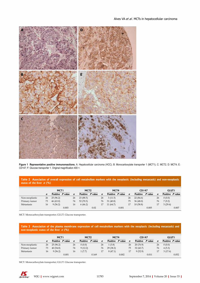
RESULTSFigure 1 illustrates positive immunoreactions for the dif-ferent markers analyzed in the different hepatic lesions. Data presented in Tables 2 and 3 show that the proteins were found in both the cytoplasm and the plasma mem-brane regions of cancer cells, at different frequencies. All
11782 September 7, 2014|Volume 20|Issue 33|WJG|www.wjgnet.com
Table 1 Immunohistochemistry protocols used to characterize the proteins expression
Code Industry Dilution Epitope retrieval Amplification system
MCT1 (sc-365501)
Santa Cruz Biotechnology, Santa Cruz, CA, United States
1:500 EDTA (1 mmol/L, pH = 8); Vectastain® Elite ABC reagent98 ℃; 20 min (Burlingame, CA, United States)
MCT2 (sc-14926)
Santa Cruz Biotechnology, Santa Cruz, CA, United States
1:200 Citrate buffer (10 mmol/L, pH = 6); 98 ℃; 20 min
Ultravision Detection System Anti-polyvalent, HRP, Lab Vision Corporation, Fremont, CA, United States)
MCT4 (sc-50329)
Santa Cruz Biotechnology, Santa Cruz, CA, United States
1:500 Citrate buffer (10 mmol/L, pH = 6); 98 ℃; 20 min
Ultravision Detection System Anti-polyvalent, HRP, Lab Vision Corporation, Fremont, CA, United States)
CD147 (sc-71038)
Santa Cruz Biotechnology, Santa Cruz, CA, United States
1:400 EDTA (1 mmol/L, pH = 8); Vectastain® Elite ABC reagent (Burlingame, CA, United States)98 ℃; 20 min
GLUT1 (ab15309)
Abcam, Cambridge, United Kingdom 1:500 Citrate buffer (10 mmol/L, pH = 6); 98 ℃; 20 min
Ultravision Detection System Anti-polyvalent, HRP, Lab Vision Corporation, Fremont, CA, United States)
Caspase 3 Diagnostic BioSystems (DBS) Pleasanton, CA, United States
1:160 Citrate buffer (10 mmol/L, pH = 6); 98 ℃; 40 min
NovoLink (Novocastra Laboratories Ltd, Newcastle Upon Tyne, United Kingdom)(3C SP03)
Keratin 19(K19, b170)
Novocastra Laboratories Ltd, Newcastle Upon Tyne, United Kingdom
1:300 Citrate buffer (10 mmol/L, pH = 6); 98 ℃; 40 min
NovoLink (Novocastra Laboratories Ltd, Newcastle Upon Tyne, United Kingdom)
Ki-67 (MIB-1)
(Dako, Glostrup, Denmark) 1:400 Citrate buffer (10 mmol/L, pH = 6); 98 ℃; 40 min
NovoLink (Novocastra Laboratories Ltd, Newcastle Upon Tyne, United Kingdom)
EGFR human (DAK-H1-WT)
(Dako, Glostrup, Denmark) 1:200 Citrate buffer (10 mmol/L, pH = 6); 98 ℃; 40 min
NovoLink (Novocastra Laboratories Ltd, Newcastle Upon Tyne, United Kingdom)
MCT: Monocarboxylate transporter; GLUT: Glucose transporter; EGFR: Epidermal growth factor receptor.
Alves VA et al . MCTs in hepatocellular carcinoma
11783 September 7, 2014|Volume 20|Issue 33|WJG|www.wjgnet.com
Table 2 Association of overall expression of cell metabolism markers with the neoplastic (including metastasis) and non-neoplastic status of the liver n (%)
MCT1 MCT2 MCT4 CD147 GLUT1
n Positive P value n Positive P value n Positive P value n Positive P value n Positive P valueNon-neoplastic 26 25 (96.2) 26 23 (88.5) 26 3 (11.5) 26 22 (84.6) 26 0 (0.0)Primary tumor 73 46 (63.0) 74 52 (70.3) 76 31 (40.8) 75 36 (48.0) 76 7 (9.2)Metastasis 16 9 (56.2) 16 6 (46.2) 17 11 (64.7) 17 10 (58.8) 17 5 (29.4)
0.003 0.02 0.001 0.005 0.007
MCT: Monocarboxylate transporter; GLUT: Glucose transporter.
Table 3 Association of the plasma membrane expression of cell metabolism markers with the neoplastic (including metastasis) and non-neoplastic status of the liver n (%)
MCT1 MCT2 MCT4 CD147 GLUT1
n Positive P value n Positive P value n Positive P value n Positive P value n Positive P valueNon-neoplastic 26 25 (96.2) 26 0 (0.0) 26 1 (3.8) 26 20 (76.9) 26 0 (0.0)Primary tumor 73 40 (54.8) 74 9 (12.2) 76 29 (38.2) 75 32 (42.7) 76 4 (5.3)Metastasis 16 9 (56.2) 16 1 (7.7) 17 8 (47.1) 17 9 (52.9) 17 3 (17.6)
0.001 0.169 0.002 0.011 0.052
MCT: Monocarboxylate transporter; GLUT: Glucose transporter.
Figure 1 Representative positive immunoreactions. A: Hepatocellular carcinoma (HCC); B: Monocarboxylate transporter 1 (MCT1); C: MCT2; D: MCT4; E: CD147; F: Glucose transporter 1. Original magnification 400 ×.
A
B
C
D
E
F
Alves VA et al . MCTs in hepatocellular carcinoma
11784 September 7, 2014|Volume 20|Issue 33|WJG|www.wjgnet.com
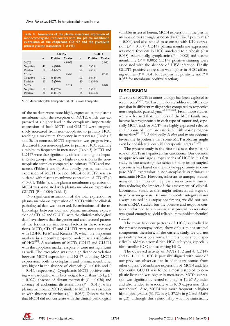
of the markers were more highly expressed at the plasma membrane, with the exception of MCT2, which was ex-pressed at a higher level in the cytoplasm. Importantly, expression of both MCT4 and GLUT1 were progres-sively increased from non-neoplastic to primary HCC, reaching a maximum frequency in metastases (Tables 2 and 3). In contrast, MCT2 expression was progressively decreased from non-neoplastic to primary HCC, reaching a minimum frequency in metastases (Table 3). MCT1 and CD147 were also significantly different among the hepat-ic lesion groups, showing a higher expression in the non-neoplastic samples compared to primary HCC and me-tastasis (Tables 2 and 3). Additionally, plasma membrane expression of MCT1, but not MCT4 or MCT2, was as-sociated with plasma membrane expression of CD147 (P < 0.001; Table 4), while plasma membrane expression of MCT4 was associated with plasma membrane expression GLUT1 (P = 0.004; Table 4).
No significant association of the total expression or plasma membrane expression of MCTs with the clinical-pathological data was observed. Examinations of the re-lationships between total and plasma membrane expres-sion of CD147 and GLUT1 with the clinical-pathological data have shown that the gender and architectural pattern of the lesions are important factors in these associa-tions. MCTs, CD147 and GLUT1 were not associated with EGFR, Ki-67 and Keratin 19, which are important markers in a recently proposed molecular classification of HCC[18]. Associations of MCTs, CD147 and GLUT1 with the apoptosis marker caspase 3, were not significant as well. The exception was the significant correlation between MCT4 expression and Ki-67 counting. MCT1 expression, both in cytoplasm and plasma membrane, was higher in the absence of cirrhosis (P = 0.044 and P = 0.015, respectively). Cytoplasmic MCT2 positive stain-ing was associated with liver weight lower than 1.5 kg (P = 0.027), absence of distant metastasis (P = 0.036) and absence of abdominal dissemination (P = 0.010), while plasma membrane MCT2, similar to MCT1, was associat-ed with absence of cirrhosis (P = 0.036). Despite the fact that MCT4 did not correlate with the clinical-pathological
variables assessed herein, MCT4 expression in the plasma membrane was strongly associated with Ki-67 positivity (P = 0.004) and also tended to associate with K19 expres-sion (P = 0.087). CD147 plasma membrane expression was more frequent in HCC unrelated to cirrhosis (P = 0.038). Additionally, cytoplasmic (P = 0.008) and plasma membrane (P = 0.005) CD147 positive staining were associated with the absence of HBV infection. Finally, GLUT1 positive expression was higher in HCC affect-ing women (P = 0.041 for cytoplasmic positivity and P = 0.033 for membrane positive reaction).
DISCUSSIONThe role of MCTs in tumor biology has been explored in recent years[3,6-9]. We have previously addressed MCTs ex-pression in different malignancies compared to respective non-neoplastic parenchyma[3,6-9,10-12,20]. From those studies, we have learned that members of the MCT family may behave heterogeneously in each type of tumor and, espe-cially MCT1 and/or MCT4, are highly expressed selected and, in some of them, are associated with worse prognos-tic markers[11,12,21]. Additionally, in vitro and in vivo evidence favors the hypothesis that some MCT isoforms might even be considered potential therapeutic targets[5,22,23].
The present study is the first to assess the possible role of MCTs in hepatocellular carcinoma. Our decision to approach our large autopsy series of HCC in this first study before assessing our series of biopsies or surgical specimens was based on the unique opportunity to com-pare MCT expression in non-neoplastic vs primary vs metastatic HCCs. However, inherent to autopsy studies, many of the tumors of the present series were advanced, thus reducing the impact of the assessment of clinical-laboratorial variables that might reflect initial steps of hepatocarcinogenesis. Because molecular integrity is not always assured in autopsy specimens, we did not per-form mRNA studies, but the positive and negative con-trols performed herein assure that protein preservation was good enough to yield reliable immunohistochemical studies.
The most frequent patterns of HCC, as studied in the present necropsy series, show only a minor stromal component; therefore, in the current study, we did not particularly focus on stroma. Future studies should spe-cifically address stromal-rich HCC subtypes, especially fibrolamellar HCC and sclerosing HCC.
The observed activity of MCTs 1, 2 and 4, CD147 and GLUT1 in HCC is partially aligned with most of our previous observations in adenocarcinomas from other organs[3]. Membrane expression of MCT4 and, less frequently, GLUT1 was found almost restricted to neo-plastic liver and was higher in metastases. MCT4 expres-sion was significantly related to a higher Ki-67 Ag index and also tended to associate with K19 expression (data not shown). Also, MCT4 was more frequent in higher histological grades (36.4% in g.1, 37.2% in g.2 and 63.6% in g.3), although this relationship was not statistically
CD147 GLUT1
n Positive P value n Positive P valueMCT1 < 0.001 1.000Negative 40 4 (10.0) 40 2 (5.0)Positive 74 56 (75.7) 74 4 (5.4)MCT2 0.766 0.434Negative 102 56 (54.9) 103 5 (4.9)Positive 10 5 (50.0) 10 1 (10.0)MCT4 0.114 0.004Negative 80 46 (57.5) 81 1 (1.2)Positive 36 15 (41.7) 38 6 (15.8)
Table 4 Association of the plasma membrane expression of monocarboxylate transporters with the plasma membrane expression of the chaperone CD147 and the glycolytic protein glucose transporter 1 n (%)
MCT: Monocarboxylate transporter; GLUT: Glucose transporter.
Alves VA et al . MCTs in hepatocellular carcinoma

11785 September 7, 2014|Volume 20|Issue 33|WJG|www.wjgnet.com
significant. These relevant data link MCT4 activity with high proliferative index and possibly to the “progenitor cell component” of HCC, pointing to a role of MCT4 in HCC progression and/or more aggressive course.
As previous studies have extensively shown, the tran-sition from high-grade dysplastic nodule to early HCC, as well as from “small, well-differentiated, indistinct border HCC” to “progressed HCC”, runs in parallel with an abrupt shift in the vascular pattern[24]. We speculate that this fact could lead to important neo-angiogenesis that is immunohistochemically identified by diffusely CD34-positive microvessels, which might correspond to the shift to a preferably glycolytic pattern of energy source through the Warburg effect. At this point, MCT4 and GLUT1 would become overexpressed, in accordance with previous evidence that MCT4 is highly expressed in response to hypoxia, mostly mediated by HIF-1 α[3,25]. Interestingly, due to the double arterial and portal vein blood inflow, the liver is per se an organ normally exposed to “hypoxia”, which is potentiated in cirrhosis and in malignant transformation[26]. In the cirrhotic liver, inflam-mation, hepatocytic lesion formation and regeneration, and neo-vessel formation are associated with the hypoxic setting, and stem cell activation is stimulated because these cells are able to resist this acidic/hypoxic microen-vironment where cancer development and progression are strongly favored[24]. In livers chronically infected by HBV and HCV, the consequent cirrhosis is associated with a remarkable decrease in oxygen supply and, as such, the microenvironment induces stabilization of HIF-1α, which promotes angiogenesis by activating the transcrip-tion of vascular endothelial growth factor and cyclooxy-genases and activating matrix metalloproteinases[26]. In summary, hypoxia induces cell damage and inflammation, limits liver regeneration and is strongly associated with HCC development[27]. Additionally, hypoxia was proved to be associated with glycolysis upregulation[28]. This result was partially demonstrated in the present study, as MCT4 and GLUT1 were more frequently expressed in primary HCC and metastasis than in non-neoplastic hepatocytes, thus corroborating previous evidence that GLUT1 is not detectable in normal epithelial tissues and benign epithelial neoplasms[28,29]. GLUT1 expression was previously demonstrated to be increased in HCC and promotes hepatic carcinogenesis[29].
MCT1 and its chaperone CD147, as well as MCT2, were found in this study to be more frequently expressed in non-neoplastic tissue than in HCC. Among the hepa-tocellular carcinomas, these proteins were significantly most commonly expressed in cases not related to HBV infection and in those occurring in non-cirrhotic livers. These observations warrant additional studies. This evi-dence points to the possible important role of MCT1 and CD147, and possibly MCT2, in the liver cell energy system. In contrast, MCT4 seems to be responsible for maintaining the glycolytic and acid resistant phenotype of cancer cells during the progression of HCC, with higher expression in advanced stages. In this context, it is
important to highlight that while MCT4 affinity for the substrate makes this protein a transporter specialized in lactate efflux, MCT1 affinity for the substrate allows this isoform to promote both uptake and efflux of lactate. Therefore, the expression of MCT1, associated with its chaperone CD147, in non-neoplastic hepatic tissue may be related to one or both lactate transport directions, but most likely with the uptake of lactate because this tissue is a gluconeogenic tissue. Following this rationale, it was also expected to find plasma membrane expression of MCT2 (the isoform specialized for substrate uptake) in non-neoplastic hepatic tissue, as previously described for normal liver[25]. However, plasma membrane expression of MCT2 was not found in non-neoplastic tissue. Instead, MCT2 was highly expressed in the cytoplasm of non-neoplastic tissue and progressively decreased towards me-tastasis. This important cytoplasmic expression of MCT2 might be related to mitochondrial pyruvate transport, as MCTs were already described to be present in the mito-chondria[30]. Because the metabolic behavior of liver cells in cancer will be adapted towards glucose consumption instead of glucose production, the expression of MCT2 will no longer be required, explaining the progressive de-crease of MCT2 expression in the cytoplasm. In fact, the portion of tumors that maintains MCT2 expression in the cytoplasm should have a metabolism more similar to that found in normal tissues and therefore be less aggres-sive. This result explains the association between MCT2 cytoplasmic expression and variables related to a less aggressive profile, such as lower liver weight, absence of metastasis and absence of abdominal dissemination.
Interestingly, the increase in plasma membrane ex-pression of MCT4 was not accompanied by CD147, similar to what was observed and expected for MCT2, but in contrast to what was observed for MCT1 (since, as expected, MCT1 was co-expressed with CD147). This information points to the existence of another MCT chaperone, as already hypothesized by our group in other studies[20]. Therefore, further studies will be important to search for the additional MCT chaper-one, as it is essential for MCT activity and may also be regulating its expression. It is important to emphasize that MCTs and their chaperones are predominantly expressed in plasma membrane, their natural location, in both normal and neoplastic cells. Thus, it is not sur-prising that the hyper-expression of MCTs and CD147 corresponded more consistently with clinical data in dif-ferent solid tumors[3].
In conclusion, our results assessed for the first time the role of MCTs in the liver and in HCC. In the present autopsy series, mostly representative of advanced HCC, MCT4 and GLUT1 were progressively highly expressed from non-neoplastic to primary HCC to metastatic hepatocellular carcinoma, in contrast to MCT2 that de-creased towards malignancy and was associated with less advanced tumors. In addition to pointing to an impor-tant role of the hypoxia pathways in the progression of HCC, these data might add HCC to the list of possible
Alves VA et al . MCTs in hepatocellular carcinoma

11786 September 7, 2014|Volume 20|Issue 33|WJG|www.wjgnet.com
beneficiaries of anti-MCT therapies. Moreover, MCT4 was strongly associated with augmented Ki-67 expres-sion, showing also a relevant trend to association with the “progenitor-cell component-related” keratin 19 and to higher histological grades. Future studies should further assess morphologically and molecularly the interactions related to the hypoxia and metabolic pathways in the de-velopment of HCC, especially approaching surgical sam-ples representative of the sequence cirrhosis - dysplastic nodules - HCC.
COMMENTSBackgroundTo concisely and accurately summarize the related background of the article and to enable the readers to gain some basic knowledge relevant to the article, thus helping them better understand the significance of the article.Research frontiersTo briefly introduce the hotspots or important areas in the research field related to the article.Innovations and breakthroughsTo summarize and emphasize the differences, particularly the advances, achievements, innovations and breakthroughs, from the other related or similar articles so as to allow the readers to catch up the major points of the article. Applications To summarize the actual application values, the implications for further applica-tion and modification, or the perspectives of future application of the article.TerminologyTo concisely and accurately describe, define or explain the specific, unique terms that are not familiar to majority of the readers, but are essential for the readers to understand the article.Peer reviewTo provide the comments from peer reviewers that most represent the charac-teristics, values and significance of the article, and allow the readers to have an objective point of view toward the article.
REFERENCES1 Jungermann K, Kietzmann T. Oxygen: modulator of meta-
bolic zonation and disease of the liver. Hepatology 2000; 31: 255-260 [PMID: 10655244]
2 Nath B, Szabo G. Hypoxia and hypoxia inducible factors: diverse roles in liver diseases. Hepatology 2012; 55: 622-633 [PMID: 22120903 DOI: 10.1002/hep.25497]
3 Pinheiro C, Longatto-Filho A, Azevedo-Silva J, Casal M, Schmitt FC, Baltazar F. Role of monocarboxylate trans-porters in human cancers: state of the art. J Bioenerg Bio-membr 2012; 44: 127-139 [PMID: 22407107 DOI: 10.1007/s10863-012-9428-1]
4 Warburg O. On the origin of cancer cells. Science 1956; 123: 309-314 [PMID: 13298683]
5 Miranda-Gonçalves V, Honavar M, Pinheiro C, Martinho O, Pires MM, Pinheiro C, Cordeiro M, Bebiano G, Costa P, Pal-meirim I, Reis RM, Baltazar F. Monocarboxylate transporters (MCTs) in gliomas: expression and exploitation as therapeu-tic targets. Neuro Oncol 2013; 15: 172-188 [PMID: 23258846 DOI: 10.1093/neuonc/nos298]
6 Pinheiro C, Longatto-Filho A, Scapulatempo C, Ferreira L, Martins S, Pellerin L, Rodrigues M, Alves VA, Schmitt F, Baltazar F. Increased expression of monocarboxylate transporters 1, 2, and 4 in colorectal carcinomas. Virchows Arch 2008; 452: 139-146 [PMID: 18188595 DOI: 10.1007/s00428-007-0558-5]
7 Pinheiro C, Longatto-Filho A, Ferreira L, Pereira SM, Etlinger D, Moreira MA, Jubé LF, Queiroz GS, Schmitt F, Baltazar F. Increasing expression of monocarboxylate trans-
porters 1 and 4 along progression to invasive cervical carci-noma. Int J Gynecol Pathol 2008; 27: 568-574 [PMID: 18753962 DOI: 10.1097/PGP.0b013e31817b5b40]
8 Pinheiro C, Longatto-Filho A, Pereira SM, Etlinger D, Moreira MA, Jubé LF, Queiroz GS, Schmitt F, Baltazar F. Monocarboxylate transporters 1 and 4 are associated with CD147 in cervical carcinoma. Dis Markers 2009; 26: 97-103 [PMID: 19597291 DOI: 10.3233/DMA-2009-0596]
9 Pinheiro C, Albergaria A, Paredes J, Sousa B, Dufloth R, Vieira D, Schmitt F, Baltazar F. Monocarboxylate transporter 1 is up-regulated in basal-like breast carcinoma. Histopa-thology 2010; 56: 860-867 [PMID: 20636790 DOI: 10.1111/j.1365-2559.2010.03560.x]
10 Pinheiro C, Longatto-Filho A, Simões K, Jacob CE, Bresciani CJ, Zilberstein B, Cecconello I, Alves VA, Schmitt F, Baltazar F. The prognostic value of CD147/EMMPRIN is associated with monocarboxylate transporter 1 co-expression in gastric cancer. Eur J Cancer 2009; 45: 2418-2424 [PMID: 19628385 DOI: 10.1016/j.ejca.2009.06.018]
11 Pértega-Gomes N, Vizcaíno JR, Miranda-Gonçalves V, Pinheiro C, Silva J, Pereira H, Monteiro P, Henrique RM, Reis RM, Lopes C, Baltazar F. Monocarboxylate transporter 4 (MCT4) and CD147 overexpression is associated with poor prognosis in prostate cancer. BMC Cancer 2011; 11: 312 [PMID: 21787388 DOI: 10.1186/1471-2407-11-312]
12 Pinheiro C, Sousa B, Albergaria A, Paredes J, Dufloth R, Vieira D, Schmitt F, Baltazar F. GLUT1 and CAIX expression profiles in breast cancer correlate with adverse prognostic factors and MCT1 overexpression. Histol Histopathol 2011; 26: 1279-1286 [PMID: 21870331]
13 Mathupala SP, Parajuli P, Sloan AE. Silencing of mono-carboxylate transporters via small interfering ribonucleic acid inhibits glycolysis and induces cell death in malignant glioma: an in vitro study. Neurosurgery 2004; 55: 1410-1419; discussion 1419 [PMID: 15574223]
14 Pinheiro C, Longatto-Filho A, Soares TR, Pereira H, Bedros-sian C, Michael C, Schmitt FC, Baltazar F. CD147 immuno-histochemistry discriminates between reactive mesothelial cells and malignant mesothelioma. Diagn Cytopathol 2012; 40: 478-483 [PMID: 22619123 DOI: 10.1002/dc.22821]
15 Ferlay J, Shin HR, Bray F, Forman D, Mathers C, Parkin DM. Estimates of worldwide burden of cancer in 2008: GLOBO-CAN 2008. Int J Cancer 2010; 127: 2893-2917 [PMID: 21351269 DOI: 10.1002/ijc.25516]
16 Montalto G, Cervello M, Giannitrapani L, Dantona F, Ter-ranova A, Castagnetta LA. Epidemiology, risk factors, and natural history of hepatocellular carcinoma. Ann N Y Acad Sci 2002; 963: 13-20 [PMID: 12095924]
17 Felipe da Silva AS. Expression analysis of EGFR and related proteins in hepatocellular carcinoma and surrounding liver tissue metastases: clinicopathologic study in autopsies. Bra-zil: PhD Thesis, Faculdade de Medicina da Universidade de São Paulo, 2013
18 Hoshida Y, Toffanin S, Lachenmayer A, Villanueva A, Min-guez B, Llovet JM. Molecular classification and novel targets in hepatocellular carcinoma: recent advancements. Semin Liver Dis 2010; 30: 35-51 [PMID: 20175032 DOI: 10.1055/s-0030-1247131]
19 Rüschoff J, Kerr KM, Grote HJ, Middel P, von Heydebreck A, Alves VA, Baldus SE, Büttner R, Carvalho L, Fink L, Jochum W, Lo AW, López-Ríos F, Marx A, Molina TJ, Olszewski WT, Rieker RJ, Volante M, Thunnissen E, Wrba F, Celik I, Störkel S. Reproducibility of immunohistochemical scoring for epi-dermal growth factor receptor expression in non-small cell lung cancer: round robin test. Arch Pathol Lab Med 2013; 137: 1255-1261 [PMID: 23270410 DOI: 10.5858/arpa.2012-0605-OA]
20 Pinheiro C, Reis RM, Ricardo S, Longatto-Filho A, Schmitt F, Baltazar F. Expression of monocarboxylate transporters 1, 2, and 4 in human tumours and their association with CD147 and CD44. J Biomed Biotechnol 2010; 2010: 427694 [PMID:
COMMENTS
Alves VA et al . MCTs in hepatocellular carcinoma

11787 September 7, 2014|Volume 20|Issue 33|WJG|www.wjgnet.com
20454640 DOI: 10.1155/2010/427694]21 de Oliveira AT, Pinheiro C, Longatto-Filho A, Brito MJ,
Martinho O, Matos D, Carvalho AL, Vazquez VL, Silva TB, Scapulatempo C, Saad SS, Reis RM, Baltazar F. Co-expres-sion of monocarboxylate transporter 1 (MCT1) and its chap-erone (CD147) is associated with low survival in patients with gastrointestinal stromal tumors (GISTs). J Bioenerg Biomembr 2012; 44: 171-178 [PMID: 22281667 DOI: 10.1007/s10863-012-9408-5]
22 Sonveaux P, Végran F, Schroeder T, Wergin MC, Verrax J, Rabbani ZN, De Saedeleer CJ, Kennedy KM, Diepart C, Jordan BF, Kelley MJ, Gallez B, Wahl ML, Feron O, Dewhirst MW. Targeting lactate-fueled respiration selectively kills hypoxic tumor cells in mice. J Clin Invest 2008; 118: 3930-3942 [PMID: 19033663 DOI: 10.1172/JCI36843]
23 Pinheiro C, Longatto-Filho A, Nogueira R, Schmitt F, Balta-zar F. Lactate-induced IL-8 pathway in endothelial cells--letter. Cancer Res 2012; 72: 1901-1902; author reply 1903-1904 [PMID: 22473315 DOI: 10.1158/0008-5472]
24 Roncalli M, Park YN, Di Tommaso L. Histopathological classification of hepatocellular carcinoma. Dig Liver Dis 2010; 42 Suppl 3: S228-S234 [PMID: 20547308 DOI: 10.1016/S1590-8658(10)60510-5]
25 Halestrap AP, Wilson MC. The monocarboxylate transporter family--role and regulation. IUBMB Life 2012; 64: 109-119
[PMID: 22162139 DOI: 10.1002/iub.572]26 Arzumanyan A, Reis HM, Feitelson MA. Pathogenic mecha-
nisms in HBV- and HCV-associated hepatocellular carcino-ma. Nat Rev Cancer 2013; 13: 123-135 [PMID: 23344543 DOI: 10.1038/nrc3449]
27 Ripoli M, D’Aprile A, Quarato G, Sarasin-Filipowicz M, Gouttenoire J, Scrima R, Cela O, Boffoli D, Heim MH, Mo-radpour D, Capitanio N, Piccoli C. Hepatitis C virus-linked mitochondrial dysfunction promotes hypoxia-inducible fac-tor 1 alpha-mediated glycolytic adaptation. J Virol 2010; 84: 647-660 [PMID: 19846525 DOI: 10.1128/JVI.00769-09]
28 Airley RE, Mobasheri A. Hypoxic regulation of glucose transport, anaerobic metabolism and angiogenesis in cancer: novel pathways and targets for anticancer therapeutics. Che-motherapy 2007; 53: 233-256 [PMID: 17595539]
29 Amann T, Maegdefrau U, Hartmann A, Agaimy A, Marien-hagen J, Weiss TS, Stoeltzing O, Warnecke C, Schölmerich J, Oefner PJ, Kreutz M, Bosserhoff AK, Hellerbrand C. GLUT1 expression is increased in hepatocellular carcinoma and pro-motes tumorigenesis. Am J Pathol 2009; 174: 1544-1552 [PMID: 19286567 DOI: 10.2353/ajpath.2009.080596]
30 Benton CR, Campbell SE, Tonouchi M, Hatta H, Bonen A. Monocarboxylate transporters in subsarcolemmal and in-termyofibrillar mitochondria. Biochem Biophys Res Commun 2004; 323: 249-253 [PMID: 15351729]
P- Reviewer: Gong Y, Hung LY, Liu TC S- Editor: Gou SX L- Editor: A E- Editor: Wang CH
Alves VA et al . MCTs in hepatocellular carcinoma

© 2014 Baishideng Publishing Group Inc. All rights reserved.
Published by Baishideng Publishing Group Inc8226 Regency Drive, Pleasanton, CA 94588, USA
Telephone: +1-925-223-8242Fax: +1-925-223-8243
E-mail: [email protected] Desk: http://www.wjgnet.com/esps/helpdesk.aspx
http://www.wjgnet.com
I S S N 1 0 0 7 - 9 3 2 7
9 7 7 1 0 07 9 3 2 0 45
3 3