characterization of conserved arginine residues on cdt1 … wild-type and rr-aa, and pro61->ser61...
TRANSCRIPT

Full Terms & Conditions of access and use can be found athttp://www.tandfonline.com/action/journalInformation?journalCode=kccy20
Download by: [Tokyoto Igaku Kenkyu Kiko] Date: 18 April 2017, At: 05:57
Cell Cycle
ISSN: 1538-4101 (Print) 1551-4005 (Online) Journal homepage: http://www.tandfonline.com/loi/kccy20
Characterization of conserved arginine residueson Cdt1 that affect licensing activity andinteraction with Geminin or Mcm complex
Zhiying You, Koji L. Ode, Mayumi Shindo, Haruhiko Takisawa & Hisao Masai
To cite this article: Zhiying You, Koji L. Ode, Mayumi Shindo, Haruhiko Takisawa & HisaoMasai (2016) Characterization of conserved arginine residues on Cdt1 that affect licensingactivity and interaction with Geminin or Mcm complex, Cell Cycle, 15:9, 1213-1226, DOI:10.1080/15384101.2015.1106652
To link to this article: http://dx.doi.org/10.1080/15384101.2015.1106652
View supplementary material Accepted author version posted online: 03Mar 2016.Published online: 03 Mar 2016.
Submit your article to this journal Article views: 294
View related articles View Crossmark data
Citing articles: 1 View citing articles

REPORT
Characterization of conserved arginine residues on Cdt1 that affect licensing activityand interaction with Geminin or Mcm complex
Zhiying Youa, Koji L. Odeb,y, Mayumi Shindoc, Haruhiko Takisawab, and Hisao Masaia
aDepartment of Genome Medicine, Tokyo Metropolitan Institute of Medical Science, Tokyo, Japan; bDepartment of Biological Sciences, Graduate Schoolof Science, Osaka University, Toyonaka, Osaka, Japan; cLaboratory of Protein Analysis, Tokyo Metropolitan Institute of Medical Science, Tokyo, Japan
ARTICLE HISTORYReceived 27 May 2015Revised 30 September 2015Accepted 7 October 2015
ABSTRACTAll organisms ensure once and only once replication during S phase through a process called replicationlicensing. Cdt1 is a key component and crucial loading factor of Mcm complex, which is a centralcomponent for the eukaryotic replicative helicase. In higher eukaryotes, timely inhibition of Cdt1 byGeminin is essential to prevent rereplication. Here, we address the mechanism of DNA licensing usingpurified Cdt1, Mcm and Geminin proteins in combination with replication in Xenopus egg extracts. Wemutagenized the 223th arginine of mouse Cdt1 (mCdt1) to cysteine or serine (R-S or R-C, respectively) and342nd and 346th arginines constituting an arginine finger-like structure to alanine (RR-AA). The RR-AAmutant of Cdt1 could not only rescue the DNA replication activity in Cdt1-depleted extracts but also itsspecific activity for DNA replication and licensing was significantly increased compared to the wild-typeprotein. In contrast, the R223 mutants were partially defective in rescue of DNA replication and licensing.Biochemical analyses of these mutant Cdt1 proteins indicated that the RR-AA mutation disabled itsfunctional interaction with Geminin, while R223 mutations resulted in ablation in interaction with theMcm2»7 complex. Intriguingly, the R223 mutants are more susceptible to the phosphorylation-inducedinactivation or chromatin dissociation. Our results show that conserved arginine residues play critical rolesin interaction with Geminin and Mcm that are crucial for proper conformation of the complexes and itslicensing activity.
KEYWORDSCdc2/Cyclin B; Cdc7 kinase;Cdk2/Cyclin A; Cdt1; cellcycle; DNA helicase; DNAreplication; DNA-bindingactivity; licensing; Mcm
Introduction
During the course of DNA replication, multiprotein complexes,called pre-replicative complexes (pre-RCs), containing Orc1–6(origin recognition complex 1–6), Cdc6, Cdt1 and Mcm2»7(minichromosome maintenance 2»7), need to be sequentiallyassembled to establish ‘licensed’ origins.1,2 Cdt1 plays a criticalrole in the licensing process to load the hexameric Mcm2»7complex (an eukaryotic replicative helicase) onto chromatinduring G1 phase. At the onset of S phase, Cdt1 is renderedinactive through ubiquitin-dependent proteolysis in a CDK-dependent and PCNA (proliferating cell nuclear antigen)-dependent manners3-7 and through the inhibitor Geminin.8
Geminin directly binds to Cdt1 and prevents the loading of theMcm2»7 complex onto DNA.8-12 This double regulationensures that Cdt1 is not present during S phase, resulting inprevention of DNA re-replication.
To strictly enforce once per cell cycle replication, it is neces-sary to coordinate the temporal and spatial regulation of licens-ing. The level of Cdt1 protein oscillates during cell cycle (highin the G1 phase, low in S phase, and high again at the M-G1transition).13 Geminin accumulates during S, G2, and Mphases, and is destabilized during G1 phase of the cell cycle.8,9
It has been proposed that Cdt1 associates dynamically with
chromatin throughout G1 phase and the licensing inhibitorGeminin is recruited simultaneously by Cdt1 onto chromatin,and that the stoichiometry of the Cdt1:Geminin complex couldact as a molecular switch that determines when the origins ofreplication are on (licensing) or off (inhibition).14-17 In addi-tion, crystallographic studies suggest the presence of 2 formsthat are recruited to chromatin. One is a “permissive” 1:2 Cdt1-Geminin heterotrimer complex that is able to load Mcm; theother is an “inhibitory” 2:4 Cdt1-Geminin heterohexamer com-plex that is unable to engage Mcms, thus preventing new pre-RCs formation.18 Phosphorylation also regulates the functionof Cdt1, since a mutant Cdt1 with a 6-amino-acid deletion,which was not efficiently phosphorylated during mitosis andwas more strongly associated with chromatin, showedenhanced licensing activity.19 It is known that Cdk (cyclin-dependent kinasea) and Cdc7 kinase are involved in regulationof Cdt1.2,6,20,21
It was proposed that 2 Cdt1 molecules recruit double hex-americ Mcm2»7 complex loading during pre-RC formation.22
Previously, we reported that mouse Cdt1 forms a complex withMcm4/6/7, Mcm2/3/4/5/6/7 and Mcm2/4/6/7 and that Cdt1significantly stimulates the DNA binding and helicase activitiesof Mcm4/6/7, an active helicase complex.23 It was reported that
CONTACT Zhiying You [email protected] Department of Genome Medicine, Tokyo Metropolitan Institute of Medical Science, Tokyo 156-8506, Japan.Color versions of one or more of the figures in the article can be found online at www.tandfonline.com/kccy.yCurrent address: Department of Systems Pharmacology, Graduate School of Medicine, The University of Tokyo, Bunkyo-ku, Tokyo, Japan
Supplemental data for this article can be accessed on the publisher’s website.© 2016 Taylor & Francis
CELL CYCLE2016, VOL. 15, NO. 9, 1213–1226http://dx.doi.org/10.1080/15384101.2015.1106652

C-terminal region of Cdt1 is required for interaction withMcm.11 More recently, charge complementarity between theC-terminal conserved residues of Cdt1 and Mcm6 was shownto be important for the Cdt1-Mcm2»7 interaction and licens-ing activity by mutagenesis analysis.23-26 However, the Cdt1-mediated Mcm2»7 chromatin loading and licensing is morecomplex, and the central domain of Cdt1 was also reported tobe required for interaction with Mcm2»7 and licensing activ-ity.17,27 Thus, it remains controversial how Cdt1 acts duringMcm loading.
Alignment of Cdt1 sequences revealed that Cdt1 R223 andarginine finger-like residues (R342, 346) are well conserved inhuman, mouse, Xenopus, and Drosophila. A mutant of Dro-sophila Cdt1 containing cystine substitution at this conservedR223 exhibits female-sterile phenotype, suggesting that it isfunctionally important.28 We mutagenized the correspondingarginine (R223) of mouse Cdt1 to cystine or serine and argininefinger-like residues (R342, 346) to alanine. We report here thatmutations of these 3 arginine residues (R-S, R-C and RR-AA)profoundly affect DNA replication, pre-RC formation, interac-tion with Mcm and Geminin and phosphorylation, suggestingthey are critical for licensing activity of Cdt1.
Results
Construction and purification of mutant Cdt1 proteins withamino acid substitutions at conserved arginine residues
We previously reported that the mouse Cdt1 forms complexeswith both Mcm2»7 and hexameric Mcm4/6/7 through directinteraction with Mcm2 and Mcm6 subunit.23 We have alignedthe sequences of Cdt1 from different species (human, mouse,Xenopus, and Drosophila) and have identified 3 conserved argi-nine residues (R223, R342 and R346) (Fig. S1). A Drosophilamutant carrying arginine to cysteine amino acid substitution atR223 exhibits female-sterile phenotype, suggesting functionalimportance of this residue.28 Furthermore, alignment with E.coli DnaA protein revealed similarity of the sequences aroundR342 and R346 of Cdt1 with those around Arg281 and Arg285of DnaA (Fig. S1B). Both arginine residues are required forDnaA functions and Arg285 residue constitutes the argininefinger.29 Thus, we mutagenized R223 of mouse Cdt1 to cystineor serine and R342/R346 to alanine (Fig. 1A). We have realizedthat the plasmids expressing the wild-type and mutant Cdt1inadvertently contained the following mutations; Ile24->Thr24in wild-type and RR-AA, and Pro61-> Ser61 in R-S/R-C. Theseamino acid changes occur at non-conserved residues and it ismore likely that they do not affect the functions of Cdt1,although we cannot completely rule out the possibility thatthey may affect the functions in some unknown way.
We expressed wild-type and mutant Cdt1 proteins using thebaculo-virus expression system and purified them through theconsecutive steps involving nickel agarose affinity chromatog-raphy, anti-Flag M2 antibody-agarose affinity chromatography,and glycerol gradient sedimentation (Fig. 1B). The purifiedCdt1 proteins appear to be almost homogeneous in silver stain-ing (Fig. 1C). However, in helicase assays, contaminated nucle-ases were detected which specifically degraded the singlestranded DNA in the fractions near the bottom (Fig. S2C,
lower). Further purification by the mono-Q column also couldnot remove the nuclease (Fig. S2A and S2B). The contaminat-ing nuclease prohibited accurate measurement of the helicaseactivity (Fig. S2B and S2C, middle). Thus, Cdt1 proteins werefurther purified by applying the first glycerol gradient fractions(11 to 14) to the second Ni-sepharose column and glycerol gra-dient fractionation (Fig. 1D). As a result, we were able to obtainfractions virtually free from contaminating nucleases. All themutants Cdt1 migrated at around 60 kDa (monomer position)in glycerol gradient as the wild-type protein, although the peakfraction of the mutants was smaller than the wild-type by onefraction. Thus, the results suggest that the mutation did not sig-nificantly affect the size or the shape of the protein.
Licensing and DNA replication activities of mutant Cdt1
In order to evaluate the licensing and DNA replication activi-ties of Cdt1, the wild type and mutated Cdt1 proteins wereassayed for their ability to restore the replication and Mcm-loading activities in Xenopus egg extract immunodepleted ofendogenous Xenopus Cdt1 (xCdt1). Depletion of endogenousxCdt1 led to loss of Mcm loading, whereas chromatin loadingof Orc and Cdc6 proteins increased (Fig. 2A, lane 3).30 Addi-tion of the wild-type Cdt1 protein to the Cdt1-depleted extractsrestored Mcm2 loading in a dose-dependent manner (Fig. 2A,lanes 4–6). However, R-S and R-C mutants showed signifi-cantly impaired Mcm2 loading activity (Fig. 2A, lanes 7–12). Incontrast, the RR-AA mutant restored the Mcm2 loading moreefficiently than the wild-type; chromatin loading was signifi-cantly restored even at 0.6 nM by RR-AA but not by the wild-type (Fig. 2A, lanes 6 and 15). The chromatin binding ofPCNA, DNA polymerase a and DNA polymerase e at 75 minwas restored 0.6 nM by RR-AA more efficiently than by thewild-type (Fig. 2A, lanes 21 and 30), consistent with the effecton Mcm loading. These results suggest that these arginine resi-dues of Cdt1 play critical roles in its licensing activity.
The differential efficacy of mutant Cdt1 proteins in Mcm-loading was recapitulated also in DNA replication assay. ACdt1-depleted extract did not replicate sperm chromatin, butDNA replication could be restored by addition of the wild-typeCdt1 protein (Fig. 2B). Consistent with previous study,31 Cdt1has an optimum concentration for DNA replication; sufficientDNA replication was not observed at a low concentration dueto inefficient Mcm loading, whereas the elongation of nascentDNA strand is inhibited at a higher concentration. Cdt1 addedat the concentration from 3.7 to 15 nM was optimum for DNAreplication. In contrast, R-S and R-C mutants showed impairedDNA replication at the concentration below 3.75 nM, consis-tent with its weak licensing activity. In contrast, the RR-AAmutant stimulated DNA replication more efficiently than thewild type, especially at low concentrations (1.86–0.23 nM)(Fig. 2B). These results indicate that the arginine finger-likemutant of Cdt1 has more potent licensing and DNA replicationactivities than the wild-type but that the R223 mutationsdecrease these activities.
We next evaluated the role of Geminin in functions of theseCdt1 mutants by codepleting Geminin. To inhibit the licensingactivity of endogenous xCdt1, approximately 100 nM of Gemi-nin was required.8,30,32,33 In mock depletion, Xenopus egg
1214 Z. YOU ET AL.

Figure 1. Purification of various recombinant Cdt1 proteins. (A) Schematic representation of Cdt1 protein showing the Geminin- and Mcm-binding motifs, and the muta-genized arginines. The two regions of Cdt1 required for association with Geminin are indicated by gray boxes. The C-terminal region of Cdt1 required for interact withMCM is indicated by a cross-hatched box. (B) Diagram explaining the procedure of purification of Cdt1 protein. (C) Wild-type or various mutant forms of Cdt1 protein,expressed in insect cells, were purified by Ni-NTA and anti-Flag antibody beads, and then were fractionated on 15-35% glycerol gradient (38,000 rpm for 17 hr). (D) Afterfirst glycerol gradient fractionation, the fractions from 10 to 14 of mCdt1 were pooled and further purified with Ni-NTA column and glycerol gradient fractionation. Eachfraction was subjected to SDS-PAGE (5–20% polyacrylamide), followed by staining with silver. Degradation products (~50 kDa) were co-purified with the full-lengthproteins in case of the mutant proteins.
CELL CYCLE 1215

extract was supplemented with sufficient recombinant Geminin(400 nM) to inhibit Mcm loading onto the chromatin (Fig. 3A,compare lanes 1 and 2). An equal amount of Cdt1 protein(wild-type or mutant) was added to Cdt1 and Geminin double-depleted extract (Fig. 3A, lanes 4, 6, 8 and 10). We found thatRR-AA could load Mcm more efficiently than the wild typeCdt1 in the absence of added Geminin, whereas the R-S or R-Cmutant could not. The presence of recombinant Geminin didnot completely inhibit the licensing activity of mutant Cdt1RR-AA (Fig. 3A, compare lanes 5 and 11). Titration of Gemi-nin in Cdt1-depleted extracts indicate that RR-AA could loadMcm on chromatin even at a high concentration of Geminin(120 nM) (Fig. 3B, lanes 11–16), while Mcm loading was inhib-ited at 40 nM of added Geminin with the wild-type Cdt1(Fig. 3B, lanes 4-9). Geminin added at a concentration as highas 600 nM, under which the Mcm loading was completely
inhibited in the egg extracts, did not completely suppressMcm loading in the presence of the RR-AA mutant Cdt1(Fig. 3C, lane 10). This result suggests that RR-AA mutantis refractory to the licensing inhibition by Geminin. Toevaluate whether the elevated licensing activity of the RR-AA mutant can be explained by its resistance to Geminin,we carefully compared the licensing activity of wild-typeand the mutant in the absence of Geminin. In xCdt1 andGeminin double-depleted extract, RR-AA activated theMcm2 loading significantly more effectively than the wildtype at high (10 nM) and low (3.3 nM) concentrations(Fig. 3C, lanes 14–25; 3 independent experiments. See alsoFig. S3). Since an additional mutation in RR-AA and thewild-type is the same, the altered function in RR-AA ismost likely mediated by the amino acid changes at the 2arginine residues. This result suggests that the arginine
Figure 2. Licensing and replication activities of various mutant Cdt1 proteins. (A) Western blot analysis of Mcm2, Orc2, Cdc6, Cdt1, pol a, pol e, and PCNA on chromatin inthe egg extract at 15 min and 75 min. S-phase entry occurs at about 40 min under the condition employed. The extracts were either mock-depleted or Cdt1-depleted incombination with buffer or wild-type/mutant Cdt1 proteins (60, 6 and 0.6 nM). Asterisk indicates a non-specific band. Actin bands stained with Ponceau S are shown asloading control to show chromatin recovery. (B) DNA replication at 60 min and 90 min in either mock-depleted extract or Cdt1-depleted extract supplemented with bufferor wild-type/mutant Cdt1 in the range of 60 nM to 0.23 nM. The replication efficiency is shown by setting the value of [a-32P]dATP incorporation in the presence of WTmCdt1 at 90 min as 100%. Two independent experiments were carried out with very similar results.
1216 Z. YOU ET AL.

finger-like mutant of Cdt1 can exert hyper-licensing activityeven in a Geminin-independent manner.
The licensing activities of mutant Cdt1 proteins werefurther examined by more carefully analyzing their abilityto counteract the inhibition of DNA replication by Gemi-nin. Addition of Geminin (160 nM) completely inhibitedDNA replication (Fig. 3D). Under this condition, additionof the wild-type Cdt1 at 60 nM restored DNA replicationactivity to the level without Geminin, while only a low orvery little DNA replication was observed in the presence of
the wild-type Cdt1 at 7.5 nM or 0.9 nM, respectively. Onthe other hand, R-S and R-C Cdt1 could not rescue theDNA replication even at 60 nM. In contrast, efficient DNAreplication was observed with RR-AA even at 0.9 nM.Taken together, these results are consistent with the conclu-sion that the R-S and R-C mutants are significantlyimpaired in licensing and replication activities, whereas RR-AA is hyperactive. The latter mutant appears to be insensi-tive to inhibition by Geminin, but is also able to stimulatethe licensing in a manner independent of Geminin. We
Figure 3. Licensing and replication activities of various mutant Cdt1 proteins in the presence of Geminin. (A) Western blot analysis of Mcm2, Orc2, Cdc6, Cdt1, and Gemi-nin on chromatin in the egg extract that was either mock-depleted or Cdt1/Geminin double-depleted in the presence of 6 nM wild-type or mutant Cdt1 (R-S, R-C, andRR-AA) proteins with (C) or without (¡) Geminin (400 nM). (B) Western blot analysis of indicated proteins in the Cdt1-depleted extract supplemented with 10 nM wild-type or RR-AA Cdt1 protein and Geminin protein at the indicated concentrations. (C) Western blot analysis of indicated proteins in the Cdt1-depleted extract in the pres-ence of various amounts of Geminin (lanes 4–10) or Cdt1/Geminin double-depleted extract supplemented with 10 and 3.3 nM wild-type or RR-AA proteins (lanes 13–25;3 independent experiments). The licensing activity of the mutant Cdt1/RR-AA is not significantly inhibited by addition of Geminin at a very high concentration. (D) Theability of the wild-type and mutant Cdt1 to restore DNA replication that has been inhibited by an excess Geminin protein was examined. Xenopus egg extract was supple-mented with 160 nM Geminin, which resulted in complete inhibition of DNA replication. Then wild-type or mutant Cdt1 proteins were added at the indicated concentra-tions and incubated in 60 and 90 min, respectively. The replication efficiency is shown by setting the value of [a-32P]dATP incorporation in the absence of mCdt1 andGeminin at 90 min as 100.
CELL CYCLE 1217

conclude that all the 3 arginine residues of Cdt1 playimportant roles in both licensing and replication.
Stimulation of helicase and DNA-binding activitiesof Mcm by Cdt1 protein
We previously reported that Cdt1 stimulates Mcm helicase.23
In order to understand how the arginine mutants affect thelicensing and replication activities, we next examined themutant Cdt1 proteins in DNA helicase assays using Mcm4/6/7.We first confirmed that the stimulation of Mcm4/6/7 helicaseactivity is correlated with the Cdt1 protein amount in glycerolgradient fractions (Fig. 4A). The intensities of the full-lengthbands in fractions #13 of the wild-type and mutant Cdt1 pro-teins in glycerol gradient fractionation were quantified and anequal amount (as a full-length polypeptide) was used for heli-case and DNA binding assays of the Mcm4/6/7 complex. Asshown in Figure 4B, all the Cdt1 mutants (R-S, R-C and RR-AA) have exhibited similar extent of stimulation of bothMcm4/6/7 helicase and single stranded DNA-binding activities(Fig. 4B, left and right; Fig. S4A). Since Cdt1 alone could notform clear protein-DNA complexes in gel-shift assays underthis condition (Fig. 4B right, lanes 15–18), the biotin-labeledsingle stranded and forked DNAs were prepared and the pull-down assay were carried out using streptavidin beads. We showthat each Cdt1 mutant protein efficiently binds to both single-stranded and forked DNA (Fig. 4C and 4D, lanes 10–13), indi-cating that all the mutant proteins bind to DNA with affinitysimilar to that of the wild-type. Nitrocellulose filter DNA bind-ing assays using labeled single-stranded and double-strandedDNA as well as the single-stranded and double-stranded DNAcellulose binding assays also showed that DNA-binding activi-ties of the wild-type and 3 mutant Cdt1 proteins are similar(data not shown and Fig. S4B).
Defective complex formation of mutant Cdt1 (R-S and R-C)and Mcm
It has been known that the licensing involves the recruitment ofMcm2»7 to chromatin by its direct interaction with Cdt1.11,23
We and others reported that mutations in the C-terminaldomain of Cdt1 reduced or abolished its interaction with Mcm,thereby inactivating chromatin loading and DNA replicationactivities.23-26 Therefore, we next set out to evaluate the interac-tion between Cdt1 and Mcm complex. The purified Mcm2alone, Mcm4/6/7 or Mcm2»7 complex was mixed with variousCdt1 (containing a Flag-tag), and immunoprecipitation usinganti-Flag antibody agarose beads, or anti-Cdt1 antibody cou-pled with protein G Dynabeads were performed. We foundthat the affinity of wild type or each mutant Cdt1 with Mcm2protein was similar (Fig. 5A, upper). Similarly, interaction withMcm4/6/7 or Mcm2»7 was not affected by the mutation(Fig. 5A, lower panels). These results indicate that the mutantCdt1 proteins physically interact with Mcm with similar affinityto that of the wild-type.
We then analyzed the complex formation using native gelelectrophoresis. Mcm2»7 migrates as a complex about600 kDa, consistent with it being a hexameric complex (Fig. 5B,lane 1). Upon addition of Cdt1, the band of the Mcm2»7
complex disappears and a slow-migrating band composed pre-sumably of Mcm2»7 and Cdt1 was detected (Fig. 5B, lane 2).Actually, the slow-migrating band was run on a second dimen-sion SDS-PAGE, the result of which confirmed that it consistedwith Mcm2»7 and Cdt1 (data not shown). The RR-AA mutantforms a similar complex with Mcm2»7 (Fig. 5B, lanes 2 and5), whereas both R-S and R-C generated bands migratingbetween Mcm2»7 and Mcm2»7-Cdt1 complexes (Fig. 5B,lanes 3–4). Thus, R-S and R-C, although physically interactingwith Mcm, may form an “incomplete” complex with Mcm,thus resulting in deficiency in licensing and DNA replication.
Phosphorylation of Cdt1
It was previously reported that Cdt1 is dephosphorylated in theabsence of active Cdc2, which resulted in its rebinding to chro-matin in G2/M phase. Under this condition, Mcm is hypophos-phorylated and its release from chromatin in G2 phase isseverely inhibited.20,34 It has also been reported that Cdt1 asso-ciates with Cdc7, and chromatin-bound Cdt1 is regulated in aCdc7-dependent manner.21 Specifically, Cdt1 is dissociatedform chromatin during S phase progression by the kinase activ-ity of the Cdc7 complex. Thus, Cdk and Cdc7 regulate theactivity of Cdt1. We first examined the phosphorylation ofCdt1 by these kinases in vitro. Equal amounts of the wild-typeand mutant Cdt1 proteins were subjected to kinase assays usingvarious kinases (Fig. 6A and B, left, lanes 1–4). Phosphorylationlevels of Cdt1 mutants by Cdk2/Cyclin A were indistinguish-able to that of the wild-type Cdt1 (Fig. 6A right, lanes 6–9).However, the phosphorylation of R-S and R-C with Cdc2/Cyclin B was significantly more vigorous than that of wild-type(Fig. 6A, lanes 11–14). Consistent with previous report, Cdt1protein was phosphorylated by Cdc7/Dbf4 but not by thekinase-defective Cdc7 kinase (KD), although the phosphoryla-tion level was lower than that by Cdk2/Cyclin A (Fig. 6B anddata not shown). Interestingly, the phosphorylation levels of R-S and R-C mutants by Cdc7 were significant higher than thatof the wild-type, whereas that of RR-AA was somewhat lower(Fig. 6B right, compare lanes 7–9 to lane 6). We have con-ducted mass spectrometry analyses of the phosphorylated resi-dues in the in vitro phosphorylated Cdt1 wild-type and mutantproteins. The ratio of phosphorylated peptides to total peptidesin the R-S mutant, phosphorylated by Cdc2/CyclinB1 or Cdc7/ASK, increased by 60 % or 25 %, respectively, compared to thenon-treated sample. In contrast, it decreased in the RR-AAmutant phosphorylated by Cdc2 or Cdc7 by 15 % or 25 %,respectively (data not shown), consistent with the results ofSDS-PAGE gel. The increased phosphorylation of R-S and R-Cby Cdc7 kinase probably accelerates release of Cdt1 from chro-matin, resulting in reduced Mcm chromatin loading as well asreduced DNA synthesis. In contrast, the ablation of Cdc7-mediated phosphorylation of RR-AA may inhibit removal ofCdt1 from chromatin during S phase progression, and mightresult in increase of Mcm-loading and DNA synthesis. Thus,we speculate that combined effect of Cdc2 and Cdc7 kinase onthe mutant Cdt1 may be related to their phenotypes.
Since CDK activities are involved in inhibiting Cdt1association with chromatin,20,35 the effect of Cdk2/Cyclin Aand Cdc2/Cyclin B on DNA binding activity was examined.
1218 Z. YOU ET AL.
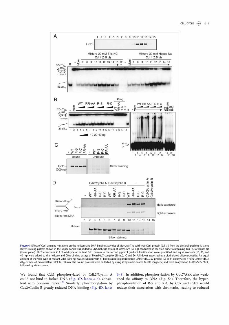
We found that Cdt1 phosphorylated by Cdk2/Cyclin Acould not bind to forked DNA (Fig. 4D, lanes 2–5), consis-tent with previous report.20 Similarly, phosphorylation byCdc2/Cyclin B greatly reduced DNA binding (Fig. 4D, lanes
6–8). In addition, phosphorylation by Cdc7/ASK also weak-ened the affinity to DNA (Fig. S5). Therefore, the hyper-phosphorylation of R-S and R-C by Cdk and Cdc7 wouldreduce their association with chromatin, leading to reduced
Figure 4. Effect of Cdt1 arginine mutations on the helicase and DNA-binding activities of Mcm. (A) The wild-type Cdt1 protein (0.5 ml) from the glycerol gradient fractions(silver staining pattern shown in the upper panel) was added to DNA helicase assays of Mcm4/6/7 (50 ng) conducted in reaction buffers containing Tris-HCl or Hepes-Na(lower panel). (B) The fractions #13 of wild-type or mutant Cdt1 protein in the second glycerol gradient fractionation were quantified and equal amounts (10, 20, and40 ng) were added to the helicase and DNA-binding assays of Mcm4/6/7 complex (50 ng). (C and D) Pull-down assays using a biotinylated oligonucleotide. An equalamount of the wild-type or mutant Cdt1 (200 ng) was incubated with 50-biotinylated oligonucleotide (37mer-dT40, 50 pmole) (C) or 50-biotinylated Y-fork (37mer-dT40/dT20-37mer, 40 pmole) (D) at 30!C for 30 min. The bound proteins were collected by using streptavidin-coated M-280 magnetic, and were analyzed on 4–20% SDS-PAGE,followed by silver staining.
CELL CYCLE 1219

Mcm loading and DNA replication, consistent with aboveresults.
Altered interaction between the RR-AA Cdt1 and Geminin
To ensure the regulated licensing during cell cycle, Gemininplays an important role by directly binding to Cdt1 and therebyinhibiting its licensing activity.9,10 The results in Xenopus eggextracts indicated that RR-AA mutant is refractory to inhibitionby Geminin. Therefore, interaction with Geminin was investi-gated using purified GST-tagged Geminin and various mutantsof Cdt1. Pull-down assay were carried out using anti-Flag anti-body agarose beads or glutathione sepharose beads (Fig. 7A).Wild type and mutant Cdt1 showed identical level of Gemininbinding in both assays (Fig. 7A), suggesting that overall affinityof Geminin to Cdt1 was not affected by the mutations.
Next, we examined the complex formation between Gemininand various Cdt1 mutants on a native protein gel (Fig. 7B).Although glycerol gradient fractionation of purified Gemininprotein showed that it was broadly distributed from 50 kDathrough 300 kDa, with a peak around the size of a dimeric com-plex (Fig. 7C), native gel electrophoresis showed that Gemininforms multiple high molecular-weight complexes appearing as aladder (Fig. 7B, lane 1), consistent with the structural studiesshowing that Geminin could form dimers and tetramers.36,37 In
the presence of Geminin and Cdt1 proteins, several new distinctbands that were not detected with Geminin alone were detected.There are no differences between the wild-type and R-S/R-Cmutant Cdt1 proteins in the patterns of complex formation(Fig. 7B, lanes 3 and 4). In contrast, we found that RR-AAformed a fast-migrating band (representing a dimeric Geminincomplex) but the band representing a tetramer of Geminin com-plex was largely gone (Fig. 7B, lane 5; Fig. S6A, lane 5). Instead,2 slow migrating bands (dot and star) were detected by anti-Cdt1 and -GST (the tag for Geminin) antibodies, suggesting theycontain both RR-AA and Geminin proteins (Fig. S6A and S6B,lane 5). In addition, slow-migrating bands (arrow) of Cdt1 weregenerated between the wild-type and R-S/R-C mutant Cdt1 pro-teins, but not in RR-AA mutant in which a more slow-migratingband (star) appeared instead (Fig. 7B; Fig. S6B, compare lanes2–4 to lane 5). These results suggest that the RR-AA mutantcould form a larger complex with Geminin. Since the effect ofGeminin on the licensing activity could vary depending on thestates of the complex18, the results could be consistent with thefact that RR-AA mutant is refractory to inhibition by Geminin.
Discussion
Here we report crucial arginine residues on Cdt1 that differen-tially affect DNA replication, licensing and complex formation
Figure 5. Physical interactions of Cdt1 with Mcm complexes. (A) Co-immunoprecipitation of Mcm and Cdt1. Co-immunoprecipitation assays were performed using anti-Flag M2 antibody agarose beads (upper; Mcm2), anti-Cdt1 antibody (middle and lower panels; Mcm4/6/7 or Mcm2»7 complex) coupled with Dynabead protein G. All ofthem were mixed with indicated proteins in buffer, and washed 4 times after 1 hr incubation. Bound proteins were eluted with 200 mg/ml Flag peptide (upper) or 0.1 Mcitric acid, pH 2.2 (middle and lower). The samples were run on SDS-PAGE, followed by silver staining. I: Input (7 % of the input); U: unbound (upper, 40 % of the unbound;middle, 20 % of the unbound; lower, 25 % of the unbound); B, bound. " indicates a non-specific band. (B) Complex formation of Mcm2»7-Cdt1 was examined on a nativegel. The indicated amount of Mcm2»7 and Cdt1 proteins were mixed and electrophoresed on a 5% native polyacrylamide gel and stained with silver.
1220 Z. YOU ET AL.

Figure 6. Phosphorylation of the wild-type and mutant Cdt1 proteins by various kinases in vitro. (A) Standard reaction mixtures for in vitro phosphorylation by Cdk2/CyclinA and Cdc2/Cyclin B kinase complex were incubated with wild-type and mutant Cdt1 proteins at the same concentration for 1 hr at 37!C, and products were analyzed on5–20% SDS-PAGE. The gel was stained with silver (left panel) and dried for autoradiogram (right panel). (B) In vitro phosphorylation assays were conducted with Cdc7/Dbf4 kinase (Cdc7 WT) and Cdc7 kinase dead mutant (Cdc7 KD). The reaction products were analyzed on 5–20% SDS-PAGE, which was silver-stained (left panel), dried,and autoradiographed (right panel). (C) The levels of Cdt1 phosphorylation in the autoradiograms in A and B were quantified by Fuji image analyzer, expressed as relativevalues (the maximum level of kinase activities taken as 100) and were plotted.
CELL CYCLE 1221

with Geminin and Mcm (Table 1). Serine or cysteine substitu-tion of 223th arginine (R-S or R-C) resulted in reduced DNAreplication/licensing activity, whereas alanine substitution of342nd and 346th arginines constituting an arginine finger-likestructure (RR-AA) resulted in enhanced activity. We show herethat 223th arginine is required for formation of the functionalMcm-Cdt1 complex and that 342nd and 346th arginines areinvolved in formation of a larger inhibitory complex with Gem-inin. Our results also indicate that arginine mutations affect theextent of phosphorylation of Cdt1 by Cdc2-CyclinB or Cdc7-ASK. Thus, conserved arginine residues of Cdt1 play crucialroles in generating proper complexes with Mcm, Geminin andalso possibly with regulatory kinases, thereby profoundly affect-ing its licensing activity.
The function of Cdt1 mutants correlates withconformational change with Geminin or Mcm
It has been proposed that a Cdt1-Geminin complex can exist in2 distinct states, ‘permissive’ and ‘inhibitory’ complexes.18 Theresults reported here support this hypothesis. The RR-AA Cdt1mutant can form a complex with Geminin that is larger thanthat generated by the wild-type, and is hyperactive in DNA rep-lication and in recruitment of the Mcm complex to chromatin.Consistent with the previous proposal, the RR-AA Cdt1 is com-petent for licensing and replication in the presence of a highlevel of Geminin (Fig. 3). The Cdt1(RR-AA)-Geminin, formingpreferentially a permissive complex, can recruit Mcm to chro-matin even at a very low concentration.
Figure 7. Physical interactions of Geminin and Cdt1. (A) Pull down assays of Geminin and Cdt1. The purified GST-tagged Geminin were mixed with Flag-tagged Cdt1, andimmunoprecipitation was performed using anti-Flag M2 antibody agarose beads (upper) or glutathione beads (lower). The associated proteins collected on beads weresubjected to SDS-PAGE, followed by silver staining. I: Input (7 % of the input); U: unbound (upper, 25 % of the unbound; lower, 33 % of the unbound); B, bound. (B) Thecomplex formation of Geminin-Cdt1 was examined on a native gel. Indicated proteins were mixed and electrophoresed on a 5% native polyacrylamide gel and stainedwith silver. The bands in the square boxes indicate the Cdt1-Geminin complexes. The arrow and dot indicate different complexes of Cdt1-Geminin, respectively. (C) Par-tially purified GST-Geminin protein was fractionated by 15–35% glycerol gradient centrifugation at 45,000 rpm for 16 hr. Proteins in the fractions were analyzed by SDS-PAGE and stained with silver. The glycerol gradient fractionation of purified Geminin protein showed that the Geminin mainly forms a dimeric complex. (D) Effect ofArg342,Arg346-Ala substitution of Cdt1 on its interaction with Geminin. See Discussion for details.
1222 Z. YOU ET AL.

It was previously reported that 210th arginine in humanCdt1 (present at the Cdt1:Geminin interface; corresponding toR223 in the mouse Cdt1) is crucial for Cdt1 licensing activityand DNA replication.18 Consistent with this, we show here thatthe R-S/R-C Cdt1 are severely impaired in DNA replicationand Mcm loading activities. Our results further show that theR-S/R-C Cdt1 are not able to generate a functional complexwith Mcm2»7, while interaction with Geminin is not affected.This suggests an unexpected possibility that the tertiary inter-face of Cdt1 with Geminin may be involved in interaction withMcm. The structural information indicated that charge com-plementarity between the C-terminal segments of Cdt1 andMcm6 is essential for Cdt1-Mcm interaction.24-26 Our resultssuggest that similar charged interactions at the central domainsof Cdt1 also are important for functional Cdt1-Mcm interac-tion as well as for licensing. This is consistent with previousdeletion analyses of Cdt1 which indicate that the centraldomain containing R223 is crucial for its functions.17,22,27
Effect of arginine mutations on phosphorylation
CDK negatively regulates pre-RC assembly by inhibiting Mcmloading and re-replication. Cdt1 protein accumulates in thenucleus during G1 phase, leading to Mcm2»7 complex loadingon chromatin. In S phase, Cdt1 activity is restrained by Gemi-nin stabilization and Cdt1 degradation.3,38 In the G2/M phases,Cdt1 starts to accumulate again, but cannot interact with chro-matin because of the inhibitory phosphorylation mediated bymitotic kinase. A recent study has shown that the mutant Cdt1deficient in mitotic phosphorylation re-associates with chroma-tin in G2/M phase, resulting in unscheduled DNA licensingand re-replication.19 This indicates that the proper phosphory-lation of Cdt1 in G2/M phase is critical for regulated licensing.It was reported before that the kinase activity of the Cdc7 com-plex is also required for Cdt1 dissociation from chromatin atthe onset of S phase.21 Thus, both Cdk and Cdc7 would regulatechromatin binding of Cdt1. We found that R-S/R-C Cdt1 arephosphorylated significantly more efficiently by Cdc2/Cyclin Band by Cdc7 in vitro. This suggests a possibility that R-S/R-CCdt1 may be hyperphosphorylated and that its association withchromatin may be inhibited, resulting in reduced licensing. Incontrast, RR-AA is poorly phosphorylated by Cdc7 in vitro,which may result in hypophosphorylation of Cdt1. This wouldfacilitate reassociation of Cdt1 to chromatin in G2/M phase
and would also be inhibitory for its release from chromatin atthe onset of S phase, leading to increased licensing and DNAreplication in Xenopus egg extracts. This would also explain theGeminin-independent replication stimulatory effect of the RR-AA mutant.
The DNA-binding domain of Cdt1 has been proposed tocontain the N-terminal and middle domains.11,14 The argininemutations did not affect the affinity to DNA (Fig. 4B & C). Onthe other hand, the phosphorylation of Cdt1 by Cdk dramati-cally inhibits its affinity to DNA (Fig. 4D). Thus, the efficacy ofCdk-mediated phosphorylation of Cdt1 may have profoundeffect on its association with DNA, which may ultimately affectits Mcm loading activity.
Structural consideration of RR-AA mutation
Crystallographic studies suggest that Cdt1 interact with Gemi-nin through 3 interfaces to form a complex.12,18 As shown inFigure 7D, Arg342 (loop-2 of Cdt1; secondary interaction) hasa van der Waals contact with Tyr95 and Pro91 of Geminin.Arg342 interacts also with Pro195 and Gly196 of the upperloop (loop-1 of Cdt1; primary interaction) of Cdt1, the majorGeminin binding interface (Fig. 7D, middle and right panels).Carbonyl group of Pro195 makes hydrogen bonding withArg342, and Gly196 is in contact with the aliphatic part ofArg342 (loop-2; Fig. 7D, middle panel). In addition, theArg346 also interacts with Glu89 of Geminin. Substitution ofArg342 and Arg346 with alanine results in loss of the primaryand secondary interactions as well as that of hydrogen bonding(Fig. 7D, right panel), which would extensively affect the nor-mal conformation of the Geminin-Cdt1 complex. This findingsupports the notion that differences in the stoichiometry ofCdt1 and Geminin can cause the formation of different Cdt1-Geminin complexes with different effect.15-18,30
We conclude that the complex formation of functionallyproficient Cdt1-Geminin and Cdt1-Mcm and proper phos-phorylation of Cdt1 are required for the regulated spatial andtemporal chromatin loading of the Mcm and DNA replication.
Materials and methods
Cloning of wild-type and mutant forms of Cdt1 genes intobaculovirus transfer vectors
Site-directed mutagenesis of the Cdt1 genes was conducted withthe QuikChange site-directed mutagenesis kit (Agilent Tech-nologies). The oligonucleotides 50- CGACCAGCTGACCG-CATGGCATCCGGCATTCAATGTGGACG-30, 50-GAGCATGCTCCACAATAGCTCTGAGACTGTGACC-30, and 50-GAGCATGCTCCACAATTGCTCTGAGACTGTGACC-30 wereused as the primers to clone the full-length Cdt1 coding regionscontaining R342R346-AA, R223-S and R223-C Cdt1 mutants,respectively, in pFastbac1.
Antibodies
The antibodies against following Xenopus proteins weredescribed previously; anti-Xenopus Mcm2 antibody,39 antibod-ies for Xenopus Cdt1, Cdc6, Orc2 and Geminin,30 anti-Xenopus
Table 1. Summary of the biochemical properties of mutants of Cdt1 protein.
WT RR-AA R-S R-C
DNA replication CC CCCC C CLicensing activity CC CCCC C CComplex formation with Mcm2~7 CC CC ¡ ¡Complex formation with Geminin CC ¡ CC CCStimulation of MCM helicase CC CC CC CCDNA-binding activity CC CC CC CCKinase activity Cdk2/Cyclin A CC CC CC CC
Cdc2/Cyclin B CC C/CC CCC CCCCdc7/Dbf4 CC C CCC CCC
The assays were performed as described in Materials and Methods. The activity ofthe wild type Cdt1 is indicated asCC in each assay; the activities of mutant Cdt1proteins are shown by the number of plus signs. No activity is shown as a minussign.
CELL CYCLE 1223

Pol e antibody,40 anti-Pol a antibody.41 Anti-PCNA antibody(PC 10) was purchased from Sigma Aldrich.
Purification of recombinant proteins
Recombinant wild-type or various mutant forms of Cdt1 pro-tein, expressed in insect cells, were purified by Ni-NTA andanti-Flag antibody beads. The peak fractions were further frac-tionated on 15»35% glycerol gradient which was run at38,000 rpm for 17 hr, as previously described.23 To removecontaminating nucleases, further purification was carried outas follows. The fractions from 10 to 14 in glycerol gradientwere pooled and treated with Benonase (final 1 U/ml). Thesamples were bound to Ni-sepharose for 1 hr at 4!C and thor-oughly washed with buffer containing 5 mM ATP and 10 mMMg-acetate. Then, Cdt1 proteins were eluted with elution buffercontaining the 0.5 M imidazole, 2 mM ATP, 2 mM Mg-acetate,and 100 mM Na-acetate. The eluates were further fractionatedby second glycerol gradient centrifugation.
The highly purified recombinant mouse Mcm4/6/7 andMcm2»7 protein complexes were prepared from insect cells aspreviously described.42 The recombinant GST-Geminin proteinwas purified from E. coli cells by glutathione sepharose beadsaccording to the manufacture’s protocol. GST-Geminin wasfurther fractionated by 15-35% glycerol gradient centrifugationat 45,000 rpm for 16 hr. The glycerol gradient fractionationindicated that GST-Geminin mainly formed a dimer. Each frac-tion was subjected to SDS-PAGE (5–20% polyacrylamide) todetect the proteins by silver staining.
Pull-down experiments
Pull-down experiments were performed using anti-Flag M2antibody agarose beads (Sigma), glutathione beads or anti-Cdt1antibody (Santa Cruz Biotechnology) coupled to Dynabeadsprotein G (Life Technologies). Indicated proteins were mixedin PBS buffer (Cdt1-Mcm interaction) or buffer containing20 mM Tris-HCl (pH 7.5), 100 mM sodium acetate, 5 mM Mg-acetate, 1 mM ATP, 1 mM EDTA, 10% glycerol, 0.01% TritonX, and proteinase inhibitor (Cdt1-Geminin interaction). After1 hr incubation, beads were washed 3 times with the abovebuffer, and then once with PBS buffer. Bound proteins wereeluted with 200 mg/ml Flag peptide or 0.1 M citric acid (pH2.2). The samples were run on SDS-PAGE, followed by silverstaining.
Complex formation analysis
The complex formation of Mcm2»7-Cdt1 and Geminin-Cdt1 were examined in a native gel as follows. 300 ng eachof Mcm2»7 and Cdt1 or Geminin and Cdt1 were mixed onice in a buffer containing of 20 mM Tris-HCl (pH 7.5),10 mM creatine-phosphate, 5 mM Mg-acetate, 5 mM DTT,and 0.01% Triton X-100, and then electrophoresed on a 5%Tris-glycine native polyacrylamide gel containing 5% glyc-erol, followed by staining with silver or western-blotting.The proteins that had been electrophoresed under a non-denaturing condition were transferred to a membrane afterthe gel was incubated in 20 mM Tris-150 mM glycine-0.1%
SDS for 10 min. The membrane was then treated with50 mM Tris pH7.5-2% SDS-0.8% 2-mercaptoethanol at 50!Cfor 30 min, washed in TBST (Tris-buffered saline, 0.1%Tween 20) and then incubated with anti-Cdt1 antibody(Santa Cruz Biotechnology). The membrane was thenwashed in TBST buffer and incubated in anti-GST antibodyto detect Geminin protein. The protein markers, thyroglobu-lin (669 kDa), catalase (232 kDa) and lactate dehehydroge-nase (140 kDa), were run side by side.
In vitro phosphorylation
The wild type and mutant Cdt1 proteins (100 ng) were incu-bated with Cdk2/CyclinA, Cdc2/CyclinB (purchased fromCarna Biosciences, Inc.), or Cdc7/ASK complex at 37!C for30 min in a reaction mixture consisting of 20 mM Tris-HCl(pH 7.5), 30 mM NaCl, 10 mM MgCl2, 1 mM ATP, and 0.01%Triton X-100, 10 mM 2-glycerophosphate, 1 mM Calyculin Ain the presence of [g-32P]ATP. The phosphorylated proteinswere analyzed by SDS-polyacrylamide gel electrophoresis, fol-lowed by silver staining. The radioactivity on the same gel wasdetected by a Bio-Image analyzer (BAS 2500; Fuji).
DNA-binding assays using biotinylated oligonucleotide
Wild-type or mutant Cdt1 (200 ng) and 50-biotinylated oligo-nucleotides were incubated at 30!C for 30 min in a buffer con-taining 20 mM Tris-HCl (pH 7.5), 100 mM sodium acetate,10 mM Mg(CH3COO)2, 1 mM DTT, 1 mM ATP, 5 mM NaF;1 mM NaVO4, and 0.01% Triton X-100. Streptavidin-coatedM-280 magnetic beads was added to the mixture and incubatedfor 30 min at 4!C. After the removal of the supernatant, thebeads were collected, washed with the buffer, and were ana-lyzed on 4–20% SDS-PAGE.
The coupled reactions of phosphorylation and DNA bindinganalysis in Figure 4D were conducted as follows. After phos-phorylation of Cdt1 with Cyclin A/Cdk2 and Cdc2/CyclinB for30 min at 30!C in the above buffer, a mixture were diluted intoabove binding buffer and biotin-labeled fork DNA (37mer-dT40/dT20-37mer) were added, and the incubation was contin-ued for 30 min at 30!C. And then, the streptavidin-coatedM-280 magnetic beads (Dynal Biotech ASA) were added to themixture and incubated for 30 min at 4!C. After the removal ofthe supernatant (unbound), the beads (bound) were washed4 times in a buffer (400 ml) containing 20 mM Tris (pH 7.5),100 mM Na-Acetate, 1 mM DTT, 0.01% Triton X-100, 5 mMNaF, 1 mM NaVO4 and 10% glycerol, re-suspended in SDSsample buffer, and were analyzed on 4–20% gradient PAGE,followed by silver staining.
Gel-shift and helicase assays
DNA helicase and DNA-binding activities were examined basi-cally as previously described.42
Replication assays
Replication assays contained 10 ml of interphase extract withFlag-peptide-eluted mCdt1 preparations or control buffer.
1224 Z. YOU ET AL.

Demembranated Xenopus sperm nuclei were added at a finalconcentration of 2,500 nuclei/ml. DNA synthesis was monitoredby [a-32P]dATP incorporation after 60 min or 90 min at 23!C.After the incubation of sperm nuclei, the samples (4 ml) wereremoved and added to the 10 ml of a reaction-stop buffer(0.25 mg/ml proteinase K, 8 mM EDTA, 80 mM Tris–HCl (pH8.0), 0.13% (w/v) phosphoric acid, 10% (w/v) Ficoll, 5% (w/v)SDS, and 0.2% (w/v) bromophenol blue). After incubation at37!C for 1 hr, the samples were diluted by addition of 40 ml ofTE (50 mM Tris-HCl (pH 8.0), 1 mM EDTA) and spotted ontoa Whatman DE81 cellulose chromatography paper (GE Health-care). The filter paper was washed with 0.5 M sodium phosphatebuffer (pH 6.8), deionized water and then 100% ethanol. Amountof 32P on the filter paper was measured by a scintillation counter.
Chromatin-binding assays in xenopus egg extracts
To isolate the chromatin fraction, sperm chromatin (2500nuclei/ml) was incubated with interphase extracts of Xenopuseggs (20 ml) at 23!C for indicated time in the figure or legends.Chromatin fraction was isolated as described previously.43
Mass spectrometer analyses of Cdt1 phosphorylatedin vitro
About 2 mg of Cdt1 protein (wild-type and mutants), eithernon-treated or phosphorylated by Cdc2/CyclinB or Cdc7/ASK,was treated with trypsin and concentrated by IMAC-C18(immobilized metal ion affinity chromatography) to improvethe detection sensitivity of phosphorylated molecules. Eachfraction was analyzed via LCMS/MS on Triple TOF 5600 Plus(AB Sciex) mass spectrometer. Analysis of the data with Pro-teinPilot 4.5beta software (AB Sciex) identified 600-1000 pepti-des with a 95% confidence at a false discovery rate of 1%. Nophosphorylation was identified on the T24 and S61 residues.
Abbreviations
Cdc7 kinase Cell division cycle 7-related protein kinaseCdk cyclin-dependent kinaseCdt1 Cdc10-dependent transcript 1Mcm mini-chromosome maintenance
Disclosure of potential conflicts of interest
No potential conflicts of interest were disclosed.
Acknowledgments
We thank Katsuhiko Kamada (RIKEN Discovery Research Institute, Japan)for structural analyses of interaction of Cdt1 (wild-type and RR-AA) withGeminin. Naoko Kakusho and Rino Fukatsu for excellent technical assis-tance. We thank the members of our laboratories for helpful discussions.
References
[1] Masai H, Matsumoto S, You Z, Yoshizawa-Sugata N, Oda M.Eukaryotic chromosome DNA replication: where, when, and how?Annu Rev Biochem 2010; 79:89-130; PMID:20373915; http://dx.doi.org/10.1146/annurev.biochem.052308.103205
[2] Fujita M. Cdt1 revisited: complex and tight regulation during thecell cycle and consequences of deregulation in mammalian cells.Cell Div 2006; 1:22; PMID:17042960; http://dx.doi.org/10.1186/1747-1028-1-22
[3] Li X, Zhao Q, Liao R, Sun P, Wu X. The SCF(Skp2) ubiquitin ligasecomplex interacts with the human replication licensing factor Cdt1and regulates Cdt1 degradation. J Biol Chem 2003; 278:30854-8;PMID:12840033; http://dx.doi.org/10.1074/jbc.C300251200
[4] Nishitani H, Lygerou Z. DNA replication licensing. Front Biosci2004; 9:2115-32; PMID:15353274; http://dx.doi.org/10.2741/1315
[5] Nishitani H, Sugimoto N, Roukos V, Nakanishi Y, Saijo M, Obuse C,Tsurimoto T, Nakayama KI, Nakayama K, Fujita M, et al. Two E3ubiquitin ligases, SCF-Skp2 and DDB1-Cul4, target human Cdt1 forproteolysis. Embo J 2006; 25:1126-36; PMID:16482215; http://dx.doi.org/10.1038/sj.emboj.7601002
[6] Arias EE, Walter JC. PCNA functions as a molecular platform totrigger Cdt1 destruction and prevent re-replication. Nat Cell Biol2006; 8:84-90; PMID:16362051; http://dx.doi.org/10.1038/ncb1346
[7] Sugimoto N, Kitabayashi I, Osano S, Tatsumi Y, Yugawa T, Nari-sawa-Saito M, Matsukage A, Kiyono T, Fujita M. Identification ofnovel human Cdt1-binding proteins by a proteomics approach: pro-teolytic regulation by APC/CCdh1. Mol Biol Cell 2008; 19:1007-21;PMID:18162579; http://dx.doi.org/10.1091/mbc.E07-09-0859
[8] McGarry TJ, Kirschner MW. Geminin, an inhibitor of DNA replica-tion, is degraded during mitosis. Cell 1998; 93:1043-53;PMID:9635433; http://dx.doi.org/10.1016/S0092-8674(00)81209-X
[9] Wohlschlegel JA, Dwyer BT, Dhar SK, Cvetic C, Walter JC, Dutta A.Inhibition of eukaryotic DNA replication by geminin binding toCdt1. Science 2000; 290:2309-12; PMID:11125146; http://dx.doi.org/10.1126/science.290.5500.2309
[10] Tada S, Li A, Maiorano D, Mechali M, Blow JJ. Repression of originassembly in metaphase depends on inhibition of RLF-B/Cdt1 bygeminin. Nat Cell Biol 2001; 3:107-13; PMID:11175741; http://dx.doi.org/10.1038/35055000
[11] Yanagi K, Mizuno T, You Z, Hanaoka F. Mouse geminin inhibits notonly Cdt1-MCM6 interactions but also a novel intrinsic Cdt1 DNAbinding activity. J Biol Chem 2002; 277:40871-80; PMID:12192004;http://dx.doi.org/10.1074/jbc.M206202200
[12] Lee C, Hong B, Choi JM, Kim Y, Watanabe S, Ishimi Y, Enomoto T,Tada S, Kim Y, Cho Y. Structural basis for inhibition of the replica-tion licensing factor Cdt1 by geminin. Nature 2004; 430:913-7;PMID:15286659; http://dx.doi.org/10.1038/nature02813
[13] Nishitani H, Taraviras S, Lygerou Z, Nishimoto T. The humanlicensing factor for DNA replication Cdt1 accumulates in G1 and isdestabilized after initiation of S-phase. J Biol Chem 2001; 276:44905-11; PMID:11555648; http://dx.doi.org/10.1074/jbc.M105406200
[14] Xouri G, Squire A, Dimaki M, Geverts B, Verveer PJ, Taraviras S,Nishitani H, Houtsmuller AB, Bastiaens PI, Lygerou Z. Cdt1 associ-ates dynamically with chromatin throughout G1 and recruits Gemi-nin onto chromatin. Embo J 2007; 26:1303-14; PMID:17318181;http://dx.doi.org/10.1038/sj.emboj.7601597
[15] Lutzmann M, Maiorano D, Mechali M. A Cdt1-geminin complexlicenses chromatin for DNA replication and prevents rereplicationduring S phase in Xenopus. EMBO J 2006; 25:5764-74;PMID:17124498; http://dx.doi.org/10.1038/sj.emboj.7601436
[16] Xouri G, Dimaki M, Bastiaens PI, Lygerou Z. Cdt1 interactions in thelicensing process: a model for dynamic spatiotemporal control oflicensing. Cell Cycle 2007; 6:1549-52; PMID:17598984; http://dx.doi.org/10.4161/cc.6.13.4455
[17] Kisielewska J, Blow JJ. Dynamic interactions of high Cdt1 and gemi-nin levels regulate S phase in early Xenopus embryos. Development2012; 139:63-74; PMID:22096080; http://dx.doi.org/10.1242/dev.068676
[18] De Marco V, Gillespie PJ, Li A, Karantzelis N, Christodoulou E,Klompmaker R, van Gerwen S, Fish A, Petoukhov MV, Iliou MS, et al.Quaternary structure of the human Cdt1-Geminin complex regulatesDNA replication licensing. Proc Natl Acad Sci U S A 2009; 106:19807-12; PMID:19906994; http://dx.doi.org/10.1073/pnas.0905281106
[19] Coulombe P, Gregoire D, Tsanov N, Mechali M. A spontaneous Cdt1mutation in 129 mouse strains reveals a regulatory domain
CELL CYCLE 1225

restraining replication licensing. Nat Commun 2013; 4:2065;PMID:23817338; http://dx.doi.org/10.1038/ncomms3065
[20] Sugimoto N, Tatsumi Y, Tsurumi T, Matsukage A, Kiyono T, Nishi-tani H, Fujita M. Cdt1 phosphorylation by cyclin A-dependent kin-ases negatively regulates its function without affecting gemininbinding. J Biol Chem 2004; 279:19691-7; PMID:14993212; http://dx.doi.org/10.1074/jbc.M313175200
[21] Ballabeni A, Zamponi R, Caprara G, Melixetian M, Bossi S, MasieroL, Helin K. Human CDT1 associates with CDC7 and recruits CDC45to chromatin during S phase. J Biol Chem 2009; 284:3028-36;PMID:19054765; http://dx.doi.org/10.1074/jbc.M803609200
[22] Takara TJ, Bell SP. Multiple Cdt1 molecules act at each origin to loadreplication-competent Mcm2-7 helicases. Embo J 2011;PMID:22045335
[23] You Z, Masai H. Cdt1 forms a complex with the minichromosomemaintenance protein (MCM) and activates its helicase activity. J BiolChem 2008; 283:24469-77; PMID:18606811; http://dx.doi.org/10.1074/jbc.M803212200
[24] Jee J, Mizuno T, Kamada K, Tochio H, Chiba Y, Yanagi K, Yasuda G,Hiroaki H, Hanaoka F, Shirakawa M. Structure and mutagenesisstudies of the C-terminal region of licensing factor Cdt1 enable theidentification of key residues for binding to replicative helicase Mcmproteins. J Biol Chem 2010; 285:15931-40; PMID:20335175; http://dx.doi.org/10.1074/jbc.M109.075333
[25] Liu C, Wu R, Zhou B, Wang J, Wei Z, Tye BK, Liang C, Zhu G.Structural insights into the Cdt1-mediated MCM2-7 chromatin load-ing. Nucleic Acids Res 2012; 40:3208-17; PMID:22140117; http://dx.doi.org/10.1093/nar/gkr1118
[26] Wei Z, Liu C, Wu X, Xu N, Zhou B, Liang C, Zhu G. Characteriza-tion and structure determination of the Cdt1 binding domain ofhuman minichromosome maintenance (Mcm) 6. J Biol Chem 2010;285:12469-73; PMID:20202939.
[27] Ferenbach A, Li A, Brito-Martins M, Blow JJ. Functional domains ofthe Xenopus replication licensing factor Cdt1. Nucleic Acids Res 2005;33:316-24; PMID:15653632; http://dx.doi.org/10.1093/nar/gki176
[28] Whittaker AJ, Royzman I, Orr-Weaver TL. Drosophila doubleparked: a conserved, essential replication protein that colocalizeswith the origin recognition complex and links DNA replication withmitosis and the down-regulation of S phase transcripts. Genes Dev2000; 14:1765-76; PMID:10898791
[29] Kawakami H, Keyamura K, Katayama T. Formation of an ATP-DnaA-specific initiation complex requires DnaA Arginine 285, a conservedmotif in the AAAC protein family. J Biol Chem 2005; 280:27420-30;PMID:15901724; http://dx.doi.org/10.1074/jbc.M502764200
[30] Ode KL, Fujimoto K, Kubota Y, Takisawa H. Inter-origin cooperativ-ity of geminin action establishes an all-or-none switch for replicationorigin licensing. Genes Cells 2011; 16:380-96; PMID:21426446;http://dx.doi.org/10.1111/j.1365-2443.2011.01501.x
[31] Tsuyama T, Watanabe S, Aoki A, Cho Y, Seki M, Enomoto T, TadaS. Repression of nascent strand elongation by deregulated Cdt1 dur-ing DNA replication in Xenopus egg extracts. Mol Biol Cell 2009;20:937-47; PMID:19064889; http://dx.doi.org/10.1091/mbc.E08-06-0613
[32] Gillespie PJ, Li A, Blow JJ. Reconstitution of licensed replication ori-gins on Xenopus sperm nuclei using purified proteins. BMC Bio-chem 2001; 2:15; PMID:11737877; http://dx.doi.org/10.1186/1471-2091-2-15
[33] Waga S, Zembutsu A. Dynamics of DNA binding of replication initi-ation proteins during de novo formation of pre-replicative complexesin Xenopus egg extracts. J Biol Chem 2006; 281:10926-34;PMID:16497662; http://dx.doi.org/10.1074/jbc.M600299200
[34] Fujita M, Yamada C, Tsurumi T, Hanaoka F, Matsuzawa K, InagakiM. Cell cycle- and chromatin binding state-dependent phosphoryla-tion of human MCM heterohexameric complexes. A role for cdc2kinase. J Biol Chem 1998; 273:17095-101; PMID:9642275; http://dx.doi.org/10.1074/jbc.273.27.17095
[35] Ballabeni A, Melixetian M, Zamponi R, Masiero L, Marinoni F,Helin K. Human geminin promotes pre-RC formation and DNAreplication by stabilizing CDT1 in mitosis. EMBO J 2004;23:3122-32; PMID:15257290; http://dx.doi.org/10.1038/sj.emboj.7600314
[36] Okorokov AL, Orlova EV, Kingsbury SR, Bagneris C, Gohlke U, Wil-liams GH, Stoeber K. Molecular structure of human geminin. NatStruct Mol Biol 2004; 11:1021-2; PMID:15378034; http://dx.doi.org/10.1038/nsmb835
[37] Thepaut M, Maiorano D, Guichou JF, Auge MT, Dumas C, MechaliM, Padilla A. Crystal structure of the coiled-coil dimerization motifof geminin: structural and functional insights on DNA replicationregulation. J Mol Biol 2004; 342:275-87; PMID:15313623; http://dx.doi.org/10.1016/j.jmb.2004.06.065
[38] Liu K, Luo Y, Lin FT, Lin WC. TopBP1 recruits Brg1/Brm to repressE2F1-induced apoptosis, a novel pRb-independent and E2F1-specificcontrol for cell survival. Genes Dev 2004; 18:673-86;PMID:15075294; http://dx.doi.org/10.1101/gad.1180204
[39] Kubota Y, Mimura S, Nishimoto S, Masuda T, Nojima H, TakisawaH. Licensing of DNA replication by a multi-protein complex ofMCM/P1 proteins in Xenopus eggs. EMBO J 1997; 16:3320-31;PMID:9214647; http://dx.doi.org/10.1093/emboj/16.11.3320
[40] Hashimoto Y, Tsujimura T, Sugino A, Takisawa H. The phos-phorylated C-terminal domain of Xenopus Cut5 directly medi-ates ATR-dependent activation of Chk1. Genes Cells 2006;11:993-1007; PMID:16923121; http://dx.doi.org/10.1111/j.1365-2443.2006.00998.x
[41] Mimura S, Takisawa H. Xenopus Cdc45-dependent loading of DNApolymerase alpha onto chromatin under the control of S-phase Cdk.Embo J 1998; 17:5699-707; PMID:9755170; http://dx.doi.org/10.1093/emboj/17.19.5699
[42] You Z, De Falco M, Kamada K, Pisani FM, Masai H. The Mini-Chro-mosome Maintenance (Mcm) Complexes Interact with DNA Poly-merase alpha-Primase and Stimulate Its Ability to Synthesize RNAPrimers. PLoS One 2013; 8:e72408; PMID:23977294; http://dx.doi.org/10.1371/journal.pone.0072408
[43] Hashimoto Y, Takisawa H. Xenopus Cut5 is essential for a CDK-dependent process in the initiation of DNA replication. EMBO J2003; 22:2526-35; PMID:12743046; http://dx.doi.org/10.1093/emboj/cdg238
1226 Z. YOU ET AL.