characteristics and immunological roles of surface layer
TRANSCRIPT

ISSN 2234-3806 • eISSN 2234-3814
https://doi.org/10.3343/alm.2018.38.3.189 www.annlabmed.org 189
Ann Lab Med 2018;38:189-195https://doi.org/10.3343/alm.2018.38.3.189
Review ArticleClinical Microbiology
Characteristics and Immunological Roles of Surface Layer Proteins in Clostridium difficile Nobuaki Mori, M.D.1,2 and Takashi Takahashi, M.D.2
Department of General Internal Medicine1, National Hospital Organization Tokyo Medical Center, Meguro-ku, Tokyo; Laboratory of Infectious Diseases2, Graduate School of Infection Control Sciences and Kitasato Institute for Life Sciences, Kitasato University, Minato-ku, Tokyo, Japan
Clostridium difficile is a major causative agent of antibiotic-associated diarrhea and has become the most common pathogen of healthcare-associated infection worldwide. The pathogenesis of C. difficile infection (CDI) is mediated by many factors such as coloniza-tion involving attachment to host intestinal epithelial cells, sporulation, germination, and toxin production. Bacterial cell surface components are crucial for the interaction between the bacterium and host cells. C. difficile has two distinct surface layer proteins (SLPs): a conserved high-molecular-weight SLP and a highly variable low-molecular-weight SLP. Re-cent studies have shown that C. difficile SLPs play roles not only in growth and survival, but also in adhesion to host epithelial cells and induction of cytokine production. Sequence typing of the variable region of the slpA gene, which encodes SLPs, is one of the methods currently used for typing C. difficile. SLPs have received much attention in recent years as vaccine candidates and new therapeutic agents in the treatment of C. difficile-associated diseases. Gaining mechanistic insights into the molecular functions of C. difficile SLPs will help advance our understanding of CDI pathogenesis and the development of vaccines and new therapeutic approaches. In this review, we summarize the characteristics and immunological roles of SLPs in C. difficile.
Key Words: Clostridium difficile, Surface layer proteins, Attachment, Innate immunity, Typ-ing, Vaccine
Received: September 29, 2017Revision received: November 7, 2017Accepted: December 28, 2017
Corresponding author: Nobuaki MoriDepartment of General Internal Medicine, National Hospital Organization Tokyo Medical Center, 2-5-1 Higashigaoka, Meguro-ku, Tokyo 152-8902, Japan Tel: +81-3-3411-0111Fax: +81-3-3412-9811 E-mail: [email protected]
© Korean Society for Laboratory MedicineThis is an Open Access article distributed under the terms of the Creative Commons Attribution Non-Commercial License (http://creativecom-mons.org/licenses/by-nc/4.0) which permits unrestricted non-commercial use, distribution, and reproduction in any medium, provided the original work is properly cited.
INTRODUCTION
Clostridium difficile was recently shown to be phylogenetically
distant from rRNA clostridial cluster I, located in cluster XI, within
the family Peptostreptococcaceae [1]. Yutin et al [2] proposed
the genus name Peptoclostridium to include all organisms in
cluster XI. However, Lawson et al [3] have proposed that Clos-tridium difficile be named Clostridioides difficile gen. nov. comb.
nov. based on phenotypic, chemotaxonomic, and phylogenetic
analyses. Thus, although this species may be renamed follow-
ing proper validation [4], in this review, we used the name Clos-tridium difficile to ensure visibility and readability, as this is the
name conventionally used.
C. difficile is a gram-positive, spore-forming, obligate anaero-
bic bacterium that is the main causative pathogen of antimicro-
bial-associated colitis. C. difficile infection (CDI) is the leading
cause of infectious diarrhea in hospitalized patients and is in-
creasingly recognized as a common cause of diarrhea in the
community [5-7]. The reported CDI-related mortality rate is 5.7–
14%, and the all-cause mortality within 30 days of CDI onset is
11–38% [8-11]. Although CDI surveillance in Asia remains lim-
ited compared with that in North America and Europe, existing
evidence suggests that CDI occurs at similar rates in Asia and
other regions in the world [12].
CDI pathogenesis involves the following multiple processes: C.
difficile is acquired through ingestion of spores shed into the en-
vironment by infected individuals with or without disease symp-
toms. Once the vegetative cells reach the anaerobic environment
1 / 1CROSSMARK_logo_3_Test
2017-03-16https://crossmark-cdn.crossref.org/widget/v2.0/logos/CROSSMARK_Color_square.svg

Mori N, et al.The role of SLPs in Clostridium difficile
190 www.annlabmed.org https://doi.org/10.3343/alm.2018.38.3.189
of the cecum and colon, they proliferate and colonize the intes-
tinal mucosa. Disruption of the intestinal microbiota, for exam-
ple by antibiotic treatment, allows the vegetative cells to pene-
trate the mucus layer and adhere to the surface of the epithelial
cells. It is through this intimate contact with host cells that a num-
ber of virulence factors produced by C. difficile promote intesti-
nal damage and disease [13].
Many prokaryotes express a surface-exposed proteinaceous
layer, termed the surface layer (S-layer), which forms a regular
two-dimensional array visible by electron microscopy [14]. S-
layers are found on both gram-positive and -negative bacteria
and are highly prevalent in archaea; they comprise one or more
types of S-layer proteins (SLPs), which are some of the most
abundant bacterial cell proteins [15]. Our knowledge of SLPs
has increased over the past decade; SLPs play important roles
not only in growth and survival but also in the interaction with
the host and its immune system. All C. difficile strains also ex-
press crystalline or paracrystalline SLPs on the outer cell surface
[16]. Recent studies have shown that C. difficile SLPs are in-
volved not only in adhesion to host intestinal cells but also in the
induction of cytokine production and the recognition of C. diffi-cile by the immune system [17-19]. In this review, we highlight
recent discoveries in the field of C. difficile SLPs and discuss their
importance in CDI pathogenesis.
SLP STRUCTURE AND LOCUS
Although the S-layer of most bacteria is comprised of one major
protein, which is modified by glycosylation in some species [20],
C. difficile expresses two distinct SLPs that form two superim-
posed and structurally different S-layer lattices [14, 21, 22], which
are not glycosylated [23]. Of the two distinct C. difficile SLPs, one
is a high-molecular-weight SLP (HMW-SLP; ~40 kDa), and the
other is a low-molecular-weight SLP (LMW-SLP; ~35 kDa) (Fig.
1) [24]. Both SLPs are exposed at the cell surface, as determined
by surface iodination and immunogold labeling and by indirect
immunofluorescence [25]. A single gene, slpA, that has a con-
served genomic location among all C. difficile strains, encodes
both SLPs. slpA encodes a precursor protein, termed SlpA, that
has three identifiable subdomains: an N-terminal secretion sig-
nal, followed by a highly variable LMW region, and a HMW re-
gion containing three tandem cell wall binding 2 (CWB2) motifs
(Fig. 1A) [26]. The precursor protein is then cleaved by prote-
ase Cwp84 to generate the two mature proteins (HMW-SLP and
LMW-SLP; Fig. 1B) [27-29]. Both HMW- and LMW-SLPs are
linked by non-covalent interactions to form a tightly associated
complex [21]. Although SLPs have been identified in all C. diffi-cile isolates, there is sequence variability between the PCR ribo-
type strains [30, 31]. HMW-SLP is highly conserved in C. diffi-cile, with up to 97% sequence identity between the strains [14];
it exhibits strong and specific binding to gastrointestinal tissues
and human epithelial cells and is most likely anchored to the
cell wall [32]. LMW-SLP, in contrast, exhibits greater sequence
variation between strains. It is not known how the sequence dif-
ferences in the LMW region influence SLP-host interaction. The
mechanism by which the HMW/LMW-SLP complex assembles
to form the mature S-layer remain unknown.
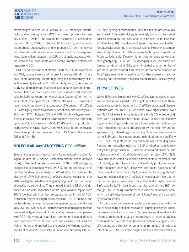
The C. difficile genome encodes 29 SlpA paralogues that com-
Fig. 1. Amino acid sequence and schematic rep-resentation of the SlpA precursor protein. (A) Ami-no acid sequence of the SlpA precursor protein in Clostridium difficile 630 (GenBank: AJP12540.1). Amino acids highlighted in black indicate the sig-nal peptide and those highlighted in gray denote
LMW SLP HMW SLP
Cwp84A B
residues involved in the interaction with the HMW-SLP. The amino acids comprising the LMW-SLP are underlined. The black triangle indi-cates the cleavage site that results in the generation of the two mature SLPs. (B) Schematic representation of the SlpA precursor protein. The black section represents the signal peptide, the light gray section denotes the conserved region within LMW-SLP, and the black triangle indicates the cleavage site of cysteine protease CWP84, which produces mature LMW-SLP and HMW-SLP. Abbreviations: SLPs, surface layer proteins; HMW, high molecular weight; LMW, low molecular weight; CWP84, cell wall protein 84.
Mori N, et al.The role of SLPs in Clostridium difficile
https://doi.org/10.3343/alm.2018.38.3.189 www.annlabmed.org 191
prise the cell wall protein family. The slpA gene is located within
a 36.6 kb cell wall protein (cwp) gene cluster; slpA is encoded
within a genomic locus including 11 of these 29 paralogues [22,
27]. A previous study using whole-genome sequencing identi-
fied a 10-kb cassette within the S-layer locus, including the slpA,
secA2, cep2, and cwp66 genes, that displays higher inter-strain
diversity than the rest of the locus [33]. This suggests that fre-
quent and independent horizontal transfer of the cwp cluster
has occurred throughout the C. difficile population.
MECHANISMS OF SLP-MEDIATED ATTACHMENT
To initiate the C. difficile colonization process in the host, C. dif-ficile must first adhere to the intestinal cells. Multiple adhesins
have been implicated in the attachment of C. difficile to the mu-
cus layer of the intestine: the flagellar cap protein, FliD; the fla-
gellin FliC; the surface-associated heat-shock-induced adhesin,
Cwp66; heat-shock protein, GroEL; fibronectin-binding proteins;
and binary toxin [34-39]. Of these, SLPs constitute a major con-
tributor to bacterial adherence [40]. A study investigating animal
and human C. difficile isolates showed that variation in the S-
layer led to variable adherence to epithelial cells [41]. As dem-
onstrated by in vitro studies, SLPs bind to Hep-2 cells, Vero cells,
and human gastrointestinal tissues [32, 42]; furthermore, chemi-
cal removal of the SLPs or treatment of C. difficile bacterial cells
with anti-SLP Fab fragments abolishes C. difficile adherence to
mouse 929 and human HeLa cells [25]. Similarly, purified SLPs
bind to intestinal tissues and several proteins of the extracellular
matrix; antibodies against HMW-SLP inhibit this adherence [32].
Merrigan et al [40] have demonstrated that pre-treatment of host
cells with purified SlpA or SlpA subunits abrogates C. difficile-
attachment in a dose-dependent manner in vitro. Conversely,
pre-treatment of viable C. difficile with anti-SlpA antibodies also
abrogates adherence. Collectively, these observations suggest
that C. difficile SLPs may contribute to colonization and infection
persistence. However, the precise host receptor that interacts
with the SLPs has yet to be identified.
IMMUNOREGULATORY ROLE OF SLPS
SLPs have emerged as a second class of C. difficile virulence
determinants, in addition to the large clostridial toxins (toxin A
and toxin B) [42]. Recent studies have begun to characterize
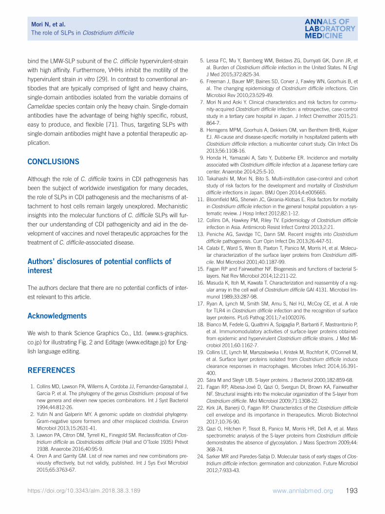
host innate and adaptive responses to C. difficile attachment
(Fig. 2) [13]. SLPs have the ability to activate pro-inflammatory
signaling through toll-like receptor 4 (TLR4) expressed on the
surface of host cells [17]. Engagement of TLR4 results in the
subsequent production of inflammatory cytokines by initiating
downstream signaling of nuclear factor-kappa beta and inter-
feron (IFN) regulatory factor 3 [40], leading to immune cell acti-
vation. In vitro studies using mouse bone marrow-derived den-
dritic cells and human monocyte-derived dendritic cells have
shown that purified SLPs induce the production of both pro-in-
flammatory [tumor necrosis factor-alpha (TNF-α), interleukin
(IL)-12, IL-23, and IL-1b] and anti-inflammatory (IL-10) cytokines
[17, 18]. Furthermore, SLPs induce the maturation of dendritic
cells characterized by the production of IL-12p70, TNF-α, IL-23,
and IL-6 and increased expression of major histocompatibility
complex (MHC) class II, cluster of differentiation (CD) 40, CD80,
and CD86 [17]. SLP-activated dendritic cells, in turn, drive strong
T helper (Th) 1 and Th17 responses characterized by the pro-
duction of IFN-gamma and IL-17 [17]. Moreover, SLPs can also
activate a clearance response in macrophages. SLPs activate
24
448
449
450
451
452
453
454
455
456
457
458
459
460
461
Fig. 2. The SLPs of Clostridium difficile activate dendritic cells and macrophages, which in462
turn produce various cytokines and chemokines. Abbreviations: SLPs, surface layer proteins;463
TLR, toll-like receptor; IL, interleukin; IFN, interferon; MIP, macrophage inflammatory 464
protein; MCP, monocyte chemotactic and activating factor; CD, cluster of differentiation;465
MHC, major histocompatibility complex; Th, T helper.466
Fig. 2. The SLPs of Clostridium difficile activate dendritic cells and macrophages, which in turn produce various cytokines and chemo-kines. Abbreviations: SLPs, surface layer proteins; TLR, toll-like receptor; IL, inter-leukin; IFN, interferon; MIP, macrophage inflammatory protein; MCP, mono-cyte chemotactic and activating factor; CD, cluster of differentiation; MHC, major histocompatibility complex; Th, T helper.
Mori N, et al.The role of SLPs in Clostridium difficile
192 www.annlabmed.org https://doi.org/10.3343/alm.2018.38.3.189
macrophages to produce IL-12p40, TNF-α, monocyte chemo-
tactic and activating factor (MCP), and macrophage inflamma-
tory protein 1 (MIP-1); upregulate the expression of cell-surface
markers (TLR2, CD40, CD14, and MHC class II); and enhance
macrophage phagocytosis and migration [19]. As monocytes
and dendritic cells play important roles in the immune response,
these observations suggest that SLPs are strongly associated with
the activation of host innate and adaptive immune defenses in
response to CDI.
CDI due to hypervirulent strains, such as PCR ribotypes 027
and 078, causes severe and recurrent diseases [43, 44]. There
have been conflicting reports regarding the predictability of in-
fection severity based on C. difficile ribotypes [45]. A previous
study has demonstrated that there is no difference in the immu-
nomodulation of monocytes and monocyte-derived dendritic
cells by SLPs between the hypervirulent (epidemic) or non-hy-
pervirulent (non-epidemic) C. difficile strains [18]. However, a
recent study has shown that sequence differences in C. difficile
SLPs are tightly linked to disease susceptibility and severity [46].
SLPs from PCR ribotypes 027 and 078, which are hypervirulent
strains, induce a more potent inflammatory response, exhibiting
up to two-fold increases in IL-6, IL-12p40, and IL10 production;
higher levels of CD80, CD40, and MHC class II; and increased
chemokine production, relative to the SLPs from PCR ribotypes
001 and 014 [46].
MOLECULAR slpa GENOTYPING OF C. difficile
Several typing systems are currently being utilized in epidemio-
logical studies of C. difficile: restriction endonuclease analysis
(REA), pulse-field gel electrophoresis (PFGE), PCR ribotyping,
multiple locus sequence typing (MLST), and multilocus variable
number tandem repeat analysis (MVLA) [47]. Focusing on the
diversity of LMW-SLP among C. difficile strains, Karjalainen et al
[48] investigated whether slpA genotyping could be used as an
alternative to serotyping. They showed that the DNA and de-
duced amino acid sequences of the slpA variable region were
100% identical within a given serogroup, as confirmed by PCR-
restriction fragment length polymorphism (RFLP) analysis and
nucleotide sequencing, whereas the inter-serogroup identity was
fairly low [48]. Kato et al [31] demonstrated that this typing method
has reliable typability and discriminatory power in comparison
with PCR ribotyping and applied it to strains isolated directly
from stool specimens. Subsequent studies have evaluated this
typing method and applied it to the analysis of various local out-
breaks of C. difficile, especially in Japan and Germany [31, 48-
61]. slpA typing is reproducible, and the results are easily dis-
seminated. This methodology is relatively low-cost; the overall
cost for generating one sequence is estimated to be less than
10 US dollars [48]. Therefore, slpA typing may be a useful method
for outbreaks occurring in a hospital setting. However, a compar-
ative study of seven C. difficile typing techniques showed that
MLVA exhibits a significantly higher discriminatory power than
slpA genotyping, PFGE, or PCR ribotyping [62]. The study per-
formed by Huber et al [63], which included a large number of
genomes, demonstrated that isolates of the same ribotype or
MLST type may differ in slpA type. For these reasons, slpA ge-
notyping has not become the global standard for C. difficile typing.
PERSPECTIVES
While SLPs have limited utility in C. difficile typing, active or pas-
sive immunization against SLPs might constitute a viable thera-
peutic strategy in the treatment of C. difficile-associated disease.
Patients with recurrent CDI episodes have significantly lower
anti-SLP IgM levels than patients with a single CDI episode [64].
Sera from CDI patients have been shown to have significantly
higher anti-SLP IgG levels than sera from carriers or healthy con-
trols, indicating that SLPs are targeted by the host immune re-
sponses [65]. Interestingly, the binding of recombinant antibod-
ies to SLPs and their components exhibited strain specificity
[66]. These reagents could potentially be useful for diagnosis.
Passive immunization using anti-SLP antibodies significantly
delays the progression of C. difficile-associated diarrhea and
prolongs survival in C. difficile-infected hamsters [67]. SLPs
have also been tested as vaccine components in hamsters, but
did not fully protect the animals, and antibody production varied
from modest to poor [68]. However, intra-rectal vaccination of
mice using the recombinant SlpA protein resulted in significantly
lower gut colonization by C. difficile in vaccinated mice than in
the control group; vaccinated mice also had a higher level of
SlpA-specific IgA in their feces than the control mice [69]. Al-
though SlpA is being examined as a vaccine candidate, prob-
lems may still arise because of the high slpA sequence variabil-
ity between strains.
As the use of conventional antibiotics is associated with the
risk of developing antibiotic resistance, targeting essential bacte-
rial virulence factors, such as SLPs, provides an alternative anti-
microbial therapeutic strategy. Interestingly, a recent study has
examined the potential of single-domain antibodies against spe-
cific targets as a strategy for enhancing immunity and reducing
infection [70]. SLP-specific single-domain antibodies (VHHs)
Mori N, et al.The role of SLPs in Clostridium difficile
https://doi.org/10.3343/alm.2018.38.3.189 www.annlabmed.org 193
bind the LMW-SLP subunit of the C. difficile hypervirulent-strain
with high affinity. Furthermore, VHHs inhibit the motility of the
hypervirulent strain in vitro [29]. In contrast to conventional an-
tibodies that are typically comprised of light and heavy chains,
single-domain antibodies isolated from the variable domains of
Camelidae species contain only the heavy chain. Single-domain
antibodies have the advantage of being highly specific, robust,
easy to produce, and flexible [71]. Thus, targeting SLPs with
single-domain antibodies might have a potential therapeutic ap-
plication.
CONCLUSIONS
Although the role of C. difficile toxins in CDI pathogenesis has
been the subject of worldwide investigation for many decades,
the role of SLPs in CDI pathogenesis and the mechanisms of at-
tachment to host cells remain largely unexplored. Mechanistic
insights into the molecular functions of C. difficile SLPs will fur-
ther our understanding of CDI pathogenicity and aid in the de-
velopment of vaccines and novel therapeutic approaches for the
treatment of C. difficile-associated disease.
Authors’ disclosures of potential conflicts of interest
The authors declare that there are no potential conflicts of inter-
est relevant to this article.
Acknowledgments
We wish to thank Science Graphics Co., Ltd. (www.s-graphics.
co.jp) for illustrating Fig. 2 and Editage (www.editage.jp) for Eng-
lish language editing.
REFERENCES
1. Collins MD, Lawson PA, Willems A, Cordoba JJ, Fernandez-Garayzabal J, Garcia P, et al. The phylogeny of the genus Clostridium: proposal of five new genera and eleven new species combinations. Int J Syst Bacteriol 1994;44:812-26.
2. Yutin N and Galperin MY. A genomic update on clostridial phylogeny: Gram-negative spore formers and other misplaced clostridia. Environ Microbiol 2013;15:2631-41.
3. Lawson PA, Citron DM, Tyrrell KL, Finegold SM. Reclassification of Clos-tridium difficile as Clostridioides difficile (Hall and O’Toole 1935) Prévot 1938. Anaerobe 2016;40:95-9.
4. Oren A and Garrity GM. List of new names and new combinations pre-viously effectively, but not validly, published. Int J Sys Evol Microbiol 2015;65:3763-67.
5. Lessa FC, Mu Y, Bamberg WM, Beldavs ZG, Dumyati GK, Dunn JR, et al. Burden of Clostridium difficile infection in the United States. N Engl J Med 2015;372:825-34.
6. Freeman J, Bauer MP, Baines SD, Corver J, Fawley WN, Goorhuis B, et al. The changing epidemiology of Clostridium difficile infections. Clin Microbiol Rev 2010;23:529-49.
7. Mori N and Aoki Y. Clinical characteristics and risk factors for commu-nity-acquired Clostridium difficile infection: a retrospective, case-control study in a tertiary care hospital in Japan. J Infect Chemother 2015;21: 864-7.
8. Hensgens MPM, Goorhuis A, Dekkers OM, van Benthem BHB, Kuijper EJ. All-cause and disease-specific mortality in hospitalized patients with Clostridium difficile infection: a multicenter cohort study. Clin Infect Dis 2013;56:1108-16.
9. Honda H, Yamazaki A, Sato Y, Dubberke ER. Incidence and mortality associated with Clostridium difficile infection at a Japanese tertiary care center. Anaerobe 2014;25:5-10.
10. Takahashi M, Mori N, Bito S. Multi-institution case-control and cohort study of risk factors for the development and mortality of Clostridium difficile infections in Japan. BMJ Open 2014;4:e005665.
11. Bloomfield MG, Sherwin JC, Gkrania-Klotsas E. Risk factors for mortality in Clostridium difficile infection in the general hospital population: a sys-tematic review. J Hosp Infect 2012;82:1-12.
12. Collins DA, Hawkey PM, Riley TV. Epidemiology of Clostridium difficile infection in Asia. Antimicrob Resist Infect Control 2013;2:21.
13. Peniche AG, Savidge TC, Dann SM. Recent insights into Clostridium difficile pathogenesis. Curr Opin Infect Dis 2013;26:447-51.
14. Calabi E, Ward S, Wren B, Paxton T, Panico M, Morris H, et al. Molecu-lar characterization of the surface layer proteins from Clostridium diffi-cile. Mol Microbiol 2001;40:1187-99.
15. Fagan RP and Fairweather NF. Biogenesis and functions of bacterial S-layers. Nat Rev Microbiol 2014;12:211-22.
16. Masuda K, Itoh M, Kawata T. Characterization and reassembly of a reg-ular array in the cell wall of Clostridium difficile GAI 4131. Microbiol Im-munol 1989;33:287-98.
17. Ryan A, Lynch M, Smith SM, Amu S, Nel HJ, McCoy CE, et al. A role for TLR4 in Clostridium difficile infection and the recognition of surface layer proteins. PLoS Pathog 2011;7:e1002076.
18. Bianco M, Fedele G, Quattrini A, Spigaglia P, Barbanti F, Mastrantonio P, et al. Immunomodulatory activities of surface-layer proteins obtained from epidemic and hypervirulent Clostridium difficile strains. J Med Mi-crobiol 2011;60:1162-7.
19. Collins LE, Lynch M, Marszalowska I, Kristek M, Rochfort K, O’Connell M, et al. Surface layer proteins isolated from Clostridium difficile induce clearance responses in macrophages. Microbes Infect 2014;16:391-400.
20. Sára M and Sleytr UB. S-layer proteins. J Bacteriol 2000;182:859-68. 21. Fagan RP, Albesa-Jové D, Qazi O, Svergun DI, Brown KA, Fairweather
NF. Structural insights into the molecular organization of the S-layer from Clostridium difficile. Mol Microbiol 2009;71:1308-22.
22. Kirk JA, Banerji O, Fagan RP. Characteristics of the Clostridium difficile cell envelope and its importance in therapeutics. Microb Biotechnol 2017;10:76-90.
23. Qazi O, Hitchen P, Tissot B, Panico M, Morris HR, Dell A, et al. Mass spectrometric analysis of the S-layer proteins from Clostridium difficile demonstrates the absence of glycosylation. J Mass Spectrom 2009;44: 368-74.
24. Sarker MR and Paredes-Sabja D. Molecular basis of early stages of Clos-tridium difficile infection: germination and colonization. Future Microbiol 2012;7:933-43.

Mori N, et al.The role of SLPs in Clostridium difficile
194 www.annlabmed.org https://doi.org/10.3343/alm.2018.38.3.189
25. Takeoka A, Takumi K, Koga T, Kawata T. Purification and characteriza-tion of S layer proteins from Clostridium difficile GAI 0714. J Gen Micro-biol 1991;137:261-7.
26. Fagan RP and Fairweather NF. Clostridium difficile has two parallel and essential Sec secretion systems. J Biol Chem 2011;286:27483-93.
27. Fagan RP, Janoir C, Collignon A, Mastrantonio P, Poxton IR, Fairweather NF. A proposed nomenclature for cell wall proteins of Clostridium diffi-cile. J Med Microbiol 2011;60:1225-8.
28. de la Riva L, Willing SE, Tate EW, Fairweather NF. Roles of cysteine pro-teases Cwp84 and Cwp13 in biogenesis of the cell wall of Clostridium difficile. J Bacteriol 2011;193:3276-85.
29. Kandalaft H, Hussack G, Aubry A, van Faassen H, Guan Y, Arbabi-Ghah-roudi M, et al. Targeting surface-layer proteins with single-domain anti-bodies: a potential therapeutic approach against Clostridium difficile-as-sociated disease. Appl Microbiol Biotechnol 2015;99:8549-62.
30. Karjalainen T, Waligora-Dupriet AJ, Cerquetti M, Spigaglia P, Maggioni A, Mauri P, et al. Molecular and genomic analysis of genes encoding sur-face-anchored proteins from Clostridium difficile. Infect Immun 2001; 69:3442-6.
31. Kato H, Yokoyama T, Arakawa Y. Typing by sequencing the slpA gene of Clostridium difficile strains causing multiple outbreaks in Japan. J Med Microbiol 2005;54:167-71.
32. Calabi E, Calabi F, Phillips AD, Fairweather NF. Binding of Clostridium difficile surface layer proteins to gastrointestinal tissues. Infect Immun 2002;70:5770-8.
33. Dingle KE, Didelot X, Ansari MA, Eyre DW, Vaughan A, Griffiths D, et al. Recombinational switching of the Clostridium difficile S-layer and a nov-el glycosylation gene cluster revealed by large-scale whole-genome se-quencing. J Infect Dis 2013;207:675-86.
34. Tasteyre A, Barc MC, Collignon A, Boureau H, Karjalainen T. Role of FliC and FliD flagellar proteins of Clostridium difficile in adherence and gut colonization. Infect Immun 2001;69:7937-40.
35. Ternan NG, Jain S, Srivastava M, McMullan G. Comparative transcrip-tional analysis of clinically relevant heat stress response in Clostridium difficile strain 630. PLoS One 2012;7:e42410.
36. Waligora AJ, Hennequin C, Mullany P, Bourlioux P, Collignon A, Karj-alainen T. Characterization of a cell surface protein of Clostridium diffi-cile with adhesive properties. Infect Immun 2001;69:2144-53.
37. Janoir C, Barc MC, Collignon A, Karjalainen T. Identification and char-acterization of a fibronectin-binding protein from Clostridium difficile. Microbiology 2003;149:2779-87.
38. Cerquetti M, Molinari A, Sebastianelli A, Diociaiuti M, Petruzzelli R, Capo C, et al. Characterization of surface layer proteins from different Clos-tridium difficile clinical isolates. Microb Pathog 2000;28:363-72.
39. Gerding DN, Johnson S, Rupnik M, Aktories K. Clostridium difficile bi-nary toxin CDT. Gut Microbes 2014;5:15-27.
40. Merrigan MM, Venugopal A, Roxas JL, Anwar F, Mallozzi MJ, Roxas BA, et al. Surface-layer protein A (SlpA) is a major contributor to host-cell adherence of Clostridium difficile. PLoS One 2013;8:e78404.
41. Spigaglia P, Barketi-Klai A, Collignon A, Mastrantonio P, Barbanti F, Rupnik M, et al. Surface-layer (S-layer) of human and animal Clostridium diffi-cile strains and their behaviour in adherence to epithelial cells and in-testinal colonization. J Med Microbiol 2013;62:1386-93.
42. Madan R and Petri WA Jr. Immune responses to Clostridium difficile in-fection. Trends Mol Med 2012;18:658-66.
43. Marsh JW, Arora R, Schlackman JL, Shutt KA, Curry SR, Harrison LH. Association of relapse of Clostridium difficile disease with BI/NAP1/027. J Clin Microbiol 2012;50:4078-82.
44. Goorhuis A, Bakker D, Corver J, Debast SB, Harmanus C, Notermans DW, et al. Emergence of Clostridium difficile infection due to a new hy-
pervirulent strain, polymerase chain reaction ribotype 078. Clin Infect Dis 2008;47:1162-70.
45. Walk ST, Micic D, Jain R, Lo ES, Trivedi I, Liu EW, et al. Clostridium dif-ficile ribotype does not predict severe infection. Clin Infect Dis. 2012;55: 1661-8.
46. Lynch M, Walsh TA, Marszalowska I, Webb AE, MacAogain M, Rogers TR, et al. Surface layer proteins from virulent Clostridium difficile ribo-types exhibit signatures of positive selection with consequences for in-nate immune response. BMC Evol Biol 2017;17:135.
47. Knight DR, Elliott B, Chang BJ, Perkins TT, Riley TV. Diversity and evo-lution in the genome of Clostridium difficile. Clin Microbiol Rev 2015;28: 721-41.
48. Karjalainen T, Saumier N, Barc MC, Delmée M, Collignon A. Clostridium difficile genotyping based on slpA variable region in S-layer gene sequence: an alternative to serotyping. J Clin Microbiol 2002;40:2452-8.
49. McCoubrey J, Starr J, Martin H, Poxton IR. Clostridium difficile in a ge-riatric unit: a prospective epidemiological study employing a novel S-lay-er typing method. J Med Microbiol 2003;52:573-8.
50. Eidhin DN, Ryan AW, Doyle RM, Walsh JB, Kelleher D. Sequence and phylogenetic analysis of the gene for surface layer protein, slpA, from 14 PCR ribotypes of Clostridium difficile. J Med Microbiol 2006;55:69-83.
51. Poilane I, Humeniuk-Ainouz C, Durand I, Janoir C, Cruaud P, Delmée M, et al. Molecular characterization of Clostridium difficile clinical isolates in a geriatric hospital. J Med Microbiol 2007;56:386-90.
52. Kato H, Kato H, Nakamura M, Iwashima Y, Nakamura A, Ueda R, et al. Rapid analysis of Clostridium difficile strains recovered from hospital-ized patients by using the slpA sequence typing system. J Infect Che-mother 2009;15:199-202.
53. Joost I, Speck K, Herrmann M, von Müller L. Characterization of Clos-tridium difficile isolates by slpA and tcdC gene sequencing. Int J Anti-microb Agents 2009;33(S1):S13-8.
54. Kato H, Kato H, Ito Y, Akahane T, Izumida S, Yokoyama T, et al. Typing of Clostridium difficile isolates endemic in Japan by sequencing of slpA and its application to direct typing. J Med Microbiol 2010;59:556-62.
55. Tagashira Y, Kato H, Senoh M, Nakamura A. Two cases of fulminant colitis due to binary toxin-positive Clostridium difficile that are not PCR ribotype 027 or type 078. J Med Microbiol 2013;62:1486-9.
56. Xiao K, Kong F, Wang Q, Jin P, Thomas L, Xiong L, et al. Multiplex PCR targeting slpA: a rapid screening method to predict common Clostridi-um difficile ribotypes among fluoroquinolone resistant clinical strains. Pathology 2013;45:595-9.
57. Niwa H, Kato H, Hobo S, Kinoshita Y, Ueno T, Katayama Y, et al. Post-operative Clostridium difficile infection with PCR ribotype 078 strain iden-tified at necropsy in five Thoroughbred racehorses. Vet Rec 2013;173: 6077.
58. Stahlmann J, Schönberg M, Herrmann M, von Müller L. Detection of nosocomial Clostridium difficile infections with toxigenic strains despite negative toxin A and B testing on stool samples. Clin Microbiol Infect 2014;20:590-2.
59. von Müller L, Mock M, Halfmann A, Stahlmann J, Simon A, Herrmann M. Epidemiology of Clostridium difficile in Germany based on a single center long-term surveillance and German-wide genotyping of recent isolates provided to the advisory laboratory for diagnostic reasons. Int J Med Microbiol 2015;305:807-13.
60. Cheng JW, Xiao M, Kudinha T, Kong F, Xu ZP, Sun LY, et al. Molecular epidemiology and antimicrobial susceptibility of Clostridium difficile iso-lates from a university teaching hospital in China. Front Microbiol 2016; 7:1621.
61. Miller-Roll T, Na’amnih W, Cohen D, Carmeli Y, Adler A. Molecular types
Mori N, et al.The role of SLPs in Clostridium difficile
https://doi.org/10.3343/alm.2018.38.3.189 www.annlabmed.org 195
and antimicrobial susceptibility patterns of Clostridium difficile isolates in different epidemiological settings in a tertiary care center in Israel. Di-agn Microbiol Infect Dis 2016;86:450-4.
62. Killgore G, Thompson A, Johnson S, Brazier J, Kuijper E, Pepin J, et al. Comparison of seven techniques for typing international epidemic strains of Clostridium difficile: restriction endonuclease analysis, pulsed-field gel electrophoresis, PCR-ribotyping, multilocus sequence typing, multi-locus variable-number tandem-repeat analysis, amplified fragment length polymorphism, and surface layer protein A gene sequence typing. J Clin Microbiol 2008;46:431-7.
63. Huber CA, Foster NF, Riley TV, Paterson DL. Challenges for standard-ization of Clostridium difficile typing methods. J Clin Microbiol 2013;51: 2810-4.
64. Drudy D, Calabi E, Kyne L, Sougioultzis S, Kelly E, Fairweather N, et al. Human antibody response to surface layer proteins in Clostridium diffi-cile infection. FEMS Immunol Med Microbiol 2004;41:237-42.
65. Sánchez-Hurtado K, Corretge M, Mutlu E, McIlhagger R, Starr JM, Pox-ton IR. Systemic antibody response to Clostridium difficile in colonized patients with and without symptoms and matched controls. J Med Mi-crobiol 2008;57:717-24.
66. Shirvan AN and Aitken R. Isolation of recombinant antibodies directed against surface proteins of Clostridium difficile. Braz J Microbiol 2016; 47:394-402.
67. O’Brien JB, McCabe MS, Athié-Morales V, McDonald GS, Ní Eidhin DB, Kelleher DP. Passive immunization of hamsters against Clostridium dif-ficile infection using antibodies to surface layer proteins. FEMS Microbi-ol Lett 2005;246:199-205.
68. Ní Eidhin DB, O’Brien JB, McCabe MS, Athié-Morales V, Kelleher DP. Active immunization of hamsters against Clostridium difficile infection using surface-layer protein. FEMS Immunol Med Microbiol 2008;52:207-18.
69. Bruxelle JF, Mizrahi A, Hoys S, Collignon A, Janoir C, Péchiné S. Immu-nogenic properties of the surface layer precursor of Clostridium difficile and vaccination assays in animal models. Anaerobe 2016;37:78-84.
70. Virdi V, Coddens A, De Buck S, Millet S, Goddeeris BM, Cox E, et al. Oral-ly fed seeds producing designer IgAs protect weaned piglets against en-terotoxigenic Escherichia coli infection. Proc Natl Acad Sci U S A 2013; 110:11809-14.
71. Siontorou CG. Nanobodies as novel agents for disease diagnosis and therapy. Int J Nanomedicine 2013;8:4215-27.