chapters 7 & 19 nucleosides, nucleotides and nucleic acids
TRANSCRIPT
10/31/13
1
Chapters 7 & 19 - Nucleosides, Nucleotides and Nucleic Acids Nucleosides, nucleotides and nucleic acids function as vitamins and coenzymes, energy carriers, second messengers, catalysts, and as genetic information transmitting and storage materials. They all contain nitrogenous bases. The two Base Families are shown below at pH 7.
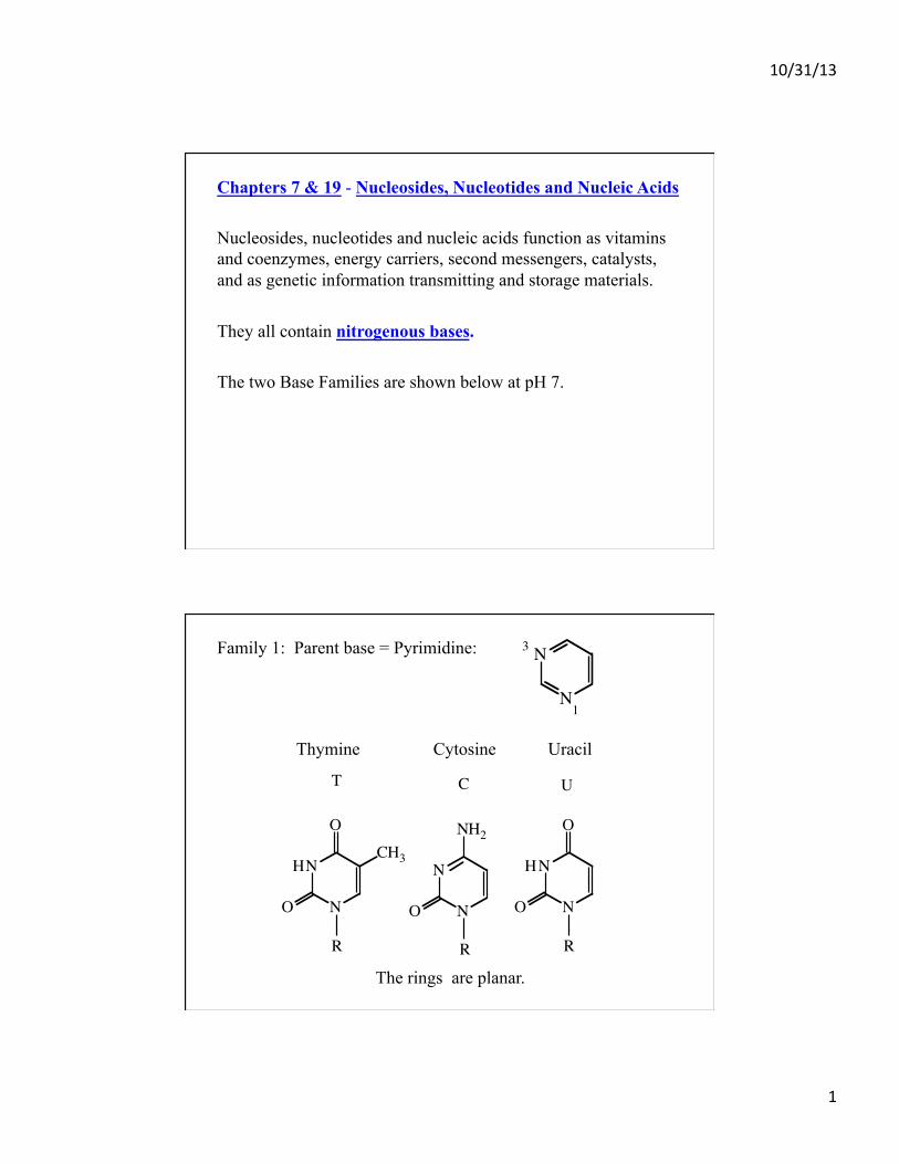
Family 1: Parent base = Pyrimidine:
Thymine Cytosine Uracil The rings are planar.
3
1
N
N
CT U
N
O
O
NH
NO
N
NH2
H
N
OCH3
O
N
R R R!

10/31/13
2
Family 2: Parent Base = Purine: Adenine Guanine Hypoxanthine These rings are puckered.
3
1
9
7N
N
N
N!
A
N
N
R
N
N
NH2
G
N
N
O
NH2
H
N
N
R
!
Minor Bases – The most common are methylated bases. Endocyclic e.g. 7-methyl-G The free bases are hydrophobic and have low water solubility at pH 7. The exocyclic NH2 are non-ionizable over pH 0 – 14, just like the NH2 in Asn and Gln. At low and high pH the endocyclic N’s ionize and the bases become more soluble.
pKa’s C N3 4.5 U N3 9.5 A N1 3.8 G N1 9.4 N7 2.4

10/31/13
3
The bases exist as resonance structures (electron delocalization) so that each bond has double bond character. They absorb uv light with a maximum near 260 nm.
=
!
A
220 240 260 280 300 3200.0
0.2
0.4
0.6
0.8
1.0
Wavelength HnmL
Absorption
U
220 240 260 280 300 3200.0
0.2
0.4
0.6
0.8
1.0
Wavelength HnmL
C
220 240 260 280 300 3200.0
0.2
0.4
0.6
0.8
1.0
Wavelength HnmL
G
220 240 260 280 300 3200.0
0.2
0.4
0.6
0.8
1.0
Wavelength HnmL
Tautomers are rapidly interconverting isomers that exist in equilibrium. This is a sort of H “delocalization”. For Uracil 3 tautomers exist.
HH
Lactam Lactim Double Lactim
Low pHpH 7
N
N
OH
HONO
N
OH
H
N
O
O
N
!
10/31/13
4
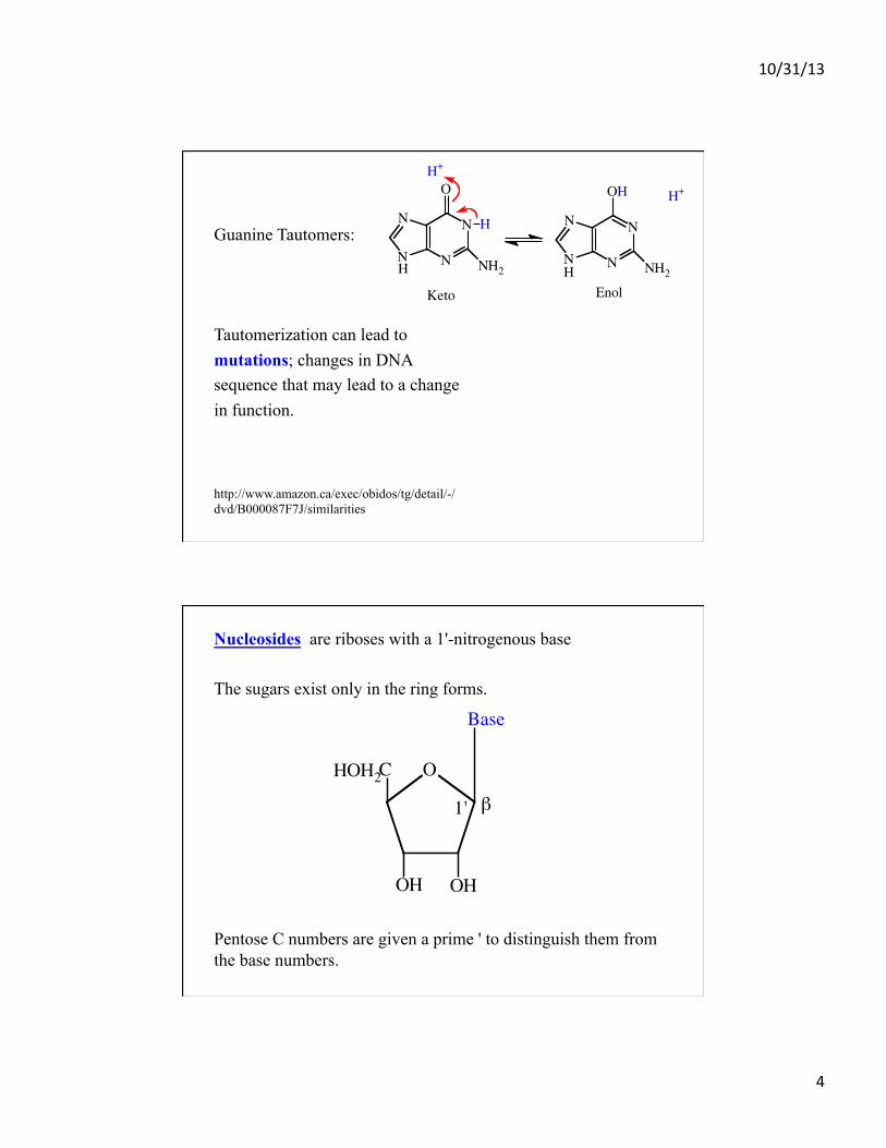
Guanine Tautomers: Tautomerization can lead to mutations; changes in DNA sequence that may lead to a change in function.
H+
HHEnolKeto
N
N NH2
OH
N
N
N
N N
N
O
NH2
H
H+
!
http://www.amazon.ca/exec/obidos/tg/detail/-/dvd/B000087F7J/similarities
Nucleosides are riboses with a 1'-nitrogenous base The sugars exist only in the ring forms. Pentose C numbers are given a prime ' to distinguish them from the base numbers.
OC
Base
OH OH
β
HOH2
1'
!

10/31/13
5
Nucleoside Naming: Ribose + Adenine = Adenosine Ribose + Guanine = Guanosine Ribose + Cytosine = Cytidine Ribose + Uracil = Uridine Ribose + Hypoxanthine = Inosine deoxyRibose + Thymine = Thymidine deoxyRibose + Adenine = deoxyAdenosine …..etc Adenosine is a local hormone and neuromodulator. Other nucleosides are mainly functional as components of nucleotides.
Nucleotides = Nucleosides + phosphate Structure: Ribose-5'-phosphate + Nitrogenous base: The phosphate oxygens have pKa’s of about 1.0 and 6.0 so are ionized at pH 7.
β
5'
--
(
O
Base
OH OH)
PO
OO
O CH2
2'
!

10/31/13
6
Nucleotide and Deoxynucleotide Naming: Adenosine 5'-monophosphate, Adenylate, AMP
Guanosine 5'-monophosphate, Guanylate, GMP
Cytidine 5'-monophosphate, Cytidylate, CMP
Uridine 5'-monophosphate, Uridylate, UMP
Inosine 5’-monophosphate, Inosinate, IMP deoxythymidine 5' monophosphate, Deoxythymidylate, dTMP deoxyadenosine 5' monophosphate, Deoxyadenylate, dAMP …….. etc.
Cells also contain nucleotides with phosphates at the 2'OH, 3'OH, and 3',5' cyclic monophosphates etc. cAMP cAMP & cGMP are second messengers important in intracellular “signal transduction”.
PO
O
O
O
CH2
OH
O-
5'
β
3'
Adenine
!
10/31/13
7
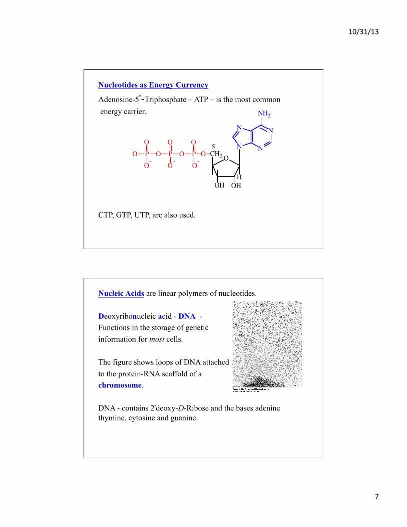
Nucleotides as Energy Currency
Adenosine-5'-Triphosphate – ATP – is the most common energy carrier. CTP, GTP, UTP, are also used.
O
OHH
CH2OPO-
OH
OOPOP-O
O O
O-O-
N
N N
N
NH2
5'
!
Nucleic Acids are linear polymers of nucleotides. Deoxyribonucleic acid - DNA - Functions in the storage of genetic information for most cells. The figure shows loops of DNA attached to the protein-RNA scaffold of a chromosome. DNA - contains 2'deoxy-D-Ribose and the bases adenine thymine, cytosine and guanine.
!

10/31/13
8
Ribonucleic acid - RNA – functions in the storage of genetic information for some viruses e.g. influenza and HIV. RNA is primarily a carrier of genetic information and is involved in some catalysis. RNA - contains D-Ribose and uridine instead of thymine.
Structure of Nucleic Acids Here is the DNA dinucleotide T-G. Phosphodiester bonds link 3'OH to 5'OH. Backbone = sugar-phosphate Side Chains = bases
OPOO-
O-
T
5'
3'
GHN
N
O
NH2N
N
H
N
N
O
O
H3C
1'
O
OHH
CH2OPOO-
O
3'
5'O
H
CH2
!

10/31/13
9
5'
1'5' end 3' end
OHP P P P P
A C G T A
!
The sequences of nucleotides is always written 5' to 3', left to right.
pApCpGpTpA = ACGTA Fewer than ~ 50 is an oligonucleotide. Greater than ~ 50 is a polynucleotide = nucleic acid.
DNA 3D Structure In 1953, it was known that: 1. DNA is the component of chromosomes that carries genetic
information.
2. # dA = # dT & # dC = # dG Watson & Crick determined the 3D structure of B-DNA in 1953 by X-ray diffraction.
10/31/13
10
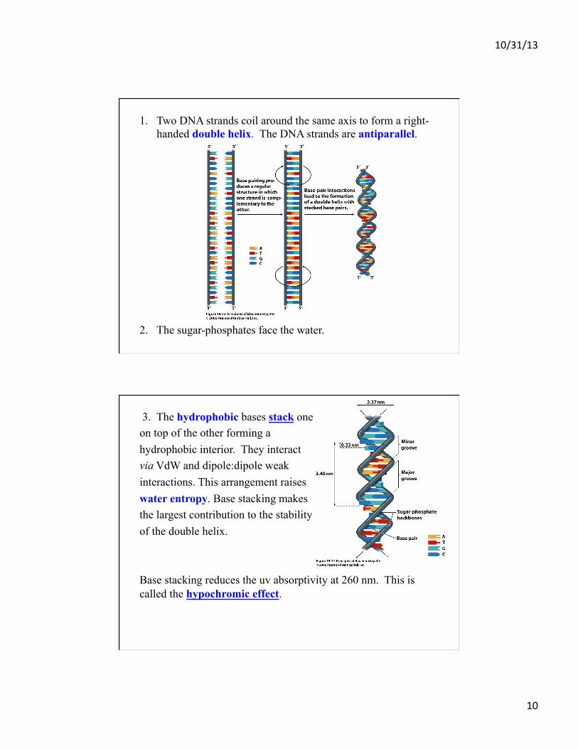
1. Two DNA strands coil around the same axis to form a right-handed double helix. The DNA strands are antiparallel.
2. The sugar-phosphates face the water.
!
3. The hydrophobic bases stack one on top of the other forming a hydrophobic interior. They interact via VdW and dipole:dipole weak interactions. This arrangement raises water entropy. Base stacking makes the largest contribution to the stability of the double helix. Base stacking reduces the uv absorptivity at 260 nm. This is called the hypochromic effect.
!

10/31/13
11
4. The bases are H-bonded. The following show Watson:Crick H-bonding. Pairing of A-T, G-C means the 2 strands have different but complementary sequences.
In DNA A:T G:C
In RNA A:U G:C
.
.!
T
A
H HN
N
N
H
R
CH3
O
O
N
NN
N
R
!
N
N
R
N
N NH
H
HO
N
NO
R
NH H
G
C
!
The bases are 0.33 nm apart. The double helix rises 3.4 nm in 1 turn = 10.5 bases. A space-filling model shows the major and minor grooves, important for binding protein..s.
A
C
G
T
T
G
C
A
5'
3'
3'
5' !
! !

10/31/13
12
The sugar-phosphate backbone has many flexible bonds so different conformations are possible. In the absence of water a more compact A-DNA is formed. The double helix is right-handed, complementary, antiparallel. Z-DNA named after Zig-Zag. It forms a left-handed, complementary, double helix.
A B Z !
Palindromes read the same forward and backward: ROTATOR NURSES RUN A Nucleic Acid “Palindrome” is: There is self-complementarity within the strands. Hairpins and cruciforms can form.
5'-T-T-A-G-C-A-C-G-T-G-C-T-A-A-3'3'-A-A-T-C-G-T-G-C-A-C-G-A-T-T-5'
!
Hairpin
Cruciform
!
10/31/13
13
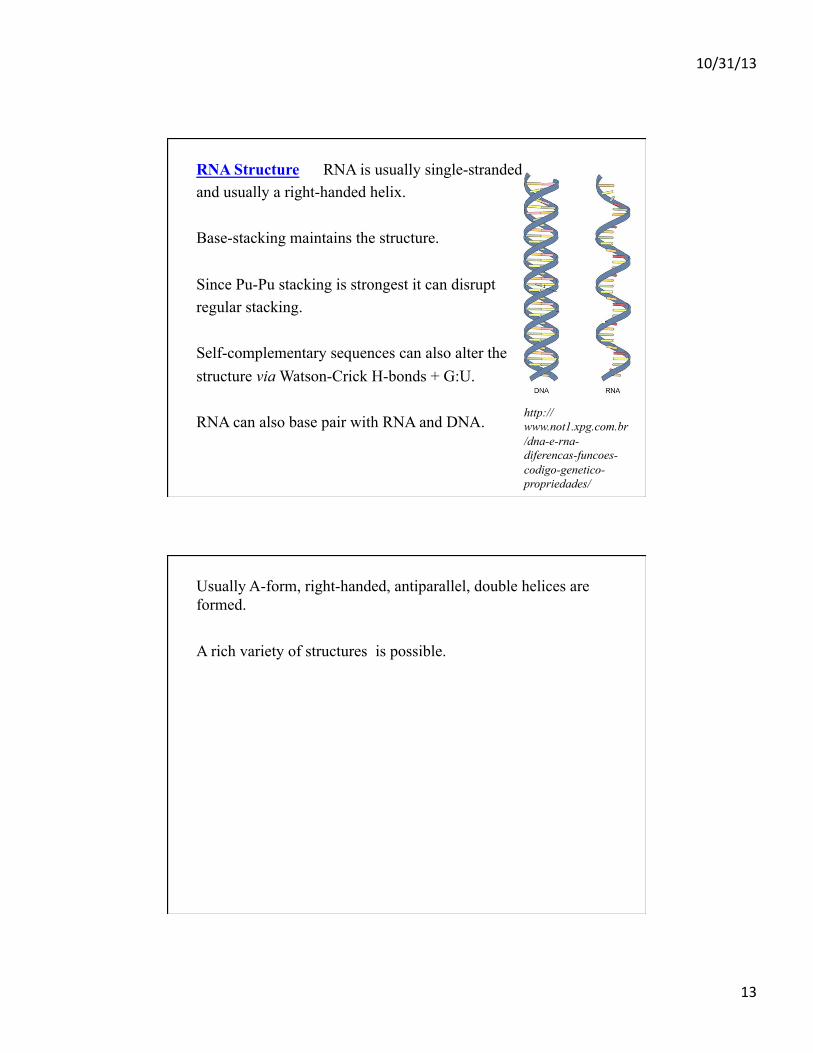
RNA Structure RNA is usually single-stranded and usually a right-handed helix. Base-stacking maintains the structure. Since Pu-Pu stacking is strongest it can disrupt regular stacking. Self-complementary sequences can also alter the structure via Watson-Crick H-bonds + G:U. RNA can also base pair with RNA and DNA.
http://www.not1.xpg.com.br/dna-e-rna-diferencas-funcoes-codigo-genetico-propriedades/
Usually A-form, right-handed, antiparallel, double helices are formed. A rich variety of structures is possible.

10/31/13
14
Connection between Structure and Function: Before a cell divides, the double helix unfolds and each strand serves as a template for the replication of a new complementary strand, resulting in 1 double helix for each new cell.
!
!
DNA is made and stored in the cell nucleus. Information contained in the DNA is transcribed into RNA in the nucleus. Proteins are made in the cytoplasm of eukaryotes according to information stored in the DNA. 1. Messenger RNA carries the genetic information from the nucleus to the cytoplasm, where it is translated into protein.
!

10/31/13
15
mRNA makes up ~5% of total cellular RNA – it is short-lived, seconds to minutes. The length depends on the gene that it encodes, usually 100’s – 1000’s of nucleotides. Monocistronic: RNA that encodes 1 polypeptide. Polycistronic: RNA that encodes 2 or more polypeptides. This is more common in prokaryotes and viruses. 2. Transfer RNA carries the amino acids to the mRNA for protein assembly. ~15% of cellular RNA. It is the “translator”.
tRNA Usually about 100 nucleotides in length with self-complementary sequences that form a “clover-leaf” structure.
tRNA
mRNA3'5'
Codon
A U C
AA
.. ..
U A G
5'
3'
Anticodon Loop
!
10/31/13
16
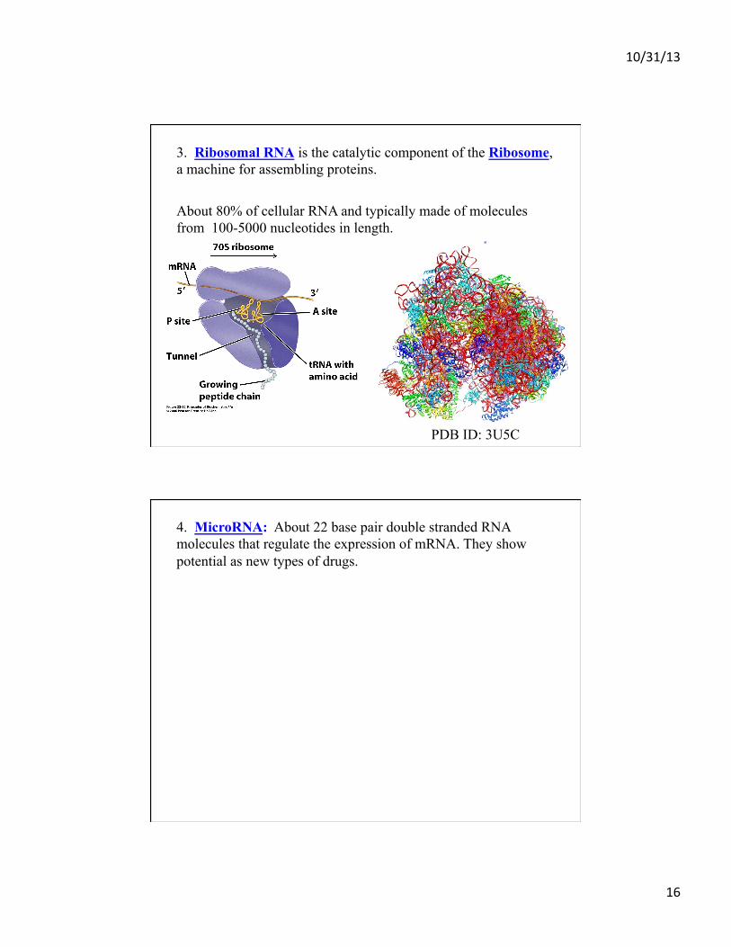
3. Ribosomal RNA is the catalytic component of the Ribosome, a machine for assembling proteins. About 80% of cellular RNA and typically made of molecules from 100-5000 nucleotides in length. PDB ID: 3U5C
!
4. MicroRNA: About 22 base pair double stranded RNA molecules that regulate the expression of mRNA. They show potential as new types of drugs.

10/31/13
17
Physical Properties of Nucleic Acids 70 – 90oC the DNA double helix denatures, H-bonds are broken, bases unstack, and the strands separate. Extremes of pH can also unfold DNA.
Renaturation at lower temperatures – it occurs in 2 steps. 1. Complementary bases pair. 2. The rest of the structure forms cooperatively; it “zips-up”.

10/31/13
18
DNA with high G:C content denatures @ a higher temperature than A:T-rich DNA. Tm is the Melting Temperature, the mid-point of the transition where the strands are 50% melted, 50% duplex.
!
Denaturation is detected by an increase in uv absorption of the single strands; hyperchromicity. Stability of Duplexes: RNA:RNA > DNA:RNA > DNA:DNA
!
10/31/13
19
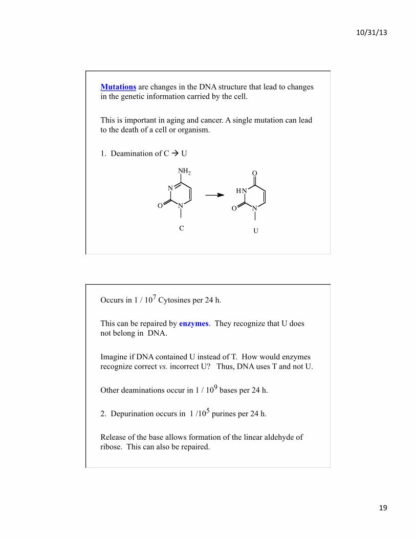
Mutations are changes in the DNA structure that lead to changes in the genetic information carried by the cell. This is important in aging and cancer. A single mutation can lead to the death of a cell or organism. 1. Deamination of C à U
C U
N
O
O
NH
NO
N
NH2
!
Occurs in 1 / 107 Cytosines per 24 h. This can be repaired by enzymes. They recognize that U does not belong in DNA. Imagine if DNA contained U instead of T. How would enzymes recognize correct vs. incorrect U? Thus, DNA uses T and not U. Other deaminations occur in 1 / 109 bases per 24 h. 2. Depurination occurs in 1 /105 purines per 24 h. Release of the base allows formation of the linear aldehyde of ribose. This can also be repaired.

10/31/13
20
3. UV light induces formation of a cyclobutane thymine dimer or 6-4 photoproduct if two T’s are stacked. This causes kinking in DNA.
CN
O
C CCH3H
CN
H
O
CN
H
OC C
CH3H
CN
OP
T
T
uv light
CN
O
CH
CN
H
O
CN
H
OC
H
CN
O
CCH3
CCH3
P
Cyclobutane thymine dimer
6
6
5
5
uv light
!
5
5
6
6
P
CCH3
CCH3OH
CN
O
CH
CN
H
O
CN
H
OHC
H
CN
O
6-4 photoproduct
4
!
4. X-rays and other radiation can also damage DNA. 5. Oxidative Damage by H2O2 and free radicals: OH., O. 6. Chemicals - nitrous acid, - alkylating agents - base analogs