chapter xv pdl 101 human anatomy & · pdf filetypes of muscle tissue ‐ classified by...
TRANSCRIPT

CHAPTER XVPDL 101 HUMAN ANATOMY &
PHYSIOLOGY
Ms. K. GOWRI. M.Pharm.,Lecturer.

Types of Muscle Tissue ‐ Classified by location,
appearance, and by the type of nervous system control or innervation.
• Skeletal muscle• Located throughout the body connected to bones and joints• Striated in appearance • Under voluntary nervous control.
• Smooth or visceral muscle• Located in the walls of organs • No striations • Under involuntary or unconscious nervous control.
• Cardiac muscle• Located only in the heart• Striated in appearance • Under involuntary or unconscious nervous control.

Skeletal Muscle
• Most skeletal muscles are connected to at least two bones– Muscles attach directly to bone
– Or muscles attach indirectly to bone through tendons
• Muscles produce movement by producing tension between its ends
• Skeletal Muscle Structure– Cellular Level
– Molecular Level

Skeletal Muscle Structure – Cellular Level
• A Skeletal muscle fiber is an individual muscle cell
• Muscle fibers are long and narrow in shape
– Sarcolemma• The plasma membrane of the muscle cell
• Surrounds the sarcoplasm
– Many nuclei (multi‐nucleated)• Located in the periphery of the muscle cell just beneath the sarcolemma

• Each muscle fiber contains various organellesspecifically designed to meet the needs of the contractile skeletal muscle fiber– Abundant mitochondria
• High demand for energy (ATP) required for muscle contraction
– Myoglobin• Protein with a high affinity for oxygen• Transfers oxygen from the blood to the mitochondria of the muscle cell
Skeletal Muscle Structure – Cellular Level

Skeletal Muscle Structure – Cellular Level
• Each muscle fiber contains:• Myofibrils – a cylindrical bundle of contractile proteins, which are called Myofilaments, within a muscle fiber
– Located in the sarcoplasm of the muscle cell
• Myofilaments – the contractile protein filaments that make up the Myofibrils
– Actin – thin filament
– Myosin – thick filament


Skeletal Muscle Structure – Cellular Level
• Sarcoplasmic reticulum (SR)– Saclike membranous network of tubules
• Elaborate form of smooth endoplasmic reticulum
– Surrounds each myofibril
– Contains terminal cisternae• Located where the SR ends, which is near the area where actin and myosin overlap
– The SR tubules and terminal cisternae store high concentrations of calcium, which is important in the process of skeletal muscle contraction


• Transverse tubules (T‐tubules)– Closely associated with SR– Connected to the sarcolemma– Penetrate the sarcolemma into the interior of the muscle cell (invaginations)
– Bring extracellular materials into close proximity of the deeper parts of the muscle fiber
• SR and T‐tubules Function– Activate skeletal muscle contraction when the muscle cell is stimulated by a nerve impulse
– Transmit nerve impulses from the sarcolemma to the myofibirls
Skeletal Muscle Structure – Cellular Level


• Sarcomere– Smallest contractile unit of the muscle fiber
– Arrangement of Myofilaments• Alternating bands of light and dark areas• Due to the organization of the actin and myosin
– Striated appearance
Skeletal Muscle Structure – Molecular Level


Sarcomere Components
• Z‐lines = borders of the sarcomere– Perpendicular to long axis of the muscle fiber
– Anchor thin myofilaments (actin)
• M‐lines – Perpendicular to long axis of the muscle fiber
– Anchor thick myofilaments (myosin)

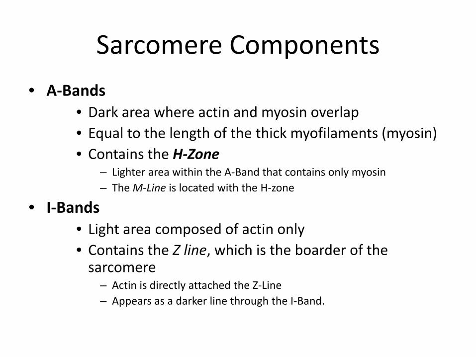
• A‐Bands• Dark area where actin and myosin overlap• Equal to the length of the thick myofilaments (myosin)• Contains the H‐Zone
– Lighter area within the A‐Band that contains only myosin– The M‐Line is located with the H‐zone
• I‐Bands• Light area composed of actin only• Contains the Z line, which is the boarder of the sarcomere
– Actin is directly attached the Z‐Line– Appears as a darker line through the I‐Band.
Sarcomere Components


Skeletal Muscle Structure – Molecular Level
• Actin
– G‐actin (globular actin) = the basic component of each actin myofilament
• Contains myosin binding sites
– The actin myofilament consists of two strands of G‐actin molecules
• The two strands of G‐action molecules are twisted together with two regulatory proteins:
– tropomyosin
– troponin


Skeletal Muscle Structure – Molecular Level
• Tropomyosin– Rod‐shaped protein that occupies the groove between the twisted strand of actin molecules
– Blocks the myosin binding sites on the G‐actin molecules
• Troponin– A complex of three globular proteins.
• One is attached to the actin molecule
• One is attached to tropomyosin
• One contains a binding site for calcium


Skeletal Muscle Structure – Molecular Level
• Myosin– Crossbridges
• Composed of a rod‐like tail and two globular heads – The tails form the central portion of the myosin myofilament
– The two globular headsface outward and in opposite directions
• Interact with actin during contraction.
• Contain binding sites for both actin and ATP– The enzyme ATP‐ase is located at the ATP binding site for hydrolysis of ATP

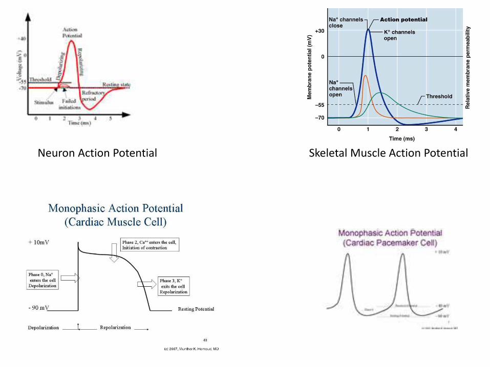
Neuron Action Potential Skeletal Muscle Action Potential

Events in Neuron Action Potential1. A threshold voltage there is immediate opening of the sodium
voltage dependent activation gate
2. At threshold there is the start of slow closure of the sodium voltage dependent inactivation gate
Result from the above two events sodium can rush into the neuron because both gates are temporarily open to sodium

As a result of the positive sodium ions rushing in through the open gates you get step 2 depolarization of the membrane

3. When the slow sodium inactivation gate closes the positive sodium ions stop rushing in and the membrane depolarizes no further – the up‐shoot stops.
4. The same voltage that operated the sodium gates also is the same voltage to initiate action of the potassium gates – however the potassium gates are very slow so they do not open till around the time that the sodium inactivation gate isclosing – thus since no further sodium is rushing In and now positive potassium is rushing out the inside of the neuron again begins to become More negative –Repolarization.

5. Just like the potassium gate was slow to open it is also slow to close – thus an overshoot of potassium moves out of the cell –causing the interior of the neuron to become more negative than at the start (Resting Membrane Potential). This overshoot is
termed “hyperpolarization.”
6. The neuron must again return to the Resting Membrane Potential state – this is a result of the Sodium /Potassium pump (3 Na out for 2 K in) action and the large intracellular molecular anions (discussed in the General electrophysiology PowerPoint).

Refractory Periods• Absolute Refractory Period – a time in which the same area of the neuron cell membrane cannot be re‐excited (fire another action potential). It is time it takes for the sodium gates to fully reset.
• Relative Refractory Period – a time immediately after the absolute refractory period in which the same area of the neuron cell membrane can be re‐excited but requires a higher voltage higher than the usual threshold voltage. During this time the potassium channels are still open – thus positive potassium is rushing out making the interior of the neuron more negative thus harder to reach the voltage of threshold.

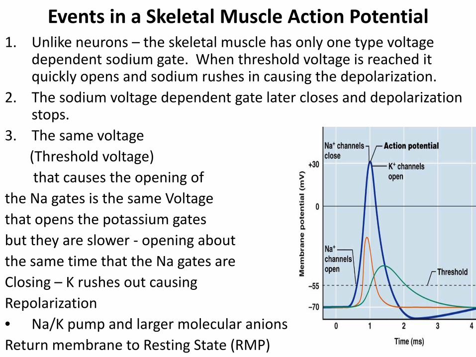
Events in a Skeletal Muscle Action Potential 1. Unlike neurons – the skeletal muscle has only one type voltage
dependent sodium gate. When threshold voltage is reached it quickly opens and sodium rushes in causing the depolarization.
2. The sodium voltage dependent gate later closes and depolarization stops.
3. The same voltage (Threshold voltage) that causes the opening of
the Na gates is the same Voltage that opens the potassium gatesbut they are slower ‐ opening aboutthe same time that the Na gates areClosing – K rushes out causing Repolarization• Na/K pump and larger molecular anions Return membrane to Resting State (RMP)

Excitation‐ Contraction Coupling• Neurons can illicit conductivity (sometimes referred to as excitation)
• This conductivity is in the form of action potentials
• Muscle can illicit the charge activity of conductivity as well as the mechanical activity of contractility
• The charge movement of conductivity (action potentials) leads to the mechanical contraction – thus the excitation must be coupled to the contraction
• Since charge activity is faster than mechanical activity –the action potentials initiate first followed fairly quickly by the initiation of the mechanical activity of contraction.

Latent Phase of ContractionThe differential in time between when the action potentials initiate and the contraction initiates is termed the latent phase of muscle contraction
Action potential initiated

Latent Phase
The latent phase involves all the events after the action potential till the myosin drags the actin over it (sliding filament) – thus causing the initiation of the muscle contraction

Actual Contraction (skeletal muscle)
Though the actionpotential only lasts1 to 3 millisecondsthe skeletalmuscle contractionlasts over 100 ms
A contraction as a result of one action potential is termed a muscle twitch. A muscle twitch has 3 periods – latent, contraction and relaxation.

100 milliseconds is 1/10 of a second• 100 milliseconds (1/10th of a second) is not a long time at all – we could not do anything muscle wise in that time frame – thus we must add (summate) together these isolated contractions (muscle twitch) – to make a longer useful contraction. The adding together of muscle twitches is termed “tetany.”
• Tetany occurs by delivering to the muscle cell‐ a rapid continuous set of action potentials.
• The action potentials can occur fast but how close together they can occur is limited by the action potentials “absolute refractory period.”