chapter two: review of literature
TRANSCRIPT

CHAPTER TWO: REVIEW OF LITERATURE
2.1 Dental Caries
Dental caries is perhaps the most ubiquitous disease that has afflicted mankind. While it
is not normally a fatal condition, it can cause a great deal of pain and distress, and the
loss of teeth has profound consequences in terms of eating, speaking, and social
behaviour in general. It remains the oldest and the most prevalent oral disease in human
history. It is only recently that the advent of daily dental care and clinician oversight
has reduced the frequency of caries within large populations (Winston & Bhaskar,
1998).
Dental caries is caused by acidic action produced by bacteria from dental biofilm and a
change in the equilibrium between demineralization and remineralization, which
favours demineralization (Tantiborjn et al., 1997). The formation of a carious lesion
does not happen all at once, but usually over several months or years (Winston &
Bhaskar, 1998).
2.1.1 Caries process The caries process is now well understood, much of it extensively described in early
dental literature. It is a simple process in concept, but complicated in details. In
general, the enamel surface is covered by a film called pellicle. As bacteria making up
the normal oral flora adhere to the pellicle, a bacterial mass called plaque is formed.
The plaque bacteria particularly Streptococcus mutans and Lactobacilli convert ingested
sugars by glycolysis to weak organic acids such as lactic, pyruvic, acetic, propionic,
formic, and butyric. The acids produced by these bacteria diffuse through the plaque
and into the tooth, leaching calcium and phosphate from enamel and eventually causing
collapse of the tooth structure and formation of a cavity (Winston & Bhaskar, 1998).
6
In general, caries is a disease caused by a group of oral streptococcal micro-organisms,
comprised primarily of Streptococcus mutans that occurs in three phases:
i) Initial interaction with the tooth surface mediated by adhesions.
ii) Accumulation of the bacteria in a biofilm and the production of glucose and
glucans by the bacterial enzyme glucosyl transferase, and
iii) Formation of lactic acid.
2.1.2 Early enamel demineralization
Enamel is the visible outer layer of the tooth. It is translucent, and can vary in color
from yellowish to greyish white. The different colours of enamel may be attributed to
variations in thickness, translucent proprieties, the quality of the crystal structure, and
surface stains. Dental enamel is composed primarily of hydroxyapatite, but it also
contains carbonate and fluoride (Dawes, 2003).
Enamel is the hardest tissue in the human body. It is almost entirely mineral by weight
(96%) but only 87% mineral by volume. Thus 13 % of the space in enamel is water and
soluble and insoluble proteins. The organic and water component of enamel allow
diffusion of ions from plaque and saliva into and out of enamel. The mineral part of
enamel consists mostly of varieties of biological apatite. Structurally, enamel is
composed of millions of rods or prisms. Each rod begins at the dentin-enamel junction
(zone between the enamel and dentine) and extends to the outer surface of the crown.
Enamel is formed by epithelial cells (ameloblasts) that lose their functional ability when
the crown of the tooth has been completed. Therefore, enamel, after formation, has no
power of further growth or repair, only mineral gain and loss.
Over the last 30 or so years, several theories have been published as explanations of the
phenomenon of subsurface dissolution of enamel sometimes observed when a
7
permeable solid such as dental enamel is subjected to acid attack either by bacteria or
diet acids. Subsurface demineralization of dental enamel during acid dissolution has
been reported many times, but its cause remains obscure
In general, an early caries lesion in enamel is observed clinically as a white opaque spot
lesion (Kidd & Joyston-Bechal, 1997). The lesion area is slightly softer than the
surrounding sound enamel and increases in whiteness when dried with air, which result
from the loss of translucency of enamel. Clinically no cavitation is evident but the
surface may be rougher than normal (Kidd & Joyston-Bechal, 1997).
2.1.2.1 Histopathology of enamel demineralization
In the past, numerous authors (Applebaum, 1940; Darling 1956; Besic, 1953; Coolidge
et al., 1955) verified subsurface demineralization. The histological appearance of the
lesion of enamel caries or demineralization has been described by Darling (1956, 1958)
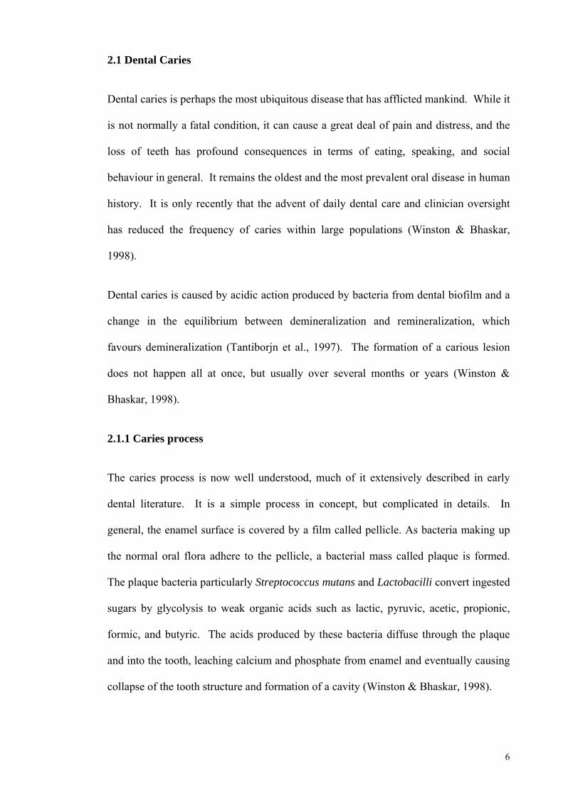
and Kidd & Joyston-Bechal, (1997) later referred to it as four zones, surface zone, body
of the lesion, dark zone and translucent zone as shown in Figure 2.1
Figure 2.1 Four zones in early enamel lesion (Adapted from Kidd & Joyston-Bechal, 1997)
8
An early enamel lesion seen under polarised light reveals four distinct zones of
demineralization. The surface zone is well mineralised by replacement ions from
plaque and saliva. Surface zone is 20µm-100µm thick. It is superficial to the positive
body lesion, in microradiography this zone appears as radioopaque zone, approximately
30 µm in depth. But the body of the lesion is poorly mineralised. Body of the lesion
appears beneath the intact surface zone and considered the largest proportion of carious
enamel in the small lesion (Kidd & Joyston-Bechal, 1997). Deeper to the body of the
lesion, a darker zone represents some demineralization, while the deepest zone, is yet
again demineralised (Kidd & Joyston-Bechal, 1997). Translucent zone of enamel caries
is not seen in all lesions. It lies between the dark zone and the normal enamel. The
width of this zone varies from 5 µm- 10 µm and has more porosity than enamel (Kidd &
Joyston-Bechal, 1997). These zones illustrate the dynamic series of events which are
occurring in the early enamel lesion.
In general early enamel caries also known as subsurface demineralization, became
more obvious at weeks 2, 3, and 4 from the initial attack, and the classic histological
zones of the white-spot lesion in polarized light could be identified (Holmen et al.,
1985). Caries is not simply a process of continued demineralization, sometimes the
lesion progresses in spite of the availability of calcium and fluoride ions (Kidd &
Joyston-Bechal, 1997).
2.1.3 Dentine Caries
The caries process in dentine involves the demineralization of the mineral component
and breakdown of the organic component of collagen fibres. The carious process in
dentine is approximately twice as rapid as in enamel. Advanced carious lesions in
dentine consist of two distinct layers having different microscopic and chemical
9
structures (Daculsi et al., 1987). The outer layer is heavily infected by bacteria which
are mainly located in the tubular spaces.
Dentine, because of its low mineral content, is more susceptible to irreversible damage
from caries process when compared with enamel (Hoppenbrouwers et al., 1986).
2.1.4 Demineralization and remineralization Phenomena Generally, the life of dental hard tissues is well understood and research has revealed
the structures and concepts involved in natural processes of the oral environment. The
nature of these tissues and how they behave under certain conditions is clear, but what is
not clear is the degree to which these natural processes can be influenced or even
accelerated. Over the course of human life, enamel and dentine undergo unlimited
cycles of demineralization and remineralization.
Minerals in dental hard tissues consist of carbonated calcium hydroxyapatite (Kautsky
& Featherstone, 1993) which differs from calcium hydroxyapatite by the substitution of
carbonate for a portion of the phosphate in calcium hydroxyapatite. Carbonated
calcium hydroxyapatite is more soluble than calcium hydroxyapatite, especially in
acidic media (Kautsky & Featherstone, 1993).
For many years pH is considered as one of the major factors that affect the imbalance in
oral cavity toward demineralization or remineralization. The most obvious theory is
that the drop in pH is the result of fermentation of carbohydrates by some plaque
bacteria. The gradual return of the pH is the result of buffers present in plaque and
saliva. Provided the pH does not drop below 5.5 the enamel remains intact, but below
this critical level, crystals of apatite dissolve (Dawes, 2003). It was suggested that the
critical pH below which enamel dissolves is not constant but rather is inversely
proportional to the concentration of calcium and phosphate in the saliva and plaque
10
fluid. Essentially, the sudden drop in pH following meals produces an undersaturation
of those essential ions (Ca2+
and PO4
3-) in the plaque fluid with respect to tooth mineral.
This promotes the dissolution of the enamel. At elevated pH, the ionic supersaturation
of plaque shifts the equilibrium the other way, causing a mineral deposition on the
tooth. The stages of caries progression are clear and in the interest of preventing
surgical intervention, early carious lesions appear to be the best opportunity for
countering this destructive process. Saliva alone has the capability to increase plaque
pH with bicarbonate although typically this process may take up to 2 hours. The
susceptibility of apatite in enamel surface layers makes it critical to control the acidity
of the plaque fluid and the Ca2+
and PO4
3- ion concentrations in saliva (Featherstone,
2000).
Just as acid was able to diffuse into the enamel and dissolve mineral, if the pH is first
neutralized, the calcium phosphate will eventually reach concentration equilibrium and
can diffuse back into the tooth if conditions are right. This reversal of the deminer-
alization process is called remineralization. Remineralization will occur if healthy
saliva first neutralizes the acid, raising the pH, and provides the needed calcium and
phosphate in solution to diffuse back into the tooth (Featherstone, 2000).
2.1.5 Secondary caries:-
Secondary or recurrent caries may be defined most simply as caries detected at the
margins of restoration (Mjör, 2005). Similar to primary caries, the enamel or root
surface adjacent to the restorative material may possess an inactive arrested lesion, an
active incipient lesion, or a frank cavitated lesion clinically (Kidd, 1990; Mjör, 1997;
Mjör, 1998; Mjör & Toffenetti, 2000).
11

The principal reason for restoration failure is secondary caries in both the permanent
and primary dentitions (Brown et al., 1988; Kidd et al., 1992). Secondary caries
accounts for approximately 60 percent of all reasons for restoration replacement,
regardless of restorative material type (Mjör, 1998; Burke & Cheung, 1999). Other
reasons include material failure, tooth fracture or defect, endodontic involvement,
prosthetic abutment utilization, technical errors, and deterioration of aesthetics quality
with tooth-colored restoratives (Burke & Cheung, 1999). The longevity of failed
restorations is variable and dependent upon the restorative material (Brown et al., 1988,
Burke & Cheung, 1999). Amalgams tend to have the greatest median and mean
survival times when compared with composite resins and glass ionomers. It must be
realized that amalgam have been available for more than 100 years; and these materials
have been refined for posterior tooth restoration. In contrast, the terms "composite
resin" and "glass ionomer" in most clinical studies encompass many different
formulations with variable strengths and weaknesses. In such studies of restoration
failure and longevity, subtypes of composite resins and glass ionomers were not taken
into account. A sequel of secondary caries is the effect on the tooth requiring
restoration replacement. With removal and replacement, the size of the restoration
changes considerably (Kidd et al., 1994; Fontana, 1995).
2.1.5.1 Histopathology of secondary caries
Histological examination of artificial and natural lesions around restorations may show
lines of demineralized tissue running along the cavity wall (Kidd, 1977; Pereira et al.,
1998). These are called wall lesions, and they are the result of microleakage. They are
very commonly seen around amalgam restorations and probably indicate initial leakage
prior to sealing of the margin (Kidd et al., 1990).
12
During the past four decades, naturally occurring and artificially induced secondary
caries around restorative materials have been characterized microscopically as two
separate parts, but interrelated lesions (Dijkman & Arends, 1992; Dionysopoulus et al.,
1994; Dionysopulus et al., 1998). The primary or outer surface lesion develops in the
enamel or root surface adjacent to the restoration; while the wall lesion forms in the
cavosurface tooth structure along the restorative interface (Kidd, 1977). The outer
surface lesion may be readily visualized in the enamel or root surface adjacent to the
restoration. The wall lesion occurs due to microleakage of oral fluids, percolation of
hydrogen ions and lytic enzymes from plaque, and bacterial colonization along the
cavosurface wall (Kidd, 1977). It was suggested that whenever a restorative material is
placed, there is a possibility for a microspace or gap to be formed between the
restorative material and the cavosurface enamel, dentine, and cementum.
Secondary caries or recurrent caries are most often located on the gingival margins of
Class II through Class V restorations (Mjör, 2005). Recurrent caries is rarely diagnosed
in Class I restorations (Mjör, 2005). The gingival part of Class V restoration is at times
challenging because of the proximity to the gingiva (Mjör & Tofenetti, 2000).
Attention must be paid to gingival part of Class V restoration during preparation the
cavity and inserting filling material, carving, and finishing (Mjör & Toffenetti, 2000).
In Class V cavities, half of restoration cover enamel and the other half cover dentine or
on thin enamel. In addition, the presence of water in dentine decrease surface energy
and prevent adhesive agent from establishing a good mechanical retention. The
gingival margin placed at the dentine-cementum exhibited more severe microleakage
than occlusal (enamel) margins (Daniela et al., 2002). They found that none of the
restorative materials completely sealed the tooth/restoration interface in Class V cavity
preparations.
13
2.1.6 Causative factors
Carious lesion of enamel is generally believed to be the end results of acid attack,
however there are many factors involved in the total description of the process.
2.1.6.1 Bacteria and dental plaque
Dental plaque is the community of microorganisms found on a tooth surface as a
biofilm, embedded in a matrix of polymers of host and bacterial origin (Marsh, 2004)
Of clinical relevance is the fact that biofilm is less susceptible to antimicrobial agents,
while microbial communities can display enhanced pathogenicity (Socransky &
Haffajee, 2002). The structure of the plaque biofilm might restrict the penetration of
antimicrobial agents, while bacteria growing on a surface grow slowly and display a
novel phenotype, one consequence of which is a reduced sensitivity to inhibitors
(Gilbert et al., 2002).
Plaque is natural and like the resident microflora of all other sites in the body
contributes to the normal development of the physiology and defences of the host.
Numerous studies have been undertaken to determine the composition of the plaque
microflora from diseased sites in order to try and identify those species directly
implicated in causing pathology. Interpretation of the data from such studies is difficult
because plaque-mediated diseases occur at sites with a pre-existing diverse resident
microflora, and the traits associated with cariogenicity such as, acid production, acid
tolerance, intracellular and extracellular polysaccharide production are not restricted to
a single species. A comparison of the properties of strains representing several
streptococcal species has shown considerable overlap in the expression of these
cariogenic traits (De Soet et al., 2000). Microorganisms in biofilms such as plaque are
in close physical contact that can increase the probability of interactions, some of which
can modulate the pathogenic potential of cariogenic bacteria.
14
The quantity and quality of saliva determines the extent to which teeth demineralized.
For example relatively fewer caries are generally found in the lower front part of the
mouth where teeth are more exposed to saliva. The type and number of caries causing
bacteria present in the mouth is also relevant. All bacteria can turn carbohydrates into
acids but certain families of bacteria such as Streptococcus mutans and Lactobacilli are
more powerful acid producers. The presence of this type of bacteria in plaque increases
the risk of decay. Some people have higher levels of decay causing bacteria than others
due to neglected or inappropriate oral hygiene.
2.1.6.2 Dietary and habits factors
Dental caries has often been described as a disease related to the consumption of diet
and acidic food and drink. Although, as a multi-factorial disease, oral bacteria, tooth
enamel composition and salivary components and consistency are also major factors.
Diet is defined as the type and amount of food eaten by an individual. The role of
dietary carbohydrates in the causation of dental caries is well established. Diet and
habits may interfere with the balance of tooth demineralization and remineralization in
several ways. The diet provides sugars and other fermentable carbohydrates, which are
metabolized to acids by plaque bacteria. Riva & Van Loveren, (2003) suggested that
the resultant low pH favours the growth of the acidogenic and aciduric bacteria
(Streptococcus mutans). In contrast, a diet lower in added sugars and fermentable
carbohydrates and high in calcium-rich cheese may favour remineralization. Sucrose
facilitates the colonization of teeth by Streptococcus mutans and their outgrowth (van
der Hoeven & Schaeken, 1995). It has been known for a long time that diet habits such
as acidic food and drinks may soften dental hard tissues (Hartles & Wagg, 1962). The
erosive activity of citric, malic, phosphoric and other acids as ingredients of beverages
and foodstuffs has been demonstrated in many in vitro and in vivo studies (Zero, 1996).
15
Further, a series of studies indicates that the erosive potential of an acidic drink is not
entirely dependent on its pH, but is also strongly affected by its titratable acid content
and by the calcium-chelation properties of the food and beverages, as they efficiently
bind released calcium. The greater the buffering capacity of the drink, the longer it will
take for the saliva to neutralise the acid (Lussi et al., 2004). Zero & Adrian, (2005)
confirmed that and they found the greater intake of the acidic drink or food, the longer it
will take for saliva to neutralize the acid.
The most common side effect of acidic beverages on enamel is erosion. Dental erosion
is the physical result of pathologic, chronic and localized loss of hard tissues from the
tooth surface by a chemical process without bacterial involvement (Ten Cate & Van
Duinen, 1996). The total titratable acid level is considered more important than pH
level, because it will determine the actual concentration H+ available to interact with
the tooth surface (Zero, 1996).
Most soft drinks contain one or two common food acidulants-phosphoric acid and citric
acid. Occasionally, other acidulants such as malic acid or tartaric acid are also used.
Animal studies have shown that phosphoric acid is very erosive at pH 2.5 but much less
so at pH 3.3. Citric, malic and tartaric acids are considered to be especially erosive
because of their acidic nature and the ability to chelate calcium at higher pH. Citric acid
was more erosive than malic acid when formulated to experimental drinks at high pH
(Hughes et al., 2000). These acids lower the surface pH and diffuse through the plaque
and into the tooth, leaching calcium and phosphate from the enamel. At this time the
plaque pH may have dropped to 4.0 – 4.5 (Winston & Bhaskar, 1998).
Infrequently reported is the acid dissolution caused by wine. The pH of wine has been
reported to range from 3 to 3.8 (Mok et al., 2001). Wine derives its acidity mostly from
tartaric and malic acids and from smaller concentrations of citric and succinic acids
16
(Touyz, 1994). Because the critical point at which enamel dissolves is reported to be a
pH of 5.0 (Barron et al., 2003), wine may play a major role in enamel demineralization
and erosion.
The frequency of consumption seems to be a significant contributor to the cariogenicity
of the diet and habit (Bowen et al., 1983). They concluded that it is not only the
frequency of ingestion that is related to the development of caries, but the time that
sugars are available to microorganisms in the mouth. The importance of frequency is
clear when caries is regarded as the outcome of the alternation of demineralization and
remineralization (Riva & Van Loveren, 2003). However, higher frequency means more
demineralization and less remineralization. The duration of the decrease in pH after
intake of cariogenic food is an important confounding factor in this relation.
Humans in different parts of the world developed certain oral chewing habits. Tobacco,
coca leaves chewing in North and South America, betel quid chewing in Southeast Asia
and qat chewing in Yemen and East Africa are some examples.
17
2.2 Qat
Qat also known scientifically as (Catha edulis) is an evergreen shrub belonging to the
family Celastraceae. It grows in Yemen and Southern Arabia as well as in certain East
African countries such as Ethiopia, Somalia, Djibouti and Kenya (Kalix, 1987). Qat
grows especially well in moist conditions and is generally cultivated along the mountain
slopes, at altitudes of 3500-7000 feet, and can vary in height from 3-15 feet. The leaves
of qat are habitually chewed by inhabitants in these regions because of its
psychostimulant effect, similar to that produced by amphetamine-like substances.
2.2.1 Qat chewing habit
Qat chewing, particularly in Yemen, is deeply rooted in tradition and forms a basis of
social interaction. It plays a dominant role in celebrations, marriages, relatives and
friends sitting. It is well spread from Eastern Africa to Southern Arabian Peninsula
particularly, Yemen. Nowadays it is cultivated in Kenya, Uganda, Tanzania, Congo,
Zimbabwe, Zambia, South Africa and Israel. Ethiopia is thought to be the country of
origin of qat use (Al-Sharabi, 2002). The initial use of qat in Yemen cannot be
determined due to lack of documentation, but the earliest uses of qat in Yemen seems to
be in the form of tea among the Muslim sofis during religious ceremonies (Al-Sharabi,
2002).
Use of qat was then modified to chewing its leaves and twigs, to absorb its active
ingredients. It is always chewed in one preferred side of the mouth. The left side of the
mouth is the most frequent preferred side. Young, fresh leaves are chewed and formed
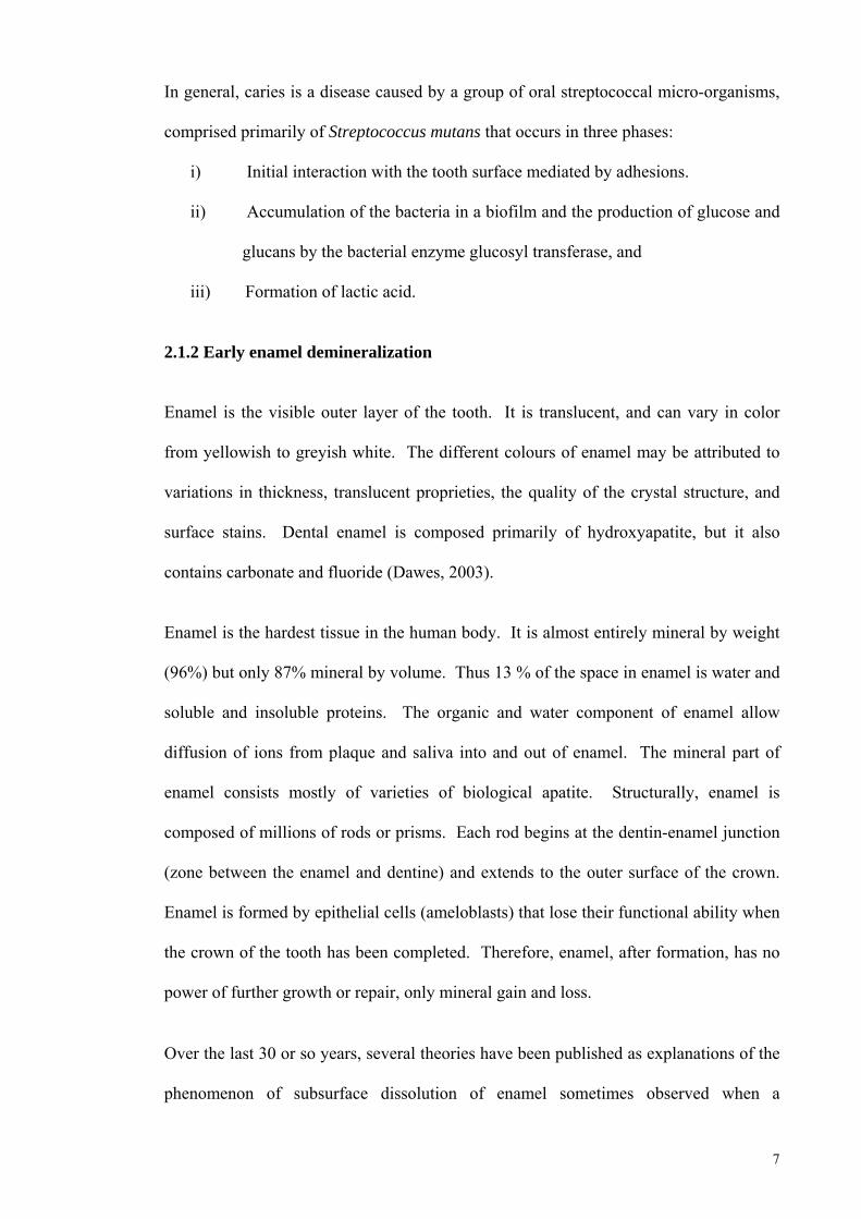
to a bolus and held in the lower buccal pouch unilaterally for three hours or longer as
shown in Figure 2.2. The saliva and leaf slurry is usually swallowed and may partly be
expectorated. The size of the bolus and time chewing varied from person to person.
18
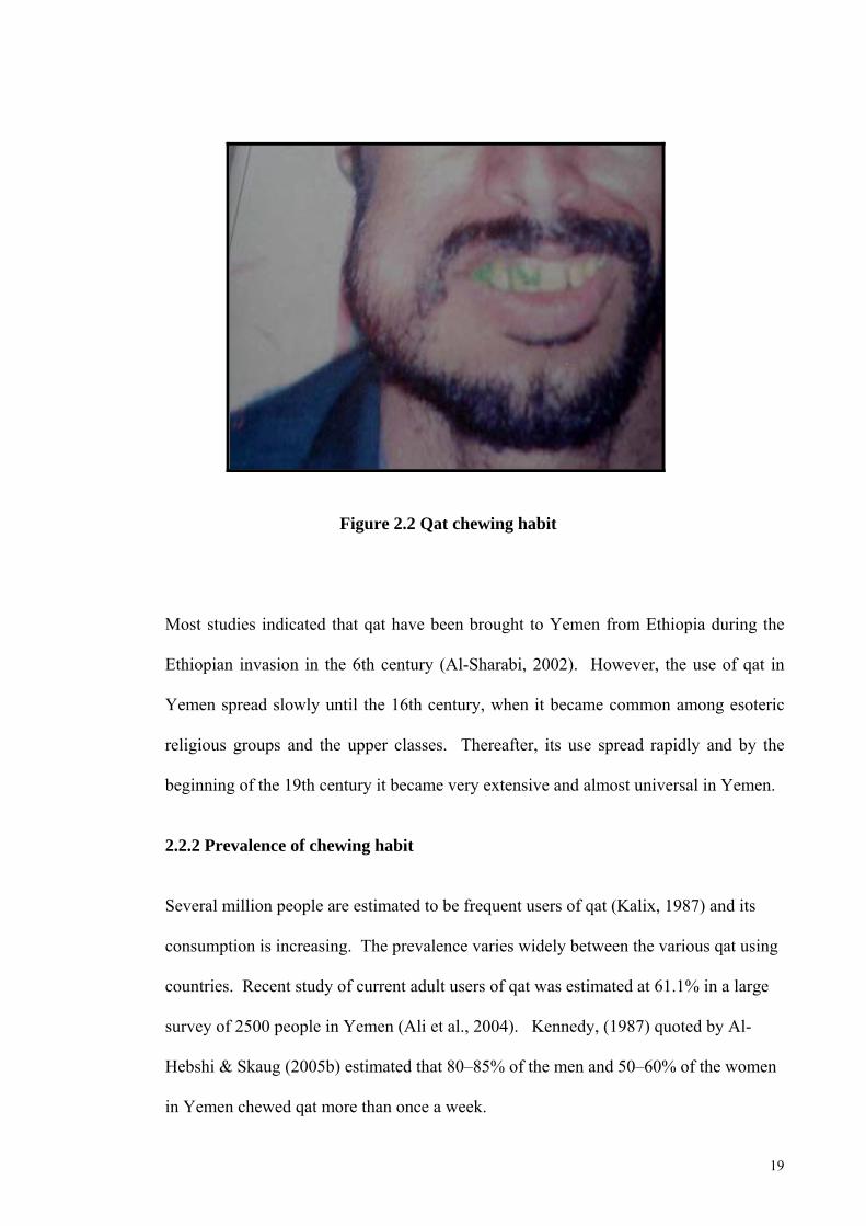
Figure 2.2 Qat chewing habit
Most studies indicated that qat have been brought to Yemen from Ethiopia during the
Ethiopian invasion in the 6th century (Al-Sharabi, 2002). However, the use of qat in
Yemen spread slowly until the 16th century, when it became common among esoteric
religious groups and the upper classes. Thereafter, its use spread rapidly and by the
beginning of the 19th century it became very extensive and almost universal in Yemen.
2.2.2 Prevalence of chewing habit
Several million people are estimated to be frequent users of qat (Kalix, 1987) and its
consumption is increasing. The prevalence varies widely between the various qat using
countries. Recent study of current adult users of qat was estimated at 61.1% in a large
survey of 2500 people in Yemen (Ali et al., 2004). Kennedy, (1987) quoted by Al-
Hebshi & Skaug (2005b) estimated that 80–85% of the men and 50–60% of the women
in Yemen chewed qat more than once a week.
19
In countries such as Yemen and Somalia many houses have special room specifically
used for chewing qat. The cultural use of communal qat chewing is common among the
Yemenite Jews in Israel ranging from twice a week to daily usage (Meir et al., 2004).
Qat also is available throughout European cities particularly in United Kingdom where
there are Yemeni, Ethiopian and Somali communities.
Qat chewing is an expensive habit. Chewers spend up to half of their monthly income
for this habit thus neglecting the family need (Kalix, 1987). At a national level,
diversion of resources toward the production or importation and marketing of qat has a
negative impact on the economy of country. The cultivation of this shrub results in the
decreased production of other more essential crops like cereals and also water
consuming thus promoting malnutrition and disease.
2.2.3 Chemistry and mechanism of action
The chemical study of qat goes back to 1887, when Fluckinger and Gerock first found
an alkaloid. The chemical composition of qat was next studied by Stokman who
described three different alkaloids: cathine, cathinine and cathidine (Al-Meshal et al.,
1985) quoted by Al-Hebshi & Skaug (2005b).
Analysis of twenty two qat samples of different origins has shown that on average a
100g of fresh qat leaves contain 36 mg cathinone, 120 mg cathine and 8 mg
norephedrine; although the concentration of these constituents varies within wide limits
(Geisshusler & Brenneisen, 1987), quoted by Al-Hebshi & Skaug (2005b). The
ascorbic acid content of qat is high, where a 100g of fresh leaves contains 325 mg of
ascorbic acid (Mustard, 1952), quoted by Al-Hebshi & Skaug (2005b) as shown in
(Table 2.1)
20
The contents of minerals and other vitamins in a 100g mixture of fresh leaves are also
shown in Table 2.1. Separation and identification of amino acids via ion-exchange and
paper chromatography detected several amino acids in Qat extracts, these amino acids
include asparaginic acid, threonine, serine, glutamic acid, proline, glycine, alanine,
valine, leucine, isoleucine, phenylalanine, tyrosine, histidine, tryptophan, ornithine and
arganine as well as α-aminobutyric acid.
Qat leaves also contain a significant amount of magnesium and reducing sugars mainly
galactose and choline to the extent of about 0.05 %. Dried qat leaves also contain
considerable amounts of tannin. Tannin belongs to phenolic compound which are
commonly found in plants. Tannin is commonly referred to as tannins acid. It is found
in grains, fruits, herbs and beverages derived from plants. Tannin concentration in qat
ranges from 3.5-9.7 g/100 g of qat leaves (Dhaifala & Santavy, 2004), as shown in
Table 2.1.
21
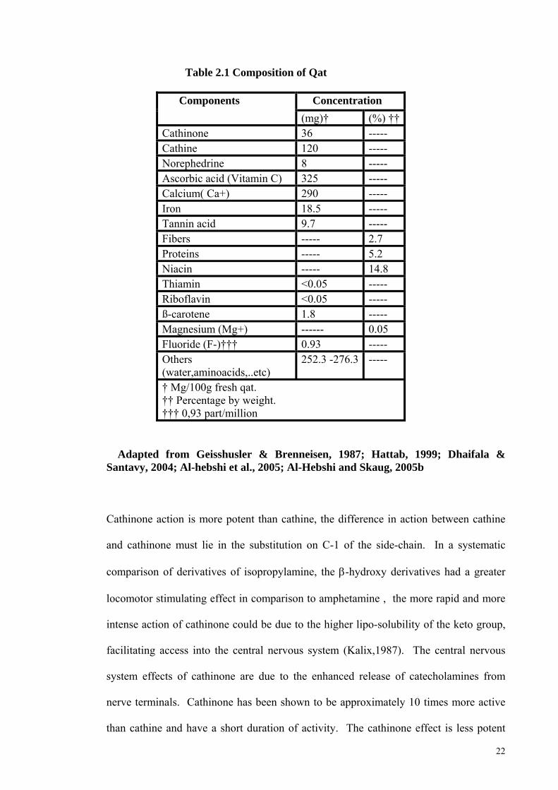
Table 2.1 Composition of Qat
Concentration Components (mg)† (%) ††
Cathinone 36 ----- Cathine 120 ----- Norephedrine 8 ----- Ascorbic acid (Vitamin C) 325 ----- Calcium( Ca+) 290 ----- Iron 18.5 ----- Tannin acid 9.7 ----- Fibers ----- 2.7 Proteins ----- 5.2 Niacin ----- 14.8 Thiamin <0.05 ----- Riboflavin <0.05 ----- ß-carotene 1.8 ----- Magnesium (Mg+) ------ 0.05 Fluoride (F-)††† 0.93 ----- Others (water,aminoacids,..etc)
252.3 -276.3 -----
† Mg/100g fresh qat. †† Percentage by weight. ††† 0,93 part/million
Adapted from Geisshusler & Brenneisen, 1987; Hattab, 1999; Dhaifala & Santavy, 2004; Al-hebshi et al., 2005; Al-Hebshi and Skaug, 2005b
Cathinone action is more potent than cathine, the difference in action between cathine
and cathinone must lie in the substitution on C-1 of the side-chain. In a systematic
comparison of derivatives of isopropylamine, the β-hydroxy derivatives had a greater
locomotor stimulating effect in comparison to amphetamine , the more rapid and more
intense action of cathinone could be due to the higher lipo-solubility of the keto group,
facilitating access into the central nervous system (Kalix,1987). The central nervous
system effects of cathinone are due to the enhanced release of catecholamines from
nerve terminals. Cathinone has been shown to be approximately 10 times more active
than cathine and have a short duration of activity. The cathinone effect is less potent
22
than amphetamine but its effect lasts longer (approximately 5 hours) than amphetamine
(Al-Meshal et al., 1985).
Tolerance to qat practically does not occur; if it does, the doses are increased only very
slowly. This may be due to the intrinsic properties of this plant or to the physical limits
on the amount that can be consumed (Kalix, 1988). There are conflicting opinions
regarding the existence of a withdrawal syndrome.
2.2.4 General effect on human
The pharmacological effect of qat in humans include mydriasis, tachycardia, elevated
blood pressure, transient facial and conjunctival congestion, headaches, hyperthermia,
increased respiration (through central stimulation, bronchodilation and counter
regulation of hyperthermia), and increased diuresis when taken in large quantities of
fluids together with qat (Halbach, 1972). The reinforcing effect of qat include:
euphoria, excitement, and insomnia.
The astringent characteristics of the tannin are believed to be involved in the delayed
intestinal absorption and might thereby contribute to some degree of malnutrition.
Moreover, constipation is the most common medical complaint of qat users and may be
attributed to both tannins and norpseudoephedrine. The anorexia associated with qat
chewing is attributed to norpseudoephedrine as a common side effect of amphetamine
type drugs (Halbach, 1972).
23
2.2.5 Effect of qat on oral health
For many years qat chewing became a challenge for dental practitioners in Yemen, East
Africa and some western countries. Many studies have associated the habit with
detrimental effects on oral hard and soft tissues.
2.2.5.1 Effect of qat on hard tissues
The first published report on the oral and dental effect of qat-chewing was by Hill &
Gibson (1987). The study was conducted on 121 Yemeni males, of whom 115 were qat
chewers. The prevalence of dental caries was low (less than 2% of all teeth were
carious). They attributed this phenomenon to the fluoride content in water in Yemen.
They claimed that qat leaves contain 360 parts/million flouride, but they didn’t mention
the analytical method. On the other hand Hattab (1999) found that qat leaves contain
negligible amount of fluoride (0.93 part/million fluoride). He purchased fresh qat
samples from Yemen suspended in deionized water, spun, and the supernatants exposed
to a chelator that decomplexes fluoride, which was assayed with an F‾ electrode coupled
to an ion analyser. Fluoride was released into whole saliva after qat chewing for 15
min. Qat suspended in stimulated whole saliva for 1.5 hour in vitro was also measured.
Total fluoride in dried qat leaves and their ash was assayed by acid-
hexamethyldisiloxane microdiffusion method. All methods demonstrated negligible
amounts of fluoride in or from qat leaves (<0.02 mg F/ml leached into water or saliva;
0.06 µF/ml in saliva after chewing, 0.93 mg total F/g in dried leaf, 2.07 mg total F/g in
ash).
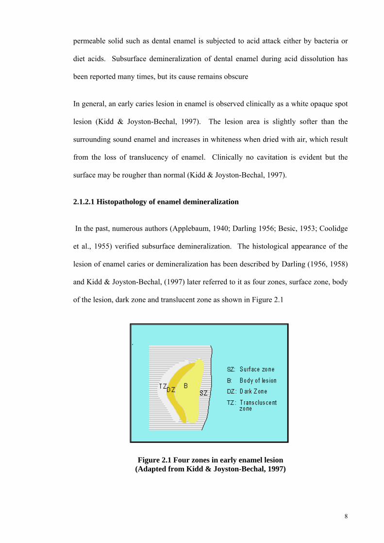
Al-Sharabi, (2002) found that there were strong association between qat chewing and
cervical caries, staining and lost of gingival attachment as shown in (Figure 2.3).
24
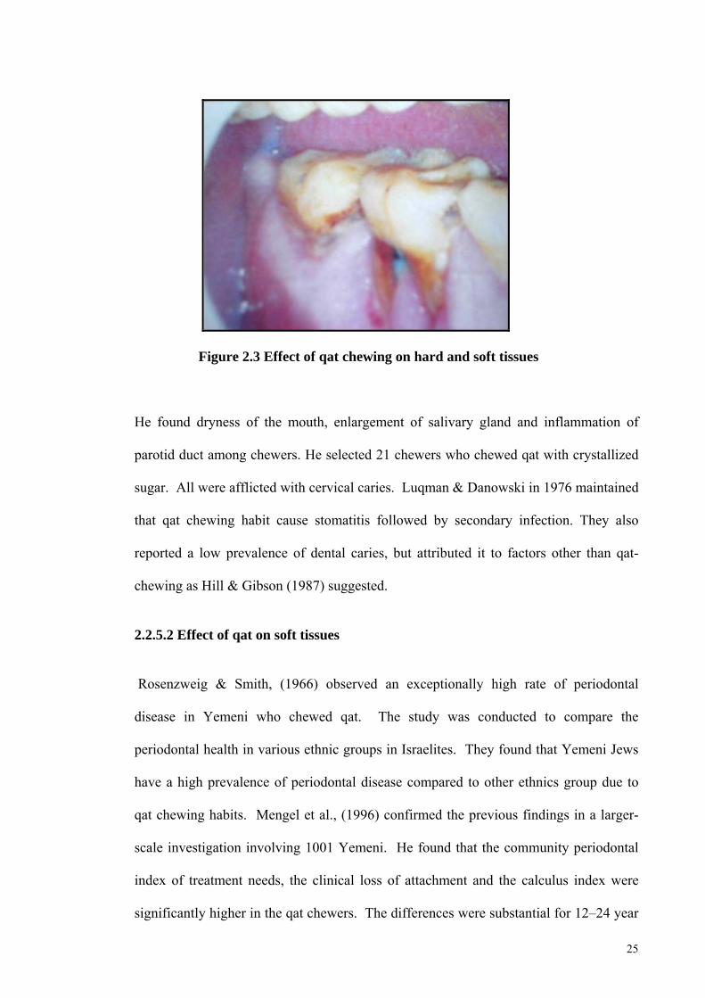
Figure 2.3 Effect of qat chewing on hard and soft tissues
He found dryness of the mouth, enlargement of salivary gland and inflammation of
parotid duct among chewers. He selected 21 chewers who chewed qat with crystallized
sugar. All were afflicted with cervical caries. Luqman & Danowski in 1976 maintained
that qat chewing habit cause stomatitis followed by secondary infection. They also
reported a low prevalence of dental caries, but attributed it to factors other than qat-
chewing as Hill & Gibson (1987) suggested.
2.2.5.2 Effect of qat on soft tissues
Rosenzweig & Smith, (1966) observed an exceptionally high rate of periodontal
disease in Yemeni who chewed qat. The study was conducted to compare the
periodontal health in various ethnic groups in Israelites. They found that Yemeni Jews
have a high prevalence of periodontal disease compared to other ethnics group due to
qat chewing habits. Mengel et al., (1996) confirmed the previous findings in a larger-
scale investigation involving 1001 Yemeni. He found that the community periodontal
index of treatment needs, the clinical loss of attachment and the calculus index were
significantly higher in the qat chewers. The differences were substantial for 12–24 year
25
age group, while insignificant for those in the 35 – 44 year age group. On the other
hand Hill & Gibson in (1987) found that the non-chewing sides showed significantly
deeper periodontal pockets than did the chewing sides, suggesting that qat had a
beneficial effect on the chewing side or a detrimental effect on the contralateral side.
Their study was conducted on 121 Yemeni males, of whom 115 were chewer.
Jorgensen & Kaimeny in 1990 found that there were generally no significant differences
in the periodontal health of 131 chewers and 199 non-chewers. The chewers showed
significantly lower lingual plaque and gingivitis scores than did the non-chewers. They
thus concluded that there was no evidence that chewing qat is detrimental to periodontal
health. Al-Hebshi & Skaug, (2005a) confirmed the study of Hill & Gibson (1987) and
found that qat chewing induce antimicrobial profile that is compatible with gingival
health. The prevalence and levels of selected periodontal bacteria in the supra- and
subgingival dental plaque of chewers and non chewers as well as of chewing sides and
non-chewing sides were compared using DNA–DNA checkerboard hybridization.
Veillonella parvula, Streptococcus intermedius and Eikenella corrodens, which are
known to be compatible with periodontal health, were found to be significantly more
prevalent and/or at significantly higher levels in the subgingival plaque of the chewers
than the non-chewers, and of the chewing sides compared to the non-chewing sides.
The periodontal pathogen of Tanerella forsythi occurred in significantly higher levels in
the subgingival plaque of the chewing sides. The effect of chewing on the supra-
gingival plaque was not pronounced, and the microbial profile induced was, as in
subgingival plaque, not incompatible with periodontal health (Al-Hebshi & Skaug,
2005a). The recession and attachment loss of gingiva and periodontal supporting
tissues may not be related to qat itself but it could be to other factors such as the
mechanical abrasion during the long period of chewing. Mucosal changes due to qat
use have also been investigated. Al-Sharabi, (2002) found that 100% of 325 chewers
26
has white lesion. Oral keratosis is common among chewers. Hill & Gibson, (1987)
found that 50% of the chewers had some degree of keratosis, while a very recent study
showed that 22.4 % of 345 chewers had keratotic white lesions, however the severity of
lesion was mild (Ali et al., 2004). Neither study suspected dysplasia or malignancy, nor
was histopathological examination carried out. In a case-control study, qat chewing was
not among the habits that showed significant association with oral leucoplakia (Macigo
et al., 1995). However, a recent report demonstrated that qat chewing had genotoxic
effects on buccal epithelial cells in a dose dependent manner, suggesting that it may
play a role in oral malignancies (Kassie et al., 2001). The effect of qat on oral
microbiology has recently been assessed. In vitro study, aqueous crude qat extracts
were shown to interfere with formation of adherent biofilms by Streptococcus mutans,
and to inhibit synthesis of water-soluble and water-insoluble glucans, which were
important for Streptococcus mutans attachment, in a dose-dependent manner. However,
the extracts did not show any antibacterial activity against the bacterium and rather
favoured its growth (Al-Hebshi et al., 2005). In another in vitro investigation involving
36 oral strains, aqueous crude qat extracts were found to possess selective antibacterial
properties in vitro. The majority of periodontal disease associated bacteria particularly
Porphyromonas gingivalis and T. forsythia were sensitive to the extracts. A few
periodontal health-associated bacteria were susceptible even at the highest concentration
tested. Actobacillus acidophilus showed a marked growth reduction in presence of the
extracts; however, none of the other cariogenic bacteria were sensitive. In addition to
their selective antibacterial properties, the extracts were also shown to possess antibiotic
resistance modifying properties; they resulted in two to four fold potentiation of
tetracycline and penicillin-G against the three resistant strains tested (Al-Hebshi et al.,
2005a).
27
2.3 Artificial Caries
A variety of in vitro methods has been developed to produce artificial enamel lesions for
use in demineralization and remineralization studies. This include the use of acidified
gels (von Bartheld, 1961; Kidd, 1977; Ingram & Silverstone, 1981; Wefel & Harless,
1984), buffered solutions (Coolidge et al., 1955; Wefel & Harless, 1984), and
incubation with natural plaque (Clarkson et al., 1984).
Wefel & Harless, (1984) found that the system that produced lesion which best mimic
natural white spot lesion was the acidified gelatine gel. They suggested that the
presence of impurities within the acidified gel, the transport of ions to and from enamel,
and perhaps the charge of the macromolecules may all help to provide an environment
ideal for remineralization phenomena to occur.
This technique involves selecting caries free teeth and coated with acid resistant varnish
leaving only a window of exposed enamel and dentine. The teeth were then subjected
to acid attack. Immersion periods varied from three days to six weeks (Kidd, 1977;
Wefel & Harless, 1984; Tantbirojn et al., 1997). The teeth were then removed from
acid gel and rinsed thoroughly with water then sectioned. After imbibitions in clearing
agent such as water and quinoline the teeth were examined. The lesion produced is not
distinguishable from that of natural caries when examined under light microscope
(Swift, 1989).
2.3.1 Evaluation techniques
Studying the speed and depth of penetration of demineralization of tooth structure is
difficult because of the different component and structure of enamel and dentine.
Enamel is a heavily mineralised material with limited porosity while dentine has
28
approximately half the mineral content and is highly porous with a relatively soft
collagen matrix.
Methods used for the analysis of enamel demineralization and remineralization include
techniques with various degrees of sophistication and quantitative capabilities. For the
last 50 years, several measuring techniques such as polarized light microscopy,
microradiography, scanning electron microscopy, microhardness, chemical analysis,
iodide absorptimetry and iodide permeability has been used for assessment
demineralization.
2.3.1.1 Polarized light microscopy
Polarized light analysis is a very sensitive technique for showing changes in hard tissues
and permitting the measurement of porosity change (Arend & Bosch, 1992). With
respect to de- and remineralization, birefringence experiments can qualitatively show
mineral loss and mineral gain. Polarized microscope make use of the birefringence of
the mineral component enamel which can resolve a beam plane polarized light into two
rays that travel at different velocities (Shellis & Poole,1985). The use of semi
quantitative polarised microscope in caries research attempts to relate the two planes of
light to the difference in pore volume (Holmen et al., 1985). It has been used in caries
research and can provide information on the lesion characteristics and pore volume in
demineralised and remineralized enamel (Kidd, 1983; Arend & Bosch, 1992; Gilmour
& Edmunds, 1998).
It has also been found that under polarized light, the enamel demineralization showed
four zones. After imbibition of the sections in water, the surface zone and the body of
the lesion are apparent. Dark zone and primary translucent zone are best seen after
imbibitions of sections in quinoline (Kidd, 1977). Caries-like lesion under polarized
29
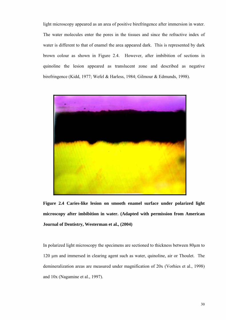
light microscopy appeared as an area of positive birefringence after immersion in water.
The water molecules enter the pores in the tissues and since the refractive index of
water is different to that of enamel the area appeared dark. This is represented by dark
brown colour as shown in Figure 2.4. However, after imbibition of sections in
quinoline the lesion appeared as translucent zone and described as negative
birefringence (Kidd, 1977; Wefel & Harless, 1984; Gilmour & Edmunds, 1998).
Figure 2.4 Caries-like lesion on smooth enamel surface under polarized light
microscopy after imbibition in water. (Adapted with permission from American
Journal of Dentistry, Westerman et al., (2004)
In polarized light microscopy the specimens are sectioned to thickness between 80µm to
120 µm and immersed in clearing agent such as water, quinoline, air or Thoulet. The
demineralization areas are measured under magnification of 20x (Vorhies et al., 1998)
and 10x (Nagamine et al., 1997).
30
For carious lesion at restoration interface, the lesions appear as two parts under
polarized light microscopy, an outer lesion and cavity wall lesion (Hals & Narnaes,
1971; Kidd, 1977; Grieve & Glyn, 1980; Gilmour & Edmunds, 1998; Nury & Alev,
2002).
2.3.1.2 Microradiography
The technique of mineral quantification by means of x-ray absorption has, in principle,
been known since the 1940s. Microradiography has been developed slowly as a suitable
method for mineral quantification in dental tissues (Arends & Bosch, 1992).
The best known type of microradiography is transverse microradiography (TMR), also
known as contact microradiography. In TMR, the sample is cut into thin slices, from 90
to 200µm for enamel or for dentine, prepared planoparallel and oriented perpendicularly
to the anatomical tooth surface.
In (LMR) Longitudinal microradiography, longitudinal tooth samples are prepared, cut
parallel to the anatomical tooth surface with a thickness of 0.5 mm. X-ray projections on
photographic film are made of these planoparallel samples, together with an aluminium
step-wedge. The resulting microradiographic images are then scanned automatically
under a densitometer.
2.3.1.3 Scanning electron microscope
(SEM) has been used to study differences in surface morphology (Ingram & Fejerskov,
1986). It has been employed alone or combined with an electron probe to study the
natural and fracture surface of enamel caries (Haikel et al., 1983).
31
It has been tried extensively but provides only qualitative information on the amount
porosity (Arends & Bosch, 1992).
2.3.1.4 Microhardness
Microhardness indentation measurement has been used to determine demineralization
and remineralization (Arends et al., 1980). An estimation of the hardness of the lesion
can be made by measuring the size of the indentation made by hardness tester (Arends
et al., 1980). In caries research, the assumption is that the measured hardness of tissue
is related to the degree of porosity of superficial layers of enamel (Purdell-Lewis et al.,
1976).
2.3.1.5 Wet chemical analysis
The determination of calcium and phosphate in solutions in which a hard tissue is
dissolved by means of an acid is in principle, a good method to quantify de- and
remineralization of the tooth tissue and the method has been used in vitro studies.
However, the analysis is destructive, and only flat samples can be used. The samples
are cut into two parts, that is, an experimental and a control part. The experimental part
can then be subjected to intraoral de-or remineralization and compared with the control
by dissolving the samples or parts of the samples in acid then determine the calcium and
phosphate content of the solution (Arends & Bosch, 1992).
2.3.1.6 Iodine absorptiometry
In this method, photons with energy of 27.4 KV resulting from the decay of a 125I
source are used to irradiate longitudinal tooth sections. The geometry of sample and
beam are analogous to the one used in LMR. The incident and the transmitted radiation
32
flux are measured with a scintillation counter. The amount of absorbed photon radiation
is a measure of the amount of mineral per unit area (kg.m¯²).
It has been shown (Almqvist et al., 1988) that the change in photon radiation due to a
dentine sample placed in the beam is linearly correlated (r = 0.83) with the amount of
Ca lost in vitro as determined by chemical analysis. This method provides quantitative
mineral loss and gain data with sensitivity, comparable to that of TMR.
2.3.1.7 Iodide permeability test
Bakhos et al., (1977) introduced a method of measuring changes in the permeability of
tooth surfaces, the iodide permeability test (IP). Such measurements are related to the
pore volume of enamel and can give, in principle, sensitive estimates of the initial stages
of de- and remineralization. Samples are completely covered with 2M KI solution for 3
min and wiped off; the window on the enamel sample is covered with water for 40s to
permit back-diffusion of iodide. The water is quantitatively recovered by an absorbent
disk. The iodide content of the disk is determined by an iodide-specific electrode and is
a measure of IP.
33