chapter six
TRANSCRIPT
Copyright © John Wiley & Sons, Inc. All rights reserved.
Chapters 6
Bone Tissue
Lecture slides prepared by Curtis DeFriez, Weber State University

Copyright © John Wiley & Sons, Inc. All rights reserved.
Introduction The skeletal system has 6 important functions:
Provide support by acting as a structural
framework and a point of attachment for
tendons and ligaments
Protect the internal organs (brain, chest, etc.)
Assist body movements (in conjunction with
muscles)
Store and release salts of calcium and
phosphorus
Participate in blood cell production
(hematopoiesis)
Store triglycerides in adipose cells of yellow
marrow

Copyright © John Wiley & Sons, Inc. All rights reserved.
Bone is a dynamic tissue – it is always
remodeling (building up and breaking down).
Like all organ systems (and as part of
the even larger musculoskeletal organ
system), the skeletal system is made
of several different tissues.
The two major tissues are bone
(osseous tissue) and cartilage.
Tissues of the Skeletal System

Copyright © John Wiley & Sons, Inc. All rights reserved.
Tissues of the Skeletal System Bone is a highly vascularized C.T. with a hard,
mineralized extracellular matrix. It is found in the
body in two different arrangements:
Compact bone – most of the bone in this
graphic is compact bone.
Spongy bone is seen as
the less organized tissue
along the left margin
(with the spicules).

Copyright © John Wiley & Sons, Inc. All rights reserved.
Tissues of the Skeletal System Compact bone is good at providing
protection and support.
It forms the diaphysis of long bones,
and the external layer of all bones.
Spongy bone is lightweight and
provides tissue support .
It forms much of the epiphysis
and the internal cavity of long bones.
Compact bone
Spongy bone
Copyright © John Wiley & Sons, Inc. All rights reserved.
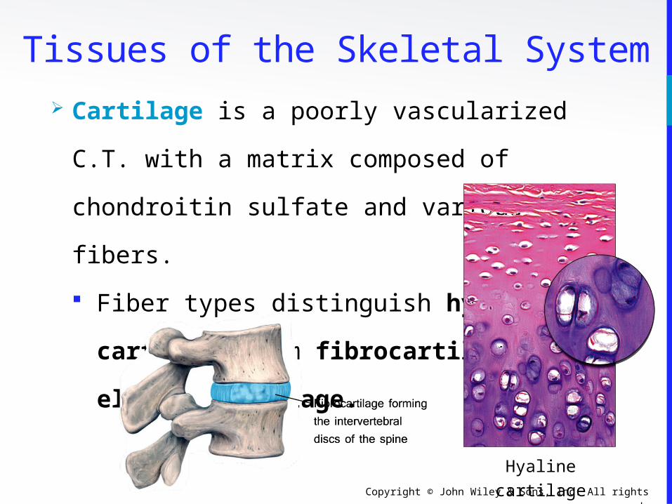
Tissues of the Skeletal System Cartilage is a poorly vascularized C.T. with a
matrix composed of chondroitin sulfate and
various fibers.
Fiber types distinguish hyaline
cartilage from fibrocartilage or
elastic cartilage.
Hyaline cartilage

Copyright © John Wiley & Sons, Inc. All rights reserved.
Articular cartilage is the thin layer of hyaline
cartilage covering the epiphysis of long bones.
Articular cartilage is found where the bone
forms an
articular (joint) surface -
where one bone
moves against another
bone.
Tissues of the Skeletal System
Hyaline cartilage is the articular cartilage of this long bone

Copyright © John Wiley & Sons, Inc. All rights reserved.
The periosteum is a tough sheath of dense,
irregular connective tissue on the outside of the
bone.
It contains osteoblasts that
help the bone grow in thickness,
but not in length.
It also assists with fracture repair
and serves as an attachment point
for tendons and ligaments.
Tissues of the Skeletal System

Copyright © John Wiley & Sons, Inc. All rights reserved.
The medullary cavity is a space within the
diaphysis of long bones that contains fatty
yellow bone marrow in adults.
The endosteum is a membrane that
lines the medullary cavity .
The endosteum is composed of
osteoclasts, osteoblasts, and
connective tissue.
Structure of Bone

Copyright © John Wiley & Sons, Inc. All rights reserved.
Tissues of the Skeletal System The perichondrium is a dense irregular connective
tissue membrane that surrounds cartilage.
Chondrocytes are cells that
form cartilage.
As we will soon see, many of
the major bones are formed
from cartilage (the remainder
do not go through a
cartilaginous stage.)
Perichondrium
Periosteum

Copyright © John Wiley & Sons, Inc. All rights reserved.
Tissues of the Skeletal System The various cells in osseous tissues are
shown in the bottom graphic:

Copyright © John Wiley & Sons, Inc. All rights reserved.
Osteoblasts are bone building cells: They
synthesize and secrete collagen fibers and
other organic components.
Osteocytes are mature osteoblasts
(maintenance).
Osteoclasts are large bone breakdown cells.
As white blood cells, osteoclasts
migrated from the bone
marrow to become “fixed
macrophages” in the
substance of the bone.
Tissues of the Skeletal System

Copyright © John Wiley & Sons, Inc. All rights reserved.
Tissues of the Skeletal System Besides bone and cartilage, the skeletal
system contains other important tissues:
Epithelium (endothelium) form
the capillary walls
Nerves (the periosteum is
especially tender)
Red marrow – hematopoiesis
Yellow marrow – fat storage

Copyright © John Wiley & Sons, Inc. All rights reserved.
Chemical Constituents of Bone Bone is 25% water, 25% organic proteins, 50%
mineral salts (hydroxyapatite crystals).
Organic constituents
• Collagen fibers provide flexibility and tensile
strength.
Inorganic hydroxyapatite crystals
(mineral salts)
• Calcium Phosphate (Ca3PO4)2
• Calcium Carbonate (CaCO3 – marble)
• Other trace elements: magnesium, fluoride,
sulfate

Copyright © John Wiley & Sons, Inc. All rights reserved.
The humerus in
the arm is a typical
long bone.
Bone Structure

Copyright © John Wiley & Sons, Inc. All rights reserved.
Bone Structure The diaphysis is the shaft or
body of a long bone.
The epiphyses form the distal
and proximal ends of a
long bone.
The metaphyses are the areas
where the epiphyses and
diaphysis join.

Copyright © John Wiley & Sons, Inc. All rights reserved.
Bone Structure In adolescents, through the end of
active growth, the epiphysis of the
long bones contains hyaline
cartilage and forms an “epiphyseal
growth plate”.
The growth plate is always
actively dividing and causing the
bone to elongate from each end.

Copyright © John Wiley & Sons, Inc. All rights reserved.
In adults, the epiphyseal cartilage is no longer
present and elongation of bones has stopped.
The epiphyseal growth plate
becomes an “epiphyseal line”,
as growing cartilage is
replaced by calcified bone.
• The epiphyseal line is
visible externally and on
X-rays.
Bone Structure

Copyright © John Wiley & Sons, Inc. All rights reserved.
Histology of Bone Tissue Compact Bone contains units called osteons or
Haversian systems formed from concentric
lamellae (rings of calcified matrix).
Interstitial lamellae
between osteons are left
over fragments of older
osteons.

Copyright © John Wiley & Sons, Inc. All rights reserved.
Outer circumferential lamellae encircle the
bone beneath the periosteum.
Inner circumferential
lamellae encircle
the medullary
cavity.
Histology of Bone Tissue
Copyright © John Wiley & Sons, Inc. All rights reserved.
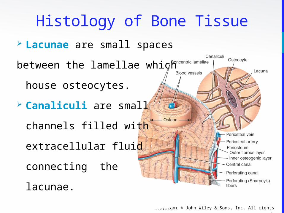
Lacunae are small spaces
between the lamellae which
house osteocytes.
Canaliculi are small
channels filled with
extracellular fluid
connecting the
lacunae.
Histology of Bone Tissue

Copyright © John Wiley & Sons, Inc. All rights reserved.
Histology of Bone Tissue Blood and lymphatic vessels
are found in the osteon’s
Central canal.
Perforating (Volkmann’s)
canals allow transit of
these vessels to the
outer cortex of the
bone.

Copyright © John Wiley & Sons, Inc. All rights reserved.
Histology of Bone Tissue Spongy bone lacks osteons. Instead, lamellae
are arranged in a lattice of thin columns called
trabeculae.
Trabeculae of spongy bone support and
protect the red bone marrow and are
oriented along lines of stress (helps bones
resist stresses without breaking).
Hematopoiesis (blood cell production)
occurs in spongy bone.

Copyright © John Wiley & Sons, Inc. All rights reserved.
Histology of Bone Tissue Within each trabecula of spongy bone are
lacunae .
As in compact bone, lacunae contain
osteocytes that nourish the mature bone
tissue from the blood circulating through the
trabeculae.

Copyright © John Wiley & Sons, Inc. All rights reserved.
Histology of Bone Tissue The interior of long bones is made up primarily of spongy
bone. The use of spongy bone lessens overall bone weight.

Copyright © John Wiley & Sons, Inc. All rights reserved.
Bone is richly supplied with blood; Periosteal
arteries and veins supply the periosteum and
compact bone.
Nerves accompany the blood
vessels (this is often the case.)
The periosteum is rich in
sensory nerves sensitive to
tearing or tension (as anyone
who has bruised their shin
will tell you!)
Blood and Nerve Supply of Bone

Copyright © John Wiley & Sons, Inc. All rights reserved.
Bone Formation Ossification or osteogenesis is the process of
forming new bone. Bone formation occurs in
four situations:
Formation of bone in an embryo
Growth of bones until adulthood
Remodeling of bone
Repair of fractures

Copyright © John Wiley & Sons, Inc. All rights reserved.
Bone Formation Osteogenesis occurs by two different methods,
beginning about the 6th week of embryonic
development.
Intra-membranous ossification produces
spongy bone.
• This bone may subsequently be remodeled
to form compact bone.
Endochondral ossification is a process
whereby cartilage is replaced by bone.
• Forms both compact and spongy bone.

Copyright © John Wiley & Sons, Inc. All rights reserved.
Bone Formation Intra-membranous ossification is the simpler of
the two methods.
It is used in forming the flat bones of the skull,
mandible, and clavicle.
Bone forms from mesenchymal cells that develop
within a membrane – without going through a
cartilage stage (recall that mesenchyme is the
tissue from which almost all other C.T. develop.)
Many ossification centers.

Copyright © John Wiley & Sons, Inc. All rights reserved.
Bone Formation

Copyright © John Wiley & Sons, Inc. All rights reserved.
Bone Formation

Copyright © John Wiley & Sons, Inc. All rights reserved.
Bone Formation

Copyright © John Wiley & Sons, Inc. All rights reserved.
Bone Formation

Copyright © John Wiley & Sons, Inc. All rights reserved.
Bone Formation

Copyright © John Wiley & Sons, Inc. All rights reserved.
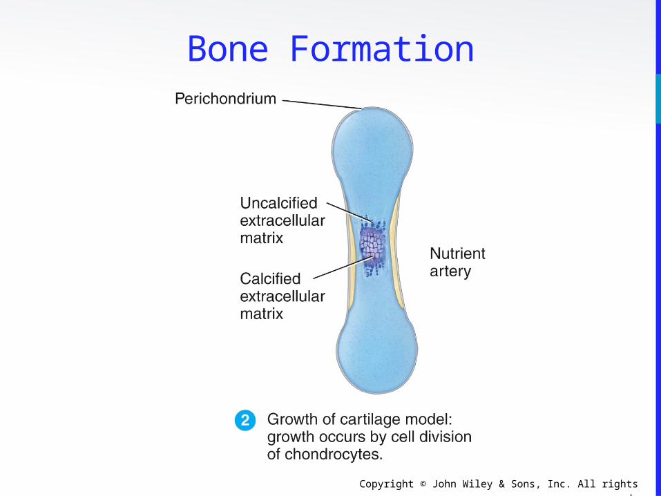
Bone Formation Endochondral ossification is the method used in
the formation of most bones, especially long
bones.
It involves replacement of cartilage by bone.
There are one primary and two secondary
centers of growth.

Copyright © John Wiley & Sons, Inc. All rights reserved.
Bone Formation

Copyright © John Wiley & Sons, Inc. All rights reserved.
Bone Formation
Copyright © John Wiley & Sons, Inc. All rights reserved.
Bone Formation

Copyright © John Wiley & Sons, Inc. All rights reserved.
Bone Formation

Copyright © John Wiley & Sons, Inc. All rights reserved.
Bone Formation

Copyright © John Wiley & Sons, Inc. All rights reserved.
Bone Formation

Copyright © John Wiley & Sons, Inc. All rights reserved.
Bone Formation

Copyright © John Wiley & Sons, Inc. All rights reserved.
Bone Formation Ossification contributing to bone length is
usually complete by 18-21 years of age.
Bones can still continue to thicken and are
capable of repair even after the epiphyseal
growth plates have closed.

Copyright © John Wiley & Sons, Inc. All rights reserved.
Bone Formation Human growth hormone is one of the body’s
many anabolic hormones. Among other things,
its secretion will stimulate bone growth, muscle
growth, loss of fat, and increased glucose
output in the liver.
The use of growth hormone has been increasing
in popularity among athletes due to the
numerous “benefits” associated with its use;
side effects are often not thought of when
young athletes use these drugs.

Copyright © John Wiley & Sons, Inc. All rights reserved.
Bone FormationInteractions Animation
Bone Formation
You must be connected to the internet to run this animation

Copyright © John Wiley & Sons, Inc. All rights reserved.
Bone Growth and Remodeling A balance must exist between the actions of
osteoclasts and osteoblasts.
If too much new tissue is formed, the bones
become abnormally thick and heavy
(acromegaly).
Excessive loss of calcium weakens the bones,
as occurs in osteoporosis.
They may also become too “soft”, as seen in
the bone diseases rickets and osteomalacia.

Copyright © John Wiley & Sons, Inc. All rights reserved.
Bone Growth and RemodelingInteractions Animation
Bone Remodeling
You must be connected to the internet to run this animation

Copyright © John Wiley & Sons, Inc. All rights reserved.
Bone Growth and RemodelingNormal bone metabolism depends on
several factors:
Minerals are an essential component.
Large amounts of calcium and phosphorus
and smaller amounts of magnesium, fluoride,
and manganese are required for bone growth
and remodeling.

Copyright © John Wiley & Sons, Inc. All rights reserved.
Bone Growth and Remodeling Vitamins are also necessary for normal bone
metabolism:
Vitamin A stimulates activity of osteoblasts.
Vitamin C is needed for synthesis of collagen.
Vitamin D is essential to healthy bones
because it promotes the absorption of calcium
from foods in the gastrointestinal tract into
the blood.
Vitamins K and B12 are needed for synthesis of
bone proteins.
Copyright © John Wiley & Sons, Inc. All rights reserved.
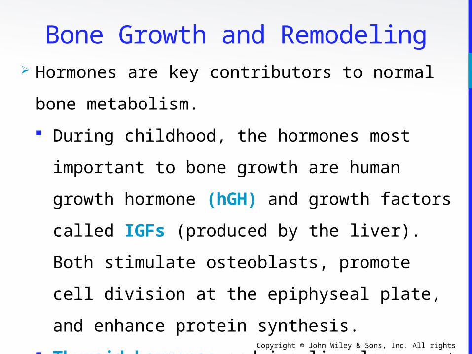
Bone Growth and Remodeling Hormones are key contributors to normal bone
metabolism.
During childhood, the hormones most
important to bone growth are human growth
hormone (hGH) and growth factors called
IGFs (produced by the liver). Both stimulate
osteoblasts, promote cell division at the
epiphyseal plate, and enhance protein
synthesis.
Thyroid hormones and insulin also promote
bone growth by stimulating osteoblasts and
protein synthesis.
Copyright © John Wiley & Sons, Inc. All rights reserved.
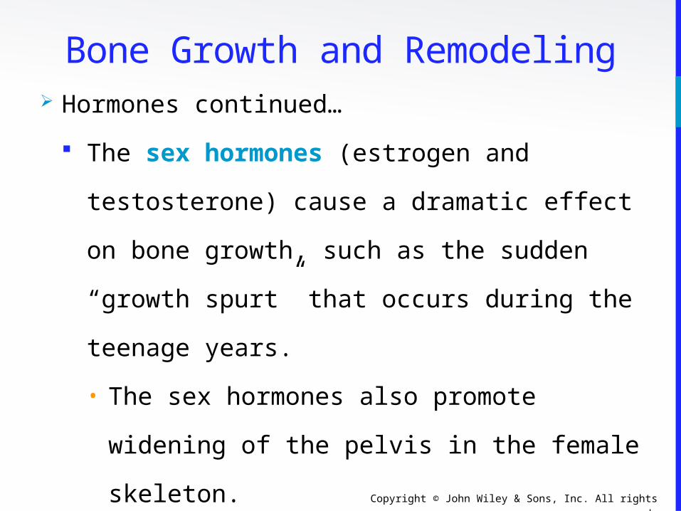
Bone Growth and Remodeling Hormones continued…
The sex hormones (estrogen and
testosterone) cause a dramatic effect on bone
growth, such as the sudden “growth spurt”
that occurs during the teenage years.
• The sex hormones also promote widening of
the pelvis in the female skeleton.
• They are also responsible for closing the
epiphyseal plates at the end of puberty.

Copyright © John Wiley & Sons, Inc. All rights reserved.
Bone Growth and Remodeling Hormones continued…
Parathyroid hormone (PTH) and calcitonin
are critical for balancing the levels of calcium
and phosphorus between blood and bone.
• Maintaining a normal serum Ca2+ level
takes precedence over mineralizing bone
(usually both can be done) – can you
suggest an explanation why this is true?

Copyright © John Wiley & Sons, Inc. All rights reserved.
Calcium Homeostasis Day to day control of calcium regulation
mainly involves:
PTH stimulates osteoclastic activity and raises
serum calcium level.
Calcitonin (thyrocalcitonin), and to a lesser
extent hGH and the sex hormones, stimulate
osteoblastic activity and lower serum calcium
level.
Vitamin D is needed for absorption of the Ca2+
and PO4– ions from the small intestine, and
reabsorption of those same ions in the kidneys.

Copyright © John Wiley & Sons, Inc. All rights reserved.
Calcium Homeostasis
The role of regulating serum Ca2+ levels and mineralizing bone
is under hormonal control, and is carefully balanced .
I made this little
diagram… but I’m not sure where I
got the figures from.
Can we reproduce
this?
PTH
CalcitoninhGH
Testosterone

Copyright © John Wiley & Sons, Inc. All rights reserved.
Calcium Homeostasis

Copyright © John Wiley & Sons, Inc. All rights reserved.
Fracture and Repair The naming of fractures can be confusing
because of the many different criteria that are
used.
Some schemes describe the anatomical
appearance of the fracture:
• Partial, complete (fx is all the way through
the bone), closed (simple), open (fx
punctures the skin), “Green stick” (a small
linear break in the bone cortex), impacted,
comminuted, spiral, transverse, displaced

Copyright © John Wiley & Sons, Inc. All rights reserved.
Fracture and Repair Anatomical appearance – like breaking a green
twig
Greenstick

Copyright © John Wiley & Sons, Inc. All rights reserved.
Fracture and Repair Anatomical appearance – the distal part is
shoved up into the proximal part.
Impacted

Copyright © John Wiley & Sons, Inc. All rights reserved.
Fracture and Repair Anatomical appearance – though not seen here,
one or both bones are “open” to the outside.
Open (compound)

Copyright © John Wiley & Sons, Inc. All rights reserved.
Fracture and Repair Naming fractures, continued…
Other fractures are classified by the disease or
mechanism which produced the fracture.
• Pathological fracture (usually from a
cancerous process or severe chronic disease),
compression fracture (produced by extreme
forces such as in trauma)
• Stress fracture (produced from repeated
strenuous activities such as running)

Copyright © John Wiley & Sons, Inc. All rights reserved.
Fracture and Repair Naming fractures, continued…
Still other fractures describe a common
pattern of injury, often involving more than
one bone, and usually denoted by an eponym
(someone’s name):
• Colles’ fracture of the distal radius
• Pott’s fracture of the distal fibula

Copyright © John Wiley & Sons, Inc. All rights reserved.
Fracture and Repair Eponyms – Colles’ is a fracture of the distal
radius ± ulna.
Colles’
Copyright © John Wiley & Sons, Inc. All rights reserved.
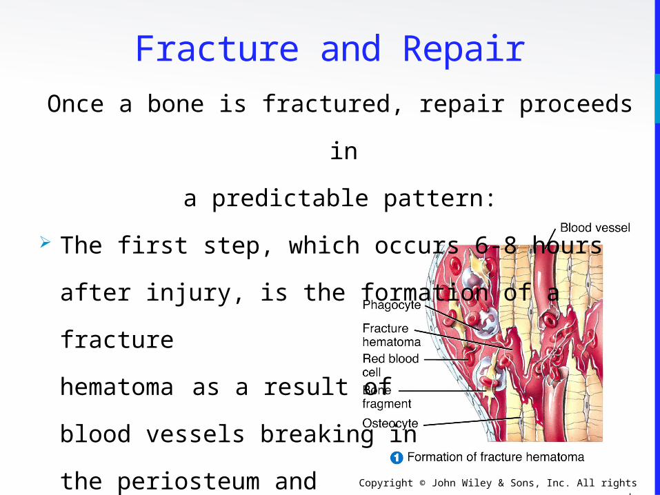
Once a bone is fractured, repair proceeds in
a predictable pattern:
The first step, which occurs 6-8 hours after
injury, is the formation of a fracture
hematoma as a result of
blood vessels breaking in
the periosteum and
in osteons.
Fracture and Repair

Copyright © John Wiley & Sons, Inc. All rights reserved.
The second and third steps involve the
formation of a callus (takes a few weeks, to as
many as six months).
Phagocytes remove cellular debris and
fibroblasts
deposit collagen to
form a fibro-
cartilaginous callus...
Fracture and Repair

Copyright © John Wiley & Sons, Inc. All rights reserved.
Fracture and Repair ... which is followed by osteoblasts forming a
bony
callus of spongy bone.

Copyright © John Wiley & Sons, Inc. All rights reserved.
Fracture and Repair The final step takes several months and is
called remodeling :
Spongy bone is replaced by
compact bone.
The fracture line
disappears, but
evidence of the break
remains.

Copyright © John Wiley & Sons, Inc. All rights reserved.
Exercise and Bone Tissue Under mechanical stress, bone tissue becomes
stronger through deposition of mineral salts and
production of collagen fibers by osteoblasts.
Unstressed bones, on the other hand, become
weaker.
Astronauts in space suffer rapid loss of bone
density.
The main mechanical stresses on bone are
those that result from the pull of skeletal
muscles and the pull of gravity (weight-bearing
activities).

Copyright © John Wiley & Sons, Inc. All rights reserved.
Aging and Bone Tissue A decrease in bone mass occurs as the level
of sex hormones diminishes during middle age
(especially in women after menopause).
Bone resorption by osteoclasts outpaces
bone deposition by osteoblasts.
Since female bones are generally smaller
and less massive than males to begin with,
old age has a greater adverse effect in
females.

Copyright © John Wiley & Sons, Inc. All rights reserved.
Aging and Bone Tissue There are two principal effects of aging on bone
tissue:
Loss of bone mass
• The loss of calcium from bones is one of the
symptoms in osteoporosis.
Brittleness
• Collagen fibers give bone its tensile strength, and
protein synthesis decreases with age.
• The loss of tensile strength causes the bones to
become very brittle and susceptible to fracture.
Copyright © John Wiley & Sons, Inc. All rights reserved.
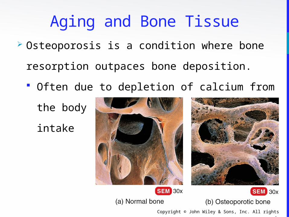
Aging and Bone Tissue Osteoporosis is a condition where bone
resorption outpaces bone deposition.
Often due to depletion of calcium from the
body or inadequate
intake

Copyright © John Wiley & Sons, Inc. All rights reserved.
End of Chapter 6
Copyright 2012 John Wiley & Sons, Inc. All rights
reserved. Reproduction or translation of this work
beyond that permitted in section 117 of the 1976
United States Copyright Act without express
permission of the copyright owner is unlawful.
Request for further information should be addressed
to the Permission Department, John Wiley & Sons, Inc.
The purchaser may make back-up copies for his/her
own use only and not for distribution or resale. The
Publisher assumes no responsibility for errors,
omissions, or damages caused by the use of these
programs or from the use of the information herein.