chapter iii diosgenin extraction and characterization in...
TRANSCRIPT
85
Chapter III
Diosgenin extraction and characterization in
Dioscorea prazeri
3.1Introduction3.1.1 The steroidal sapogenin pathway3.1.2 Diosgenin Chemistry3.1.3 Medicinal Significance of Diosgenin3.1.4 Extraction of active component Diosgenin3.1.5 Characterisation of active component Diosgenin
3.2Materials and Method3.2.1 Extraction and characterisation of D. prazeri3.2.2 Standard Stock Solution and Calibration curve1.2.4 Pre- processing of plant material1.2.5 Diosgenin extraction: Hydrolysis and neutrilisation - Method I1.2.6 Diosgenin extraction; Hydrolysis and neutrilisation; Method II1.2.7 Diosgenin Extraction1.2.8 Isolation of Diosgenin Pure Fraction from D. prazeri1.2.9 TLC for Diosgenin assessment with D. prazeri plant extract
3.3Results3.3.1 Preprocessing3.3.2 Hydrolysis3.3.3 Neutrilisation3.3.4 Soxhlet extraction3.3.5 HPLC analysis and the chromatographic conditions standardized for Diosgenin3.3.6 Diosgenin assay3.3.7 Diosgenin assay from D. prazeri plant extract -TLC
3.4Discussion
3.5References III
86
3.1 Introduction
Saponins are a diverse group of compounds widely distributed in the plant kingdom,
which are characterized by their structure containing a triterpene or steroid aglycone and
one or more sugar chains. Consumer demand for natural products coupled with their
physicochemical (surfactant) properties and mounting evidence on their biological
activity (such as anticancer and anticholesterol activity) has led to the emergence of
saponins as commercially significant compounds with expanding applications in food,
cosmetics, and pharmaceutical sectors. Saponins, are glycosides widely distributed in the
plant kingdom, and include a diverse group of compounds characterized by their steroidal
or triterpenoid aglycone structure with one or more sugar chains. Their structural
diversity is reflected in their physicochemical and biological properties, which are
exploited in a number of traditional (as soaps, fish poison, and molluscicides) and
industrial applications (Price et al., 1987; Oakenfull, 1981; Fenwick et al., 1991;
Hostettmann and Marston, 1995; Oakenfull and Sidhu, 1989). Research has established
saponins as the active components in many herbal medicines (Liu and Henkel, 2002;
Alice et al., 1991).
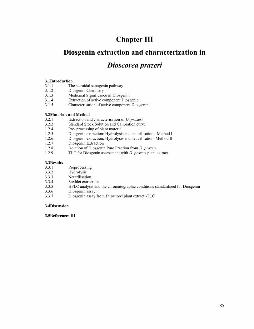
Fig. 3. 1 Diosgenin
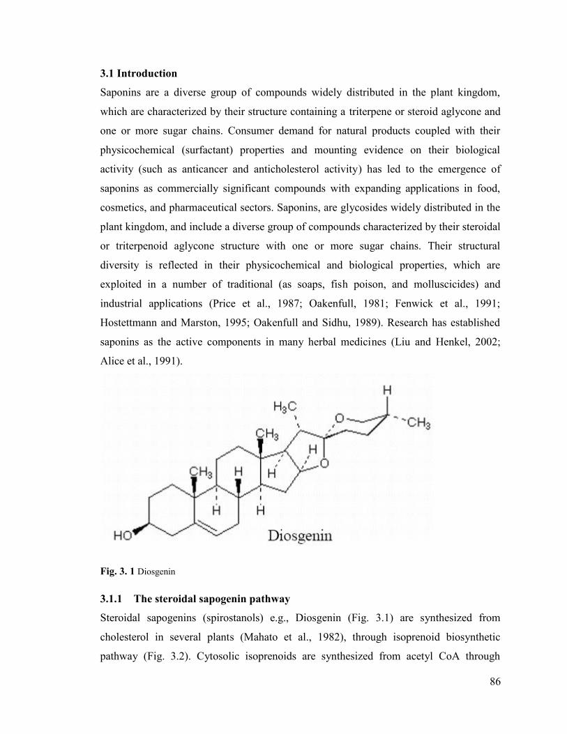
3.1.1 The steroidal sapogenin pathway
Steroidal sapogenins (spirostanols) e.g., Diosgenin (Fig. 3.1) are synthesized from
cholesterol in several plants (Mahato et al., 1982), through isoprenoid biosynthetic
pathway (Fig. 3.2). Cytosolic isoprenoids are synthesized from acetyl CoA through
87
intermediate formation of mevalonate, isopentanyldiphosphate, dimethylallyldiphosphate,
isopentenyl diphosphate, geranyldiphosphate, faresnyl diphospahte, sqaulene,
cycloartenol and leads to steroidal sapogenin in broad view. Saponins are glycosides
containing one or more sugar chains on a triterpene or steroid aglycone backbone called a
sapogenin. Steroidal saponins in which the side chain is held open by glycoside formation
(furostanols) are naturally occurring glucosides in several plant species (Sharma et al.,
1982). These glycosides are converted in vitro to spirostanols by the elimination of the
glucose molecule at Carbon positioned ‘26’ lead to ring closure by the action of
glucosidases.
Fig. 3. 2 Diosgenin Biosynthetic Pathway.
88
3.1.2 Diosgenin Chemistry
Steriods form an important group of compounds based on the fundamental saturated
tetracyclic hydrocarbon: 1,2- cyclopentanoperhydrophenanthrene (sterane or gonane).
According to their chemical structure, the wide array of steroid molecules may be divided
into several groups as Sterols, Brassinosteroids and Sapogenins. All these compounds
have basic structural skeleton or nucleus of four fused rings of 17-carbon atoms but they
differ in chemical groups or side chain attached to the basic skeleton and double bond at
specific position in the nucleus (Asolkar and Chadha., 1979). Saponins are catagorised
according to number of sugar chains in their structure. Bidesmosidic saponins have two
sugar chains with one attached through an ether linkage at C- 26 are furastanol saponins.
The nature of the aglycone and the functional groups on the aglycone backbone and
number and nature of the sugars can vary greatly resulting in a very diverse group of
compounds (Price et al., 1987; Hostettmann and Marston, 1995)
Diosgenin has spiroketal side chain attached at positions 16 and 17 of the sterane and
has a double bond at 5-6. It has a hydroxyl group at 3rd position; hydroxyl groups are
mostly found combined with sugars, making the compounds water soluble and highly
saponaceous. It is a steroidal sapogenin that is isolated from plants and is structurally
similar to cholesterol.
Diosgenin is obtained entirely from natural sources (Yams of dioscorea spp.) since the
synthetic product is not an attractive proposition commercially. Although total synthesis
of a few steroid drugs has been achieved, until recently none could compete economically
feasible method of isolation of Diosgenin from naturally occurring compounds.
3.1.3 Medicinal Significance of Diosgenin
Diosgenin, one of the most important secondary metabolites present in D. prazeri tuber is
a pharmaceutically important steroidal sapogenin. It is a precursor of sex hormones
(progesterone), corticosteroids (corticosone) and contraceptives (Onwueme, 1978;
Coursey, 1967). Over 50 species have been cultivated till now for commercial extraction
of the compound. Diosgenin is among the ten most important sources of steroids and is
also the most often prescribed medicine of plant origin (Fowler, 1984). Diosgenin
induces apoptosis in cancerous cells by cyclooxygenase up-regulation and in HeLa cells
89
by caspase pathway (Huo et al., 2004). Dioscin, a derivative compound from Diosgenin,
has been reported to induce apoptosis in HeLa cells through caspase-9 and caspase-3
pathway (Cai et al., 2002). It causes an inhibition of growth of fibroblast-like
synoviocytes in human rheumatoid arthritis with apoptosis induction associated with
cylooxygenase-2 up-regulation (Liagre et al., 2004). Diosgenyl saponins induce apoptosis
and mitotic arrest in human leukemia cell lines (Ming-Jie, 2004). Diosgenin has both
antioxidant property and anticholesterolomic activity. It has been reported to have various
effects such as hypocholesterolemic action in rat (Accatino et al., 1998), antioxidant
activity in HIV patients with dementia and apoptosis through cyclooxygenase activity in
osteosarcoma cells (Moalic et al., 2001). Cholesterol-lowering activity of saponins, which
was demonstrated in animal (Matsuura, 2001), and human trials were attributed to
inhibition of the absorption of cholesterol from the small intestine, or the re-absorption of
bile acids (Oakenfull and Sidhu, 1990). Pharmaceutical applications of saponins include
as raw materials for production of hormones (Blunden et al., 1975), immunological
adjuvants (Kensil et al., 2004), treatment of cognitive impairment (Chuang et al., 2011)
and as drugs. Saponins have also been reported to be the active ingredients in various
natural health products, such as herbal extracts (Balandrin, 1996). The diverse
physicochemical and biological properties of saponins have been successfully exploited
in a number of commercial applications in food, cosmetics, agriculture and
pharmaceutical sectors. Market trends showed increasing evidence of the use of natural
ingredients for their biological activity and have increased the demand for saponins in
recent years (Ozlem and Mazza, 2007). In cosmetics, due to their surface active
properties, saponins are being utilized as natural surfactants in cleansing products in the
personal care sector such as shower gels, shampoos, foam baths, hair conditioners and
lotions, bath/shower detergents, liquid soaps, baby care products, mouth washes, and
toothpastes (Indena, 2005;Brand and Brand, 2004;Olmstead, 2002). Saponins and
sapogenins are also marketed as bioactive ingredients in cosmetic formulations with
claims to delay the aging process of the skin (Yoo et al., 2003; Bonte et al., 1998) and
prevent acne (Bombardelli et al., 2001).
90
3.1.4 Extraction of active component Diosgenin
In addition to well-established analytical methodologies, new technologies and
approaches are also being investigated to overcome processing challenges posed by the
complex nature and diversity of this unique class of compounds. While common trends
can be identified, process development is carried out for each raw material as the
composition of the plant material and the saponin mixture will affect the process
considerably. The first step in the processing of saponins involves their extraction from
the plant matrix. As in any extraction process, the extraction solvent, extraction
conditions (such as temperature, time, pH, solvent to feed ratio), and the properties of the
feed material (such as composition and particle size) are the main factors that determine
process efficiency and the properties of the end product. Sample pretreatments, extraction
methods and extraction solvents were a few significant steps during the extraction
procedures and holds importance in its application and for its activity.
The processing methods and temperatures used for drying have considerable
effect on the quality of the medicinal plant materials. Shade drying or drying at lower
temperatures are the preferred method for drying plant material since it can maintain or
minimize loss of color of the plant material; and the lower temperatures can prevent the
loss of volatile substances in the plant materials (Ibanez et al., 2003, Bartram, 1995).
However, plants can be dried in a number of ways like drying ovens or at room
temperature, solar dryers, indirect fire, baking, lyophilization, microwave or infrared
devices. Pre-selection, peeling the skins of roots and rhizomes, boiling in water,
steaming, soaking, pickling, distillation, fumigation, roasting, natural fermentation, and
treatment with lime and chopping are some of the common processing practices. All
processed medicinal plant materials should be protected from contamination and
decomposition as well as from insects, rodents, birds and other pests, and from livestock
and domestic animals. Medicinal plant preparations can be prepared in several ways that
usually vary based upon the plant being used, and sometimes, the condition for which it
is being used. These preparations can be in the form of infusions, decoctions, tinctures,
macerations, fresh juices, etc. Some other methods include hot baths, powdered plants,
steam inhalation and even aromatherapy. Hence the processing of the material needs
significant attention and standardisation to obtain high yield. (Bartram, 1995)

91
A few analytical methods for the Diosgenin estimation from plant material were
mentioned as follows. These protocols refer to use pulversied plant material of
approximately 8.0 gm of fresh tubers/whole plantlet. The hydrolysed sample for 4 h with
hydrochloric acid were filtered using Qualigen filter paper No. 615 and w ashed with
distilled water until the residue was acid free. The washed residue was extracted with
petroleum ether (Boiling point: 60-80°C) in a soxhlet extractor for 4-6 h. The solvent was
evaporated and the residue dissolved in HPLC grade light petroleum ether and
isopropanol (12:1). It was then filtered into a measuring flask using a sample clarification
kit (Millipore, Bedford, MA) consisting of a 10 mL syringe, filter holder and Millipore
filters (0. 5 mM) (Dixit et al., 2003). Another general method consist of using fresh
tubers, which were cleaned under running tap water and dried by wiping with clean cloth
/tissue. The whole plantlet/ tubers were chopped and dried. The dried tubers/whole
plantlet were powdered and mixed with 50 mL of distilled water with simultaneous
stirring for 10 minutes in round bottom flask. To the slurry add distilled water and
concentrated hydrochloric acid in accord to maintain 5% of acid concentration (w/v). The
flask fixed with condenser was refluxed on a boiling water bath for 2 hour 30 minutes to
3.0 hour to complete the hydrolysis. After the hydrolysis, this slurry was allowed to attain
room temperature and filtered in a Buchner funnel under vacuum. The residue was
washed with distilled water till the filtrate is free from acid. The acid free residue was
transferred to Petri dish and dried in an oven at 100°C at 6 hours. The dried residue was
extracted with n-hexane in a soxhlet apparatus for 8 hours. The extracted solvent
containing Diosgenin was concentrated, chilled on ice (0°C) and filtered. The mother
liquor obtained after filtering was again concentrated, chilled on ice and re-extracted.
Diosgenin obtained from extractions were pooled and weighed after drying (for 2 hours
at 80°C) temperature and values were expressed on dry weight basis (Nandi, 1980).
Solubility enhancement has important implications for the bioactivity and
processing of saponins. Solubility of saponins is also affected by the properties of the
solvent (as affected by temperature, composition and pH). While water, alcohols
(methanol, ethanol) and aqueous alcohols are the most common extraction solvents for
saponins, solubility of some saponins in ether, chloroform, benzene, ethyl acetate, or
glacial acetic acid has also been reported. The complex structure of saponins may

92
undergo chemical transformations during storage or processing which in turn may modify
their properties/activity. The glycosidic bond (between the sugar chain and the aglycone),
and the interglycosidic bonds between the sugar residues can undergo hydrolysis in the
presence of acids/ alkali, due to hydrothermolysis (heating in presence of water) or
enzymatic/microbial activity resulting in the formation of aglycones, prosapogenins,
sugar residues or monosaccharides depending on the hydrolysis method and conditions
(Hostettmann and Marston, 1995). Incomplete acid hydrolysis yields saponins, while
complete acid hydrolysis was found to produce the constituent aglycone.
The saponin content of plant materials is affected by the plant species, genetic
origin, the part of the plant being examined, the environmental and agronomic factors
associated with growth of the plant, and post-harvest treatments such as storage and
processing (Fenwick et al., 1991), hence all these parameters require optimization and
standardization to obtain high yeild.
3.1.5 Characterisation of active component Diosgenin
i. Thin Layer Chromatography analysis with Dioscorea prazeri extracts (Dp Extract)
TLC is a simple, quick procedure that gives the chemist a quick answer as to how many
components are in a mixture. It supports the identity of a compound in a mixture when
the Rf of a compound is compared with the Rf of a known compound. It is particularly
valuable for qualitative determination of small amounts of impurities. The finger print
profile of the plant is easier to study based on the separation of colored bands, number
and Rf values. Here the chromatographic analyses were used for the qualitative analysis
of Diosgenin in D. prazeri.
ii. Diosgenin analysis by HPLC of Dioscorea prazeri extracts (Dp Extract)
The active principle compound Diosgenin was necessarily to be characterized on
HPLC/Mass spec to evaluate its status in cryopreserved and transgenic samples. A
comparative study of the Diosgenin content of these plants along with control plants need
to be assessed for biochemical stability. Diosgenin estimation was carried out with
Dioscorea prazeri whole plantlet, leaf, root and tuber extracts. For plotting standard
93
curve, Diosgenin (standard) was procured from Sigma, USA (~98% pure). The
Extraction and HPLC conditions were critical and essentially to be standardised with
various solvent systems, isocratic or gradient to obtain better yield from the plant, D.
prazeri. The diosgenin content of tubers of Dioscorea prazeri obtained from diverse
provinces and from various plant species showed various concentration of Disogenin as
per literature review (Nandi, 1980).
Dioscorea prazeri obtained from different region were found to have various level
of Diosgenin content and maximum at reproductive stage as mentioned along the region
as Jammu & Kashmir, Meghalaya, Assam, West Bengal, Tamil Nadu and U.P, was
reported with 1.6 %, 1.8%, 2.0%, 2.2%, 1.6% and 1.9% as respectively. The sapogenin
content from various sources as mentioned as Dioscorea prazeri (Diosgenin) with 1-
2.0%, Costus speciosus (Diosgenin) with 0.8-1.2%, Kallastroemia sp (Diosgenin) with
0.5-0.6%, Trigonella foenum (Hecogenin) with 0.02-0.04%, Agave cantala (Hecogenin)
with 1.10-1.20%, Solanum khasianum (Solasodine) with 1.5-2.5%, Soyabean
(Stigmasterol) with 0.02-0.05% and Smilax (Smilagenin) with trace amount.
3.2 Materials and Method
3.2.1 Extraction and characterisation of D. prazeri
The experimental plant materials used for extraction and characterisation studies of the
compound, Diosgenin of Dioscorea prazeri were obtained from plants that were
transferred to green house and grown in control environmental conditions. Tubers of D.
Prazeri obtained from In vitro grown, wild plant, cryopreserved plants, transgenic plants;
whole plantlet of D. prazeri; leaves of D. prazeri; stems of D. prazeri; roots of D. prazeri;
tubers of the Dioscorea alata; whole plantlet of D. alata and the leaves of D. alata were used for
the analysis f the active secondary metabolite. The tubers obtained from various stages of
growth phases of
D. prazeri were analysed for the study on Diosgenin content. Various solvent system
were used for the study like Ethanol, Methanol, Acetonitrile, Chloroform, Petroleum
ether, n-Hexane, 2-Isopropanol, Analytical grade water, Hydrochloric acid (2.5 N) and
Sodium hydroxide for achieving neutrilisation point during the experiment.

94
The instruments used were Soxhlet extraction unit, Roto evaporator (BUCHI),
Nitrogen evaporator, Lyophilizer (Maxi Lyo), Vaccum Centrifuge (Eppendorf), Hot air
vacuum oven (Scientek, Alpha systems), TLC apparatus, HPLC system (Schimadzu) and
Soxhlet apparatus (Biosox).
3.2.2 Standard Stock Solution and Calibration curve
A standard stock solution of Diosgenin was prepared in HPLC grade Methanol. Working
standard solutions in a range 1-5g were prepared by dilution from this stock solution.
Calibration curve was prepared based on peak areas of 5 concentration runs. Linearity
was obtained in the concentration range of 1-5 g. The equation and the good to fitness
(R2) were calculated. All data were processed using LC-Solution software (Shimadzu,
Japan).
3.2.3 Chromatographic Conditions
Chromatographic analysis was carried out on Shimadzu Series LC-20 AT liquid
chromatographic system, equipped with a diode array detector SPD-M20A, and a pump
of LC-20AT. Chromatographic separations were performed on C18 Column (AtlantisR d
C18 5m 4.6x250mm column) and standardized the column temperature for the
Diosgenin estimation .The peaks were resolved at a range of wavelength from 190 to 235
nm. Isocratic and gradient methods were used for the standardisation of method for
analyzing the Diosgenn content. The flow rate was adjusted to1 mL min-1. At the end of
each run, the column was rinsed with pure solvents.
1.2.4 Pre- processing of plant material
The experimental plant materials obtained from wild plants and the acclimatised in vitro
grown plants were rinsed thoroughly in running tap water for 15 minutes. The tubers
were blot dried and incised into thin uniform slices for the temperature exposure in same
pace. The tubers were weighed to obtain the fresh weight. The complete drying of the
plant material was performed with various temperature conditions range from 25 ºC to
100 ºC for optimisation. The plant material was dried for various durations ranging from 2
hours to two days. The dried plant materials was pulverized and stored at room temperature for
short term and at –20 ºC for long term. Pre-processing showed great impact on attaining
high yield of Diosgenin.
95
1.2.5 Diosgenin extraction: Hydrolysis and neutrilisation - Method I
i. Hydrolysis
The powdered materials were hydrolyzed with 2.0N to 2.5N Hydrochloric acid (Merck).
Hydrolysis was carried out in various temperature conditions ranging from 75 ºC to 100
ºC for 2 to 6 hours in water bath for the optimization. The conical flask was loosely
covered with sterile cotton plug). The extract was centrifuged at 25 ºC in 4000 rpm for 25
minutes. The extracts were filtered with a suction pump in Whatman filter paper (No. 42;
0.42-micron size).
ii. Neutrilisation and Drying
The slurry was washed with HPLC grade water to make it acid free (Nb. The slurry was
washed for 7 times and centrifuge at centrifuged at 4000 rpm for 15 minutes each time
and filtered to get the left out residue in the supernatant) each time the filtrate was kept
for a period of 40 minutes in the water after mixing it thoroughly). The pH was
neutrilised from pH1.69 to pH7.0.The slurry was kept for drying in sterile petriplate at
various temperatures ranging from 37 ºC to 78 ºC for a period of 2 hours to 4 days for
complete removal of water.
1.2.6 Diosgenin extraction; Hydrolysis and neutrilisation; Method II
i. Hydrolysis
The plantlets were dried at 45ºC to 55 ºC for two days and were were pulverised and
weighed. These materials were hydrolysed at 95ºC for 3 hours 30 minutes in 2.0N
hydrochloric acid, in water bath.
ii. Neutrilisation and Drying
The extracts were allowed to cool. The pH on post hydrolysis (1.69) was neutralized (7.0)
with 2N sodium hydroxide and was centrifuged at 4000 rpm for 25 minutes at 25 ºC.
These extracts were centrifuged at 4000 rpm for 15 minutes at room temperature and the
supernatant was decanted. The residue was dried at 55-78 ºC for optimising the
conditions on post-hydrolysis.

96
1.2.7 Diosgenin Extraction
The extraction was carried out at room temperature using n- Hexane, Acetonitrile,
petroleum ether and methanol for 4 to 6 hours with the hydrolysed plant extracts and with
the non-hydrolysed plant extracts. Soxhlet extraction was carried out with the same
solvents at various temperatures below the melting point of the active component. The
temperature for the HPLC analysis ranged from 23°C to 40˚C. The extract obtained was
dried and re-dissolved in various solvents as petroleum ether, chloroform: isopropanol
(12:1) and methanol for HPLC analysis to obtain Diosgenin. The experiments were
conducted to obtain the consistent yield and to obtain the active component. Detailed
experiments were carried out with two different solvents petroleum ether and methanol.
i. Extraction of Diosgenin with aqueous Acetonitrile
a. Procedure I
The hydrolysed plant materials were extracted at room temperature with aqueous
acetonitrile (50% v/v; Analytical grade), and sonicated for 10 minutes at amplitude of
60% (digital Branson sonifier). The sonicated sample was centrifuged at 4000 rpm for 15
minutes at room temperature (20 ºC). The supernatant was taken and roto-evaporated at
145 mbar, 95 rpm at 63ºC. The dried sample was stored at –20ºC and for analysis it was
re-dissolved in aqueous acetonitrile and made up to 10 mL using a volumetric flask. The
extract was filtered using a 0.2-micron filter and the sample was analysed on HPLC with
various gradient and isocratic methods to resolve the peak of Diosgenin from the plant
extract.
b. Procedure II
The extraction was carried out using soxhlet apparatus with solvent aqueous acetonitrile
for Diosgenin. The plant materials were extracted with aqueous acetonitrile at 80 ºC for 2
hours using Biosox apparatus. The extraction was carried out subsequent to acid
hydrolysis for 2 to 4 hours at 75ºC in a water bath. The soxhlet extracts of the plant were
further used for biochemical analysis.

97
ii. Extraction of Diosgenin with Petroleum Ether
a. Procedure I
Diosgenin was extracted from dried sample with petroleum ether for 4 hours at room
temperature. The petroleum ether phase was separated after centrifugation. The solvent
was rotoevaporated at 50ºC at a pressure of 145 mbar and 100 rpm. The dried plant
extract was stored at -20 ºC. The extract was dissolved in 1 mL of chloroform and
analysed for Diosgenin on High Performance Liquid Chromatographic system.
b. Procedure II
The dried plant materials of D. prazeri were subjected to soxhlet extraction with
petroleum ether at 60 ºC for 5 hours and subsequently concentrated at 75ºC to 80ºC for 1
to 2 hours in Biosox Unit (Techno Reach). On post- distillation the sample volume was
around 5mL, which was then roto-evaporated in water bath at 50ºC for 20 minutes with
pressure of 300PSi and cooled. The completely dried extracts were re-dissolved prior to
HPLC analysis and sonicated for 15 minutes. The extract was analysed on HPLC. The
tuber extract of Dioscorea alata was used as a negative control. The samples were
analysed for Diosgenin content on HPLC. The chromatographic peaks were compared
with the standard (Diosgenin) peak obtained. The yield of Diosgenin was calculated.
iii. Extraction of Diosgenin with n-Hexane
a. Procedure I
The hydrolysed residue was extracted with normal hexane (Boiling point: 50 ºC) in a
soxhlet extractor for 4-6 h. The solvent was evaporated. The extracts obtained was HPLC
analysed
iv. Extraction of Diosgenin with Absolute Methanol
a. Procedure I
D. prazeri plant materials were dried, pulversized and subjected to soxhlet extraction
with absolute methanol (HPLC grade) at 80 ºC for 4 hours and then re-distilled at 80 ºC
to 100 ºC in Biosox Unit (Techno Reach). On post- distillation the sample volume was
around 10mL, which was then roto-evaporated in water bath at 50ºC for 20 minutes with

98
pressure of 300PSi and cooled. The sample was completely dried using nitrogen
evaporator and was re-dissolved in various solvents and sonicated. The re-dissolved
sample was filtered using 0.2 µm Filters. The extract was analysed on HPLC. The tuber
extract of Dioscorea alata was used as a negative control. The samples were showed
steroidal sapogenin peak with the mobile phase. The retention time and peak of the
samples were compared with the standard (Diosgenin). The yield of the extract was
calculated.
1.2.8 Isolation of Diosgenin Pure Fraction from D. prazeri
The fraction of pure Diosgenin compound was isolated from D. prazeri plant extracts
using extraction methods standardised here with High Performance Liquid
Chromatographic analysis (Shimadzu). The chromatographic separations were performed
using C18 Column (AtlantisR d C18 5m; 4.6x250mm column) at specific optimised
temperature, 35ºC. The filtered (0.2 µm) extracts enriched with Diosgenin was analysed
with a flow rate of 1 mL min-1 on HPLC. The fractions of Diosgenin were collected
according to the retention time from calibrated curve with standard (Diosgenin;
SIGMA)). The fractions were spiked with the standard for the confirmation of D. prazeri
diosgenin fractions collected. The pure fractions were collected in sterile vials and
vacuumfuged (Centrifuge+Vacuum) and the white powdery compound of Diosgenin
isolated was further studied for its activity.
1.2.9 TLC for Diosgenin assessment with D. prazeri plant extract
i. Procedure 1
Weighed 1.5 g of plant material of D. prazeri and added 25 mL of Petroleum ether
(Merck, boiling point 60-80ºC) and extracted the metabolites using reflux condenser
(Servell instruments, Bangalore) for about 1.5 hours. The volume of the extract was
reduced to 1/20th of its original volume and was filtered. The plant extract (5 µL) along
with Standard (Diosgenin) was spotted on precoated preparative TLC plate (coated with
solid adsorbent of Silica gel 60 F 254 10cm x5 cm size).

99
ii. Procedure 2
Weighed 1.5 g of plant material of D. prazeri and to this add 12.5 mL of Chloroform
instead of Petroleum ether (Merck, boiling point 620C). The plant materials were
extracted for the metabolite Diosgenin using reflux condenser (Servell instruments,
Bangalore) for about 1.5 hour. The extracts obtained were filtered and reduced the
volume of the extract to 1/5th of its original volume by evaporating the extract on the
water bath. 5 µL of extract was spotted near the bottom of the pre-coated preparative
TLC plate made up of solid adsorbent of Silica gel 60 F 254 10 cm x 5 cm size along
with standard (Diosgenin).
iii. Development of Chromatogram
The solvent was transferred to chromatographic chamber to form a shallow pool of
homogenous mobile phase, of 5-6 mm depth so that only the bottom of the plate touches
the liquid. The chamber was closed and allowed to stand at constant temperature,
protected from direct sunlight for 15 minutes so that it would be saturated. The spots
were allowed to dry in plates and placed vertically in the chamber, ensuring that the
points of application were above the surface of the mobile phase. The chamber was
closed and allowed to develop chromatogram at room temperature. The mobile phase
slowly rose through the TLC plate by capillary action till the specified distance.
Chromatographic bands appeared when the equilibrium was attained between the
molecules of the component and the molecules of the solution, with the solvent system.
The separation of the components was based on the solubility and the strength of the
adsorption to the adsorbent on plate. The plate was removed from the developing
chamber, marked the solvent front and was dried. The components of the mixture were
observed. The compounds that were not intrinsically colored were observed under UV
(The plate fluoresces under UV lamp except the region in which the organic compound
are existing). An alternative method of detection was using anisaldehyde as a detection
reagent. The Rf values were calculated based on the distance travelled by the solute and
the solvent. As an additional step of visualisation the chromatogram was developed with
Iodine vapour. It was further treated with detective reagent, anisaldehyde for further
observation. Mobile phase used for TLC analysis was Chloroform: and Petroleum ether
to the ratio of 60:40 (v/v).
100
Distance traveled by soluteRf Value = -----------------------------------
Distance traveled by solvent
3.3 Results
3.3.1 Preprocessing
The plant materials of Dioscorea prazeri were dried at various temperature conditions in
pre-processing of the extraction of steroidal sapogenin (Fig. 3.3). The sun drying was
observed to decrease the yeild of Diosgenin when compared to vacuum drying.
Diosgenin yield was observed as 0.6±0.1% with this processing. The Diosgenin yield was
reduced to 0.5-0.8% in comparison with the natural content of 2.2-2.4% of this secondary
metabolite when the temperature for drying was set higher than 75ºC, according to the
data obtained on HPLC analysis. The sliced tubers were dried at 55 ºC for 36±3 hours in
hot air oven, which exhibited consistent value on Diogenin analysis. The water content of
the plant material was calculated to an average of 88.5±1% from 25 sets of experiments
for extraction and characterisation for biochemical stability.
3.3.2 Hydrolysis
Diosgenin from the plant material was extracted at room temperature using Acetonitrile,
n-Hexane, Petroleum ether, Chloroform and Methanol. However the yield of Diosgenin
obtained from extraction in room temperature was as low as 0.4± 0.1% with petroleum
ether and normal hexane to maximum level of 0.7% with methanol. Chloroform was
found to interfere with C-18 column and with the mobile phase during HPLC analysis,
and was subsequently not preferred as a solvent system during further extraction
procedures. The dried plant materials were pulverized and followed by hydrolysis with
2.0N hydrochloric acid for 3 hours 30 minutes. 1 to 2 hours of treatment were not found
to be sufficient to hydrolyse the compound completely. 3.30 hours to 6.0 hours were
yielded the same content of Diosgenin. So the optimised temperature studied was 3.30
minutes. Neutralized the hydrolysed plant material with sodium hydroxide (2.0 N) and
these were soxhlet extracted for Diosgenin (Fig. 3.4). The extracts were given prominent
peak of the compound of interest.
101
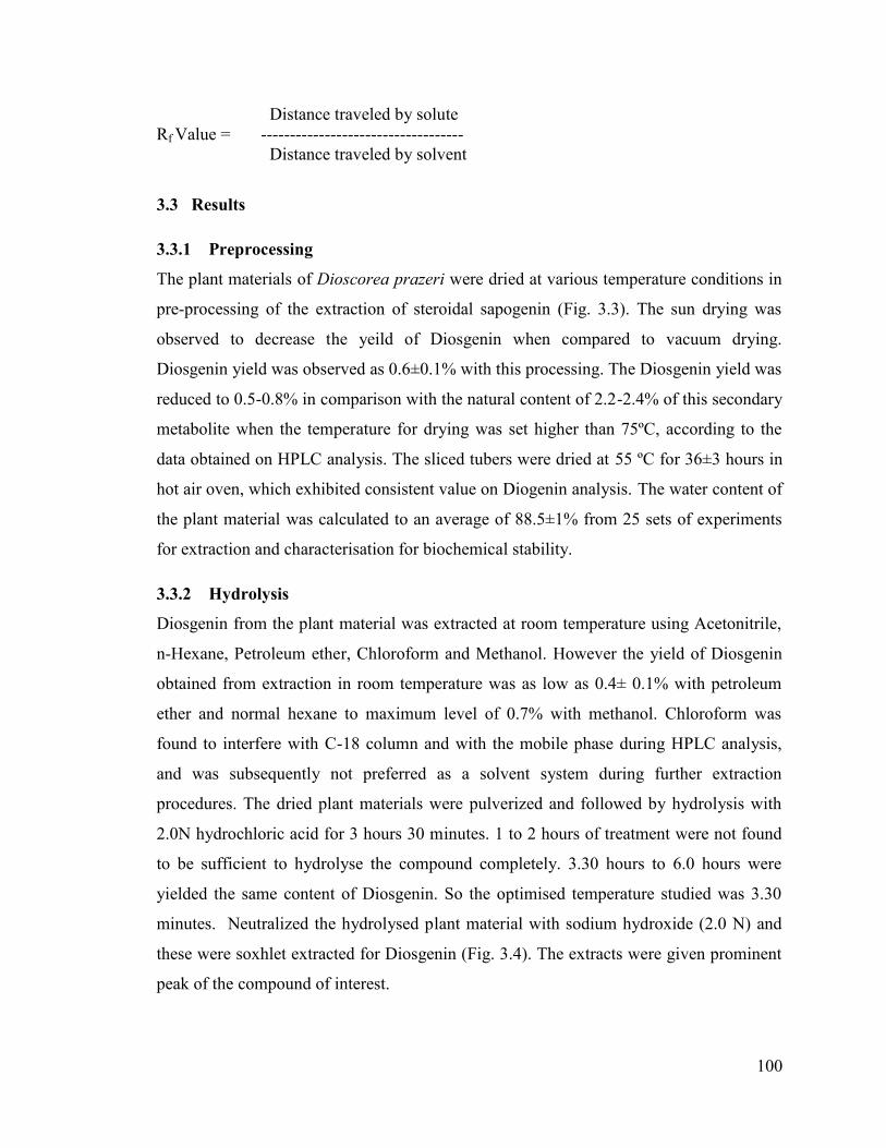
Fig. 3. 3 The various stages of Diosgenin extraction and characterisation of D. prazeri (Whole
plant materials) and D. alata (tubers).
(A) In vitro raised D. prazeri; (B), Tubers of D. alata and D. prazeri (In vitro plants and Wild
plants), Roots, and Whole Plantlet f D. prazeri (The plant material was dried under controlled
temperature conditions); (C) Pulverised plant material; (D) Plant material for hydrolysis in
hydrochloric acid (2.0 N).
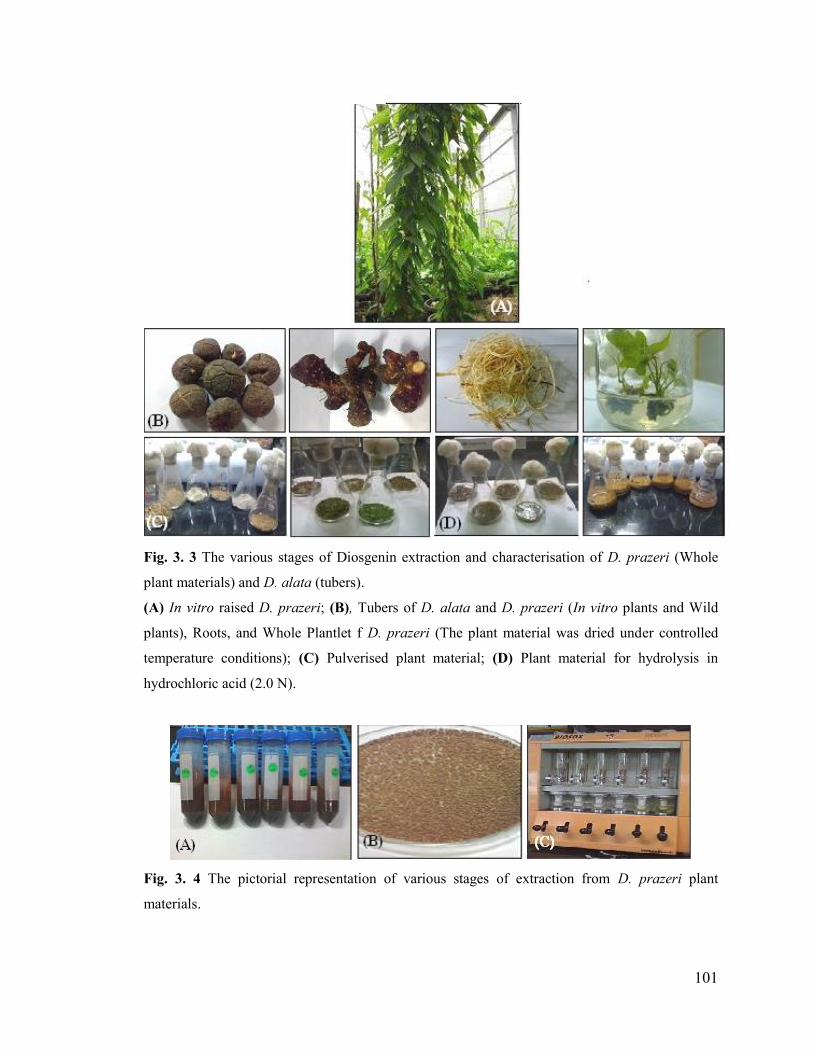
Fig. 3. 4 The pictorial representation of various stages of extraction from D. prazeri plant
materials.
102
(A) The neutrilisation of hydrolysed plant material; (B) The plant material dried at 55 ºC; (C)
Soxhlet extraction of steroidal sapogenin of D. prazeri in BIO SOX unit (Techno reach)
3.3.3 Neutrilisation
The neutirlisation with sodium hydroxide (2.0 N) was an appropriate method for
neutrilisation of the hydrolysed material. The slurry was turned black with higher
concentration of NaOH. The wastage of hydrolysed material was minimised with the
procedure of neutrilisation with restricted amount of NaOH. Continuous rinsing of the D.
prazeri extract with water for neutralising pH after hydrolysis lead to loss of the active
component to a greater extent, up to 1.2% compared to alternative methods used.
3.3.4 Soxhlet extraction
The Steroidal sapogenin, Diosgenin was extracted with BIOSOX unit with various
solvent systems. Following extraction the solvent was redistilled to obtain the
concentrated volumes of compound of interest. The extract was lyophilised completely to
obtain the dried fraction enriched with active compound of interest. The acetonitrile
extract did not give prominent peak of Diosgenin on analysis. The extraction was carried
out with petroleum ether and methanol (Analytical grade; Merck) for the studies due to
the high yield of Diosgenin and the consistency of data. So the procedure was
standardised with petroleum ether and methanol to extract the active component of D.
prazeri. The yield obtained was as high as 2.4% to 2.8% with absolute methanol and
2.0% to 2.4% with petroleum ether.
The standard curve was plotted with Diosgenin (standard) procured from Sigma, USA
(~98% pure). As a part of the study the control plantlets micropropagated and the tubers
obtained from it were compared with the wild plants to find out the variation and the data
assessed indicated fidelity at biochemical level.
The protocol for extraction of active component of the plant, steroidal sapogenin
(Diosgenin) with Methanol and an alternative procedure with Petroleum Ether were
standardized in this study as follows.
(D)(C)

103
i. Extraction of Diosgenin with HPLC grade Petroleum ether
The plantlets were dried at 45ºC for two days and hydrolysed with 2N hydrochloric acid
at 95ºC for 3½ hours were given high yield of Diosgenin. The pH on post hydrolysis (1.0)
was neutralized (7.0) with 2N sodium hydroxide. The extract was centrifuged and the
residue was dried at 55ºC in hot air oven. The dried sample was subjected to soxhlet
extraction with petroleum ether at 60 ºC for 5 hours and then re-distilled at 75ºC to 80ºC
for 1 hour in Biosox Unit (Techno Reach). On post- distillation the sample volume was
around 5mL, which was then roto-evaporated in water bath at 50ºC for 20 minutes with
pressure of 300Psi. The sonication for 15 minutes improved the yield.
The extract was analysed on HPLC. The tuber extract of D. alata was used as a
negative control that did not give any fraction of Diosgenin. The analysed samples were
exhibited conspicuous peak of Diosgenin with the mobile phase. The Diosgenin peak of
the samples was compared with the standard (Diosgenin) peak obtained. The yield of the
extract was calculated and found to be 2.2 ±0.2%.
ii. Extraction of Diosgenin with HPLC grade Methanol
The plantlets were dried at 45ºC for two days and hydrolysed at 95ºC for 3½ hours in 2N
hydrochloric acid. The pH on post hydrolysis (1.0) was neutralized (7.0) with 2N sodium
hydroxide. The extract was centrifuged and the residue was dried at 55ºC for 36 hours in
hot air oven showed consistent yield. The dried extracts were subjected to soxhlet
extraction with methanol at 80 ºC for 6 hours (Biosox Unit Techno Reach) and the
concentrated extracts enriched in diosgenin were obtained. The temperature above 100 °C
during soxhlet extraction showed lower level of Diosgenin in comparison with 60°C to
80 °C. On post- distillation the sample was completely dried by roto-evaporation
apparatus in water bath at 50ºC with pressure of 300PSi and cooled. The dried plant
extract was dissolved in HPLC grade methanol. The yield of the extract was calculated.
The extract was analysed on HPLC with methanol as a mobile phase. The samples
injected were showed prominent peak with the yield of 2.4% of Diosgenin. The
chromatogram of the sample was confirmed with that of the standard peak.
Extraction with methanol showed consistency in results and 0.4% higher yeild
than petroleum ether. When the extract was dissolved in chloroform than methanol or
104
petroleum ether, the Diosgenin peak obtained was interrupted by chloroform peak on
analysis.
3.3.5 HPLC analysis and the chromatographic conditions standardized for
Diosgenin
i. Standard Stock Solution
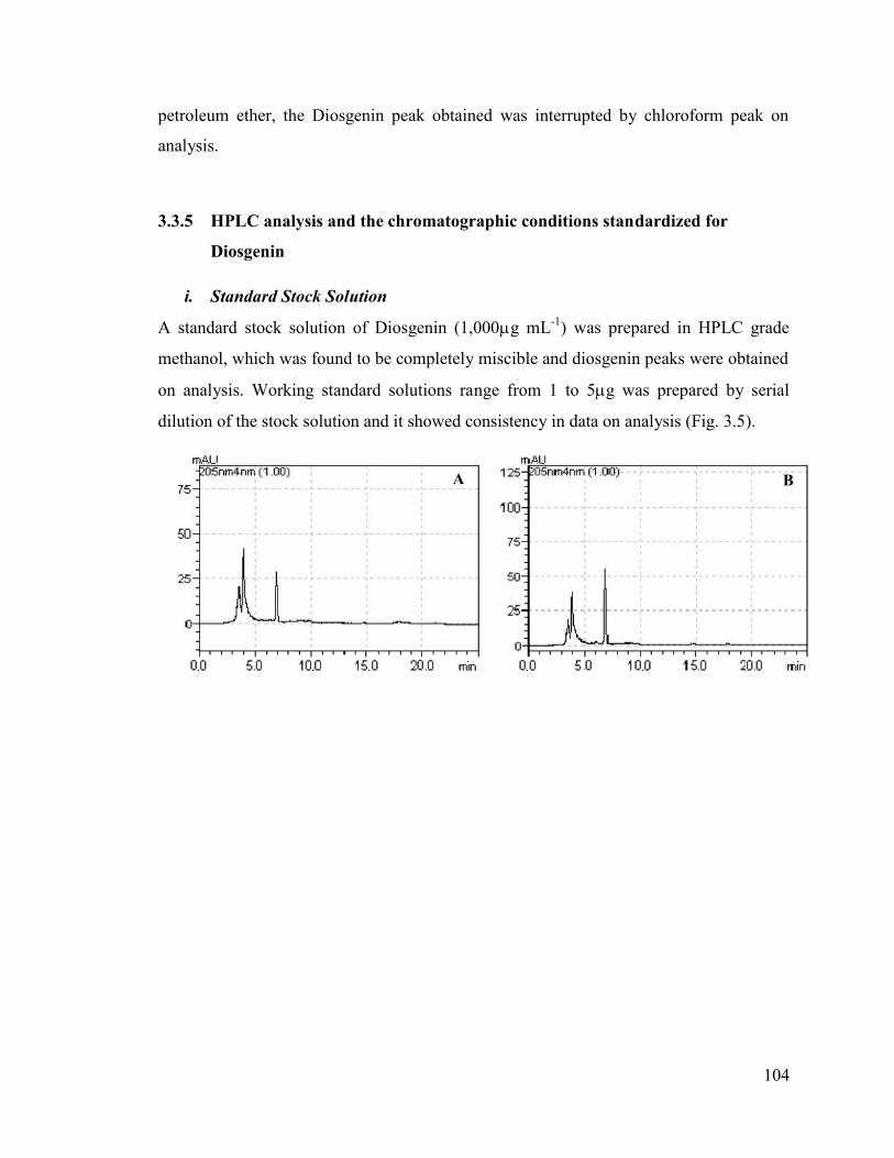
A standard stock solution of Diosgenin (1,000g mL-1) was prepared in HPLC grade
methanol, which was found to be completely miscible and diosgenin peaks were obtained
on analysis. Working standard solutions range from 1 to 5g was prepared by serial
dilution of the stock solution and it showed consistency in data on analysis (Fig. 3.5).
A B
105
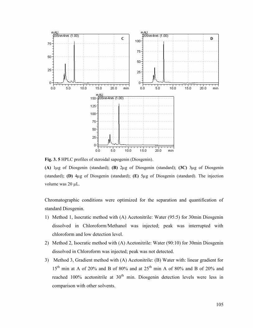
Fig. 3. 5 HPLC profiles of steroidal sapogenin (Diosgenin).
(A) 1g of Diosgenin (standard); (B) 2g of Diosgenin (standard); (3C) 3g of Diosgenin
(standard); (D) 4g of Diosgenin (standard); (E) 5g of Diosgenin (standard). The injection
volume was 20 µL.
Chromatographic conditions were optimized for the separation and quantification of
standard Diosgenin.
1) Method 1, Isocratic method with (A) Acetonitrile: Water (95:5) for 30min Diosgenin
dissolved in Chloroform/Methanol was injected; peak was interrupted with
chloroform and low detection level.
2) Method 2, Isocratic method with (A) Acetonitrile: Water (90:10) for 30min Diosgenin
dissolved in Chloroform was injected; peak was not detected.
3) Method 3, Gradient method with (A) Acetonitrile: (B) Water with: linear gradient for
15th min at A of 20% and B of 80% and at 25th min A of 80% and B of 20% and
reached 100% acetonitrile at 30th min. Diosgenin detection levels were less in
comparison with other solvents.

106
4) Method 4, Isocratic method with (A) Acetonitrile: Water (80:20) for 25 min flow rate
at 1mLmin-1.Peak was detected at RT 17-18th min at the wavelength 190 and 210 nm.
5) Method 5, Isocratic method with Methanol (100%) was done for standard Diosgenin
dissolved in Methanol and also maintaining the column temperature to 35oC. The
better resolution resolved chromatographic peak for Diosgenin with absorption
maxima was detected at 205 nm. The retention time was observed to be 6.9 to 7.1
minute.
Calibration curve was prepared based on peak areas of 5 concentration runs in
triplicates. The chromatographic analysis data on statistical analysis showed the
significance with good to fitness (R2) value, 0.999 (Fig. 3.6). The chromatographic peaks
of diosgenin showed maximum absorption at 205nm when compared to the
chromatogram at wavelength ranging from 190 to 235 nm (Fig. 3.7). The peak was
resolved with an isocratic mobile phase of absolute methanol.
ii. Chromatographic Conditions
Chromatographic analysis was carried out on Shimadzu Series LC-20 AT liquid
chromatographic system, equipped with a diode array detector SPD-M20A, and a pump
of LC-20AT. Chromatographic separations were performed on C18 Column (AtlantisR d
C18 5m 4.6x250mm column) at optimised temperature at 35°C, for Diosgenin
extraction. The analysis at room temperature (25°C) was giving inconspicuous
chromatographic peak of Diosgenin. When the temperature was higher than optimum
level, at 40°C the yield obtained was showing a depletion of 0.6 to 1.0% in Diosgenin
conent in detection level. The wavelength selected was 205 nm. An isocratic flow of
100% Methanol (HPLC grade) was resolved Diosgenin peak from the extract. The flow
rate was adjusted to 1mL min-1. At the end of each run, the column was rinsed with pure
Methanol. All data were processed using LC-Solution software (Shimadzu, Japan).
Comparative overlay of the sample and the standard showed the integrity of the sample
analyzed (Fig. 3.8).

107
Fig. 3. 6 Comparative overlay of chromatogram of Diosgenin with different concentration (1, 2,
3, 4& 5µg) with an injection volume of 20µL.
(A) Chromatographic representation; (B) Calibration curve

108
Fig. 3. 7 The various absorbance spectra of Diosgenin and 205 nm showed the maximum
absorbance.
Fig. 3. 8 HPLC profile depicting variation in concentration of Diosgenin according to the age of
in vitro propagated plants.
(A) Tubers of 1.0 year old plant; (B) Tubers of 1 year 6 months old plant; (C) Tubers of 2.0 years
old plant; (D) Tubers of 3.0 years old plant.

109
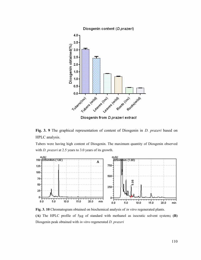
3.3.6 Diosgenin assay
The Diosgenin content was analysed from different parts of D. prazeri like leaves, roots,
stems and tubers. The stems of D. prazeri were observed with negligible amount of
Diosgenin. The roots exhibited lesser amount of Diosgenin, 0.4% when compared to
leaves with 1.2% and the tubers showed the maximum content of Diosgenin of 2.8%.
Tubers of a few plants in reproductive stages showed as high as 2.9% of Diosgenin
content on HPLC analysis (Table 3.1) (Fig. 3.9).
Even though the plants produced Diosgenin at younger stages, the yield of active
component was very low in comparison with the older plants (Fig. 3.10). The yield of
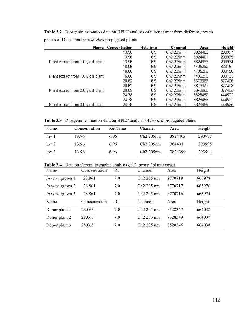
Diosgenin was ranging from 0.7±0.2% to 2.6±0.2% from the age of 12 weeks to 3 years.
The results obtained from the average of five replicates (Table 3.2). The plants were
observed to have lesser amount of Diosgenin at its early growth phases and reached a
maximum level at 3 years.
The purified fraction of Diosgenin was obtained by HPLC analysis and the
fractions were collected and verified by spiking with the standard. The pure fractions
obtained were completely dried and stored at -20 ºC. The fraction was used for the further
studies on its activity along with the soxhlet extract of D. prazeri.
The extraction and characterisation of the active component, Diosgenin from D. prazeri
was optimised. The study was conducted with various plant parts, in which the tubers
were found to have the highest content of steroidal sapogenins. The active component at
various growth phases was studied, wherein 2.5 to 3 years old plants resulted in the
highest yield. An optimised isocratic solvent system of absolute methanol for resolving
the chromatogram of Diosgenin (Fig. 3.11) was employed. The biochemical stability was
analysed with the wild grown donor plant and micropropagated plants during in vitro
propagation studies, and fidelity assessment for recovered and regenerated plants on
cryopreservation (Table 3.3 & 3.4). Enhancement of the compound was monitored during
the genetic transformation studies on D. prazeri.
110
Fig. 3. 9 The graphical representation of content of Diosgenin in D. prazeri based on
HPLC analysis.
Tubers were having high content of Diosgenin. The maximum quantity of Diosgenin observed
with D. prazeri at 2.5 years to 3.0 years of its growth.
Fig. 3. 10 Chromatogram obtained on biochemical analysis of in vitro regenerated plants.
(A) The HPLC profile of 5µg of standard with methanol as isocratic solvent system; (B)
Diosgenin peak obtained with in vitro regenerated D. prazeri

111
Fig. 3. 11 Comparative overlay of Diosgenin (Standard) and the Sample (Extract from tubers of
in vitro propagated plants).
Table 3.1 The various D.prazeri parts used for Diosgenin assay and the content (HPLC)
SI. No. Plant material *Diosgenin content on HPLC analysis (%)
1 In vitro tubers 2.85±0.1
2 Wild plant tubers 2.4±0.2
3 In vitro leaves 1.3±0.1
4 Wild plant Leaves 1.1±0.1
5 In vitro roots 0.43±0.04
6 Wild plant roots 0.40±0.02*The data is on an average value ± standard deviation of 5 replicates
112
Table 3.2 Diosgenin estmation data on HPLC analysis of tuber extract from different growth
phases of Dioscorea from in vitro propagated plants
Table 3.3 Diosgenin estmation data on HPLC analysis of in vitro propagated plants
Name Concentration Ret.Time Channel Area Height
Inv 1 13.96 6.96 Ch2 205nm 3824403 293997
Inv 2 13.96 6.96 Ch2 205nm 384401 293995
Inv 3 13.96 6.96 Ch2 205nm 3824399 293994
Table 3.4 Data on Chromatographic analysis of D. prazeri plant extractName Concentration Rt Channel Area Height
In vitro grown 1 28.861 7.0 Ch2 205 nm 8770718 665978
In vitro grown 2 28.861 7.0 Ch2 205 nm 8770717 665976
In vitro grown 3 28.861 7.0 Ch2 205 nm 8770716 665975
Name Concentration Rt Channel Area Height
Donor plant 1 28.065 7.0 Ch2 205 nm 8528347 664038
Donor plant 2 28.065 7.0 Ch2 205 nm 8528349 664037
Donor plant 3 28.065 7.0 Ch2 205 nm 8528346 664038
113
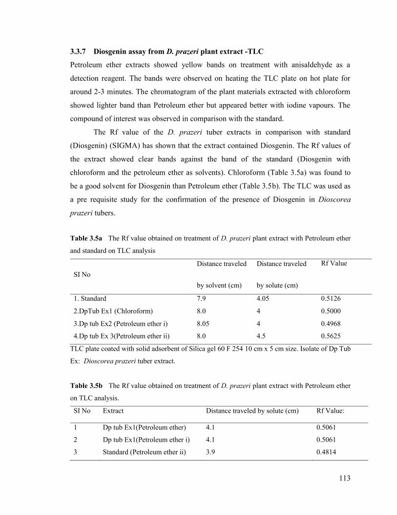
3.3.7 Diosgenin assay from D. prazeri plant extract -TLC
Petroleum ether extracts showed yellow bands on treatment with anisaldehyde as a
detection reagent. The bands were observed on heating the TLC plate on hot plate for
around 2-3 minutes. The chromatogram of the plant materials extracted with chloroform
showed lighter band than Petroleum ether but appeared better with iodine vapours. The
compound of interest was observed in comparison with the standard.
The Rf value of the D. prazeri tuber extracts in comparison with standard
(Diosgenin) (SIGMA) has shown that the extract contained Diosgenin. The Rf values of
the extract showed clear bands against the band of the standard (Diosgenin with
chloroform and the petroleum ether as solvents). Chloroform (Table 3.5a) was found to
be a good solvent for Diosgenin than Petroleum ether (Table 3.5b). The TLC was used as
a pre requisite study for the confirmation of the presence of Diosgenin in Dioscorea
prazeri tubers.
Table 3.5a The Rf value obtained on treatment of D. prazeri plant extract with Petroleum ether
and standard on TLC analysis
SI NoDistance traveled
by solvent (cm)
Distance traveled
by solute (cm)
Rf Value
1. Standard 7.9 4.05 0.5126
2.DpTub Ex1 (Chloroform) 8.0 4 0.5000
3.Dp tub Ex2 (Petroleum ether i) 8.05 4 0.4968
4.Dp tub Ex 3(Petroleum ether ii) 8.0 4.5 0.5625
TLC plate coated with solid adsorbent of Silica gel 60 F 254 10 cm x 5 cm size. Isolate of Dp Tub
Ex: Dioscorea prazeri tuber extract.
Table 3.5b The Rf value obtained on treatment of D. prazeri plant extract with Petroleum ether
on TLC analysis.
SI No Extract Distance traveled by solute (cm) Rf Value:
1 Dp tub Ex1(Petroleum ether) 4.1 0.5061
2 Dp tub Ex1(Petroleum ether i) 4.1 0.5061
3 Standard (Petroleum ether ii) 3.9 0.4814

114
TLC plate was coated with solid adsorbent of Silica gel 60 F 254 10 cm x 5 cm size.Distance traveled by solvent in all the cases was found to be 8.1 cm.
PlantMaterials
Solvent Extraction
Diosgenin
Fig. 3.12 The pictorial representation of whole process of Diosgenin extraction and
characterization from D. prazeri.
115
3.4 Discussion
Diosgenin, a plant steroid (5-spirostan- 3-ol) has been used for various steroidal drugs
since they have isolated in 1930s (Yang, 1981; Afrose S., 2010; Akinpelu D., 2008).
Steroidal drugs are considered to be some of the costliest and most important medicines
used throughout the world today. With recent reports of Diosgenin’s function in inducing
differentiation of erythroleukemia through changing lipoxygenase activities (Beneytout et
al., 1995) and inducing apotosis and cell cycle arrest in the human oesterosascoma 1547
cell line (Moalic et al., 2001), the value of Diosgenin has further increased. The most
recent finding concerning Diosgenin was found to inhibit migration and invasion of
human prostate cancer PC-3 Cells by reducing matrix metalloproteinases expression.
These findings reveal new therapeutic potential for Diosgenin in anti-metastatic therapy
(Pin-Shern Chen, 2011). So the enhancement and its characterization hold high
significance from D. prazeri, an indigenous source.
The TLC was conducted for the detection of compound Diosgenin with reference to
standard as instigation for the enhancement of secondary metabolite in D. prazeri.
Diosgenin (Standard) and the tuber extracts of D. prazeri was dissolved in chloroform
and petroleum ether and detected in TLC using petroleum ether as a solvent system as it
showed the value as 0.49 as Rf value as mentioned (Wang., 2011; Benjamin., 1984) The
iodine vapor and anisaldehyde were used as detection reagents that showed the presence
of Diosgenin.
The percentages of Diosgenin obtained from the seventy four accessions on HPLC
analysis, of D. polygonoides on this work are in the range from 0.02 to 2.64%, which is
significant since there are several literature data where the Diosgenin contents found to
be low as exemplify as with D. polygonoides (0.2%); (Coursey et.al., 1981) Dioscorea
althaeoides (0.2-2.3 %); Dioscorea prazeri (1.92%); Dioscorea villosa (1.3%); 2 years
old Dioscorea zingiberenzis (0.18-0.55%), several Dioscorea species (0.04-0.93%),
among others (Nino, 2007). Furthermore, this heterogeneity found on the steroidal
sapogenin contents as mentioned might depend on factors such as the genotype, the
physiological state, the climatic conditions as well as the geographic localization of
plants as stated (Dinan et.al., 2001). These findings correlate with the determination of
the steroidal sapogenin contents by HPLC, where significant differences were found
116
depending on the origin and part of the plant used for extraction (Oleszek et al., 2002;
Ganzera et al., 2001). Hence it was essential to standardize a protocol to obtain higher
concentration of Diosgenin from D. prazeri and was achieved with higher yield in this
study. This study on D. prazeri was carried out with reference to certain important
factors influence the characterization of the compound like the choice of plant material,
conditions of the growth, and geographical locations and it was crucial for the further
research conducted on enhancement of Diosgenin. These optimized conditions will be
used for the biochemical assay on Diosgenin of transgenic plants.
Dioscorea prazeri was one among the main natural source of Diosgenin
manufacturing (Behera, 2010), but now it is listed in rare species due to exploitation. The
high therapeutic characteristics of the compound, Diosgenin can be utilized by the
preservation of the plant germplasm, micropropagation and enhancement of the
compound in natural source. The conditions for estimation of Diosgenin were
standardised using D. prazeri plant and obtained high yield. This can be utilized for
extraction and characterization of the active compound. The plant re-established to the
natural environment through this study and the high content of Diosgenin can be utilized
for medicinal and commercial application.

117
3.5 References III
1. Accatino, L., Pizzaro, M., Solis, N., Koenig,C.S. (1998). Effects of Diosgenin a plant -derivedsteroid, on bile secretion and hepatocellular cholestasis induced by estrogens in the rat. Hepatology.28:129-140
2. Afrose,S., Hossain,S., Maki1,T., Tsujii, H. (2010). Hypocholesterolemic Response to Karaya Saponinand Rhodobacter capsulatus in Broiler Chickens. Asian-Aust J Anim Sci 23: 733 – 741
3. Akinpelu, D. A., Aiyegoro, O. A.and Okoh, A. I.(2008). In vitro antimicrobial and phytochemicalproperties of crude extract of stem bark of Afzelia africana (Smith). African Journal of Biotechnology.7: 3665-3670
4. Alice,C.B., Vargas,V.M.F., Silva,G.A.A.B., Siqueira,N.C.S., Schapoval,E.E.S., Gleye,J.,Henriques,J.A.P., Henriques,A.T. (1991). Screening of plants used in south Brazilian folk medicine. J.Ethnopharmacol. 35:165–171
5. Asolkar,L.V., Chadha,Y.R. (1979). Diosgenin and other steroid drug precursors.pp.2-17
6. Balandrin, M. F., Waller, G. R., Yamasaki, K. (eds). (1996). Commercial utilization of plant-derivedsaponins: An overview of medicinal, pharmaceutical, and industrial applications. Saponins Used inTraditional Medicine. Plenum Press, New York. pp. 1-14
7. Bartram T. (1995). Encyclopaedia of Herbal Medicine. Grace: Dorset
8. Behera,K.K., Saho, S., Prusti, A. (2010) Biochemical Quantification of Diosgenin and Ascorbic Acidfrom the Tubers of Different Dioscorea Species Found in Orissa Libyan Agriculture Research Center.Journal International. 1: 123-127
9. Beneytout JL, Nappez C, Leboutet MJ, Malivvand G (1995). A plant steroid, Diosgenin a newmegakaryocytic differentiation induce of HEL cell. Biochem. Biophys. Res. Comm. 207: 398-404
10. Benjamin, T.A.L, TAMIR, I., ROKEM,S., Israel GOLDBERG,I.(1984). Isolation and characterizationof an intermediate steroid metabolite in Diosgenin biosynthesis in suspension cultures of Dioscoreadeltoidea cells. Biochem J. 219: 619-624
11. Blunden,G., Binns,W.W., Perks.F. (1975). Commercial collection and utilization of mearl. Econ Bot.29: 140-145
12. Bombardelli, E., Morazzoni, P., Cristoni, A., and Seghizzi, R. (2001). Pharmaceutical and cosmeticformulations with antimicrobial activity. US Patent Application 2001/0046525 A1
13. Bonte, F., Meybeck, A. and Massiot, G. (1998). Method of treatment for combating the effects ofaging on the condition of skin and hair. US Patent 5,770,223.
14. Brand, H. and Brand, E. (2004). A weighty issue. Soap, Perfumery & Cosmetics, Asia. pp. 27-31
15. Cai, J., Liu, M., Wang, Z., Ju, Y. (2002). Apoptosis induced by Dioscin in HeLa cells. Biol PharmBull. 25: 193-196
16. Chen, P., Shih Y, Huang, H., Cheng, H. (2011). Diosgenin, a Steroidal Saponin, Inhibits Migrationand Invasion of Human Prostate Cancer PC-3 Cells by Reducing Matrix MetalloproteinasesExpression. Plosone.6: 1-10
118
17. Chuan-Sung Chiu, C., Chiu, Y., Wu, L., Chun Lu, T., Huang, T., Hsieh,T., Lu, C., Peng, W.(2011).Diosgenin Ameliorates Cognition Deficit and Attenuates Oxidative Damage in Senescent MiceInduced by D-Galactose. The American Journal of Chinese Medicine. 39: 551–563
18. Coursey, D.G. 91967). Yams an account of the Nature, Origin cultivation and utilization of the usefulmembers of the Diocoreaecaea. Longmans, London.
19. Dinan, L., Harmatha, J., Lafont, R. (2001).chromatographic procedures for he isolation of plantsteroids. J Chromatogr A.935:105-123
20. Dixit, S., Mandal, B.B., Ahuja S., Srivastava, P.S. (2003). Genetic stability assessment of plantsregenerated from cryopreserved embryogenic tissues of Dioscorea bulbifera L. using RAPD,biochemical and morphological analyses. Cryoletters. 24: 77-84
21. Fenwick, G. R., Price, K. R., Tsukamoto, C., and Okubo, K. 1991. Saponins. In: J.P.F. D’Mello,C.M., Duffus, J.H., Duffus.(eds), Toxic Substances in Crop Plants. The Royal Society of Chemistry,Cambridge. pp. 285–327
22. Fenwick, G.R., Price, K.R., Tsukamoto, C. and Okubo, K. (1992).Saponins. In: D’Mello, J.P.F.,Duffus, C.M. and Duffus, J.H. (eds), Toxic Substances in Crop Plants. The Royal Society ofChemistry, London. pp. 285–327
23. Fowler, M.W. (1984). Commercial applications and economic aspects of mass plant cell culture. In:Mantell S.H., Smith, H. (eds), Plant Biotechnology. Cambridge Univ. Press, Cambridge. pp. 3-37
24. Ganzera, M., Bedir, E., Khan, I.A. (2001): Determination of steroidal saponins in Tribulus terrestrisby high performance liquid chromatography and evaporative light scattering detection. Journal ofPharmaceutical Sciences, 90: 1752-1758
25. Hostettmann, K., Marston, A. (1995). Saponins: Chemistry and Pharmacology of Natural Products.Cambridge University Press, Cambridge, UK
26. Huo, R., Zhou, Q., Wang, B., Tashiro, S., Onodera, S., Ikejima, T. (2004). Diosgenin inducesapoptosis in HeLa cells via activation of caspase pathway. Acta Pharmacologica Sinica. 25: 1077-1082
27. Ibanez, E, Kubatova, A., Senorans, F.J, Cavero, S., Reglero, G., Hawthorne, S.B. (2003). Subcriticalwater extraction of antioxidant compounds from rosemary plants. J Agric Food Chem. 51: 375-382
28. Indena.(2005). Horse chestnut saponins. http://www.indena.com/pdf/cosmLeaf. pdf, accessed24/8/2005
29. Kensil, C. R., Mo, A., Truneh, A. (2004). Current vaccine adjuvants: an overview of a diverse class.Front Biosci. 9:2972-2988
30. Liagre, B., Pascale.V, Cecile, C., Chaissoux L, J., Beneytout L, J. (2004). Diosgenin, a plant steroid,induces apoptosis in human rheumatoid arthritis synovyocytes, with cyclohexogenase-2overexpression. Arthritis Res Ther. 6:373-383
31. Liu, J., Henkel, T. (2002). Traditional Chineese medicine (TCM): are polyphenols and saponins thekey ingredients triggering biological activities? Curr. Med. Chem. 9: 1483-1485
32. Mahato, S.B., Ganguly,A.N., Shahu,N.P.(1982).Phytochemistry. 8: 1445-1447

119
33. Matsuura, M. (2001). Saponins in garlic as modifiers of the risk of cardiovascular disease. J. Nutr.131:1000-1005
34. Ming-Jie. (2004). The mitotic arresting and apoptosis inducing effects of diosgenyl saponins onHuman Leukemia cell lines. Biol Pharm Bull. 27: 1059-1065
35. Moalic, S., Liagre, B., Corbiere, C., Bianchi, A., Dauca, M., Bordji, K., Beneytout, J.L. (2001). Aplant steroid, Diosgenin induces apoptosis, cell cycle arrest and COX activity in osteosarcoma cells.FEBS Lett, 506:225-230
36. Nandi, R.P. (1980). D.Phil. Disect (sc). Burdwan University, India.
37. Nino, J., Jimenez, Mosquera, D.A., Yaned, M.C. (2007). Diosgenin Quantification by HPLC in aDioscorea polygonoides Tuber Collection from Colombian Flora. J Braz Chem Soc.18: 1073-1076
38. Oakenfull, D. (1981). Saponins in food-A review. Food Chemistry. 6: 19-40.
39. Oakenfull, D., Sidhu, G. S. (1990). Could saponins be a useful treatment for hypercholesterolemia?Eur J Clin Nutr. 44: 79-88
40. Oakenfull, D.G. and Sidhu, G.S. (1989). Saponins. In: Toxicants of Plant Origin Vol. II. Cheeke, P.R.(eds), Glycosides. CRC Press Inc., Boca Raton. pp. 97–141
41. Oleszek, W. A. (2002). Chromatographic determination of plant saponins. J Chromatogr A, 967: 147–162
42. Olmstead, M. J. (2002). Organic toothpaste containing saponin. US Patent 6,485,711 B1.
43. On Wueme. (1978). The tropical tuber crops. John Wiley, New York. Pp. 234
44. Ozlem, G., Mazza,G. (2007). Saponins: Properties, Applications and Processing. Critical Reviews inFood Science and Nutrition. 47:231–258
45. Price, K.R., Johnson, I.T. and Fenwick, G.R. (1987). The chemistry and biological significance ofsaponins in foods and feeding stuffs. CRC Critical Reviews in Food Science and Nutrition 26: 27–135
46. Sharma,S.C., Chand,R., Bhatti,B.S., Sati,O.P.(982).Planta Med.46: 48-51
47. Wang, L., Wang, X., Yuana, X., Zhaoa, B. (2010). Simultaneous Analysis of Diosgenin andSarsasapogenin in Asparagus officinalis Byproduct by Thin-layer Chromatography. Phytochem Anal.22: 14–17
48. Yang, M.H. (1981). Steroidal sapogenins from plants of Dioscorea. Chin Trad Herb Drugs. 12: 41-48
49. Yoo, B. H., Kang, B. Y., Yeom, M. H., Sung, D. S., Han, S. H., Kim, H. K. and Ju, H. K. (2003).Nanoemulsion comprising metabolites of ginseng saponin as an active component and a method forpreparing the same, and a skin care composition for anti-aging containing the same. US PatentApplication 2003/0175315 A1.