chapter i cfiemicaf investigations of the red a@ chontfria...
TRANSCRIPT

Chapter I
Cfiemicaf investigations of the red a@ Chontfria armata
Chondria armata: A review.
Marine environment has proven to be a rich source of natural products with novel
structures; it is also a potentially rich source of therapeutically useful agents.
Among the marine organisms marine algae are one of the largest producers of
biomass in the marine environment that produce a wide variety of chemically
active metabolites in their surroundings, potentially as an aid to protect themselves
against other settling organisms. These active metabolites, also known as biogenic
compounds, produced by several species of marine macro- and micro-algae, have
antibacterial, antialgal, antimacrofouling and antifungal properties, which are
effective in the prevention of biofouling and have other likely uses, e.g. in
therapeutics. The isolated substances with potent antifouling activity belong to
groups of fatty acids, lipopeptides, amides, alkaloids, terpenoids, lactones,
pyrroles and steroids. These biogenic compn'unds have the potential to be
produced commercially using metabolic engineering techniques. Therefore,
isolation of biogenic compounds and determination of their structure could
provide leads for future development of not only, environmentally friendly
antifouling agents, but also serve as new and more effective therapeutic agents.'
Algae are important as primary producers of organic matkr at the base of the food
chain. They also provide oxygen for other aquatic life. Algae may contribute to
mass mortality of other orpnisms, in cases of algal blooms, but they also
contribute to economic well-being in the form of food, medicine and other
products. In tropical regions, coralline algae can be as important as corals in the
formation of reefs. Approximately there are about 30,000 known species of algae,
but the actual number of species probably exceeds this Today the algae are
classified into seven Phyla, based on their colour, type of chlorophyll, form of
food storage substance and cell wall composition2 (Table I):
18

Table 1: Classification of Algae
x
SEVEN PHYLA OF ALGAE PHYLUM THALUS FORMAT PHOTOSYNTHETIC
PIGMENTS FORM OF F0(
STORAGE Chlorophyta (Green Algae)
Unicellular, Colonial, Filamentous, and Multicellular
Chlorophylls a and b, Carotenoids Starch
Phaeophyta (Brown Algae)
Multicellular Chlorophylls a, and c, Carotenoids, Fucoxanthin
Laminarin (an o carbohydrate)
Rhodophyta (Red Algae)
Multicelluar Chlorophylls a, Phycobilins, Carotenoids
Starch
Bacillariophyta (Diatoms)
Mostly Unicellular, Some Colonial
Chlorophylls a and c, Carotenoids, Xanthophyll
Chlorophylls a and c, Carotenoids
Leucosin (an of carbohydrate)
Starch Dinoflagellata (Dinoflagellates)
Unicellular
Chrysophyta (Golden Algae)
Mostly Unicellular, Some Colonial
Chlorophylls a and c," Xanthophyll, Carotenoids
Chlorophylls a and b,Carotenoids, Xanthophyll
Laminarin (an o carbohydrate)
Paramylon (a Starch)
Euglenophyta (Euglenoids)
Unicellular
CELL WALL COMPOSITION Polysaccharides,
Primarily Cellulose
Cellulose with Alginic Acid
Cellulose or Pectin, many with Calcium Carbonate Pectin; many with Silicon
Dioxide Cellulose
Cellulose
No Cell Wall, Protein-rich Pellicle
19

Traditionally, the red algae (Rhodophyta) were divided into two classes the .
Bangiophyceae and Florideophyceae. Alternatively, a single class, the
Rhodophyceae and two subclasses, Bangiophyeidae and Florideophycidae are
used. Based on ultrastructure and molecular evidence the Bangiophyceae is now
accepted as a paraphyletic group, while the Florideophyceae is considered to be
monophyletic based on two synapomorphic characters presence of a filamentous
gonimoblast and tetrasporangia3. Since this chapter deals with the identification of
chemical constituents of the alga Chondria armata, collected off Goa coast (India)
during the low tides a review of literature on the metabolites from this alga is
presented here. The alga belongs to phylum Rhodophyta, class Florideophyceae
and family Rhodomelaceae
Domoic acid (DA) (1) an insecticidal agent, was the first compound to be isolated
from Chondria annata in Japan4, and is named after the Japanese word for this
seaweed, "domoi". It was later identified in the rhodophytes, Alsidium corallinurn,
from the east coast of Sicily5, and Chondria baileyana, from southern Nova Scotia
and PEI, Canada6 It is also known to be a constituent of Amansia glomerata,
Digenea simplex and Vidalia obtusiloba, all belonging to the family
Rhodomelaecae7. DA belongs to a group of amino acids called the kainoids,
which are classed as neuroexcitants or excitotoxins that interfere with
neurotransmission mechanisms in the brain. The first stnicture of domoic acid was
proposed in 1958, which was later revised by NAIR study in 1966 8. However, it
was not until 1982 that the correct structure with absolute configuration for (-)-
domoic acid (1) was finally determined by stereospecific total synthesis 9 and
confirmed by X-ray analysis °. DA, compound responsible for the insecticidal
activity of C armata was 14 times more potent than DDT when administered
subcutaneously into the abdomen of American cockroach". Additional related
compounds, isodomoic-acids (2-5) 12 and domoilactone (6-7) were also discovered
from the insecticidal fraction of the alga 13- 14. The insecticidal activities of
isodomoic acids were much weaker than that of domoic acid but comparable with
that of DDT13 while both lactones were found to be inactive" Zairian and co-
workers" reported two new isomers of isodomoic acid (8) and (9), along with the
20

known isodomoic acids (2,3,10) and (11) from Kyushu Island . Their structure
was deduced on the basis of ESI-MS and 'H NMR spectral analysis including 'H-
'H correlation spectroscopy and NOE correlation spectroscopy. Domoic acid is
also known to be vermifuge in a single dose as low as 20mg and inhibits
ovulation. It also exterminates Oxyris and Ascaris 4.16. These useful properties of
domoic acid are associated with certain disadvantages_ Domoic acid is also
present in edible mussels Mytiulis edulis and whenever there has been episodes of
shellfish poisoning, domoic acid has been identified as the causative substance".
It acts by causing neuronal depolarization; the resultant short-term memory loss is
symptomatic of domoic acid poisoning. Other symptoms include dizziness, nausea
and vomiting, ultimately leading to coma and brain damage or death in the most
severe cases.
Chondria armata from the Japanese waters is also reported to contain
hypoxanthine (12), L-glutamic acid (13) and D-aspartic acid (14)". Hypoxanthine
is a naturally occurring purine derivative and one of the products of the action of
xanthine oxidate on xanthine. It is occasionally found as a constituent of nucleic
acid where it is present in the anticodon of tRNA in the form of its nucleoside
inosine_ L-Glutamic acid is found in virtually all living organisms. It is one of the
major amino acids in plant and animal proteins, and is also involved in many
physiological functions. It acts as nemotransmitters in the brain_ Humans readily
metabolize ingested L-glotarnic acid so that concentration in the body remain
constant.
An a-amino acid, citrulline (15), [2-amino-5-(carbamoyl aminojpentanoic acid
and isoglutamic acid (16) (3-amino &tack acid) are also known to be a
constituent of this alga 19. The name is derived from citrullus, the Latin word for
watermelon, from which it was first isolated'. It is made from ornithine and
carbamoyl phosphate in one of the central reactions in the urea cycle. Glutamic
acid is present in a wide variety of foods and is responsible for one of the five
basic tastes of the human sense of taste (umami), especially in its physiological
form, the sodhim salt of glutamate at neutral pH. Ninety-five percent of the
dietary glutamate is metabolized by intestinal cells in a first pass 2I . Overall,
21

glutamic acid is the single largest contributer to intestinal energy. As a source for
umami, the sodium salt of glutamic acid, monosodium glutamate (MSG) is used
as a food additive to enhance the flavor of foods, although an identical effect can
be achieved by mixing and cooking together different ingredients rich in this
amino acid and other umami substances as well.
Preliminary screening of the chloroform extract of C. armata collected from Goa
(west coast of India) showed antiviral, antibacterial, and antifungal activities 2223 .
Continued research aimed at the chemistry and bioactivity of this alga by
Govenkar et al resulted in the isolation of fatty acids, a novel ester, steroids and an
alkaloid. The fatty acids were identified as myristic acid (C14112802), pentadecylic
acid (C15H3002), palmitic acid (C16113202), stearic acid (C 18113602), 5-palmitoleic
acid (C161-13432), 4-palmitoleic acid (C161-13002) and oleic acid (C1gH3402) using
gas chromatograph-mass spectrometer (GC-MS) equipped with a cross linked
methyl silicone capillary Hewlett-Packard column (1,=25 m & i.d 0 2 mm) 24. The
free sterols, were possessing A 5, 313-hydroxy nucleus and were identified as
cholest-5-en-313-ol.(17), 24-methylene- ,cholest-5-en-313-o1(18), 2413-ethy1 cholest-
5,22-dierte-311-o1(19), 24f-ethyl cholest-5-en-30-ol(20), 23k-methyl cholest-5-en-
3fl-ol (22) and 23-methyl 5a-cholmtan-311-o1(22). Acetylation of the sterol
mixture was also carried out and the corresponding steryl acetates obtained were
analyzed by GC-MS25 .
Caulerpin (23), a dimer of indole-3-acetic acid is also present in this alga along
with a fatty ester, pentyl hentriacontannate(24) 26_ The pigment caulerpin is a well
known constituent of the green algae of genus Crudes-pa2729_ It displays a
moderate in vitro antittnnor activity, acts as a plant growth regulator like its
monomeric counterpart and indole-3-acetic acid (auxin) 3° and inhibits the
multixenobiotic resistance (MXR) pump in algae 31 .1n the root elongation test with
germinated lettuce seedling, the activity of caulerpin was slightly weaker than
that of auxin but stronger than those of indole-3-pyruvic acid and indole-3-acrylic
acid. The corresponding dicarboxylic acid form of (23) also showed similar
potency.3°
22

COOHrj
H
(4)
H
(2)
HOOC
COON
HOOC
(1 )
H
(3)
(7)
HOOC
Subsequently, Cimino group32 reported a new class of bromotriterpenes, Armatols
A-F. Their structures were characterized by spectroscopic techniques, in particular
1D- and 2D-NMR. including HMQC and HMBC experiments. They also
concluded that the triterpenoids polyethers identified from Laurencia and the
armatols could arise from (6S,7S,10R,1 1R,14R,15R,18S,19S)-squalene
tetraepoxide, a common precursor. However, from a biogenetic point of view, the
discoveries of several molecules with different stereocenters suggest the
hypothesis that the biosynthesis of these molecules may occur in a not concerted
way. Interestingly, Fernandez et al, also reported the strong cytotoxic properties of
these squalene-derived compounds, suggesting that further biological assay should
be directed to an evaluation of this activity 33.
23

(19)
R=
(21)
HN \
I
(12) 0
(13)
H
(9) 0
0
NH2
(14)
OH HO
H . ..NH2
COOH
(20)
R= rYY (22)
HO
0 (24)
24

R3 R4
R1=Me, R2=0H, R3=H, R4=Br (armatol B) R1=OH, R2=Me, R3=1:1, R4=Br (armatol C) R1=Me, R2=0H, R3=Br, R4=H (armatol D) R1=OH, R2=Me, R3=Br, R4=H (armatol E)
OH 0
25

Section 1
Lipids — glyceroCpuls and" steroids of the
red *a Chondria annata.
1.1: Lipid constituents of the red alga Chondria armata
Marine organisms produce a variety of lipids because of their characteristic living
environments. Lipids are major source of metabolic energy and essential materials
for the formation of cell and tissue membranes. They are very important in
physiology, reproductive processes of marine animals and reflect the special
biochemical and ecological condition of the marine environment. The interest of
chemist, biochemist and biotechnologists in lipids from marine organisms has
been stimulated, in particular, by the recognition that polyunsaturated fatty acids
(FA) are important for human health and nutrition. They are required for
reproduction and growth. The relative proportion and composition of FA in
marine organisms are characteristic for every species and genus and also depends
on the environmental conditions.
The principal role of neutral lipids, which in marine organisms consist
predominantly of triacylglycerols and wax esters, is as an energetic reserve of FA
that are destined either for oxidation to provide energy (ATP) or for incorporation
into phospholipids. Phospholipids are the building blocks for the membrane lipid
bilayer. FA provide the hydrophobic interior of all cell membranes, forming an
impermeable barrier to water and polar molecules and separating the cell contents
from the extracellular medium. The physical properties of the membranes are
determined by the individual lipids within the FA components of the lipids and
their interaction with proteins and sterols_ Membrane lipids other than
phospholipids are the glycolipids.
Glycolipids as mentioned are ubiquitous compounds in the cell membrane of most
cell types. There are two major classes of glycolipids: glycosphingolipids and
glycoglycerolipids. Glycosphingolipids, in which the carbohydrate moiety is
linked to a •ceramide lipid moiety, have been more widely studied.
Glycosylceramides play an important role in many fields of cell biochemistry such
as molecular recognition. In addition, ceramides from marine organisms have
excited great attention as signal transducers, and some of them have been
recognized as possessing antimicrobial and cytotoxic activities.
26

Glycosphingolipids, are tumor markers for various neoplasms and are markers of
maturation or differentiation of cells in adults and embryonic tissues. Changes in
composition, metabolism and organization of glycosphingolipids in the cell
membrane are some of the most common biological changes associated with
neoplastic transformation 34-35
In contrast, .the class of glycoglycerolipids (i.e. glycosyl glycerides) has received
less attention in the recent literature. In these glycolipids the carbohydrate is 0-
glycosidically linked to carbon-3 of diacyl or monoalkyl-monoacyl glycerolipid. 36
Glycoglycerolipids are common components of various plant tissues and bacterial
cell walls. In bacteria, mono and diglycosyldiacyl glycerols containing glucose
galactose and mannose are most commonly seen 36. In plant cells, galactosyl and
digalactosyldiacyl glycerols are the most common glycoglycerolipids. Although
acylated and sulfonated variants, as well as trigalactosyldiacyl glycerols have also
been found36.
Glycolipids constitute an important class of membrane lipid that are synthesized
by both prokaryotic and eukaryotic organisms 37 . They are reported to exhibit
diverse biological functions. There is currently considerable interest in both,
intracellular and extracellular glycolipids specially galactosyl glycolipids as
antitumor promoters in cancer chemoprevention.
This section presents a full account of the structural elucidation of major
galactosylglycerols isolated from the chloroform soluble fraction of crude
methanolic extract of red alga Chondria armata (Kiitz.) Okamura. The chloroform
fraction, which was subjected to gel chromatography over Sephadex LH2O using
methanol as mobile phase gave, in order of polarity fractions PF 1 _3, apparently
homogenous on TLC, yielding purplish pink spots on spraying with methanolic
sulphuric acid. This resulted in the isolation of three major glycolipids. The flow
chart and TLC of the purified fractions PF1.3 is given beiow(Scheme I, II). Their
structure was elucidated by multidimensional nuclear magnetic resonance (NMR)
techniques like 1 1-1, 'H correlation spectroscopy (COSY), '1-1, 'H total correlation
spectroscopy (TOCSY), 13C heteronuclear multiple quantum coherence
27

Alga sample (3.5 Kg)
Extracted with MeOH, Filtered and concentrated.
Crude extract
'Fractionation
Chloroform 123g
1 Gel Chromatography Sephadex LH-20 (1:1 MeOH: CHC1 3)
I Jr F-I F-II Silica gel P.E in E.A (20-30%) Steroids Silica gel
P.E in E.A (1:1) Silica gel
I (2:98-MeOH: CHC13) Silica gel
I (5:95-MeOH: CHC13)
PF-1 PF-2
PF-3 Scheme I: Sequential organic extraction, isolation and purification of the polar glycolipids.
Scheme II: TLC of the polar glycolipids (PF 1 .3).
28

(HMQC) and I H, I3C heteronuclear multiple bond correlation (HMBC)
complemented by electrospray ionization mass spectrometry (ESI-MS) in the
positive ion mode.
Major glycolipids were identified as (2R)-2-0- (5,8,11,14-eicosatetranoy1)-3-0-a-
D-galactopyranosyl-sn-glycerol (GL2), its pentacetate (GL 1) and (2S)-1-0-
(pal m itoy1)-2-0-(5,8,11,14,17-ei cosapentanoy1)-3-0-13-D-galactopyranosyl -s n
glycerol (GL3). Additionally, six minor glycolipids were also identified on the
basis of ESI-MS. These include, a 1,2-di-Oacy1-3-0-(acyl-6'-galactosyl)-glycerol
(GLI a), sulfonoglycolipids 2-0-pal m itoy1-3-0- (6'-sulfoqu inovopyranosyl)-
glycerol (GL2.) and its ethyl ether derivative (GL2b), 1- 01e0y1-2-palmitoy1-3-0-
galactosyl glycerol (GL3.), 1,2-diacyl phosphatidyl glycerol (GL 3b) and 3-
digalactosy1-2-palmitoyl glycerol (GL3c).
Structural characterization of PF1:
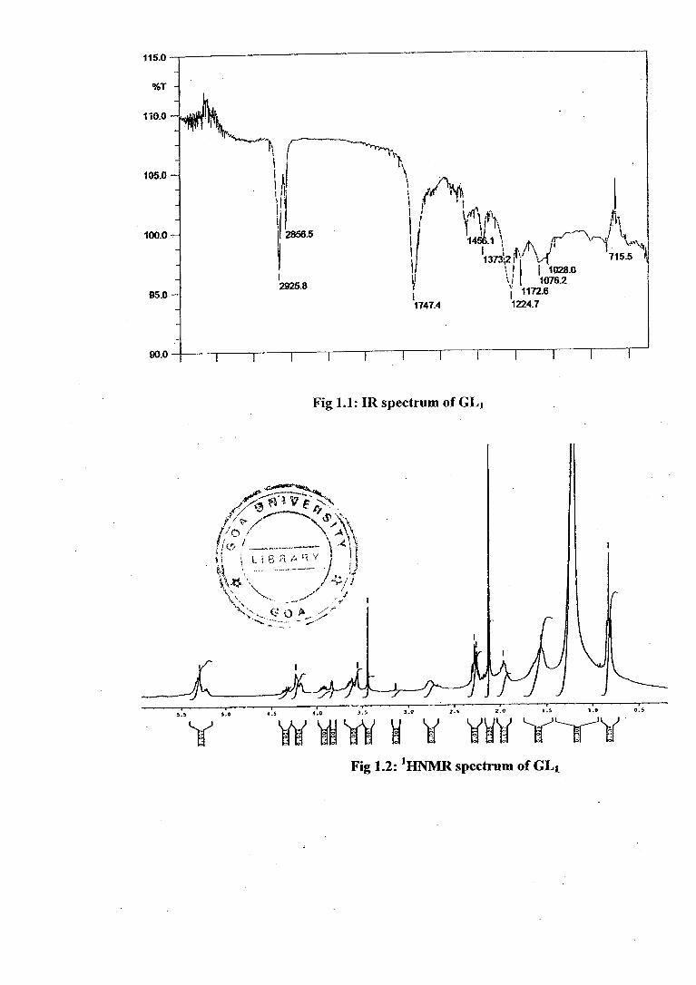
The IR spectrum (Fig 1.1) of the purified PF 2 showed absorption hands at 2925,
2856 cm- 1 for aliphatic chain and 1747, 1224 cm -1 for the presence of ester group.
It also gave protonated molecular ion peak -IM+1-11 + at .m/z 751 .in. its .ESI-MS
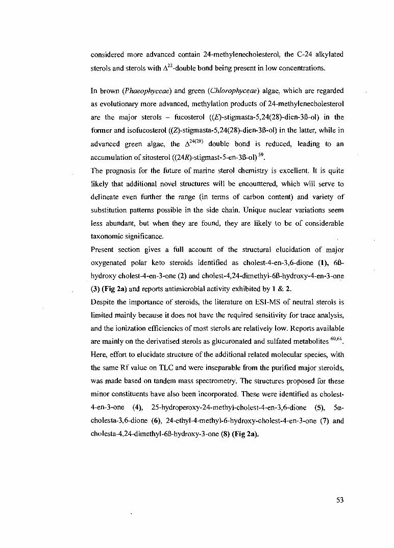
spectrum (Fig 1.7). The presence of spin systems corresponding to one hexose,
glycerol and fatty acid were readily identified from the 1D and 2D homonuclear
correlation (COSY) NMR spectra -. Thus, the I HNMR spectrum (Fig1.2)
(300MHz, CDC1 3) and I3CNMR data including DEPT experiments (Fig1.3, Table
1) was in agreement with diacylated monogalactosyl glycerol (MGDG) with the
fatty acyl chain being evident by the presence of a triplet due to a terminal methyl
at 6 0.827, a broad methylene signal at 6 1.202 [(CI-12M of aliphatic chain,
multiplets at 6 2.268, 1.967 and 1.562 assigned to three methylenes linked a, 13
and y to the ester carbonyl functionality. A broad multiplet at 6 2.7 arises from
allylic methylene protons and the olefenic methine protons were evident at 6
5.293. A sharp singlet at 6 2.12 was attributed to acetyl methyls.
The presence of glycerol moiety was also confirmed by heteronuclear multiple
HMQC (Heteronuclear multiple quantum coherence) experiment, which showed
29

AcO
4.23 4.39
3.61 0
4.178 A 3.55 OAc A 3.56
3.96
OAc
/(Thl 1.202 0.827
1.96 2.78
two doublets arising from C-3 and C-1. The signals at 8 4.22 and 4.35 correspond
to the substitution at C-1 (8 62.2) by an O-acyl group and the doublet at 8 3.56 and
3.96 was assigned to C-3 (8 68.2) of glycerol substituted by the a-galactose
residue. The glycerolipid structure was confirmed by the presence of a
characteristic signal at 8 70.0/5.23 (C-2) having a distinct a-shift to lower field for 13C and 1 H nuclei when substituted by an O-acyl group, this being a fingerprint for
glycolipids containing glycerol as alcohol rather than sphingosine 38 .
1 H-'H COSY, TOCSY (Fig1.4) (total correlation spectroscopy) and HMQC
(Fig1.5) correlations allowed assignment of sugar carbons and protons (Table-1).
1 H- 1 H COSY and TOCSY correlation of the anomeric proton at 8 4.178 with the
sn-3 protons at 8 3.56 and 3.96 established connectivity of the sugar moiety with
the glycerol. The anomeric proton at 8 4.178 with a coupling constant of 2.1 Hz
indicated a glycosidic configuration of the sugar linkage with the glycero1 39.
TOCSY correlations are illustrated in Fig la.
4.35
Fig la: TOCSY correlations of GL1
Long-range heteronuclear multiple bond correlation (HMBC)(Fig 1.6) diagnostic
correlations were observed between the ester carbonyls at 8 173.8 and 8 173.5 and
C-1 and C-2 of glycerol indicating the linkage. The complete assignments of all
the HMBC correlations are shown in Fig lb.
30

Fig lb: HMBC correlations of GL I
Tablel 1: 1 11 , "CNMR, TCOSY and HMBC of GLI
Carbon No.
1HNMR ö,1, ppm
13CNMR Sc, ppm
TOCSY Correlations
HMBC Correlations
1 4.22,4.35 62.2 I-12 C1"', C2 2 5.23(m) 70.0 141, H3a,
H3b -
3 3.56(d, 3.6Hz) 3.96(d, 6.0Hz)
68.2 H2 C 1 ,C 1 '
1' 4.17(d, 2.1Hz) 103.0 H2',H2 C2' 2' 3.52 71.6 H1', H3' C3', C4' 3' 3.85(b, s) 67.9 H2', 1-14' C4' 4' 3.55(d, 3.6Hz) 73.1 H3' C3', CS' 5' 3.61(m) 723 H6' C1', C6' 6' 4.23, 4.39 62.2 H5' C5' 1" - 173.5 - - 2" 2.28 34.1 H3" C3", Cl", C4" 3" 138 24.8 142" C2" 7" 1.96, 2.78 27.1 H8" C8", C9" 8" 531(d, 5.4Hz) 129.6 H7", H9" C7" 9" 531 128.7 H8", H10" C10" 18" 0.82(t, 6.9Hz) 14.0 H17" C16", C17"
(CH2)n 1.20(bs) 29.1-30.8 l' 173.8 C2'" 2'" 2.12(s) 22.6 Cl"'
0=C-CH3 2.12 22.6 Acetyls -Sugar.
The stereochemistry at C-2 was assigned to be R by comparison of the coupling
constant values between H-2/H-3a (J=3.6Hz) and H-2/H-3b (J=6.0Hz)
respectively with those of published data 40 ' 41 ' 42 . On the basis of the above data the
major component of PF 1 was identified as pentacetate of (2R)- 2-045,8,1 1,14-
eicosatetranoy1)-3-0-a-D-galactopyranosyl-sn-glycerol (GL 1). The fragmentation
31

observed in MS/MS spectrum of GL1 (Fig 1.7), is well in agreement with the
structure assigned. The pseudomolecular ion at m/z 751 generated a series of
daughter ions at m/z 691, 631, 571 and 511 reflecting successive loss of four
acetic acid molecules. The presence of fifth acetyl group was evident from the
elimination of yet another acetic acid molecule yielding sodiated fragment at m/z
473. Alternately, the ion at m/z 473 might have originated, as diprotonated
sodiated fragment ion, after the elimination of arachidonate ion. This ion on
elimination of fifth acetic acid molecule would lead to ion at m/z 413. This
confirmed the presence of acetylated hexose linked to the glycerol moiety, with
the latter being diesterified by acetic acid and eicosatetraenoic acid. The proposed
structure of GI, ' along with identified fragments is represented in (Scheme!).
511 Na 473 4-
Ac0 0 Ac 571
691
Scheme!: Mass fragmentation of GL 1
There is a solitary reference in the literature on the identification of 2-O-a—D-
galactopyranosyl glycerol hexacetate from Ruellia britoniana E. Leonard
(Acanthaceae)43 . The acetylated galactoglycerolipid is being reported here for the
first time from a marine source.
ESI-MS of PF1, though apparently homogenous on TLC, showed some
heterogeneity by the presence of an additional related molecular species with m/z
1017. Based on the fragmentation pattern observed in MS/MS (Fig 1.8) it was
characterized as 1,2-di-O-acyl-3-0- (6-acylgalactosyl)-glycerol GL1 a .
MS/MS studies of the [M+H] + ion at m/z 1017 (Fig1.8) resulted in three major
diagnostically important daughter ions at m/z 481, 735 and 761. The ions at m/z
735 and 761 reflect the neutral losses of the sn-1 and sn-2 substituent as free Cis 1
and C16-0 carboxylic acid respectively is supporting the presence of palmitic and
oleic acyl moieties in the molecule. The intensity differences of these various ions
indicated the position of the different fatty acid moieties, as the substituent
position at sn-2 fragments comparatively easily". This leaves a mass for the core
OAc
0 0
32

H 29 C 17 i o 8 HO
O H
423
441 2H
761
481
735 --+-1
• 295
O
C H 33 33
I • I 0
/ 13
0
J
+H
of the molecule of 481 amu. Such a mass can be explained by a substituted hexose
connected to a glycerol backbone after elimination of fatty acyl groups from the
protonated molecular ion [M + H] +. This is further supported by the presence of
an additional fragment ion at m/z 441, which reflects the loss of acyl groups (C18.1
and C16:0) from the molecular ion along with the glycerol backbone together
corresponding to a total mass of 577 amu. Fragment ion at m/z 423 results from
the cleavage between C-1 of hexose and C-3 of glycerol. Cleavage of the
molecule between C5-C6 of the sugar leads to sodiated fragment at m/z 313 which
corresponds to the third acyl substituent (C18:3) along with C-6 of sugar which
possibly seems to be galactose. The ion at m/z 295 results from the cleavage
between Cl-C2 of glycerol. Furthermore, there were a number of fragments in the
upper mass region at intervals of about 14 amu. These correspond to
fragmentation along the fatty acid acyl chains. On the basis of this fragmentation
pattern of the molecular species with the pseudomolecular ion at m/z 1017 we
propose the structure of the molecule as being 1-oleoy1-2-palmitoy1-3-0-
(linoleny1-6'-galactosyl)-glycerol (GLI a) that along with identified fragments is
illustrated in Scheme 2.
Scheme2: Mass fragmentation of GLi a.
Structural characterization of PFz:
A similar approach was adopted for PF 2 that showed physicochemical
characteristics of glycolipids. The IR spectrum (Fig 2.1) of the purified PFz
showed absorption bands at 3409.9 cm' for the presence of a hydroxyl groups (-
OH), 2922.0, 2852.5 cm-1 for the aliphatic chain and 1737.7, 1172.6 cm' for the
presence of ester group. Its 1 HNMR (Fig2.2) and 13CNMR (Fig2.3) data including
33

DEPT experiments differed from that of PF 1 only by the absence of signals for the
acetyl groups (Table-2) indicating GL2 to be deacetylated derivative of GL1.
Tablet: 1 H , 13CNMR, COSY and HMBC of GL2
Carbon No.
1HNMR Ou, ppm
' 3CNMR 8c, ppm
COSY Correlations
HMBC Correlations
1 4.00, 4.39 62.2 H2 l'", 2 2 5.30 70.0 H1, H3a, H3b - 3 3.51 (b)
3.91 (b) 67.9 H2 1,1'
1' 4.25(d, 4.5 Hz) 103.6 H2' 2' 2' 3.63 72.4 H1', H3' 3', 4' 3' 3.90 67.9 H2', H4' 4' 4' 3.69 73.1 113' 3', 5' 5' 3.50 (b) 71.6 H6' l', 6' 6' 4.30, 4.39 62.2 H5' 5' l" - 173.9 - - 2" 2.35 34.1 H3" 3", 1", 4" 3" 1.63 27.2 H2" 2", 4" 7" 2.04 (b) 29.3 H8" 8", 9" 8" 5.39 (b) 128.8 H7", H9" 7" 9" 5.39 (b) 130.0 H8", H10" 10" 18" 0.82 14.1 1117" 16"
(CHA 1.28 (b) 29.3-31.9
The structure is also confirmed by COSY (Fig 2.4), HMQC (Fig 23) and HMBC
(Fig2.6) spectral data (Table 2). COSY and HMBC correlations are illustrated in
Fig 2a and Fig 2b respectively.
4.30
Fig 2a: COSY correlations of GL2
34

4.30 4.393 HO 62.2 OH
4.0 4.396 62.2 -4)
Fig 2b: H1VIBC correlations of GL 2
This was further supported by its ESI-MS (in Me0H) which exhibited
pseudomolecular ion [M + H]+ at m/z 541 consistent with the molecular formula
of C29114909 (PF2). The MS/MS at m/z 541 (Fig2.7) showed peak at m/z 179 for
loss of a sugar unit. Subsequent loss of the four water molecules from the hexose
led to the base peak at m/z 107. The cleavage of the molecule between C-3 of
glycerol and oxygen linking it to the hexose gives the fragment ion at m/z 343
with simultaneous elimination of water molecule. The sodiated ion at m/z 204
results from the attachment of two hydrogens to the hexose moiety.
Elimination of the fatty acyl chain and hydroxyl group at C-1 of glycerol leads to
the sodiated ion at m/z 243, which is characteristic of monogalactosyl glycerols.
Fragmentation of the ester bond leads to the ion at m/z 239. Similarly the
fragment at m/z 223 could be explained as being formed by cleavage of C2-C3
bond of glycerol backbone and cleavage between the oxygen and carbonyl of
carboxylate group. The ion at m/z 267 results from the addition of sodium to the
fragment derived from the McLafferty rearrangement in the acyl moiety. Thus the
structure of major component from PF 2 was established as 2-045,8,11,14-
eicosatetranoy1)-3-0-a-D-galactopyranosyl-sn-glycerol GL2 .The fragment ions
peaks observed for GL 2 are illustrated in Scheme 3.
35

Scheme 3: Mass fragmentation of GL2
The ESI-MS examination of PF2 when taken in a dilution solvent (as given under
experimental section) showed additional peaks at m/z 601 and 629 corresponding
to pseudomolecular ions of the sodium salt Na( l- form) of sulfonoglycolipids [M —
H + 2Nar. An effort was made to elucidate their structure by tandem mass
spectrometry of these molecular species.
Thus, MS/MS of the pseudomolecular ion at m/z 601(fig 2.8) exhibited the most
abundant product ions at m/z 519 and 497 that has a mass difference
corresponding to likely loss of sulfono group (82 amu) as sulfonic acid group
(SO3H) and as sodium salt (SO 3Na) respectively. The product ion observed at m/z
345 appears to have originated by the loss of fatty acyl side chain as
corresponding acid (palmitic acid, C16:0). Cleavage between C-3 of glycerol and
the oxygen at the anomeric carbon of hexose results in the simultaneous formation
of the fragments at m/z 313 and 273. The later ion is formed with the loss of two
hydrogens. The ion at m/z 273 losses one water molecule to yield the fragment
259amu. The ion at m/z 165 results from the elimination of sodium sulphonate
group from the sulfonoquinovopyranosyl moiety and cleavage between C-3 of
glycerol and the oxygen at the anomeric carbon with the attachment of three
hydrogens. Subsequent elimination of three water molecules leads to the ion at
m/z 111. Based on fragmentation pattern the glycolipid with pseudomolecular ion
[M — H + 2Na]+ at m/z 601 was characterized as 2-0-palmitoy1-3-0-(6'-
sulfoquinovopyranosyl)-glycerol GL 2„. The proposed structure along with its
identified fragments is shown in Scheme3.
36

259-- -H2O 273 ----- --
-SO 3H
0 H -H;0 -so 3 Na
145 — - - — 165
-314,0 111 -4c
4.H
313
-SO ,H H 547 -44
0
0 13
313
0 H
-CH 2 50 3 H • H 2 O a.— 515
-CH 2 S0 3 H 2H 2 0 497
Scheme3: Mass fragmentation of GL2a.
A similar fragmentation pattern was observed for the sulfonoquinovosyl
molecular species with pseudomolecular ion at m/z 629 led to the structure GL2b
as represented in (Fig 2.9). From the fragmentation observed it is interesting to
note that the difference of 28 amu observed between the two sulfonolipids is not
because of the difference in the fatty acid chain length as expected but seems to be
due to the ethoxy group at C-1 of glycerol. The presence of sulfono group is
further reinforced by the presence of 13C NMR signal for CH2 attached to sulphur
at 53.6 ppm, as an impurity in PF2. The glycerolipids MGDG
(monogalactosyldiacylglyceride) and DGDG (digalactosyl diacylglyceride) are
uncharged species while SQDG (Sulfoquinovosyldiacylglyceride) is negatively
charged at neutral pH. This explains their presence in admixture as sodiated
adducts. The fragment ions peaks observed for GL2 b are illustrated in Scheme 4.
Scheme4: Mass fragmentation of GL2b.
Structural characterization of PF 3 :
ESI-MS of the major component of this fraction was consistent with the sodiated
molecular ion [M+Na] +at m/z 799[calc.799.5336, obsr.799.5 532] corresponding
to the molecular formula of C 45H76O 10Na. Hydroxyl and ester carbonyl
519
497
37

0
2.053-1.659 0.882
H04.247,4.271
34.83)
5.28 1 3.538 2,6 4.21,4.38
3.617 0 2.13- 1.6C24.5 09
0 Fig 3a: TOCSY correlations of GL3
functionalities were indicated by IR absorption at 3413.8, 1732.0 and 1166.8 cm -1
(fig.3.1). Its 'HNMR (Fig.3.2) , I3CNMR and DEPT(Fig 3.3) (Table -3) closely
resembled those of PF2 except that the 13C signals due to the unsaturation in the
fatty acid moiety were more distinct.
The tandem MS/MS spectrum of ion at m/z 799 is illustrated in Fig.3.7 and it
represents (2S)-1-0-palm itoy1-2-0-eicosapentanoy1-3-0-13-D-galactosyl- sn-
glycerol GL3. HMQC, TOCSY and HMBC spectra of GL3 are represented in
(Figs. 3.4, 3.5, 3.6) respectively. As evident, the TOCSY spectrum is
characteristic of glyceroglycolipid with the spin systems of glycerol, sugar and the
constituent fatty acids. TOCSY and HMBC correlations are illustrated in Fig 3a
and Fig 3b respectively.
62.0 0
4.247 4.271 88.1 HO ' ..,/ 173. 33.5,24.9 1&4
HO 73.5 0
3.65,3.8 0 2.053- 659
^0.882
3.538 4.26 1(2 4 1 &28 62.9
74.6 104.0 75.2 4.21,4.38 25.6-29.7
k...3.893 3.617 ..--Thl, '3.4k -----\
69.2 OH71'3 •
1.68
173.5 27.2 5.37,5.5.37,5.3
0 127-132 29.3-31.4
Fig 3b: HMBC correlations of GL3
HO ' 913 °
38

Table 3: ill , 13CNIVIR, COSY and H1VIBC of GL3
Carbon No.
I HNMR 81-1, ppm
13CNMR 8c, ppm
COSY Correlations
HIVIBC Correlations
1 4.21, 4.38 62.9 H2 1' 2 5.28 70.2 H1, H3a, H3b 3 3 3.65
3.83 (d, 6Hz) 68.1 1-12 1', 2'
1' 4.26(d, 7.2 Hz) 104.0 1-12' 2' 2' 3.61 71.3 H1', 1-13' 3' 3' 3.89 69.2 1-12', H4' 4' 3.53 74.6 H3' 5' 3.64 73.5 H6' 1' 6' 4.24, 4.27 62.0 H5' 5' 1" - 173.5 - - 2" 2.30(d, 7.8Hz) 343 H3" 3", I ", 4" 3" 1.68m 25.6 H2" 2", 4" 4" 2.09(q, 6Hz) 27.2 3",5"
5",6",8" ,9",11"---
5.37-5.38 (cluster)
127.0-132.0 (10 d)
4", 7"
7", I 0"--- 2.83( br dd) 29.3-31.4 (4t)
H8" 5", 6"
19" 2.02(m) 22.7 18", 20" 20" 0.97(t, 7.5) 14.1 H19" 18", 19" I"' - 173.6
2"',3"' 2.05-1.65(m) 33.5,24.9
13"' 1.25 25.6-29.7
16"' 0.88(t, 6.9Hz) 18.4
The main fragmentation pathway corresponding to concomitant elimination of
two fatty acyl moieties yielded ion at m/z 243 characteristic of MGDG 45. Ions
reflecting neutral loss of eicosapentanoate and palmitate as free fatty acids are
evident from the fragment ions at m/z 497 and m/z 543 respectively. Elimination
of the palmitoyl acyl group as an acid from the pseudomolecular ion [M+H] + led
to the ion at m/z 521. Additional ions are observed due to loss of three molecules
of water from the sugar moiety yielding protonated ion at m/z 109. Loss of two
water molecules along with hydroxymethyl group from the sugar moiety produced
ion at m/z 97 and loss of arachidonate ion results in ion at m/z 301. The fragment
ions peaks observed for GL 3 are illustrated in Scheme 5.
39

+Na 203
+H
HO
HO) 0 0
OH \
1 1 0 OH I I 1
185 -H
243 +Na
543 0 A
0 I
0 H
Scheme5: Mass fragmentation of GL3.
ESI-MS of PF3, though apparently homogenous on TLC, showed some
heterogeneity in its mass as evidenced by the presence in the ESI-MS of several
other ions of related molecular species besides the main component. The MS/MS
of sodiated molecular ion [M+Na1 at m/z 779 (Fig.3.8) yielded fragments at m/z
497 and m/z 523 indicative of loss of C18:1 and C16:0 fatty acyl groups from the
molecule as free fatty acids respectively. The presence of galactosyl sugar moiety
was evident from the ion at m/z 243. The intensity of the signals led to the
placement of palmitic acid at sn2 position 46,47,48. Taken together, the structural
analysis for the molecular species with ion at m/z 779 is consistent with 1- oleoy1-
2-palmitoy1-3-0-galactosyl glycerol GL 3a. The proposed structure along with its
identified fragments is shown in Scheme 6. 497
0
II C 17 H 33
0
0
4-- 523
* Na 243
Scheme 6: Mass fragmentation of GL3a
The CID daughter ion spectrum of the molecular species at m/z 691 is illustrated
in (Fig 3.9) and it represents 1,2-diacyl phosphatidyl glycerol. The main
fragmentation pathway observed here is the formation of ion at m/z 413
originating from the loss of 278 amu corresponding to the loss of C18:3 as free
fatty acid. The ions at m/z 171 and m/z 189 are consistent with the cleavage at
C12 of y-linolenic acid as free acid and as ketene respectively 49. The fragment at
40

0
H 0 HO
C H 3 (CH 2 ) 14
m/z 171 could also arise from phosphoglycerol moiety. The most intense ion at
m/z 301 was attributed to the concomitant elimination of palmitoleoyl and
phosphatidyl groups along with the glycerol backbone as depicted in (Fig 3.9) or
elimination of linoleic acid as sodium salt. The abundance of the ion at m/z 301 as
compared to the ion at m/z 413 is consistent with the notion that neutral loss of the
fatty acid at sn-2 is sterically more favorable than the analogous loss at sn-1
position 46,47,48. Thus structure GL3b was proposed for the molecular species with
[M+Na]+ ion at m/z 691.The schematic fragmentation pattern is shown in scheme
7.
H 0
0 H
0
0
Scheme 7: Mass fragmentation of GL3b
Tandem MS scanning experiment of protonated molecular species at m/z 655
yielded the most prominent ion at m/z 301 reflecting loss of 354 amu which is
probably due to digalactosyl unit present and a much less intense fragment at m/z
377 corresponding to the loss of palmitoyl group as sodium palmitate from the
molecule. The relative abundance of the ions placed the palmitoyl group at sn-2
position. The spectrum is consistent with 3-digalactosyl-2-palmitoylglycerol GL 3c
represented in (Fig.3.10). The proposed structure along with its identified
fragments is shown in Scheme 8.
H
• Na
Scheme 8: Mass fragmentation of GL3,
41

Meth an olysis of PF1.3:
In order to identify the acid substituents at C-1 and C-2 of component glycolipids
of PF1 _3 , methanolysis was performed in anhydrous methanol with excess of
Na2CO3 . All the three fractions yielded the same glycoside 3-O-D-
galactopyranosyl-sn-glycerol and methyl esters of corresponding fatty acids. The
mixture of the reaction product was analyzed by ESI-MS in the positive ion mode.
Thus, for example, the ESI-MS of PF 3 gave pseudomolecular ions at m/z 183,
277, 309, 301, 334 and 389. Analysis of each of these ions by tandem mass
spectrometry established their identity. Thus the ion at m/z 183 corresponded to
the attachment of two hydrogens to the sugar moiety [M+2Hr. 3-0-D
galactopyranosyl glycerol as sodium adduct was observed at m/z 277. Deacylated
glycolipid with the sodiated sugar moiety was evident as protonated molecular ion
at m/z 309. The fragment at m/z 301 represented the presence of eicosapentanoate.
Thus, the fragmentation observed in MS/MS of ion at m/z 309, a fragment
common as product of hydrolysis of PF3_3, is shown in (Scheme 9). In order to
establish the nature of the sugar moiety as D-galactose, the glycolipids were
subjected to acid hydrolysis and the compound identified by TLC with standard
sugars as described in experimental section. The optical rotation of the sugar
obtained by hydrolysis was well in agreement with the values reported for D-
galactose.
42

309.1608
100 120 140 160 180 200 220 240 260 280 300 320
mlz, amu
Scheme 9. ESI—MS/MS of hydrolysed product with m/z 309 from the fraction PF 1 _3 along with the fragmentation pattern
Antimicrobial activity of PEI-3:
Bergsson et al. (2001)5° have studied the susceptibility of Candida albicans to
several fatty acids and their 1-glycerides. They observed that capric acid, 10
carbon saturated fatty acid, causes the fastest and most effective killing of all the
three strains of C. albicans tested. Lauric acid, a 12 carbon saturated fatty acid,
was the most active acid at lower concentrations. Subsequently, Frentzen et al.
(2003)51 reported on the medium chain fatty acids of 8-12 carbon atoms exhibiting
antibacterial and antifungal properties, which are enhanced when these acids are
esterified with glycerol. The same authors also state sucrose esters as being less
effective in inhibiting the fungal growth. Based on these reports it is expected that
pathogens would be sensitive to glycolipids. This led us to evaluate the pure
fractions PF 1_3 of the present investigation, isolated and identified from the red
alga, Chondria armata, against different strains of pathogenic microorganisms, for
antibacterial and antifungal activities and compares them with the commercially
available antibiotics (Table-4).
43

Table-4: Antimicrobial activity of glycolipids from Chondria armata
Antibacterial Antifungal
Frac
tions
noo •3-
P. a
erug
inos
a
S. a
ureu
s IPIA1
mauiray
s K
lebs
iell
a sp
.
V. c
hole
rae
A. f
um
igat
us
Fus
ariu
m s
p.
C. n
eofo
rman
s
A. n
iger
Rodh
otor
ula
sp. •ds
vip.wooN
C. a
lbic
ans
PFI. - - - - - - - - - - - - - -
PF2 . +(st) - +(st) + +(st) - 1 1 - 1 1 + - -
PF3 . - - - - + 4 1 1 - 2 - - - 4
Stan
dard
10 11 + 6 4 10 - - - - - - 4
Numbers indicate the zone of inhibition in mm from center of imbued disk
(-) No activity , (+) Weak activity. +(st) It shows activity but zone of inhibition is not very clear
As evident, from the above Table-4 all the bacteria and fungi tested were resistant
to PF1 at the dose tested (65 µg/ml). PF 2 showed mild inhibitory activity against
the bacteria tested except P. aeruginosa and K. pneumoniae, at 250 1.T/disc being
also weakly active against the fungi, A. fumigatus, C. neoformans, A, niger and .
Rhodotorula sp. PF3, at 130 1.T/disc, was as effective as standard Nystatin and
antibiotic Streptomycin, against the yeast Candida albicans and bacteria
Klebsiella sp. respectively. Considerable activity was also expressed by PF 3
against the fungus, Cryptococcus neoformans, strain resistant to Nystatin. PF 3
showed mild activity against the bacteria Shigella flexineri and V. cholerae and
the fungus Aspergillus fumigatus. All the three compounds were ineffective
against the multidrug resistant strains tested. Results indicate that acetylation
inactivates the molecule and the activity is greatly influenced by the anomeric
configuration of glycosidic linkage. Compounds with [3 configuration being more
effective than the glycosides with a configuration. Antimicrobial activity of
glycoglycerolipids is being reported here for the first time.
44

Discussion:
Three major galactoglycerolipids have been isolated and identified, in the native
form, from the red alga C. armata using NMR complemented with mass
spectrometry. Six minor glycolipids have also been identified on the basis of
electrospray ionization tandem MS/MS spectrometry alone. Methanolysis of the
glycolipids yielded galactosylglycerol, which on ESI-MS provided a
pseudomolecular ion at m/z 309 representing deacylated glycolipid with the
sodiated sugar moiety. Recently, Shao et al. (2002) 97 reported the presence of a
new sulfonoglycolipid, crassicaulisine, with palmitoyl and myrsitoyl as acyl
groups, from the red alga belonging to the same genus Chondria crassicaulis.
Acyl groups in PF1-3 were characterized as the corresponding acids or
carboxylate ions (ESI-MS), and the principal components were arachidonic acid
in PF1-2, palmitic acid, and eicosapentaenoic acid in PF3. There were minor
components, which include C16:1, C18:1, and C18:3 acids.
It is of interest to note that polyunsaturated fatty acids eicosapentaenoic acid
(EPA) and arachidonic acid (AA) are present in the alga in bound form as acyl
substituents in galactosyl acyl glycerols. In agreement with previous reports,
palmitic acid seems to be the major fatty acid in sulfonoglycolipids of marine
algae. Contrary to the reports of Choi et al. (1999) 98 in glycolipids of marine
algae, the glycosidic linkage could be ot/f3 and the sugar moiety is attached,
mainly, to C-3 of sn-glycerol.
GLla is the first example of the natural occurrence of acyl glycerol acylated at the
sn-1, sn-2 and 6' positions. The presence of acyl glycerol acylated at the sn-1 and
6' positions of mannobiosyl is known from the bacteria Arthrobacter atrocyaneus
and Microcoleus luteus. 99" °°
In recent years, glycoglycerolipid analogues have gained importance in cancer
chemo prevention because of the promising inhibitory effect exhibited by them on
tumor promoting activity. The fatty acyl chain length, its position and the nature
of sugar moiety influence the activity. Galactosyl glycerols are reported to be
45
more potent than the corresponding glucosylglycerols with the same structural
features 101,102. The anomeric configuration does not seem to affect the activity 1133 .
MGDGs, containing (7Z, 10Z)-hexadecadienoic acyl group, from the green alga
Chlorella vulgaris are reported to exhibit anti-tumor promoting effect l ". SQDG
from algae inhibits DNA-polymerase and HIV-reverse transcriptase 1135 ' 106' 1437 . It is
well known that biological activity of marine macrophytes is related to the
essential polyunsaturated fatty acids (PUFAs), which are the abundant
components of macrophytic glycolipids 108.109,110
The red algae are reported to have high levels of polyunsaturated fatty acids,
mainly EPA and AA 1 ", but the contents vary within the same genus. Chondria
dasyphylla (Wood) Ag. is reported to have equal contents of EPA and AA
whereas in Chondria decipiens EPA predominates 112 . Further, in red algae PUFAs
belonging to C20 series are reported to be mainly concentrated in MGDG 113 . This
has in fact been observed in the present investigation, with EPA and AA being the
constituent fatty acids of major glycolipids identified in PF1-3, and is well in
agreement with our earlier communication on the fatty acids from the alga C.
armata, where C20 acids were not detected as free fatty acids 114 .
Glycoglycerolipids occur widely and copiously in vascular plants 115, certain
green seaweeds 116 ' 117 ' 118 cyanobacteria 119, marine dinoflagellates 120, and the
freshwater alga C. vulgarism . As to the glycoglycerolipids of red algae,
hydroxyeicosapentaenoyl galactosyl glycerols are known from the temperate red
alga Gracilariopsis lemaneiformis121 , and MGDG, DGDG, and SQDG are
reported from Gracilaria verrucosa122, which is also known to contain
sulfoquinovosylmonogalactosyl glycerol (SQMG) (GL2a). This SQMG is also
reported to be a constituent of cyanobacterium Synechocystis PCC 6803 123 and
lichenized basidiomycetes, Dictyonema glabratuml 24 . 2-0-a-D-
galactopyranosylglycerol is a metabolite of Laurencia pinnatifida125 and 2,3-
dipalmitoyl sulfonoglycolipid has been identified in Laurencia pedicularioides
and is reported to be the major glycolipid in red algae 126 . Recently, Shao et al.
(2002)97 reported the presence of a new sulfonoglycolipid, crassicaulisine, in the
red alga C. crassicaulis. Taxonomically, genus Laurencia and C. armata belong
46

to the same family, Rhodomelaceae, but in the present investigation C. armata did
not contain either of the glycolipids.
Interestingly, palmitic acid has been found to be the most abundant fatty acid
present in the sulfonoglycolipids of marine origin 127,128,105,122,129. The two
sulfonoglycolipids of the present investigation provide yet another example of a
glycolipid which contains palmitic acid as the only fatty acid component. Palmitic
acid was described as having hemolytic activity in sea urchin eggs 13° and was
presumed to be playing a unique role in algal physiology 127 .
Sulfonoquinovosyl acyl glycerols, in particular compounds with C18 fatty acid on
the glycerol moiety, may be clinically promising antitumor or immunosuppressive
agents 131 .
EXPERIMENTAL SECTION: •
General experimental procedures:
Sephadex LH2O (Pharmacia) and silica gel (60-120 mesh) [Qualigens] were used
for gel filtration and column chromatography respectively. Precoated Kieselgel 60
F254 TLC plates (Merck) were used for analytical TLC. Compounds were
visualized as purplish spots on spraying with 5% methanolic sulphuric acid
followed by heating at 100 °C. Solvent system for TLC I and II was light
petrol/ethyl acetate (6:4) and (1:1) respectively and TLC III was methanol:
chloroform (5:95).
Mass spectrometry:
Mass spectra were recorded, in the positive mode, on a QSTARXL MS/MS
Applied Biosystems, Switzerland equipped with Analyst Software. The
declustcring potential and the collision energy were optimised for MS/MS
experiments so as to cause fragmentation of the selected molecular ion species as
47

evident by the appearance of fragment ions and decrease in the intensity of the
molecular ion. ESI-MS was carried out by dissolving the compounds in methanol
as solvent. ESI-MS of PF 2 was taken in methanol as well as dilution solvent.
Dilution Solvent:
It was prepared as follows: 15.4 milligrams of ammonium acetate was dissolved
in 49.9 ml of water. To this solution was added a mixture of 49.9m1 of methanol,
0.1 ml of formic acid and 0.1 ml of acetonitrile.
NMR:
I II, 13C, COSY, HMQC and HMBC experiments were recorded, in CDC13, on a
Bruker (Avance 300) spectrometer with TMS (tetramethylsilane) as internal
standard.
Biological material:
The alga was collected during the low tides from coastal waters of Goa, west coast
of India [15° 51' N to 15°54' N and 73° 51' E to 73° 52' E] during the pre-
monsoon periods. The alga, sample no. 1316, identified by Geeta Deshmukh,
CIFE, Mumbai has been deposited at NIO Repository and Taxonomic Center.
Extraction and isolation of glycolipids:
The red alga, Chondria armata (3.5kg, dry wt.) was cleaned and extracted thrice
with methanol using a sonicator (15mins) at room temperature. The combined
methanolic extracts were evaporated under reduced pressure at 37°C temperature
to a certain minimum volume (-200m1), and then partitioned into chloroform, n-
butanol and water-soluble fractions.
The chloroform fraction (123g) was fractionated, initially on a column of
Sephadex LH2O with methanol (500m1) as eluant collected in fractions of 20m1
each. The fractions obtained were examined by TLC (solvent:light
petrol:ethylacetate, 1:1,v/v, spray: 5% methanolic sulfuric acid) and combined
according to their profile. Fractions yielding purplish spots were then purified by
48

repeated silica gel chromatography using petroleum ether (60-80 °C): ethyl acetate
(1:1) to give PF 1 (4mg, Rf — 0.52 in solvent I), and methanol: chloroform (2:98)
yielded PF2, [a]p = -16 ° (c = 0.02, CHC13, Rf -0.45 in solvent II; yield 13mg).
Further elution of the same column with methanol: chloroform (5:95) yielded PF 3
falD = -20° (c = 0.02, CHCl 3, Rf -0.175 in solvent III, yield 23mg). Final
purification was done on RP-18 column with methanol as eluant. As the neutral
glycolipids yielded purplish pink spots with methanolic sulphuric acid, all the
constituents, from chloroform soluble fraction, showing purplish pink spots on
TLC were purified.
Methanolysis of glycolipids (PF1 -3):
PF1.3, 2 mg each were dissolved in anhydrous methanol (1m1) and an excess of
sodium carbonate was added. The solution was stirred at room temperature
overnight, filtered and the solvent evaporated. The residue was analysed by ESI-
MS in methanol. Tandem mass was taken at collision energy between 30-35 eV.
Acid hydrolysis of glycolipids (PF1 -0:
Each fraction (4-8mg) in 5 ml of 2% H2SO4 in methanol was refluxed for 3 hours.
This was followed by the addition of 4 ml of water to the reaction mixture.
Methanol was removed in vaccuo and the aqueous solution extracted with
chloroform and then neutralized with barium hydroxide. Precipitated barium
sulphate was filtered through celite, water removed in vaccuo and the residue
dissolved in 1 ml of water. TLC (butanol:acetic acid: water; 5:1:4) showed a
single major spot identical with D-galactose. The NMR data do not distinguish
between L and D forms of the glycosyl moieties. The D form of the
monosaccharide dominates in living organisms; the only occurrence of L
galactose is in agar-agar52 . For confirmation of configuration of sugar residue PF1-
3 were hydrolysed with 2M TFA (triflouroacetic acid) at 110 °C for 3 hours,
following concentration to dryness under stream of nitrogen. The product was
then filtered through Sephadex G-10 (Pharmacia) using MeOH:H20 (1:1) as the
mobile phase. Fractions (5 ml) were collected and monitored on TLC plates using
49
butanol: acetic acid: water (5:1:4) as the solvent system for development. Rf value
of the sugar thus obtained was equivalent to the standard D galactose . Fractions
containing sugar (galactose) were combined, concentrated on a rotavapor and their
optical rotation measured. It was found to be (+)150 ° [Literature(+)150.71 in case
of hexose from PF1-2 and +52 °(literature: (+)52.8 °) for sugar from PF3. These
results indicated that all the three samples yielded D-galactose having a
configuration in PF1-2 and p configuration for sugar in PF3.
Antibacterial assays:
Antibacterial activity was determined against six Gram negative bacteria
(Escherichia coli, Pseudomonas aeruginosa, Salmonella typhii Shigella flexineri,
Klebsiella pneumoniae and Vibrio cholerae) and one Gram positive bacteria
(Staphylococcus aureus) using the paper disk assay method 53 . The sterile paper
disk of 6mm diameter impregnated with 65gg/disk of PFi and 130gg/disk of PF2
were placed on agar plates containing the test microorganisms. In all cases, the
concentration was approximately 1.2x10 8 CFU/gl. The impregnated disks were
placed on the medium suitably spaced apart and the plates were incubated at 37°C
for 24 hrs. Disk of Streptomycin (1 Ogg/ml) was used as a positive control. The
diameter (mm) of the growth inhibition halos caused by the sample was examined.
Antifungal assay:
Antifungal activity was determined against Aspergillus fumigatus, Fusarium sp.
Cryptococcus neoformans, Aspergillus niger, Rhodotorula sp., Nocardia sp. and
Candida albicans using the paper disk assay method as previously described in
the antibacterial assay. The sterile disk was impregnated with the compound
(65µg/disk of PF1 and 130 gg/disk of PF2). The inoculum concentration was
0.5x10³-2.8x10³ CFU/ml. Nystatin (100 gg/disk) was used as positive control. The
plates were incubated at 24 °C for 18 h. The diameter (mm) of growth inhibition
halos caused by the compound was examined.
50

0.0
Fig1.5: HMQC spectrum of GL1
Fig1.6: HMBC spectrum of GL1

1000 j
9004
8007•
700
600
500
400 ,,
300
200 1 413.2785. 473
100 149 1328 1j, _Lc. .,.
'
inta
nstty
. cou
nts
;11.3620
2978
691 4243
751 4890
700
800 100 200 300 400 500 600
rntz, arrJ
671.379 631 3099
1-- 1389
1300
1200 •
1100
Fig 1.7:ESI-MS spectrum of GILL
Inte
nsity
, cou
nts
-57.0709 1101
100 911825
90J I 1
80 --841/ 68
11 I 701
1111 60
20 .1151 339.3003
10 . 295.2147` 1.3101
1017.7447
t 'r• • ••_
200 300 400 500 600 700 800 amu
100 900 1000 1100
Fig1.8: MS/MS spectrum of GL1 3
5.5 5.0 4:5 0,5
Fig 1.2: I HNIVIR spectrum of GL 1
115.0
%T —
I
ifel.o
(PI !I _ \
14Db1 1 ir- --'--- r--Ii 'kt I 1 137321 4,11\
,/-f 715.5 I 1028.0
\ t 10762 1172.8
1747.4 1224.7
90.0
Fig 1.1: IR spectrum of GL 1

Fig 1.3: 13CNMR and DEPT spectra of GL1
Fig 1.4: TOCSY spectrum of GL1

70.0 r-r i-r . r •.;.
1500.0 1250.0 1000.0 750.0 500 1/an
4000.0 3500.0 3000.0 2500.0 2000.0 1750.0 k0-50
85.0
80.0 -]
75.0 -1
; 721.3
•
1041.5
1085.8 1172.6 737.7
I' \
' 13790")
1465.8
I 3409.9
I I
I: !I
1 2852.5
2922.0
5:0 0:5 3.0 2:5 1 ..0 0.5
Fig 2.1: IR spectrum of GL2
Fig 2.2: 1HNMR spectrum of GL2

L
Fig2.5: HMQC spectrum of GL2
•Cip.
r !•.•
Fig2.6: HMBC spectrum of GL2

3.0
2.0
1.0
Inte
nsity
. cou
nts
13.0 1
12.0-i
11.0-
10.0 -
9.0-
8.0-4
7.0 -
6.0
5.0 -
4.0
Inte
nsi
ty, c
oun t
s
50 -, _-107.0485
454
401
351
30-'
25-.
201 204.1219 541.0987 _-263.1343 15 15110371
10 239.0785 __267.0244
:
2 0737 ; "g -- 343.2047
a._44 4 i• i.• , , i • 1_
100 160 200 250 300 350 400 450 500 550 600 m/z. amu
Fig 2.7:ESI-MS spectrum of GL2
345.1102 519.2523
601.3512._
164.9228
313.2525
i 61.0133 135.0638 259.0946 • :.: I II I ; 1 , , . ■
111.0944 1: 14 )4 235.0759 273.0 I 539 I , .
if III'. It I H II T: i IiI I:I 'II I , inli ' Y
II I ,,' ;':•',{ I) : 011 I' ii !!! l!' if !. I , ■ CI
11 li ii il II
:: hi 1 . I P
4 ;.' II I ; I!lp
r 1}' . , -r-' . " 50 100 150 200 250 300 350 400
raiz. amu
497.2588
I I:
H kI I
450 500 550
Fig2.8: MS/MS spectrum of GL2a

Fig2.9: MS/MS spectrum of GL2b 0.0
e.4T
05.0
"0.0
• 00.0
9.5.o
; 5.0 •
0.0
. 0.0 —
J 5.0
0.0 --,
1463.9
1, P.-
1164.8 1732.0
I I 2852.5
2923.9
1076.2
•0.0
I
4000.0 3500.0 3000.0 2500.0 2000.0 1750.0 1500.0 1250.0 1000.0 750.0 500
k0-55
1/an
Fig 3.1: IR spectrum of GL3

r-' , ,„
Fig 3.2: iHNMR spectrum of GL3
Fig 3.3: "CNIVIR spectrum of GL3

Fig 3.5: TOCSY spectrum of GL 3

!••
;;;•--
Fig3.6: HMBC spectrum of GL 3
0727345
242$007
027.3411
11.0093 1 3122068 5832930 )•:
r Ili 156.0648
.1° 1 inosia _mini
I 1 795532
li t ':' 1 .7 ' : .' ' ,,,, 1 ,1 i 451 .706 . i 1 76
1691S2'1t1 767.5701 ■
.4r tlf 7 .14111 301.5722,2212364 4152360
P .ri)4122,4WNP.314 ;33::;41JNIVii;.iiiiii'ii■ifliii4'4.)4;'•1'35'.1,....1115•,tr.t...''''' ...;...i.- 5.P. ":...11.1 ■zi.1412....i
200 750 252 AV 467 WO .. 5f .' . 6,6 ' 550"'' rsn me 0
Fig 3.7:ESI-MS spectrum of GL3

32 61.0352
30 i ;
97.0289
25 '
185.0473
• 523.3545
243.0668 497.3210
C
c .
Fig3.8: MS/MS spectrum of GL3.
301.1649 •
280
2601
240
220
200 •
180
160 1
140 I
C 120 C
413.3056
100 1
80 ;
60 I 189.0463
40 • 171 0188 .
70 691.6035
G I 1. . •
1( v dn :4on 3.S0 OC: 650 500 550 600 650 700
Fig3.9: MS/MS spectrum of GLTh
382 •
350
300 1
250 •
200 •I
150 1
301.1673
100
50 1
261 .0590 I
235. °853.• .221 .0'748 377.2985 161.0093 655.3918 I .
50 100 150 200 250 300 350 460 450 560 550 600 650 700 !,127.
Fig3.10: MS/MS spectrum of GL3,

1.2:Sterols constituents of the red alga Chondria armata:
Plants .produce variety of different sterols, which represent a group of compounds
that are alcoholic derivatives of cyclopentanoperhydrophenanthrene. These are
essential constituents of cell membranes in animals and plants. Cholesterol is the
sterol of mammalian cells, whereas multiple sterols, or phytosterols, are produced
by plants, with sitosterol, campesterol, and stigmasterol being most common.
Plant sterols, although structurally similar to cholesterol, are not synthesized by
the human body. They are very poorly absorbed by the human intestine. The
specific plant sterols that are currently incorporated into foods intended to lower
blood cholesterol levels are extracted from soybean oil or tall (pine tree) oil.
Additional sources of plant sterols may be available in the near future. The plant
sterols currently incorporated into foods are esterified to unsaturated fatty acids
(creating sterol esters) to increase lipid solubility, thus allowing maximal
incorporation into a limited amount of fat. Some plant sterols currently available
are saturated, to form the stanol derivatives, sitostanol and campestanol, which
after esterification form stanol esters. 54 .
Oxysterols are oxygenated derivatives of cholesterol with a very short half-life
relative to cholesterol. As a consequence they are present in very low
concentrations in all mammalian systems, almost invariably accompanied by 10 3
-to 106 -fold excess of cholesterol. Oxysterols are important intermediates in a
number of hepatic and extrahepatic catabolic pathways, most of which generate
water-soluble bile acids as final products. Based on largely indirect evidence, and
in spite of their low levels in vivo, oxysterols are generally believed to be
important physiological mediators of cholesterol-induced effects. Perhaps the best
support for this model is the existence of nuclear receptors that bind these
compounds with high affinity and the fact that oxysterols potently regulate the
expression of sterol-sensitive genes in vitro. 55
51

The occurrence of sterols in marine organisms can be discussed in various ways.
Schmitz in his review56 simply used a chemical approach by discussing the sterol
structures in terms of carbon content. While convenient from a chemical
standpoint, such a presentation has no bearing on biosynthesis or biological
function and none was intended in that review. Goad 57 used a taxonomic approach
starting at the bottom of the evolutionary tree with algae and fungi, and then
proceeding via sponges, coelenterate animals and other intermediate phyla to the
chordates. In addition to emphasizing the chemotaxonomic potential of marine
sterol analyses - demonstrated in a striking fashion in sponges by Bergquist 58 , this
approach offers important clues to the possible origin of certain sterols in the food
chain. The taxonomic approach offers some assistance in designing appropriate
experiments for biosynthetic studies, which are much more complicated than in
terrestrial organisms. To paraphrase an infamous limerick, when wondering about
the origin of marine sterols we usually do not know "who is doing what, with
which, and to whom." As Goad correctly pointed out 57 , the existence of a given
sterol in a specific marine organism may be due to one or more of the following
four processes:
(1) De novo sterol biosynthesis via acetate, mevalonate and squalene.
(2) Dietary origin without further chemical modification.
(3) Dietary origin of sterols followed by chemical modification.
(4) Result of symbiotic relationship between host and symbiont (e.g.
algae,fungi, bacteria).
Over the last decades, there have been many investigations on the chemical
composition of algal lipids, including the composition of sterols. However,
taxonomic classifications based on these data were not always fully substantiated,
especially earlier ones, obtained by unsophisticated analytical methods. Still, some
taxonomic conclusions based on sterol composition are available 59 . Different
classes of algae have a distinct sterol composition. For the evolutionary lower red
algae (Rhodophyceae), cholesterol and in some cases, its biogenetic precursor
cholesta-5,24(25)-dien-313-ol are the major sterols, while those of the same class
52
considered more advanced contain 24-methylenecholesterol, the C-24 alkylated
sterols and sterols with A22-double bond being present in low concentrations.
In brown (Phaeophyceae) and green (Chlorophyceae) algae, which are regarded
as evolutionary more advanced, methylation products of 24-methylenecholesterol
are the major sterols — fucosterol ((K-stigmasta-5,24(28)-dien-3B-ol) in the
former and isofucosterol ((Z)-stigmasta-5,24(28)-dien-3B-ol) in the latter, while in
advanced green algae, the A 24(28) double bond is reduced, leading to an
accumulation of sitosterol ((24R)-stigmast-5-en-3B-ol) 59 .
The prognosis for the future of marine sterol chemistry is excellent. It is quite
likely that additional novel structures will be encountered, which will serve to
delineate even further the range (in terms of carbon content) and variety of
substitution patterns possible in the side chain. Unique nuclear variations seem
less abundant, but when they are found, they are likely to be of considerable
taxonomic significance.
Present section gives a full account of the structural elucidation of major
oxygenated polar keto steroids identified as cholest-4-en-3,6-dione (1), 6B-
hydroxy cholest-4-en-3-one (2) and cholest-4,24-dimethy1-6B-hydroxy-4-en-3-one
(3) (Fig 2a) and reports antimicrobial activity exhibited by 1 & 2.
Despite the importance of steroids, the literature on ESI-MS of neutral sterols is
limited mainly because it does not have the required sensitivity for trace analysis,
and the ionization efficiencies of most sterols are relatively low. Reports available
are mainly on the derivatised sterols as glucuronated and sulfated metabolites 60,61 .
Here, effort to elucidate structure of the additional related molecular species, with
the same Rf value on TLC and were inseparable from the purified major steroids,
was made based on tandem mass spectrometry. The structures proposed for these
minor constituents have also been incorporated. These were identified as cholest-
4-en-3-one (4), 25-hydroperoxy-24-methyl-cholest-4-en-3,6-dione (5), 5a-
cholesta-3,6-dione (6), 24-ethyl-4-methyl-6-hydroxy-cholest-4-en-3-one (7) and
ch o lesta-4,24-di methy1-6B-hydroxy-3 -one (8) (Fig 2a).
53

R2 =
1 Y= H ; X= =0 ; R=R1
2a Y= H ; X=1-0Ac ; R=R1
3 Y= CH3 ; X=P-OH ; R=R2
4 Y= H ; X= II ; R= R1
5 Y=H ; X= =0 ; R=R3
6 Y=H ; X= =0 ; R=Ri
7 Y= CH3 ; X=—OH ; R=R4
R=
z
2 Y= H ; X= =0 ; Z H
8 Y= CH3 ; ; Z= CH3
Fig. 2a: Structures of the compounds 1 to 8 from Chondria armata
Results and discussion:
The chloroform soluble fraction on filtration over Sephadex LH20 followed by
chromatography over a silica gel column gave, in order of their polarity,
compounds 1-2 apparently homogeneous on TLC. Compound 1, a crystalline
solid, Rf = 0.76 [solvent system, 30:70 (ethyl acetate:petroleum ether)] m.p.
124°C, [124-125°C] 62 coupled with a [M+H] + peak at m/z 399 in the ESI/MS
spectrum IR(Fig.2.1) and NMR(Fig.2.2) data suggested a molecular formula of
C27H4202, indicating seven degrees of unsaturation. The I3CNMR spectrum of
1(Fig.2.3, Table-1) together with the information from a DEPT spectrum
(Fig.2.3), showed the presence of 27 well-resolved signals of which 5 were
methyls, 10 methylenes, 7 methines (one olefinic) and 5 non-protonated carbons
(one olefinic and two ketonic). These data were consistent for a monounsaturated
diketosteroid. A. comparison of these data with those of cholest-4-en-3,6-dione
reported for the diketosteroid synthesized from cholesterol tetrahydropyranyl
54

under non aqueous conditions 63 established the structure of compound 1 as
cholest-4-en-3,6-dione. The structure is well in agreement with the fragmentation
observed in its tandem mass spectrum of ion at m/z 399.3.
ESI-MS/MS of compound 1 with the pseudomolecular ion [M-1-1-1] + at m/z 399
(Fig. 2.4,C) exhibited a base peak at m/z 109 (corresponding sodium and
potassium adducts were observed at m/z 131 and 147 respectively) resulting from
the simultaneous cleavage of C5-05 and C9-C1 0 bonds in ring B. The base' peak on
elimination of C-19 methyl led to the sodiated adduct ion at m/z 117. Cleavage of
C5-C7 bond along with C9-C10 bond yielded protonated fragment at m/z
137(corresponding sodium adduct appeared at 159) which is characteristic of
steroidal 4-en-3,6 diketones 64. Ring C cleavage (fission of C9-C11 and C8-C14
bonds) produced protonated fragments at m/z 177 (sodiated ion at 199) and m/z
223. Fission of C11-C12 and C8-C14 bonds of ring C generated the protonated ion at
m/z 191. Ring D cleavage (fission of C14-C 15 and C13-C17 bonds) resulted in
protonated ion at m/z 155. Ring A cleavage (fission of C1-C10 and C3-C4 bonds)
yields the protonated fragment ion at m/z 341. Thus, the structure of compound (1)
was confirmed as cholest-4-en-3,6-dione. Positive ESI-MS spectrum of compound
(1) showed additional pseudomolecular ions at m/z 385, 401 and 445 besides its
protonated pseudomolecular at m/z 399 (Fig. 2.4,A). Based on the fragmentation
observed, these molecular species have been identified and the structures are
proposed.
The pseudomolecular ion peak at m/z 385 identified as choler-4-en-3-one (4)
when subjected to CID at collision energy of 40 KeV (Fig.2.5,A) showed the most
abundant ion at m/z 123, characteristic of 4 4-3-keto-steroids, and ion at m/z 261
which losses the side chain to produce fragment at m/z 149. The base peak on
elimination of C-19 methyl group yields protonated ion at m/z 109. The ions at
m/z 367 and 357 represent ions generated by the loss of water and alkene
respectively from the protonated molecule. Ring A cleavage at C1-C10 and C3-C4
resulted in the formation of ion at m/z 329. Fragment ion at m/z 177 is formed by
splitting of C11-C12 and Cg-C 14 bonds. Similar cleavage but now involving C12-C13
and C8-C14 bonds gave ion at m/z 189. Ion at m/z 259 represents loss of side chain
along with C18 methyl. Ring D cleavage yields ion at m/z 247.
55

CID spectrum of the pseudomolecular ion at m/z 445 is represented in (Fig.2.5,B).
Initial loss of oxygen from the peroxy group in the side chain (ion at m/z 413) is
associated with the loss of either one methyl (ion at m/z 399) or two methyls (ion
at m/z 385). The latter fragment in turn losses either one water molecule or two
water molecules resulting in the formation of ions at m/z 367 and 349 respectively.
Concomitant elimination of side chain along with one water molecule gives ions
at m/z 269; this fragment could also result from ring C cleavage along C8-C 14 and
C 12-C13 bonds. Fission of C8-C14 and C 11 -C 12 gives rise to ion at m/z 255 and when
cleavage of ring B at C 9-C 10 is associated with the fission of C5-C6, it yields ion at
m/z 339, whereas cleavage associated with C6-C 7 bond or C8-C9 bond leads to
fragments m/z 137 or m/z 151 respectively. Ring D cleavage along the C 13-C 17
and C14-C15 produces ion at m/z 201 which in turn losses two methyls with the
formation of ion at m/z 171; same cleavage associated with the loss of two water
molecules and a methyl generated ion at m/z 259. Based on this fragmentation
structure 25-hydroperoxy-24-methyl-cholest-4-en-3,6-dione was proposed for the
compound (5).
The CID spectrum of the [M-I-H] + precursor ion at 401 (Fig.2.4,B) produced key
ions at m/z 383 and 365 by successive elimination of one and two water molecules
respectively. Loss of side chain followed by elimination of water generates ions
at m/z 289 and m/z 271 respectively. Ring D cleavage at C13-C17 and C14-C15
accompanied by elimination of one water molecule gives ion at m/z 229, which
subsequently losses two methyl groups resulting in the formation of fragment at
m/z 199. Ring C cleavage at C11-C12 and C8-C 1 4 produced sodiated ion at m/z 215
whereas cleavage of the same ring at C12-C13 and C8-C 14 resulted in the formation
of the ion at m/z 207 which generated ion at m/z 189 with the loss of one water
molecule. Ring B cleavage between C 9-C 10 and C7-C8 results in the sodiated ion
at m/z 175 and the fission of C9-C 10 and C 5-C6 bonds leads to the protonated ion at
m/z 111 which in turn loses a methyl group to produce fragment at m/z 95. The
structure 5a-cholesta-3,6-dione (6) is well in agreement with the fragmentation
observed for this molecule.
Compound (2) was obtained also as crystalline solid, next to compound (1) in
elution, melting point 194°C [lit 192-195°C] 65 analysed for C271-14402 which was
56

supported by pseudomolecular ion [M+Hr at m/z 401 in its ESI-MS spectrum
(Fig.2.13, B ). It showed hydroxylic (3479.3 cm 1 ), and a-13 unsaturated ketonic
absorption (1681.8 cm 1) in its IR spectrum (Fig. 2.6). The 1 H(Fig.2.7) and 13C
NMR (Fig.2.8) spectra were typical of a sterol. The 13C NMR indicated a
secondary alcohol function with a doublet at 8 73.245 in addition with a carbonyl
resonating at 200.443 ppm and olefinic singlet and doublet at 168.528 and
126.282 ppm respectively.
The 'HNMR (300MHz, CDC13, Table- 1) of (2) showed in addition to
hydroxymethine proton at 8 4.35, the presence of an olefinic proton singlet at
5.81(11-4), two tertiary methyls resonating as singlets at 8 0.744 (11 3-18) and
1.379 (113-19). A doublet at 8 0.929 (J=6.3Hz) was assigned to C 21 methyl group
and a pair of doublets at 8 0.876 and at 8 0.873 due to isopropyl methyls. These
data suggested that compound (2) is a hydroxyketosteroid.
The location of the functional groups was deduced by a combination of COSY
(Fig.2.9), HMQC(Fig.2.10) and HMBC(Fig.2.11), 2D NMR experiments. From
the 1 H- 1 H COSY H-4-11-6-11(2)-2—H(2 )-1 and H-6-H(2)-7 spin system were
inferred and by TOCSY additional H-6-H( 2)_7-H-8-H9 system was deduced.
HMBC correlation of 11 3-19 at 61.379 allowed identifying C-1, C-5, C-9 and C-
10. The olefinic proton at 8 5.81 showed HMBC connectivity with C-10 (37.084)
and C-6 (73.245) while the hydroxymethine proton at 8 4.35 correlated with C-
5(168.528), C-7 ( 38.557) and C-8 (29.709). HMBC correlations are illustrated in
Fig 2b.
0
Fig 2b : HMBC correlations of compound 2
1 1-1- 1 H COSY had already established 11(2)-2—H(2)-1 connectivity, thus,
completing the assignmefits of rings A and B with the placement of the carbonyl
57
and hydroxyl fnnctionalities at C-3 and C-6 respectively. HMBC correlation was
also observed for H(3)-18 at 6 0.744 with C-12 (39.479), C-13(42.501) and C-17
(56.151).
The side chain linked to C-17 was assigned by the long range correlation observed
for H3-21 at 6 0.929 with C-22 (36.114) and C-17(56.151) carbons and 11 3-26 with
C-27(22.541), C-25(27.996) and C-24 (39.479). Thus, based on the above data the
structure of (2) was established as cholest-6-hydroxy-3-one.
The stereochemistry was assigned on the basis of NOESY data. An NOE was
observed between H-6 at 6 4.3 and H-7 proton at 6 1.379 which had NOE
correlation with I-I-9 at 6 0.858 which served to assign a 13 orientation to the —OH
group. NOE was also observed between H-4 at 6 5.81 and H-6 further confirming
the 13 orientation of the —OH group. Additional evidence comes from the melting
point 194°C and the optical rotation [a] li, = +42.5 (c = 09,CHC1 3) observed for
compound (2) well in agreement with the reported values 66 for cholest-613-
hydroxy-4-en-3-one. Fragmentation observed in ESI/MS/MS of compound (2)
further confirmed the structure assigned.
Tandem mass spectrometry of protonated compound (2), [M+H] + 401.3 (Fig.2.13,
B), gave a base peak at m/z 383 resulting from dehydration of the molecule.
Subsequent elimination of side chain produced fragment at m/z 271. Elimination
of a second water molecule originated by protonation of the ketonic group and
abstraction of the hydrogen from the adjacent carbon, led to the fragment m/z 365.
which yielded ion at m/z 253 on the loss of side chain. Loss of two water
molecules associated with the cleavage of ring C (C8_14 and C 9_ 11 ) generated the
sodiated ion at m/z 243 and loss associated with ring A cleavage (C3H 4) produced
ion at m/z 325. Loss of ketene from ring A along with two methyl groups from the
pseudomolecular ion gave ion at m/z 329. Ion at m/z 109 resulted from the
cleavage of the bond at C5-C 6 and C9-C 10 producing ion at m/z 81 on the loss of
ethylene from ring A and ion at m/z 325 on simultaneous loss of water and methyl
group. Loss of methyl group alone with the attachment of two hydrogens yielded
ion at m/z 95.
58

Table-1 13C NMR (300MHz) data of compounds 1 and 2 in CDC13
Carbon No. Compound 1
13CNMR
Compound 2
I3CNMR 1 HNMR HMBC
1 35.5 37.9 2.02
2 33.9 34.2 2.41
3 199.4 200.4 -
4 125.4 126.2 5.81 C10, C6
5 161.0 168.5 -
6 202.2 73.2 4.35 C5, C7, C8
7 46.7 38.5 1.37
8 34.2 29.7 1.25
9 50.9 53.6 0.85
10 39.7 37.0 -
11 20.8 20.9 1.37
12 39.1 39.4 1.12
13 42.5 42.5 -
14 56.5 55.8 0.88
15 23.9 24.1
16 27.9 27.9
17 55.9 56.1 0.88
18 11.8 12.0 0.74 C12, C13, C17
19 17.4 19.4 1.37 Cl, C5, C9, C10
20 35.5 35.7 1.50
21 18.6 18.6 0.92, J = 6.3Hz C22, C17
22 36.0 36.1
23 23.7 23.8
24 39.4 39.4 1.12
25 27.9 27.9
26 22.5 22.7 0.87 C27, C25, C24
27 22.7 22.5 0.87
The 1 1-1NMR data of compound (2) from repeat collection showed the presence of
an additional sharp singlet at 6 1.601. This was attributed to a methyl group on a
double bond and hence it was placed at C4. Since there was no separate doublet
for C28 methyl it was presumed that it coincides with C21 doublet centred at 6
59

0.914. This assumption is reinforced by the presence of additional signal for C24
carbon at 6 41.3 in the I3CNMR of compound (2). The positioning of the second
methyl group at C24 stems from the fact that biogenetically 24-methylene
cholesterol is precursor of 24 alkylated steroids and it is one of the major steroids
in Chondria armata 67. On the basis of NMR signals observed it was concluded
that the compound (2) contained cholest-4,24-dimethyl-6-hydroxy-4-en-3-one (3)
in minor quantities. This is further supported by the presence of additional
molecular species with [M+H] + ions at m/z 429 corresponding to the molecular
mass of (3) and ions at m/z 431 and 465 besides the pseudomolecular ion [M+H] +
at m/z 401 for compound (2) in its ESI-MS (Fig.2.12,A). The fragmentation
observed for molecular species with protonated molecular ion m/z 429 is well in
agreement with cholest-4,24-dimethyl-6-hydroxy-4-en-3-one.
The genesis of some of the major peaks observed in the mass spectrum of steroid
(3) has been briefly discussed. The MS/MS spectrum of molecular species with
[M-I-H] + ion at m/z 429 at collision energy of 40 V is shown in (Fig.2.13,A). The
ions at m/z 411 and 393 represent [(M+H) +-1-120] and [(M+H)+-2H20]. The
fragment ion at m/z 359 and 73 seems to have originated from the cleavage of
C23-C29 bond of the side chain with the rearrangement of a hydrogen atom. Loss
of one and two water molecules from the former ion results in the formation of
ions at m/z 341 and 323 respectively. Elimination of the side chain produces ion at
m/z 301. Ring D cleavage (C13-C17 and C14-C15) along with the side chain
produces appreciable amount of ion at m/z 247. Ring B cleavage at C 9-C io and C7-
C8 with the initial loss of one water molecule gives rise to ion at m/z 149 but with
simultaneous loss of methyl group leads to ion at m/z 135. Cleavage of C 9-C 10 and
C 5 -C6 bonds with simultaneous elimination of alkene from ring A produces ion at
m/z 95. Thus, based on the fragmentation pattern observed the molecule with
pseudomolecular ion at m/z 429 were confirmed as 4,24 dimethyl-6-hydroxy
cholest-4-en-3-one. Further confirmation of the presence of this molecular species
arises from the presence of the sodiated ion at m/z 493 in the mass spectrum of
acetylated compound (2).
Product ion spectrum of sodiated molecular ion at m/z 493 is illustrated in
(Fig.2.13, C). Initial loss of acetic acid from the sodiated molecular ion (ion at m/z
60

433) is followed by the loss of ketene (ion at m/z 391), which in turn losses C 5 1111
from the side chain leading to the formation of the fragment at m/z 320. The ion at
m/z 184 was interpreted as being formed by the loss of acetic acid with the
concomitant cleavage of side chain and ring B at C5-C6 and C 9-C 1 0. Two
additional fragment ions at nilz 449 and 434 were formed presumably by the loss
of an acyl group followed by loss of methyl from the molecule. Fission of ring B
at C5-C6 and C9-Cio produces the sodiated ion at m/z 145. Same cleavage gave
ions at m/z 107 and m/z 105 (base peak) with the loss of methyl and water
respectively. The protonated side chain ion is evident at m/z 128. The ion at m/z
241 is derived from the loss of side chain, ketene from ring A and acetic acid from
the molecule. Ring D cleavage at C13-C17 and C14-C15 bonds result in the sodiated
ion at m/z 325. Cleavage of ring C at C12-C13 and C8-C14 as well as fission of ring
B at C7-C8 and C9-C10 leads to the ion at m/z 209. Cleavage of ring C at C8-C14
and C 9-C 11 with initial loss of acetic acid produced ion at m/z 175. Ring B fission
at C 9-C10 and C6-C7 bonds yielded ion at m/z 195 which in turn losses either acyl
or methyl groups to generate ions at m/z 135 and m/z 181 respectively.
The MS-MS analysis of the [M+Hj ion at m/z 431 is shown in (Fig.2.12,C).The
protonated molecular species at m/z 431, itself representing the base peak, on
successive loss of water molecule and methyl group generated ions at m/z 413 and
399 respectively. The loss of an isopropyl group from the side chain with
simultaneous elimination of water molecule produces ion at m/z 369. The ion at
m/z 395, representing removal of two water molecules from the pseudomolecular
ion, yields ion at m/z 353 when associated with the cleavage at C24-C25.
C23-C24 cleavage followed by elimination of water produced ions at m/z 361 and
343 respectively. Splitting of side chain with initial loss of two water molecules
gave rise to ion at m/z 269, whereas splitting with elimination of a methyl group
and a water molecule led to the formation of ion at m/z 271. Another ion at m/z
243 resulted from the ring D cleavage between C 13-C 17 and C14-C15 with the initial
loss of water. Ring C cleavage at C12-C13 and C8-C14 produced ion at m/z 209.
The cleavage at C 9-C 11 and C8-C14 with initial loss of one and two water
molecules yielded sodiated ion at m/z 199 and protonated ion at 159. Ring B
cleavage at C 5-C6 and C9-C 10 generates the protonated ion at m/z 125 which losses
61

ethylene from ring A yielding fragment at m/z 97. From the foregoing discussion
the molecular species with the [M+1-1] ion at m/z 431 was identified as 4,24-
dimethyl-cholestan-3-one-6-ol (8).
Molecular species with sodiated molecular ion at m/z 465 was identified as 24-
ethy1-4-methy1-6-hydroxy-cholest-4-en-3-one (7) on the basis of fragmentation
observed. The most intense signal i.e. sodiated ion at m/z 334 results from the
cleavage of the side chain at C20-C22 with the initial loss of one molecule of water
which decomposes further with the loss of a methyl group to gives product ion at
m/z 319. Dehydration of the sodiated molecule with the loss of a water molecule
results in the formation of ions at m/z 447 and loss of two water molecules from
the parent ion produced ion at m/z 406. Loss of ketene and a methyl group along
with two hydrogens gives ion at m/z 421, which further losses either one methyl or
an isopropyl to give ions at m/z 406 and m/z 378 respectively. The latter
decomposes further with elimination of either one water molecule to yield m/z 361
or ethyl group from the side chain to produce ion at m/z 351.A characteristic
feature that has been observed in the fragmentation of steroids of present
investigation is that oxygen when present in the molecule either as a ketonic group
or a hydroxyl group it is eliminated as water molecule.
In our previous paper 67 we have reported the presence of cholesterol (12.02%)
and its alkylated derivatives 24-methylene cholesterol (11.5%), 23-methyl
cholesterol (9%), 23-methyl cholestanol (6.7%), 24-p-ethylcholest-5,22-diene-3-
P-ol (4%) and 24-P-ethylcholesterol (18.02%) in the alga Chondria armata. In
the present investigation, four additional new oxysterols cholest-4,24-dimethy1-
6B-hydroxy-4-en-3-one (3), 25-hydroperoxy-24-methylcholest-4-en-3,6-dione (5),
24-ethyl-4-methy1-6-hydroxy-cholest-4-en-3-one (7) and 4,24-dimethyl-
cholestane-3-one-6-ol (8) together with four known ketosteroids cholest-4-en-3,6
dione (1), 6-B-hydroxy cholest-4-en-3-one (2) and cholest-4-en-3-one (4), 5a-
cholest-3,6-dione (6) , are being reported from the same source. It is clear from
the observations that this red alga is rich in 24 alkylated steroids with the
dominance of 243-ethyl cholesterol. Usually cholesterol, and in some cases, its
biogenetic precursor cholesta-5,24(25)-diene-3P (desmosterol) dominate in the
62

lower red algae68'57 '69 . Some of the evolutionary higher red algae contain 24-
methylene cholesterol, a precursor of the sterols alkylated at C24, and the sterol
with a C22 double bond, in low concentrations 70 . But, in the present investigation
24-methylene cholesterol is one of the major sterols in this alga and this explains
the presence of sterols with alkylated side chain in Chondria.
4-Methyl sterols occur widely in sediments being main contribution from
dinoflagellates. Virtually all marine and fresh water dinoflagellate species
biosynthesise 4-methyl sterols with different unsaturated patterns for the nucleus 105 , A7, A8(14), A 14 A17(20), , for the side chain [(A 22, A24(28)1 and different alkyl
substitution patterns for the side chain [(no substituent, 23-methyl, 24-methyl,
23,24-dimethyl, 24-ethyl, A 22(23)-23,24-dimethyl]. There is a solitary reference in
the literature on the presence of 4-methyl sterol in the freshwater plant Utricularia
neglecta L 71 (Lactibulariaceae) that is reported to contain high proportions of 4-
methyl sterols with the predominance of citrostanol (4-a-methyl-24-ethyl-5a-
cholestan-3p-co. 4-methyl sterols are being reported here for the first time from a
marine alga.
Steroidal ketones with 4-en-3-one or 4-en-3,6-dione were isolated mainly from
marine sponges 72-79 , the hard coral Dendrophyllia cornigera 80, the callus tissue
of plants 81,82, Phoenix dactylifera 83 , Typha latifolia 84 , and queen bee ovary 85 .
Oxygenated fucosterols are also known from the brown alga, Turbinaria conotdes
86 and the sea grass Cymodocea nodosa 8° . 5a-Cholestane-3,6-dione-11-hydroxy-
5a-cholestane-3,6-dione are also known as constituents of the red alga
Acantophora spicifera 87 ' 88 . However, they are being reported for the first time
from algae of genus Chondria.
There are recent reports of the identification of 6a isomer of compound (2) from
the red alga Hypnea musciformis 89 and of the presence of 613 isomer in a marine
sponge Iotrochoto birotulata 79 . Ours is the first report of natural occurrence of the
13 isomer of compound (2) in an alga. These metabolites are known as microbial
transformation products of cholesterol by the microorganisms Coriolus hirstus 63 ,
Pseudomonas strain ST-200 90 and enzymatic oxidation product of A4-cholest-313-
ol by soyabean lipoxygenase 91 .
63

The origin of oxidized sterols has been questioned for a long time 92 . As the work
was carried out with fresh organisms rather than air dried material in order to
avoid possible autoxidation and autoxidation of cholesterol is known to give 7a
and 713 hydroxy cholesterol, it is possible that this oxysterol could be
biosynthesized from cholesterol via photosensitized oxygenation or a related
mechanism 90 . Alternately, bromoperoxidases which are widespread in the marine
algae and are found particularly in red algae 93 could be responsible for the
oxidation of steroids of the present investigation. Sheu and coworkers (1999) have
studied the cytotoxic activity of the oxygenated fucosterols from the brown alga
Turbinaria conoides, the oxygenated desmosterols from the red alga Glaxaura
marginata94'95 and the oxygenated clerosterols from green alga Codium arabicum
96 using P388, KB, A-549 and HT-29 cell lines. Based on ED 50 values against
these four cell lines they concluded that the compounds with 4-en-3,6-dione
moiety seem to be more potent growth inhibitors than the compounds with 4-en-
3:-one moiety.
As mentioned, the crude methanolic extract with a LD 50 value of 17.8 mg/kg
exhibited promising antiviral activity. It is now well known that sterols with
ketonic function in 3,6 position are cytotoxic. In analogy with the literature reports
cytotoxic activity is expected for these compounds and it would also partly
explain the antiviral activity observed in the crude extract of the alga.
The absence of the facilities for the cytotoxic activity, precluded any possibility of
obtaining bioactivity data to allow a full assessment of its toxicological
significance to be made but, the antimicrobial activity of the major steroids1-2 has
been evaluated (Table-2). Compound (1) showed mild antibacterial activity as
compared to the standard Streptomycin against all the bacteria tested including
multidrug resistant strains. Considerable activity was expressed by the metabolite
against the yeast, Candida albicans and fungi Aspergillus fumigatus and
Rhodotorula as compared to the standard, Nystatin, which was ineffective against
both. It was a better candidate against the fungus, Cryptococcus neoformans and
Aspergillus niger as compared to the control. Weak activity against the fungi
Fusarium and Nocardia was also observed. Compound (2) was practically
64

inactive against all the microbes tested. Antimicrobial activity of these steroids is
being reported here for the first time and since it is not promising it was not
pursued further.
Table-2: Antimicrobial activity exhibited by compounds 1 and 2.
Bacteria Antibacterial
activity
Fungi Antifungal
activity
MDR Bacteria MDR
Activity
1 2 1 2 1 2
E. col i + + A. fumigatus 4 - S.pyogenes - +
P. aeruginosa + - Fusarium 1 - Acinetobacter 1(st) -
S. aureus 1-2(st) - C. neoformans 4(st) - S. typhi 1(st) -
S . typhi 1 - A. niger 3-4 -
S. flexineri 1 - Rhodotorula 1-2 1-2(st)
Klebsiella sp. 2-3(st) - Nocardia sp. 1 +
V. cholerae 1 - C. albicans 1.5 -
Numbers indicate the zone of inhibition in mm from center of imbued disk
(-) indicates No activity,(+) indicates Weak activity.
+(st) indicates that it shows activity but zone of inhibition is not very clear.
Experimental:
General:ESUMS and NMR spectrometry:
NMR was recorded on Bruker Avance 300MEz spectrometer in CDC1 3 with TMS
as internal standard. Electrospray ionization experiments were performed on a
QSTARXL MS/MS Applied Biosystems/MDS Sciex Instruments (Canada)
equipped with Analyst Software. Recording of ESI/MS and MS/MS spectra was
done as given in section 1 under glycolipids of this chapter.
65

Isolation of sterols from Chondria armata (Kutz):
After extraction and fractionation of the crude methanolic extract as given in
section 1 under glycolipids of this chapter, the chloroform fraction (123g) was
filtered, initially on a column of Sephadex LH2O with methanol (500m1) as eluant
collected in fractions of 20m1 each. The fractions obtained were examined by TLC
(solvent - light petrol:ethyl acetate, 7:3, v/v, spray: 5% methanolic sulfuric acid)
and combined according to their profile. Fractions yielding bluish spots were then
purified by repeated silica gel chromatography using petroleum ether (60-
8.0°C):ethyl acetate (7:3) to give the above mentioned sterols. The only difference
being that the steroids were eluted from silica gel column using ethyl
acetate:petroleum ether (30:70) and same solvent system was used for TLC.
66

746.4
00.0 -:
2809.9
2956.7
. . . . /. . . . /. . . . / .. . . / . . . . / . . . . / I I ' I 4000.0 350120 3000.0 ZSOMO 200M0 175M0 156M0 1250.0 . 1000.0 75M0 50(
Wan
4M0
20.6
MO
1670.2
Fig 2.1: IR spectrum of compound 1
7:5 5•5 5.0 4.5 4.3 0.3 2.3 1.5 0.0
Fig 2.2: IHNMR spectrum of compound 1
610
700 .90 • 160: I/0 069 150 140 1;0 4•0 4 0
Fig 2.3: I3CNMR and DEPT spectra of compound 1

3993162 1500
1000
(B) 000 0222
20.0
.401 3296
2000
t3
•
z; ft 4_41
C
2-0
0-0
11 7-00
1 20
106-0070 10-0
136_1106
147_0673
711 00-0411
700
60
199_1264
_cssmo 161 _cs
7'7_1
117. 40144"
223-132 209_1773
260 240 160 200 • ■
3430 400 420
137-
131
341 _14520 307-1 44-
...
320
500 385.3156
1813171 4452526
0 . .. / , ...... . I 350 360 370 380 390 480 410 420 430 440 450 460
oh Bo
Fig. 2.4: (A) - Positive ESI-MS of the compounds from Chondria ormolu; (B & C) MS/MS of the ions at m/z 401.3[M+111 and 399.3[M+Hj f of the sterols along with their proposed structures
Inte
ns
ity,
co
un
ts
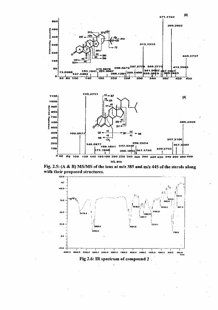
371.1762 (8)
560
399.2062
445.2727
287_0779 349.2779 260.0470 413.2983 170_9976
7 7 151.1041 201_0047 341_ _ 73.0365137.0484 255.1201 26°- 45° 339.2r 5 I 3r-2845
60 joio od 1 100 ' 226 '' 266 306 '340 3B3 420 450
0 60 80 100 120 140 160180 200 220 240 260 280 300 320 340 360 360 400
mlz, amu
Fig. 2.5: (A & B) MS/MS of the ions at miz 385 and miz 445 of the sterols along with their proposed structures.
125.0 ]-
56.7
100,0 -
75.0
50.0
1 1
25.0
0.0
-25,0
-1*)Ni4
3479.3 .
1215.1 I/681.8
2952.8 758.0
1 4000.0 3600.0 3200.0 2800,0 2400.0 2000.0 1800,0 1800.0 1400.0 1200.0 1000.0 800.0 600.0
1./crn
Fig 2.6: IR spectrum of compound 2 .
e 480
3 400
320 C
40
160
80
O
313_1310

Fig 2.7: 1HNMR spectrum of compound 2
ALO
Fig 2.8: 13CNIVIR and DEPT spectra of compound 2
Fig 2.9: COSY spectrum of compound 2.

Ppm
10
20
•
• . 1:n 2:5 201.5 1.0 • 5 4 It
1 10
120
130
;40
40
On
611
711
on
Fig 2.10: HMQC spectrum of compound 2.
"Pm
7 JU „
Oil 0.0 V V 1
0
t
0
PPM
20
407
511.
60 •
•
10.
SIP
120 -
710.
1511-
1 60 -
170-
Fig 2.11: HMBC spectrum of compound 2.

431 -3391
060
LI 480 C 3 0.400
320 0 C 1 240 C
160
SO
334.1500 64
(C)
413 32=9
465.3007
(B)
240.0926 107.0793 145.0926 171 . 1068
60 I .
..
120 180 200 290
378_14.00
406.1239 361_1310 447.1179
41R1 . 498 35).238
320 360 400 440 480
• 40
0 40
32 0 C S 24 C
16
O
31 9.1209
61336
(A)
C 250 C 9
8 " 193
0
•
•
100
4319311 415.13X
50 I 0
355 360 34 720 725 is is is is 400 405 410 115 420 425 CPI 491 46 410 rntz, arnu
Fig.2.12: (A) - Positive ESI-MS of the compounds from Chondria armada; (B & C) MS/MS of the ions at m/z 4651M+Na] + and 4311M+Hr of the sterols along with their proposed structures.

(B)
401.3293
366. 69
() •
90
BO
70-
60 3 o .
60
ato -
30-
20
10
0 100
lc)
383_3174
169-1070
800- 95 0779
I.
136.1061 ir 81_06 19
09.0931 400- I I 0
60 80 100 140 180 220
271:1950
260 300 340 380 420 440
429_20357
329-271. 006. 217 14 82
263_1807 325-7rr67 183 211-1 8 .1314 r43,-,696126 -1782
J
35 9 - 70
341 _0071
247. 3-0303 14-0-0340 317- 120
-, 100-0020 301 _ 1209 97 195-07012
1170 J
2,17 2 52
1 50 200 2430 3430
MiZ,
000
600 -
3 .4,30
accoco -
E 2 0 0 -
II 00
30
3600-
3200-
2800- c 7 0 2400- 9 p 2000-
;1600- -
C
1200 -
393_32V
29145 411
.400
99245
4.45.
Fig.2.13: (A, B & C) MS/MS of the ions at m/z 429[IV1+11] +, 401 and 493[M+Na]± of the sterols along with their proposed structures.

References:
1. Punyasloke B, Phillip CW (review), Planta 2004, 219: 561-578.
2. Jerry GJ, The world of biology, chapter 27, Algae (plantlike) and
Funguslike protests, 2006.
3. Garbary DJ, Gabrielson PW, Taxonomy and evolution. In Biology of the
red algae (K.M. Cole and R.G. Sheath, eds.). Cambridge University Press,
Cambridge, 1990: 477-498.
4. Takemoto T, Daigo K. Chemical Pharmacology Bulletin 1958, 6: 578-
580.
5. Impellizzeri G, Mangiafico S, Oriente G, Piattelli M, Sciuto, S, Fattorusso
E, Magno S, Santacroce C, &lea D. Phytochern. 1975, 14: 1549-1557.
6. Laycock MV, De Freitas ASW, Wright TLC. J. Appl. PhycoL 1989, 1: 113-
122.
7. Sato M, Nakano T, Takeuchi M, Kanno N, Nagahisa E, Sato Y.
Phytochem.1996, 42: 1595- 1597.
8. Takemoto T, Daigo K, Kondo Y, Kondo K. Yakugaku-zasshi 1966,
86(10): 874-877.
9. Ohfune Y, Tomita M. J. Am. Chem. Soc. 1982,104: 3511-3513.
10.Ohfune K, Takemoto T, Maeda M, In Y, Doi M, Inoue M, Ishida T.
Biochem. Biophys. Res. Commun. 1994, 187: 325-331.
11.Maeda M, Kodama T, Tanaka T, Ohfune Y, Nomoto K, Nishimura K,
Fujita T. J. Pesticide Sci. 1984, 9: 27-32.
12.Maeda M, Kodama T, Tanaka T, Yoshizumi H, Takemoto T, Nomoto K.
:Tennen Yuki Kagobutsu Toronkai Koen Yoshishu. 1985,79:350.
13.Maeda M, Kodama T, Tanaka T, Yoshizumi H, Takemoto T, Nomoto
Fujita T. Chem. Pharm. Bull. 1986, 34(11): 4892-4895.
14.Maeda M, Kodama T, Tanaka T, Yoshizumi H, Takemoto T, Nomoto K,
Fujita T. Tetrahedron Lett. 1987, 28(6): 633-636.
15.Zaman L, Arakawa 0, Shimosu A, Onoue Y, Nishio S, Shida Y, Noguchi
T. Toxicon. 1997, 35: 205-212.
16.Daigo KJ, Pharm.Soc.Japn.1959, 79: 350.
67

17.Wright JLC, Boyd RK, De Freitas ASW, Falk M , Foxall RA, Jamieson
WD, Laycock MV, McCulloch AW, McInnes AG .Can. J. Chem. 1989 67:
481.
18.Takemoto T, Daigo K, Sai T. Yakugaku Zassh .1965, 85: 83-5.
19.Takemoto T, Daigo K, Sai T. Yakugaku Zasshi Jan 1965, 85: 33-7.
20. Koga Y, Odaka S. J. Tokyo Chem.Soc.1914, 35: 519.
21. Reeds PJ, Burrin DG, Stoll B, Jahoor F. Journal of Nutrition 2000, 130
(4s): 978S-982S.
22.Naqvi SWA, Solimabi W, Kamat SY, Fernandes L, Reddy CVG, Bhakuni
DS, Dhawan BN. Botanica Marina 1980, 24: 51-55.
23. Kamat SY, Solimabi W, D'Souza L, Naik CG, Ambiye V, Bhakuni DS
Goel AK, Garg HS, Srimal RC. Botanica Marina 1992, 35: 161-164.
24. Govenkar MB, Wahidulla S. Botanica Marina 1999, 42: 3-5.
25. Govenkar MB, Wahidulla S. Botanica Marina 2000, 43: 201-204.
26. Govenkar MB, Wahidulla S. Phytochemistry 2000, 54: 979-981.
27. Aguilar-Santos G, Doty MS, In: Freudenthal RD (Ed.), Marine
Technology Society, Washington D.C, 1968:173.
28. Maiti BC, Thomson RH, Mahendran M, Chem. Res. 1978, S: 126.
29. Aguilar-Santos G, J Chem. Soc. 1970, C: 842.
30. Raub MF, Cardellina IIJH, Schwede JG. Phytochemistry, 1987, 26: 619-
620.
31. Ayyad SEN, Badria FA, Alex .1, Pharm. Sci. 1994, 8: 217.
32. Ciavatta LM, Wahidulla S, D'Souza L, Scognamiglio G, Cimino G.
Tetrahedron 2001, 57: 617-623
33.Fernandez 1.1, Souto ML, Norte M, (review). Nat. Prod. Rep, 2000, 17:
235-246.
34.Hakomuri S. Adv.Cancer.Res. 1989, 52: 257-331.
35.Hakomuri S. Cancer. Res. 1996, 56: 5309-5318.
36. Sastry PS. Adviipid.Res. 1974, 12: 251-310.
37. Kates M. In: M. Kates (Ed.), Handbook of Lipid Research, Vol.6. Plenum
press, New York, 1990: 1-122 and 235-320.
68

38. Sassaki GL, Gorin PAG, Tischer CA, Iacomini M. Glycobiology. 2001,
11: 345-3512
39. Dabrowski J, Hanfland P, Egge H. Biochemistry 1980, 19: 5625-5658.
40. Murakami A, Nakamura Y, Khoshimizu K, Ohigashi H. J. Agri. Food.
Chem., 1995, 43: 2779-2783.
41. Oshima Y, Yamada S, Matsunaga K, Moriya T, Ohizumi Y. J. Nat. Prod.
1994, 57: 534- 536.
42. Reshef V, Mizrachi E, Maretzki T, Silberstein C, Loya S, Hizi A, Carmeli
S. J. Nat.Prod. 1997, 60: 1251-1260.
43. Ahmad VU, Choudhary MI, Clardy J. J. Nat. Prod. 1990, 53: 960-963.
44. Murphy RC, Harrison KA. Mass Spectrom. Rev. 1994, 13: 57-75.
45. Welti R, Wang X, Williams TD. Analytical Biochemistry, 2003, 314:149-
152.
46. Hankin JA, Wheelan P, Murphy RC. Arch Biochem Biophysi. 1997, 340:
317-330.
47. Blair IA, Methods in Enzymology. 1990, 187: 13-23.
48. Waugh RJ, Murphy RC. J. Am. Soc. Mass. Spectr. 1996, 7: 490-499.
49. Kim HY, Choi JS, Yoo JS, Park MY, Kim SM. Anal. Biochem. 1999, 267:
260-270.
50. Bergsson G, Arnfinnsson J, Steingrimsson 0, Thorma,r H. Antimicrobial
agents and Chemotherapy .2001, 45: 3209 - 12.
51. Frentzen M, Weier D, Feussner I. Reports on Symposia and Congresses.
Eur. J. Lipid scis, Technol. 2003, 105: 784-792.
52. Mathur CK, van Holder KE, In Biochemistry. The Benjamin/Cumming
Publishing Co. Inc. Redwood City, 1990: 260-297.
53. El-Masry HA, Fahmy RH, Abdelwahed ASH. Molecules, 2000, 5: 1429-
1438.
54. Alice H, Lichtenstein DSc, Richard J, Deckelbaum, MD. American Heart
Association 2001, 103: 1177-1179.
55. Bjorkhem I. J. Clin. Invest 2002, 110: 725-730.
69

56. Schmitz FJ, in Marine Natural Products (P. J. Scheuer, ed.), Academic
Press, New York, Vol. I ,Chapt,5, 1978.
57. Goad LI , in Marine Natural Products (P. J. Scheuer, ed.), Academic Press,
New York, Vol. II,Chapt,2,1978.
58. Bergquist PR, Hofheinz W, Oesterhelt G, Biochem. Syst. Ecol., in press.
59. Kapetanovic R, Sladic D, Popov S, Zlatovic M, Kijajic Z, Gasic MJ J.
Serb. Chem. Soc. 2005, 70 (12): 1395-1400.
60. Chatman K, Hollenbeck T, Hagey L, Vallee M, Purdy R, Weiss F, Sinzdak
G. Ana/. C hem. 1999, 71: 2358-2363.
61. Griffiths WJ, Liu S, Yang, Y, Purdy R, *van J. Rapid Comm. Mass
Spectrom. 1999, 13: 1595-1610.
62. Fieser LF,. Org. Synth. Coll. 1963, 4:189-191.
63. Parish EJ, Kizito SA, Peng J, Heidepriem RW, Livant P. Chemistry and
Physics of Lipids. 1995, 76: 129-133.
64. Zaretskii Z,. Mass Spectrometry of Steroids (1976). Halstead Press and
Israel University Press.
65. . Wada K, Ishida T. Phytochem. 1974, I3: 2755-2757.
66. Miyake A, Tomoeda M. J.Chem. Soc. [Perkin It 1972: 663.
67. Govenkar M, Wahidullah S. Bot. Mar.2000, 43: 201-204.
68. Al Easa HS, Kornprobst JM, Rizic AM. Phytochem. 1995, 39: 373.
69. Elyakov GB, Stonic VA. In Steroids from Marine Organisms,
Kamemitslcii, A.V., (ed). Nauka: Moscow (in. Russian). 1988: 70.
70. De Rosa S, Kamenarska Z, Bankova V, Stefanov K, St. Dimitrova-
Konaklieva, Najdenski H, Tzvetkova I, Popov SZ.. Naturforsch. 2001,
56C: 1008.
71. Klink G, Dreier F, Buchs A, Gulacar FO. Org. Geochem. 1992, 18: 757.
72. Guella G, Mancini I, Pietra F. Comp. Biochem. Physiol. 1988, 90B: 113-
115.
73. Migliudo A, Picciali V, Sica D. J. Nat. Prod 1990, 53: 1262-1266.
74. Sheikh YM and Djerassi C. Tetrahedron. 1974, 30: 4095-4103.
75. Aiello A, Fattorusso E, Magno S, Menna M. J Nat. Prod 1991, 54: 281 -
285.
70

76. Tischler M, Ayer SW, Anderseb RJ, Mitchell IF, Clardy J. Can J Chem.
1987, 66: 1173-1178.
77. Mishra PD, Wahidullah S, D'Souza L, Kamat SY. Indian J. Chem. 1997,
35B: 719-721.
78. Guerriero A, Debitus C, Pietra F. Hely. Chico. Acta. 1991, 74: 487-494
79. Li •LY, Zhi-Wei Deng, Hong-Zheng Fu, Jun Li, Wen-Han Lin, Peter
Proksch. J. Asian Nat. Prod. Res. 2005, 7: 115-120.
80. Kontiza I, Abatis D, Malakate K, Vagias C, Roussis V. Steroids. 2006,
71: 177-181.
81. Weber N. Phytochemistry. 1977, 16: 1849-1851.
Itokawa H, Akasu M, Fujita N. Chem. Pharm. Bull. 1973, 21: 1386-13ff7.
83. Fernandez MI, Pedro JR, Seoane E. Phytochemistry. 1983, 22: 2087-208ff.
84. Greca MD, Monaco P. J Nat.Prod. 1990, 53: 1430-1435.
85. Barbier M.J. Chem. Ecol. 1987, 13: 1681-1687.
86. Sheu HI, Wang GH, Sung PJ, Duh CY. j. Nat. Prod. 1999, 62: 224-227:
87. Wahidulla S, D'Souza L, Patel J. Phytochemistry. 1987, 26: 2864-2865.
88. Prakash 0, Roy R, Bhakuni DS, Wahidulla S, Kamat SY. J Nat Prod
1989, 52: 686-692.
Buftel-Ponce V, Etahiri S, Guyot M Bioorg. & Med. Chem. Lett. 2002,12:
1715-1718.
90. Noriyuki D, Rikizo A. Biochem. j. 1999, 341: 621-627.
91. Teng JI, Smith LL. J. Steroid Biochem. 1976, 7: 577.
92. Rodriguez AD. Tetrahedron. 1995, 51: 4571-4618.
93. Hewson WD, and Hager PL. J Phycol. 1980, 16: 340.
94. Sheu JH, Huang SY, Duh CY: J. Nat. Prod 1996, 59: 23-26.
95. Sheu HI, Huang SY, Wang GH, Duh CY. j. Nat. Prod 1997; 60: 900-903.
96. Sheu JH, Liaw CC, Duh CY. J. Nat. Prod. 1995, 58: 1521-1526.
97. Shao Z, Cal J, Ye, Q and Guo, Y. J. Asian Nat. Prod. Res. 2002, 4, 205-
209.
98. Choi SJ, Lee ID, Lee KW, Kim DS, Choi HD and Son WB. Bull. Korean
Chem. Soc. 1999,20, 1121-1122
71

99.Bultel-Ponce V, Debitus C, Blond A, Cerceau C and Guyot M.
Tetrahedron Lett. 1997, 38, 5805-5808.
100. Niepel T, Meyer H, Wray V and Abraham WR. Tetrahedron, 1997,53,
3593-3602.
101. Colombo D, Scala A, Taino IM, Toma L, Ronchetti F, Tokuda H, Nishino
H, Nagatsu A and Sakakibara J. Bioorg. Med. Chem. Lett., 1996,6, 1187-
1190.
102. Colombo D, Compostella F, Ronchetti F, Scala A, Toma L, Mulcainaka T,
Nagatsu A, Konoshima T, Tokuda H and Nishino H. Cancer Lett., 1999,143,
1-4.
143.Colombo D, Compostella F, Ronchetti F, Scala A, Toma L, Tolcuda H and
Nishino H. Eur. J. Med. Chem., 2000,35, 1109-1113.
104.Morimoto T, Nagatsu A, Murakarni N, Sakakibara J, Toktida Nishimo
and Iwashima A. Phytochemistiy, 1995,40, 1433-1437.
105.Gustafson KR, Cardellina JH, Fuller RW, Weislow OW, Kiser RF, Shader
KM, Patterson GML and Boyd MR. Nat. Cancer Inst. 1989,81, 1254-
1258.
106. Loya S, Reshef V, Mizrachi E, Silberstein C, Rachamim YCS and Ifizi A.
J Nat. Prod, 1998, 61, 891-895.
107.0hta K, Mizushina Y, Hirata N, Takemura M, Sugawara F, Matsukage A,
Yoshida S and Sakaguchi K. Chem. Pharm. Bull. (Tokyo), 1998,46, 684-
686.
108. Khotimchenko SV. Phytochemistry, 1993,32, 1203-1207.
109.Goncharova SN, Sanina NM and Kostetsky EY. Biochem. Soc. Trans.,
2000,28, 887-890.
to. Sanina NM, Kostetsky EY and Goncharova NV. Biochem. Soc. Trans.,
2000, 28, 894-897.
llt.Khotimchenko SV and Svetashev VI. Biol. Muraya 1987, 6, 3-15
112. Khotimchenko SV and Vaskovsky VE. Bot. Mar., 1990,33, 525-528.
113.Sanina MN, Goncharova NS and Kostetsky EY. Phytochemistly, 2004,65,
721-730.
14.Govenkar MB and Wahidulla S. Bot. Mar., 1999,42, 3-5.
72
115. Van Hummel 1-1C. Fortschr. Chem. Org. Naturst., 1975,32, 267-295.
tt6.Arao T and Yamada M. Phytochemistry 1989, 28, 805-810.
117.Falsone GF, Cateni E, Vrech L, Birkofer V, Lucchini H , Wagner 0,
Seligmann L and Coassini, L..1. Ag. Naturoforsch., 1994,49b, 1297.
lig. Mancini I, Guella G, Defant A, Luz Candenas M, Armesto PC, Depentori
D and Pietra F. Hely. Chem. Acta, 1998,81, 1681-1691.
119. Reshef V, Mizrachi E, Maretzki T, Silberstein C, Loya S, Hizi A and
Carmeli S. J. Nat. Prod.1997,60, 1251-1260.
120. Oshima Y, Yamada SH, Matsunaga K, Moriya T and Ohizumi Y. J. Nat.
Prod. 1994,57, 534-536.
121. Jiang DZ and Gerwick WH. Phytochemisoy, 1990, 29, 14334440.
122. Son WB. Phytochemistry.1990,29, 307-309.
123. Kim HY, Choi JS, Yoo JS, Park MY and Kim SM. Anal. Biochem.,
1999,267, 260-270.
124. Sassaki GL, Gorin PAG, Tischer CA and Iacomini M. Glycobiology,
2001,11, 345-351.
125.Aprin Rr, Durham LJ, Kanazawa Y and Safe S. J chem. Soc. 1967 (C),
1346-1347.
126.Siddhanta AK, Mody KH, Rarnavat BK, Chauan VD, Sharma M and Garg
SH. Bot. Mar., 1995,38, 329-331.
127. Fusetani N and Hashimoto Y. Agric. BioL Chem., 1975,39, 2021-2025.
128. Araki S, Sakurai T, Oohusa T, Kayama M and Sato N. Plant Cell PhysioL,
19&9,30, 775-781.
129. Siddantha AK, Ramvat BK, Chauvan VD, Achari B, Dutta PK and
Pakrashi SC. J Agric. Bot. Mar., 1991,34, 365-367
130.Fusetani N and Hashimoto Y. Hemolysin in a green alga Ulva pertusa. In
Ohsaka, A, Hayashi; K., and Sawai, Y. (eds), Animal Plant and Microbial
Toxins. 1976,Vol. 1, pp. 325-331.
131. Aoki S, Ohta K, Yamazaki T, Sugawara F and Sagakuchi K. FEBS .1
2005, 272, 2132-2140.
73