chapter 4 vibrational spectra and normal coordinate...
TRANSCRIPT
Chapter 4
Vibrational Spectra and Normal Coordinate Analysis of Acetic Acid Cyclohexyl Ester
INTRODUCTION
Acetic acid is a weak organic acid having the m u l a CH,COOH. It is a
clear, colourless liquid and pungent odor. It is miscible with water, alcohol,
glycerol, and ether. It is a combustible liquid. It has moderate fire risk. Pure
acetic acid is moderately toxic by ingestion and inhalation but dilute material is
approved for food use. It is strong irritant to skin and tissue.
Acetic acid is a tonnage chemical of broad applications. Among the
industrial products made h m it, are acetic anhydrides, numerous acetates and
many organic chemicals used in photography, drugs and plastics. It is also used
as a coagulant for rubber latex and as a solvent.
Acetic acid cyclohexyl ester, derivative of acetic acid, is a colourless
liquid with characteristic d o u r having the formula C,H,,02 It has the boiling
point 1770C. It is immiscible in water. It is miscible with most lacquer solvents
and with halogenated and hydrogenated hydrocarbons. It is soluble in alcohol but
insoluble in water and combustible. Besides, it reacts with strong oxidants
causing fire and explosion hazard. Acetic acid esters are used in large quantities
as solvents for plastics, lacquers, resins and gums. It is also called cyclohexyl
acetate.
Acetic acid cyclohexyl ester was synthesised by Wadsworth and
Emmons 111. It is used as solvent for nitrocellulose, cellulose ether, bitumens,
metallic soaps, basic dyes, blown oils, crude rubber, many natural and synthetic
resins and, gums and lacquem. This alicyclic chemical compound has been
widely studied by various scientists.
The X-ray stmctures and proton and carbon- 13 NMR spectra have been
obtained by Baldwin and others 121 for two d~cyclohexyl esters and one
mcyclohexyl esters : namely, di-2-methyl cyclohexyl succinate, dicyclohexyl-3,
4-furandi carboxylate and tricyclohexyl-1,2,3, propanetricarboxylate. Cyclohexyl
azide was synthesized and the vibrational spectra recorded in several phases
Including liquid at various temperatures, amorphous and crystalline at 90K [3]
Senyavin and others [4] reported the vibrational spectra, structure and force fields
of perfluorinated cyclo and bicycloalkanes.
However, there is no report about the vibrat~onal spectra and analysis of
acetic acid cyclohexyl ester in the literature. Hence, an attempt has been made in
the present work to record the FTIR and FTR spectra of acetic acid cyclohexyl
ester and to study the complete vibrational analysis.
4.1 EXPERIMENTAL DETAILS
The FTIR spectra of acetic acid cyclohexyl ester were recorded on
Bmcker IFS 66V FTIR ipectmmeter in the region 4000-200 cm-I. The FT Raman
spectra of the same compound were also recorded on the same instrument with

FRA 106 Raman module equipped with Nd:YAG laser source operating at
1.06 pn line with a scanning speed of 30 cm-I mid' of spectral width 20 cm-I.
The hluencies for al l sharp bands were accurate to * 1 cm-'. The molecular
structure of this compound is given in Fig.4.l. The recorded spectra of acetic acid
cyclohexyl ester is shown in Figure 4.2.
4.2 THEORETICAL CONSIDERATIONS
The geometrical symmetry possessed by the molecule helps to determine
and classify the actual number of fundamental vibrations of the system.
The observed spectrum is explained on the basis of C, point group symmetry.
The 66 fundamental vibrations are distributed as T,, = 46a' + 20 a".
All the modes are active in both Raman and infrared. Assignments have
been made on the basis of relative intensities, magnitudes of the frequencies and
polarization of the Raman lines. The vibrational assignments are discussed in
terms of the potential energy distribution which was obtained from the evaluated
potential constants.
4.3 NORMAL COORDINATE ANALYSIS
With the modified computer program developed in this laboratory on the
basis of Fuhrer et al., program 151, the normal co-ordinate analysis were camed
Out using Wilson's F-G matrix method. The simple general valence force field
was adopted for both in plane and out of plane vibrations. The structural

Fig.4.1 Structure of ACETIC ACID CYCLOHEXYL ESTER


param- are taken from related molecules and Suttanls table [6]. The initial set
of force constants were refined by keeping a few interaction constants fixed
throughout the refinement process. The assignment to all the in plane and out of
plane fundamentals are made on the basis of intensities of Raman and IR bands,
normal coordinate analysis and on comparison with those of similar molecules.
The normal co-onhate calculations have been performed to obtain
vibrational frequencies and the potential energy distribution for the various
modes. In the normal cosrdinate analysis, the potential energy distribution plays
an important role for the characterization of the relative contributions from each
internal coordinates to the total potential energy associated with a particular
normal coordinate of the molecule.
4.4 POTENTIAL ENERGY DISTRIBUTION
A normalised potential energy distribution can be expressed as
F,,L2,k PED = -
h..
where F,, are the force constants defined by damped least square technique, Lik
the normalized amplitude of the associated element (i,k) and 7\, the eigen value
corresponding to the vibrational kquency of the element k.
The PED contribution corresponding to each of the obsewed Muencies
over 10% are alone listed in the present work.
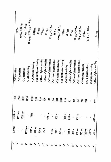
4.5 RESULTS AND DISCUSSION
The observed fhquencies along with their relative intensities of acetic
acid cyclohexyl ester and probable assignments are presented in table 3.1.
The assignment of fhquencies is made as follows.
Stretching vibrations
C-H stretching
The i & a d bands at 3010 cm-' and 2944 cm" have been assigned to C-H
asymmetric stretchings in CH,. The C-H symmetric vibration in CH, is assigned
to Raman band at 293 1 cm". The PED obtained for this CH symmetric stretching
mode in CH, shows that it is pure mode with 94% is contributed by symmetric
C-H vibration.
This molecule gives rise to eleven C-H stre.tching modes which are
assigned to 2677, 2688, 2700, 2712, 2756, 2788, 2800, 2844, 2855, 2868 and
2905 cm". As expected the PED shows that these CH stretching modes are
dominated by pure stretching characters except at 2855 cm-' and 2712 cm-' where
there are little contributions from CC stretching along with C-H stretching. These
values are good agreement with the calculated hquencies and literature values.
C =; 0 and C-0 Stretching
All esters have two strong characteristic bands one due to the C = 0
stretchmg vibration and the other due to the C-0 stretching vibration [7].
The C = 0 and C-0 &etching modes are assigned at i n M band at 1738 cm-'
and at 1538 c m - I which agree with calculated values.
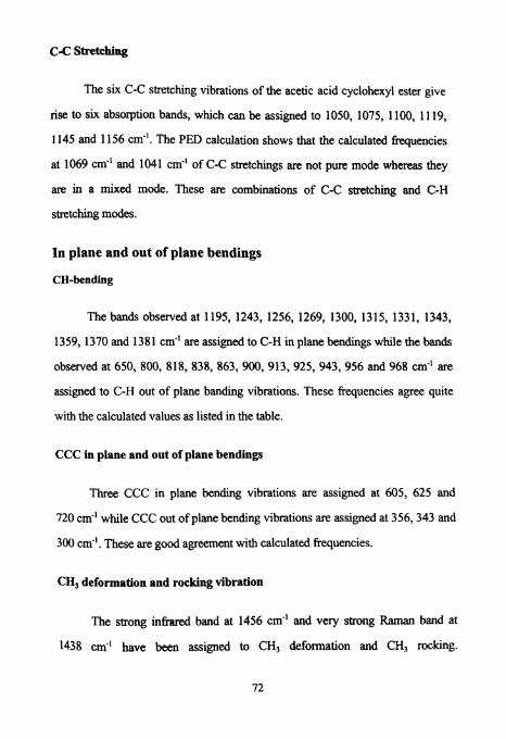
C-C stretching
The six C-C stretching vibrations of the acetic acid cyclohexyl ester give
rise to six absorption bands, which can be assigned to 1050, 1075, 1100, 1 119,
1145 and 1156 cm-I. The PED calculation shows that the calculated Wuencies
at 1069 cm-' and 1041 cm-I of C-C stretchings are not pure mode whereas they
are in a mixed mode. These are combinations of C-C stretching and C-H
stretching modes.
In plane and out of plane bendings
CH-bending
The bands observed at 1195, 1243, 1256, 1269, 1300, 1315, 1331, 1343,
1359, 1370 and 1381 cm-' are assigned to C-H in plane bendings while the bands
observed at 650, 800, 818, 838, 863,900, 913, 925, 943, 956 and 968 cm-I are
assigned to C-H out of plane banding vibrations. These frequencies agree quite
with the calculated values as listed in the table.
CCC in plane and out of plane bendings
Three CCC in plane bending vibrations are assigned at 605, 625 and
720 cm'l while CCC out of plane bending vibrations are assigned at 356,343 and
300 cm-I. These are good agreement with calculated frequencies.
CH, deformation and rocking vibration
The strong infrared band at 1456 cm4 and very strong Raman band at
1438 cm-' have been assigned to CHI deformation and CHI rocking.
The weak hfiard at 438 cmA' and Raman band at 388 cm" have been assigned to
CH, wagging mode and CH, torsion [8,9].
CCC ring breathing and trigonal bending
The skeleton of the molecule also gives some characteristic absorption
wave number such as ring breathing and CCC trigonal bending. In the present
case, these are assigned at 856 cm-' and 1019 cm-' which agree quite well with
literature values [lo- 121.
Conclusion
A complete vibrational spectm and analysis is reported in the present work
for the first time for acetic acid cyclohexylester. The close agreement between the
observed and calculated frequencies confirm the validity of the present
assignment


W.S. Wadsworth and E.D.Emmons. Org. Syn. 45,44, (1965).
C.R.Baldwin, M.M.Britton, S.C. Davies, D.G.Gillies, D.L. Hughes, G.W.Smith, L.H. Sutcliffe, J. Mol. Str. 403, 14,1997).
D.Sulzle, A. Gatial, A.Karlsson, P.Klaeboe and C.J.Nielson, J. Mol. Str. 174,207, (1988).
V.M. Senyavin, 1.V.Kochikov and G.M.Kuramshii, J. Mol. Str. 410, 463, (1997).
H. Fuhrer, V.B. Karther, K.L. Kidel, P.J.Krugdel and H.H.Manstch Computer program for infrared and spectrometry, normal coordinate analysis, Volume 5, National Research Council, Ottowa, Canada (1976).
L.E.Sutton, The interatomic bond distances and bond angles in molecules and ions, London Chem, Soc London (1 983).
D. Steele and A.Muller, J. Phys. Chem. 95,6163, (1991).
J.K.Katon and F.F.Bentley Spectrochim. Acta, 19,639 (1963).
J.L. Lucier and F.F. Bentley, Spectrochim Acta, 20, 1, (1964).
S.Mohan, A.R. Prabakaran and S.Prameela Indian J. Pure and App. Phy 29,672, (1991)
George Socrates Infiared and Raman Chmcteristic group frequencies table & Charts Third edition John Wiley & Sons Ltd (2001).
G. Varsangi, Assignments for vibrational spectra of seven hundred benzenz derivates, volume no. 7, Adam Hilger London (1974)
C.N.RRao, Chemical Applications of Infrared Spectroscopy (Academic press, New Yo&) (1963).
L.J. Bellamy, Infrared spectra of complex molecules (John - Wiley, New Yo&) (1959).
15. B.P. Straughan and S.Waker 'Spectroscopy', V01.2., (Chapman and Hall), (1976).
16. Stele, 'Interpretation of vibrational spectra (Chapman and Hall, London), (1975).
17. R.M. Silverstein and G.C. Bassler, Spectrometric identification of organic compounds, Wiley, New York (1964).
18. F. Scheinmann, Ed. An introduction of Spectroscopic methods for the identification of organic compound. Pergamon Press, New York (1970).