chapter 4 the human body - jones & bartlett...
TRANSCRIPT

9/11/2012
1
Slide 1
Chapter 4
The Human Body
Slide 2
Overview Anatomic Terms
The Anatomic Position Descriptive Anatomic Terms
Body Systems Respiratory System Circulatory System Musculoskeletal System Nervous System Skin Digestive System Endocrine System
Slide 3
Anatomic Terms
Copyright © 2013 by Jones & Bartlett Learning, LLC, an Ascend Learning Company

9/11/2012
2
Slide 4
Anatomic Terms
The anatomic position
Slide 5
Anatomic Terms
Anatomic planes Midline
Midclavicular line
Midaxillary line
Slide 6
Descriptive Anatomic Terms
Prone The patient lying flat
on the stomach
Copyright © 2013 by Jones & Bartlett Learning, LLC, an Ascend Learning Company

9/11/2012
3
Slide 7
Descriptive Anatomic Terms
Supine The patient lying
flat on the back
Slide 8
Descriptive Anatomic Terms
Fowler’s position The patient lying on
the back with a bend at the hips
• Full Fowler’s
• Semi-Fowler’s
Slide 9
Descriptive Anatomic Terms
Trendelenburg position The patient lying flat on
the back, on an incline, and with feet elevated approximately 12 inches above the head
Copyright © 2013 by Jones & Bartlett Learning, LLC, an Ascend Learning Company

9/11/2012
4
Slide 10
Descriptive Anatomic Terms
Shock position The patient lying flat on
the back, bent at the hips with feet lifted approximately 12 inches off of the ground
Slide 11
Body Systems
Slide 12
Respiratory System
The respiratory system takes oxygen from the air and makes it available for the blood to transport to every cell and rids the body of excess carbon dioxide
Copyright © 2013 by Jones & Bartlett Learning, LLC, an Ascend Learning Company

9/11/2012
5
Slide 13
Respiratory System
The airway Upper airway
• Extends from the mouth and nose to the trachea
Lower airway• Extends from the trachea to the alveoli
Slide 14
Upper Airway
Slide 15
Upper Airway
Nose and mouth Pharynx
• Oropharynx
• Nasopharynx
Epiglottis • Leaf-shaped structure that prevents food and liquid from
entering the trachea during swallowing
Copyright © 2013 by Jones & Bartlett Learning, LLC, an Ascend Learning Company

9/11/2012
6
Slide 16
Lower Airway
Slide 17
Lower Airway
Trachea (windpipe)
Cricoid cartilage Firm cartilage ring forming the lower portion of the
larynx
Larynx (voice box)
Bronchi • Two major branches of the trachea to the lungs; bronchus
subdivides into smaller air passages ending at the alveoli
The lungs
Diaphragm
Slide 18
Respiratory Terminology
Ventilation The movement of air
Respiration The exchange of gases
Copyright © 2013 by Jones & Bartlett Learning, LLC, an Ascend Learning Company

9/11/2012
7
Slide 19
Ventilation Inhalation (active)
Diaphragm and intercostal muscles contract, increasing the size of the thoracic cavity
Diaphragm moves slightly downward, flares lower portion of rib cage
Ribs move upward/outward
This creates a negative pressure in the chest cavity
Slide 20
Ventilation
Air flows into the lungs because of the negative pressure
Slide 21
Ventilation
Exhalation Diaphragm and
intercostal muscles relax, decreasing the size of the thoracic cavity
• Diaphragm moves upward
• Ribs move downward/inward
Air is expelled from the lungs
Copyright © 2013 by Jones & Bartlett Learning, LLC, an Ascend Learning Company

9/11/2012
8
Slide 22
Respiration
Alveolar respiration Gas exchange in the lungs
Cellular respiration Gas exchange in the tissues of the body
Slide 23
Alveolar Respiration
Alveolar/capillary exchange Oxygen-rich air enters the alveoli during each
inspiration
Oxygen-poor blood in the capillaries passes into the alveoli
Oxygen enters the capillaries as carbon dioxide enters the alveoli
Slide 24
Cellular Respiration
Capillary/cellular exchange Cells give up carbon dioxide to the capillaries
Capillaries give up oxygen to the cells
Copyright © 2013 by Jones & Bartlett Learning, LLC, an Ascend Learning Company

9/11/2012
9
Slide 25
Alveolar and Cellular Respiration
Slide 26
Normal Breathing
Normal respiration should be effortless
Slide 27
Normal Respiratory Rates
Adult—12-20/minute
Child—15-30/minute
Infant—25-50/minute
Copyright © 2013 by Jones & Bartlett Learning, LLC, an Ascend Learning Company
9/11/2012
10
Slide 28
Assessing Breathing
Rate
Rhythm
Quality
Breath sounds
Chest expansion
Effort of breathing
Depth (tidal volume)
Slide 29
Effort of Breathing
Accessory muscles Additional muscles used to draw air into the chest
Includes the muscles of the neck, abdomen, and chest
Use of accessory muscles is a sign of
respiratory distress!
Slide 30
Tidal Volume
The amount of air exchanged in one breath
Copyright © 2013 by Jones & Bartlett Learning, LLC, an Ascend Learning Company

9/11/2012
11
Slide 31
Considerations for
Infants and Children
Slide 32
Adults versus Children–Respiratory Anatomy
Mouth and nose In general, all structures are smaller and more
easily obstructed than in adults
Slide 33
Adults versus Children –Respiratory Anatomy
Tongue Infants’ and children’s tongues take up
proportionately more space in the mouth than adults
Trachea (windpipe) Narrower tracheas that are obstructed more easily by
swelling Softer and more flexible in infants and children
Cricoid cartilage Less developed and less rigid
Chest wall is softer Tend to depend more heavily on the diaphragm for breathing
Copyright © 2013 by Jones & Bartlett Learning, LLC, an Ascend Learning Company

9/11/2012
12
Slide 34
Circulatory System
Slide 35
Circulatory System
The heart pumps blood to the body organs through the cardiovascular system
This process is so vital to life that any interruption for more than a few minutes can mean death to the individual
Slide 36
Circulatory System
Heart
Blood vessels Arteries Veins Capillaries
Blood
Copyright © 2013 by Jones & Bartlett Learning, LLC, an Ascend Learning Company

9/11/2012
13
Slide 37
Heart Structure and function
Atrium• Right
Receives blood from the veins of the body and the heart
Pumps oxygen-poor blood to the right ventricle
• Left Receives blood from the
pulmonary veins (lungs)
Pumps oxygen-rich blood to left ventricle
Ventricle• Right
Receives blood from the right atrium
Pumps oxygen-poor blood to the lungs
• Left Receives blood from the
left atrium
Pumps oxygen-rich blood to the body
Slide 38
Heart
Slide 39
Heart
Structure and function Valves prevent backflow of blood
Copyright © 2013 by Jones & Bartlett Learning, LLC, an Ascend Learning Company

9/11/2012
14
Slide 40
Heart
Structure and function Cardiac conductive system
• Heart is more than a muscle
• Specialized contractile and conductive tissue in the heart
• Electrical impulses create coordinated contraction
Automaticity• The ability of cardiac muscle cells to generate their own
impulses
Slide 41
Blood Vessels Arteries
Arterioles
Capillaries
Venules
Veins
Slide 42
Arteries Carry blood away from the heart to the rest of the body Major arteries
Coronary • Vessels that supply the heart with blood
Aorta• Major artery originating from the heart, lying in front of the spine in the thoracic
and abdominal cavities Pulmonary
• Artery originating at the right ventricle; carries oxygen-poor blood to the lungs Carotid
• Major artery of the neck Femoral
• Major artery of the thigh Radial
• Major artery of the lower arm Brachial
• An artery of the upper arm Posterior tibial Dorsalis pedis
Copyright © 2013 by Jones & Bartlett Learning, LLC, an Ascend Learning Company

9/11/2012
15
Slide 43
Arteries
Slide 44
Arterioles
The smallest branches of an artery leading to the capillaries
Slide 45
Capillaries
Tiny blood vessels that connect arterioles to venules
Found in all parts of the body
Allow for the exchange of nutrients and waste at the cellular level
Copyright © 2013 by Jones & Bartlett Learning, LLC, an Ascend Learning Company

9/11/2012
16
Slide 46
Capillaries
Slide 47
Venules
The smallest branches of a vein leading to the capillaries
Slide 48
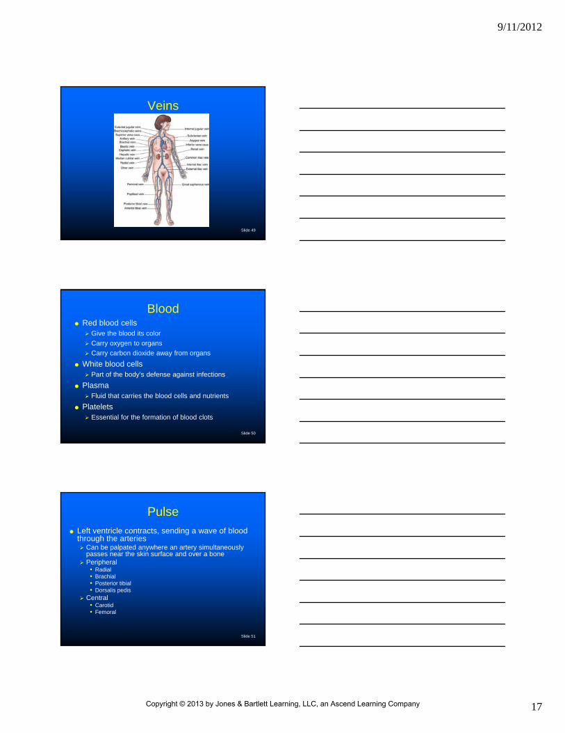
Veins
Vessels that carry blood back to the heart
Major veins Pulmonary vein
• Carries oxygen-rich blood from the lungs to the left atrium
Venae cavae• Superior
• Inferior Carries oxygen-poor blood back to the right atrium
Copyright © 2013 by Jones & Bartlett Learning, LLC, an Ascend Learning Company
9/11/2012
17
Slide 49
Veins
Slide 50
Blood Red blood cells
Give the blood its color
Carry oxygen to organs
Carry carbon dioxide away from organs
White blood cells Part of the body’s defense against infections
Plasma Fluid that carries the blood cells and nutrients
Platelets Essential for the formation of blood clots
Slide 51
Pulse
Left ventricle contracts, sending a wave of blood through the arteries Can be palpated anywhere an artery simultaneously
passes near the skin surface and over a bone Peripheral
• Radial• Brachial• Posterior tibial• Dorsalis pedis
Central• Carotid• Femoral
Copyright © 2013 by Jones & Bartlett Learning, LLC, an Ascend Learning Company

9/11/2012
18
Slide 52
Pulse
Slide 53
Blood Pressure
A measure of the pressure exerted against the walls of the arteries during contraction and relaxation of the heart Systolic
• Pressure exerted against the walls of the artery when the left ventricle contracts
Diastolic• Pressure exerted against the walls of the artery when the
left ventricle is at rest
Slide 54
Perfusion
The process of delivering oxygenated blood to the organs and removing waste products and carbon dioxide
Cellular respiration
Copyright © 2013 by Jones & Bartlett Learning, LLC, an Ascend Learning Company

9/11/2012
19
Slide 55
Perfusion
Shock (hypoperfusion) Widespread inadequate tissue perfusion
Slide 56
Shock
Signs and symptoms Pale or cyanotic skin Cool or cold skin Rapid weak pulse Altered mental status Rapid breathing Nausea and vomiting Low or decreasing blood pressure
• A LATE SIGN!
Slide 57
Musculoskeletal System
Copyright © 2013 by Jones & Bartlett Learning, LLC, an Ascend Learning Company

9/11/2012
20
Slide 58
Skeletal System
The skeletal system is the scaffolding of the body
Gives the body shape and rigidity
Protects the vital internal organs
Enables movement
Slide 59
The Skull
Skull Houses and protects the
brain
Orbit
Nasal bone
Maxilla
Mandible
Zygomatic bone
Slide 60
Spinal Column
Cervical (neck) —7 vertebrae
Thoracic (upper back)—12 vertebrae
Lumbar (lower back)—5 vertebrae
Sacral (back wall of the pelvis)—5 vertebrae
Coccyx (tailbone)—4 vertebrae
Copyright © 2013 by Jones & Bartlett Learning, LLC, an Ascend Learning Company

9/11/2012
21
Slide 61
Spinal Column
Slide 62
Thorax
Ribs 12 pairs
Attached posterior to the thoracic vertebrae• Pairs 1-10 are attached anterior to the sternum
• Pairs 11 and 12 are floating
Sternum (Breastbone) Manubrium (superior portion of sternum)
Body (middle)
Xiphoid process (inferior portion of sternum)
Slide 63
Thorax
Copyright © 2013 by Jones & Bartlett Learning, LLC, an Ascend Learning Company

9/11/2012
22
Slide 64
PelvisIlium
Ischium
Pubicsymphysis
Acetabulum
Slide 65
Upperextremity
Lowerextremity
Slide 66
Lower Extremities
Femur (thigh)
Patella (kneecap)
Tibia (shin, lower leg)
Fibula (lower leg)
Medial and lateral malleolus
Tarsals and metatarsals (foot)
Calcaneus (heel)
Phalanges (toes)
Copyright © 2013 by Jones & Bartlett Learning, LLC, an Ascend Learning Company

9/11/2012
23
Slide 67
Lower Extremities
Patella
Tibia
Fibula Femur
Slide 68
C4-35Tarsals
Metatarsals
Phalanges
Foot
Slide 69
Upper Extremities
Clavicle (collar bone)
Scapula (shoulder blade)
Acromion (tip of shoulder)
Humerus (superior portion of upper extremity)
Olecranon (elbow)
Radius (lateral bone of forearm)
Ulna (medial bone of forearm)
Carpals (wrist)
Metacarpals (hand)
Phalanges (fingers)
Copyright © 2013 by Jones & Bartlett Learning, LLC, an Ascend Learning Company

9/11/2012
24
Slide 70
Upper Extremity
Slide 71
Joints
Where bones connect to other bones
Types Ball and socket
Hinged
Slide 72
Hinge Joint
Copyright © 2013 by Jones & Bartlett Learning, LLC, an Ascend Learning Company

9/11/2012
25
Slide 73
Ball and Socket Joint
Slide 74
Muscular System
Slide 75
Muscular System
Function Gives the body shape
Protects internal organs
Provides for movement
Copyright © 2013 by Jones & Bartlett Learning, LLC, an Ascend Learning Company

9/11/2012
26
Slide 76
Muscular System
Types Voluntary
Involuntary
Cardiac
Slide 77
Muscular System
Voluntary (skeletal) Attached to the bones
Form the major muscle mass of the body
Controlled by nervous system and brain
Can be contracted and relaxed by the will of the individual
Responsible for movement
Slide 78
Muscular System
Involuntary (smooth) Found in the walls of the
tubular structures of the gastrointestinal tract and urinary system, as well as the blood vessels and bronchi
Control the flow through these structures
Carry out the automatic muscular functions of the body
Individuals have no direct control over these muscles
Respond to stimuli such as stretching, heat, and cold
Copyright © 2013 by Jones & Bartlett Learning, LLC, an Ascend Learning Company

9/11/2012
27
Slide 79
Muscular System
Cardiac Found only in the heart
• Involuntary muscle Has its own supply of blood
through the coronary artery system
Can tolerate interruption of blood supply for only very short periods
• Automaticity—the ability of the muscle to contract on its own
Slide 80
Nervous System
Slide 81
Nervous System
Function Controls the voluntary and involuntary activity of
the body
Copyright © 2013 by Jones & Bartlett Learning, LLC, an Ascend Learning Company

9/11/2012
28
Slide 82
Nervous System
Central nervous system Brain
Spinal cord
• Cranial nerves (12)• Spinal nerves
cervical (8)thoraic (12)lumbar (5)sacral (5) coccyx (1)
PeripheralNervous System
• Brain• Spinal cord
CentralNervous System
Slide 83
Nervous System
Peripheral nervous system Sensory
• Impulses carry information from the body to the brain and spinal cord
Motor• Impulses carry
information from the brain and spinal cord to the body
Slide 84
Skin
Copyright © 2013 by Jones & Bartlett Learning, LLC, an Ascend Learning Company

9/11/2012
29
Slide 85
Skin
Protects the body from the environment, bacteria, and other organisms
Helps regulate the temperature of the body
Senses heat, cold, touch, pressure, and pain; transmits this information to the brain and spinal cord
Slide 86
Skin
Layers Epidermis
• Outermost layer of skin Dermis
• Deeper layer of skin containing sweat and sebaceous glands, hair follicles, blood vessels, and nerve endings
Subcutaneous layer
Slide 87
Digestive System
Copyright © 2013 by Jones & Bartlett Learning, LLC, an Ascend Learning Company

9/11/2012
30
Slide 88
Digestive System Provides the body with
energy from food
Foods passes through hollow organs from the stomach to the anus
Nutrients are absorbed into the bloodstream through the small intestine
Slide 89
Endocrine System
Slide 90
Endocrine System
Secretes chemicals, such as insulin and adrenaline, responsible for regulating body activities and functions
Copyright © 2013 by Jones & Bartlett Learning, LLC, an Ascend Learning Company

9/11/2012
31
Slide 91
Summary
Anatomic Terms The Anatomic Position Descriptive Anatomic Terms
Body Systems Respiratory System Circulatory System Musculoskeletal System Nervous System Skin Digestive System Endocrine System
Copyright © 2013 by Jones & Bartlett Learning, LLC, an Ascend Learning Company