chapter 4 cell structure - saddleback college · chapter 4 cell structure. biology ... cross...
TRANSCRIPT

Cytology = the study of cells
Cellular basis of life:
•Basic unit of life
•Lowest level with all attributes of life
•Organisms composed of one or more cells
•Cell structure correlated to function
•All cells are related
Chapter 4 CELL STRUCTURE

BIOLOGY
Chapter 4 CELL STRUCTURE

Which cellular structure is common
to all 3 domains of life?
a) Nucleus
b) Endoplasmic reticulum
c) Mitochondria
d) Phospholipid bilayer cell membrane
e) Endocytotic vesicles

Figure 4.6
EukaryoticProkaryotic

Figure 6.3
Brightfield
(unstained specimen)
Brightfield
(stained specimen)
50
m
Confocal
Differential-interference-
contrast (Nomarski)
Fluorescence
10 m
Deconvolution
Super-resolution
Scanning electron
microscopy (SEM)
Transmission electron
microscopy (TEM)
Cross section
of cilium
Longitudinal section
of cilium
Cilia
Electron Microscopy (EM)
1
m1
0
m5
0
m
2 m
2 m
Light Microscopy (LM)
Phase-contrast
Cellular observations
microscopy

Figure 6.4TECHNIQUE
Homogenization
Tissue
cells
Homogenate
Centrifugation
Differential
centrifugation
Centrifuged at
1,000 g
(1,000 times the
force of gravity)
for 10 min Supernatant
poured into
next tube
20,000 g
20 min
80,000 g
60 minPellet rich in
nuclei and
cellular debris
150,000 g
3 hr
Pellet rich in
mitochondria
(and chloro-
plasts if cells
are from a plant)
Pellet rich in
“microsomes”
(pieces of plasma
membranes and
cells’ internal
membranes) Pellet rich in
ribosomes
Cellular fractionation
•To study organelle function

Figure 4.5
• size 1 – 10 µm
• Nucleoid
• Circular DNA plasmid
• Cell wall
• Gram +
• Gram -

Prokaryotes
• DNA not membrane
bound
• Lack membrane bound
organelles
• No histone proteins
• Peptidoglycan
• Widespread
• Size (0.5 – 5 µm)
• Bacteria or Archaea

Fig. 27-2
(a) Spherical(cocci)
1 µm
(b) Rod-shaped(bacilli)
2 µm
(c) Spiral
5 µm
Diplo-
Staphylo-
Strepto-

Prokaryotic Reproduction
• Binary Fission • Genetic Diversity via
Horizontal Gene Transfer• Transformation
• Transduction
• Conjugation

Fig. 27-3
Cellwall
Peptidoglycanlayer
Plasma membrane
Protein
Gram-positive
bacteria
(a) Gram-positive: peptidoglycan trapscrystal violet.
Gram-negativebacteria
(b) Gram-negative: crystal violet is easily rinsed away,revealing red dye.
20 µm
Cellwall
Plasma membrane
Protein
Carbohydrate portionof lipopolysaccharide
Outermembrane
Peptidoglycanlayer
Cell Surface Structures
Hans Christian Gram Gram Staining
LPS component
•O polysacch antigens for ID (E. coli O157:H7)
•Lipid A endotoxin toxic (fever/shock)
antibiotics

Fig. 27-3c
Gram-positive
bacteria
Gram-negativebacteria
20 µm

Figure 6.6
Outside of cell
Inside of cell0.1 m
TEM of a plasma membrane
• Phospholipid bilayer• Cholesterol • Proteins • Carbohydrates
Hydrophilicregion
Hydrophobicregion
Hydrophilicregion
Carbohydrate side chains
ProteinsPhospholipid
(b) Structure of the plasma membrane
8 m
cholesterol
Outside cell
Inside cell

Figure 4.7 Why are cells so small?
• Efficiency in:• Acquisition of nutrients
• Disposal of wastes
• What makes this possible?• High surface areas to volume ratio

a) One largecell.
b) Eight smallcells.
c) Cell withmicrovillion one surface.
Cell size & plasma membrane shape affect SA:V
• Which cell has the larger SA?
• Larger Vol?
• Larger SA:V ratio?

Figure 4.8Figure 4.8

Cell structure reflects eukaryotic cell’s function
a) A portion of several musclecells of the heart (X 1,500).
b) Nerve cells of the centralnervous system (X 830).
c) Cells lining a tubule of a kidney (X 250).
• How are these cells similar?
• What makes these cells different?
Figure 6.8b
Animal Cells
Cell
Nucleus
Nucleolus
Human cells from lining
of uterus (colorized TEM)
Yeast cells budding
(colorized SEM)
10
m
Fungal Cells
5
m
Parentcell
Buds
1 m
Cell wall
Vacuole
Nucleus
Mitochondrion
A single yeast cell(colorized TEM)


Figure 6.9aNucleus
Rough ER
Nucleolus
Chromatin
Nuclear envelope:
Inner membrane
Outer membrane
Nuclear pore
Chromatin
Ribosome
Porecomplex
Close-upof nuclearenvelope
• Nuclear Envelope
• Nucleus• Genetic control ctr
• DNA synthesis
• RNA synthesis
• Nuclear pores

Figure 4.12
• Chromatin
a) DNA + histone proteins (8) nucleosome
b) Replicated chromosome

NucleolusRibosome production
• Free or bound
» Protein synthesis
Figure 3.15A transmission electron micrograph (X 6,000) of
the nucleus of an animal cell
Nucleolus
Nuclear
membrane
Nuclear
pores
Nuclear
membrane

Figure 3.15A transmission electron micrograph (X 6,000) of
the nucleus of an animal cell
Nucleolus
Nuclear
membrane
Nuclear
pores
Nuclear
membrane
Nucleolus/nucleoliRibosome production
Free or bound
Protein synthesis
Figure 6.10
0.25 m
Free ribosomes in cytosol
Endoplasmic reticulum (ER)
Ribosomes bound to ER
Large
subunit
Small
subunit
Diagram of a ribosomeTEM showing ER and
ribosomes

Figure 6.11
Smooth ER
Rough ER
ER lumen
CisternaeRibosomes
Smooth ERTransport vesicle
Transitional ER
Rough ER 200 nm
Nuclear
envelope
Endoplasmic Reticulum
• Rough ER
• Smooth ER

Endomembrane Organelles• Rough Endoplasmic
Reticulum (RER)
– Ribosomes
– Protein synthesis
Endomembrane Organelles
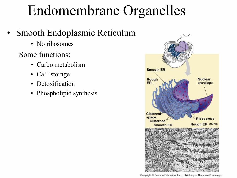
• Smooth Endoplasmic Reticulum• No ribosomes
Some functions:
• Carbo metabolism
• Ca++ storage
• Detoxification
• Phospholipid synthesis

cis face
(“receiving” side of
Golgi apparatus)
trans face
(“shipping” side of
Golgi apparatus)
0.1 m
TEM of Golgi apparatus
Cisternae
Endomembrane Organelles• Golgi apparatus/body/complex “warehouse”
• Receives
• Modifies
• Stores
• Ships

Figure 6.15-3
Smooth ER
Nucleus
Rough ER
Plasma
membrane
cis Golgi
trans Golgi
Endomembrane System

Figure 6.13a
Nucleus
Lysosome
1 m
Digestive
enzymes
Digestion
Food vacuole
LysosomePlasma membrane
(a) Phagocytosis
• Lysomsome
• Contains hydrolytic enzymes
• Breaks down “stuff”
• Intracellular digestion of nutrients
Endomembrane
Organelles

Figure 6.13bVesicle containing
two damaged
organelles1 m
Mitochondrion
fragment
Peroxisome
fragment
Peroxisome
VesicleMitochondrion
Lysosome
Digestion
(b) Autophagy
Endomembrane
Organelles• Lysomsome
• Contains hydrolytic enzymes
• Breaks down “stuff”
• Intracellular digestion of nutrients
• “Garbage man” dead organelles
• Programmed cell destruction
• Tay Sachs Disease

Figure 6.14
Central vacuole
Cytosol
Nucleus
Cell wall
Chloroplast
Central
vacuole
5 m

Figure 6.15-1
Smooth ER
Nucleus
Rough ER
Plasma
membrane
Endomembrane System

Figure 6.15-2
Smooth ER
Nucleus
Rough ER
Plasma
membrane
cis Golgi
trans Golgi
Endomembrane System

Figure 6.15-3
Smooth ER
Nucleus
Rough ER
Plasma
membrane
cis Golgi
trans Golgi
Endomembrane System

Enzymes responsible for
biosynthesis of membrane lipids
would be located in what part of
the cell?
a) endoplasmic reticulum.
b) nucleus.
c) lysosomes.
d) Golgi.
e) plasma membrane

Endomembrane Organelles• Lysomsome
– Contains hydrolytic enzymes
– Breaks down “stuff”
• Intracellular digestion of nutrients
• “Garbage man” dead organelles
• Programmed cell destruction
• Tay Sachs Disease

Endomembrane Organelles
• Vacuoles
– Food
• Temp. storage of food
– Contractile
• Expels waste

Types of Vesicles• Storage & shipping vesicles
• Secretory vesicles
• Endocytic vesicles
– Vacuoles
– Food
– Contractile
• Expels waste
• Peroxisomes
– Contain enzymes that detoxify
• Lysosomes
– Contain digestive enzymes Bacterium
Plasma
membrane
Golgi
apparatu
s
Peroxisome
Alcohol
Harmless
waste
Cell toxic
waste
Lysosome
Residual
body

Endomembrane System
A membrane protein synthesized in
the rough ER may be directed to:
a) peroxisomes.
b) lysosomes.
c) mitochondria.
d) all of the above

Golgi
Brefeldin A is a drug that disrupts transport from
the ER to the Golgi apparatus. What other
organelles and membranes are affected?
A. lysosomes, vacuoles, plasma membrane
B. lysosomes, peroxisomes, plasma membrane
C. vacuoles, mitochondria, plasma membrane
D. lysosomes, vacuoles, nuclear membrane
E. all intracellular organelles and membranes

Figure 6.16
NucleusEndoplasmic
reticulum
Nuclear
envelope
Ancestor of
eukaryotic cells
(host cell)
Engulfing of oxygen-
using nonphotosynthetic
prokaryote, which
becomes a mitochondrion
Mitochondrion
Nonphotosynthetic
eukaryote
Mitochondrion
At least
one cell
Photosynthetic eukaryote
Engulfing of
photosynthetic
prokaryote
Chloroplast
Endosymbiotic Eukaryotic Origins

Intermembrane space
Outer
membrane
DNA
Innermembrane
Cristae
Matrix
Free
ribosomes
in the
mitochondrial
matrix
(a) Diagram and TEM of mitochondrion (b) Network of mitochondria in a protist
cell (LM)
0.1 m
Mitochondrial
DNA
Nuclear DNA
Mitochondria
10 m
Mitochondrion/mitochondria
• Double membrane
• Inner membrane
• Cristae energy production
• Matrix energy production
• DNA 1 chromosome
• Binary fission
• All aerobic eukaryotes

Figure 6.17a
Intermembrane space
Outer
DNA
Innermembrane
Cristae
Matrix
Free
ribosomes
in the
mitochondrial
matrix
(a) Diagram and TEM of mitochondrion
0.1 m
membrane
Mitochondrion/mitochondria

RibosomesStroma
Inner and outermembranes
Granum
1 mIntermembrane spaceThylakoid
(a) Diagram and TEM of chloroplast
DNA
Chloroplasts
• Double membrane• Thylakoid (granum/grana)
• Sunlight NRG chemical NRG
• Photosynthetic pigments
• Stroma
• Uses chemical NRG
• Sugar production
• Own DNA

According to the endosymbiont
theory, which of the following
organelles were once endosymbiotic
prokaryotic organisms?
a) Mitochondria and lysosomes
b) Mitochondria and chloroplasts
c) Chloroplasts and Golgi apparatus
d) Golgi apparatus and ribosomes
e) Ribosomes and lysosomes

Figure 6.19
Chloroplast
Peroxisome
Mitochondrion
1 m
Peroxisome
• Single membrane
• Plants & animals
• Detoxifies cells
• H2O2

Figure 6.21
ATPVesicle
(a)
Motor protein
(ATP powered)
Microtubule
of cytoskeleton
Receptor for
motor protein
0.25 mVesiclesMicrotubule
(b)

Table 6.1
Column of tubulin dimers
Tubulin dimer
25 nm
Actin subunit
7 nm
Keratin proteins
812 nm
Fibrous subunit (keratins
coiled together)
10 m 10 m 5 m

Microtubule Function
Taxol, a drug approved for treatment of breast
cancer, prevents depolymerization of microtubules.
What cellular function that affects cancer cells
more than normal cells might taxol interfere with?
a) maintaining cell shape
b) cilia or flagella
c) chromosome movements in cell division
d) cell division (cleavage furrow formation)
e) cytoplasmic streaming

Figure 6.22
Centrosome
Longitudinalsection ofone centriole
Centrioles
Microtubule
0.25 m
Microtubules Cross section
of the other centriole

Figure 6.23
Direction of swimming
(b) Motion of cilia
Direction of organism’s movement
Power stroke Recovery stroke
(a) Motion of flagella 5 m
15 m

Figure 6.24
Microtubules
Plasmamembrane
Basal body
Longitudinal sectionof motile cilium
(a)
0.5 m 0.1 m
0.1 m
(b) Cross section ofmotile cilium
Outer microtubuledoublet
Dynein proteins
Centralmicrotubule
Radialspoke
Cross-linkingproteins betweenouter doublets
Plasma membrane
Triplet
(c) Cross section ofbasal body

Figure 6.25Microtubule
doublets
Dynein protein
ATP
(a) Effect of unrestrained dynein movement
Cross-linking proteins
between outer doubletsATP
Anchorage
in cell
(b) Effect of cross-linking proteins
(c) Wavelike motion
1
2
3

Figure 6.27
Muscle cell
Actin
filament
Myosin
Myosin
filament
head
(a) Myosin motors in muscle cell contraction
0.5 m
100 m
Cortex (outer cytoplasm):
gel with actin network
Inner cytoplasm: sol
with actin subunits
(b) Amoeboid movement
Extending
pseudopodium
30 m(c) Cytoplasmic streaming in plant cells
Chloroplast

Figure 6.27a
Muscle cell
Actinfilament
Myosin
Myosin
filament
(a) Myosin motors in muscle cell contraction
0.5 m
head
Locomotion

Figure 6.27b
100 m
Cortex (outer cytoplasm):
gel with actin network
Inner cytoplasm: sol
with actin subunits
(b) Amoeboid movement
Extending
pseudopodium
Cytoplasmic streaming

Figure 6.27c
30 m(c) Cytoplasmic streaming in plant cells
Chloroplast
Cyclosis/Cytoplasmic streaming

EXTRACELLULAR FLUIDCollagen
Fibronectin
Plasmamembrane
Micro-filaments
CYTOPLASM
Integrins
Proteoglycancomplex
Polysaccharidemolecule
Carbo-hydrates
Coreprotein
Proteoglycanmolecule
Proteoglycan complex
Cytoskeleton & extracellular matrix

Figure 6.32
Tight junctions prevent
fluid from moving
across a layer of cells
Tight junction
Tight junction
TEM0.5 m
TEM1 m
TE
M
0.1 m
Extracellular
matrixPlasma membranes
of adjacent cells
Space
between cells
Ions or small
molecules
Desmosome
Intermediate
filaments
Gap
junction
Cellular Junctions• Tight
• Anchoring
• Gap

Figure 6.28
Secondary
cell wall
Primary
cell wall
Middle
lamella
Central vacuole
Cytosol
Plasma membrane
Plant cell walls
Plasmodesmata
1 m
Plasmodesmata

Figure 6.UN01
Nucleus
(ER)
(Nuclear
envelope)

Semen & Ovum