chapter 4 a tour of the cell. copyright © 2004 pearson education, inc. publishing as benjamin...
TRANSCRIPT

CHAPTER 4CHAPTER 4
A Tour of the Cell

Copyright © 2004 Pearson Education, Inc. publishing as Benjamin Cummings
• If you stacked up 8000 cell membranes, they would be only as thick as a page in this book
• Every second, your body produces about 2 million red blood cells

Copyright © 2004 Pearson Education, Inc. publishing as Benjamin Cummings
• An electron microscope can visualize objects a million times smaller than the head of a pin
• The cells of a whale are about the same size as the cells of a mouse

Copyright © 2004 Pearson Education, Inc. publishing as Benjamin Cummings
• Antibiotics are one of the great marvels of modern medicine
BIOLOGY AND SOCIETY: DRUGS THAT TARGET CELLS
– Treatment with these drugs will kill invading bacteria
– The drugs don’t harm the human cells of the host

Copyright © 2004 Pearson Education, Inc. publishing as Benjamin Cummings
• Cells are the building blocks of all life
THE MICROSCOPIC WORLD OF CELLS
• Cells must be tiny for materials to move in and out of them fast enough to meet the cell’s metabolic needs

Copyright © 2004 Pearson Education, Inc. publishing as Benjamin Cummings
• Organisms are either:
– Single-celled, such as most bacteria and protists
– Mult-icelled, such as plants, animals, and most fungi
Copyright © 2004 Pearson Education, Inc. publishing as Benjamin Cummings
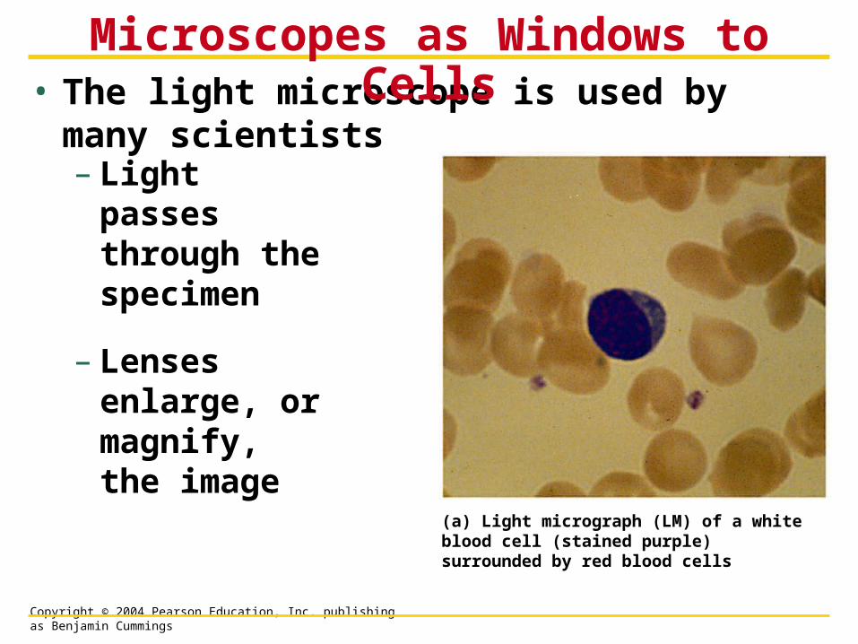
• The light microscope is used by many scientists
Microscopes as Windows to Cells
– Light passes through the specimen
– Lenses enlarge, or magnify, the image
(a) Light micrograph (LM) of a white blood cell (stained purple) surrounded by red blood cells

Copyright © 2004 Pearson Education, Inc. publishing as Benjamin Cummings
• Magnification
– An increase in the specimen’s apparent size
• Resolving power
– The ability of an optical instrument to show two objects as separate

Copyright © 2004 Pearson Education, Inc. publishing as Benjamin Cummings
• Cells were first discovered in 1665 by Robert Hooke
• The accumulation of scientific evidence led to the cell theory
– All living things are composed of cells
– All cells form from previously existing cells

Copyright © 2004 Pearson Education, Inc. publishing as Benjamin Cummings
• The electron microscope (EM) uses a beam of electrons
– It has a higher resolving power than the light microscope

Copyright © 2004 Pearson Education, Inc. publishing as Benjamin Cummings
• The electron microscope can magnify up to 100,000X
– Such power reveals the diverse parts within a cell
Human height
Length of somenerve andmuscle cells
Frogeggs
Chickenegg
Plant andanimalcells
NucleusMost bacteriaMitochondrion
Smallest bacteria
Viruses
Ribosomes
Proteins
Lipids
Smallmolecules
AtomsU
na
ide
d e
ye
Lig
ht
mic
ros
co
pe
Ele
ctr
on
mic
ros
co
pe

Copyright © 2004 Pearson Education, Inc. publishing as Benjamin Cummings
SEM• The scanning electron
microscope (SEM) is used to study the detailed architecture of the surface of a cell
(b) Scanning electron micrograph (SEM) of cilia (above)And a white blood cell

Copyright © 2004 Pearson Education, Inc. publishing as Benjamin Cummings
TEM
• The transmission electron microscope (TEM) is useful for exploring the internal structure of a cell
(c) Transmission electron micrograph (TEM) of a white blood cell & cilial

Copyright © 2004 Pearson Education, Inc. publishing as Benjamin Cummings
• The countless cells on earth fall into two categories
The Two Major Categories of Cells
– Prokaryotic cells
– Eukaryotic cells

Copyright © 2004 Pearson Education, Inc. publishing as Benjamin Cummings
Cell Size is Limited• Surface to Volume Ratio limits upper size
• Larger cells have less surface area relative to volume

Copyright © 2004 Pearson Education, Inc. publishing as Benjamin Cummings
• Prokaryotic and eukaryotic cells differ in several respects
Prokaryotic cell Nucleoid region
Eukaryotic cell Nucleus Organelles

Copyright © 2004 Pearson Education, Inc. publishing as Benjamin Cummings
• Prokaryotic cells
– Are smaller than eukaryotic cells
– Lack internal structures surrounded by membranes
– Lack a nucleus

Copyright © 2004 Pearson Education, Inc. publishing as Benjamin Cummings

Copyright © 2004 Pearson Education, Inc. publishing as Benjamin Cummings
Prokaryoticflagella
Nucleoid region (DNA)
RibosomesPlasmamembrane
Cell wall
Capsule
Pili

Copyright © 2004 Pearson Education, Inc. publishing as Benjamin Cummings
A Generic Animal Cell

Copyright © 2004 Pearson Education, Inc. publishing as Benjamin Cummings
• An idealized plant cell

Copyright © 2004 Pearson Education, Inc. publishing as Benjamin Cummings
• Surrounded by a double membrane called the nuclear envelope
Structure and Function of the Nucleus
– It contains
– chromatin -a DNA-protein structure
– a nucleolus - which produces ribosomal parts
– Nucleoplasm
• Occurs only in eukaryotic cells

Copyright © 2004 Pearson Education, Inc. publishing as Benjamin Cummings

Copyright © 2004 Pearson Education, Inc. publishing as Benjamin Cummings
Nuclear Pore Complex
Allows movement of material into and out of nucleus

Copyright © 2004 Pearson Education, Inc. publishing as Benjamin Cummings
• Ribosomes build all the cell’s proteins
– Are not membrane bound
Ribosomes

Copyright © 2004 Pearson Education, Inc. publishing as Benjamin Cummings
• DNA controls the cell by transferring its coded information into RNA
How DNA Controls the Cell
– The information in the RNA is used to make proteins
Synthesis ofmRNA in thenucleus
1
2 Movement ofmRNA intocytoplasm vianuclear pore
3 Synthesis ofprotein in thecytoplasm
DNA
mRNA
Nucleus
Cytoplasm
mRNA
Ribosome
Protein

Copyright © 2004 Pearson Education, Inc. publishing as Benjamin Cummings
• Many of the membranous organelles in the cell belong to the endomembrane system
– Endoplasmic reticulum - rough and smooth
– Golgi Apparatus
– Lysosomes
– Vacuoles
THE ENDOMEMBRANE SYSTEM: MANUFACTURING AND DISTRIBUTING
CELLULAR PRODUCTS

Copyright © 2004 Pearson Education, Inc. publishing as Benjamin Cummings
• The endoplasmic reticulum (ER)
The Endoplasmic Reticulum
– Greek for ‘network within a cell’
– Produces an enormous variety of molecules
– Is composed of smooth and rough ER
Nuclearenvelope
Ribosomes
Rough ERSmooth ER

Copyright © 2004 Pearson Education, Inc. publishing as Benjamin Cummings
• The “roughness” of the rough ER is due to ribosomes that stud the outside of the ER membrane
Rough ER

Copyright © 2004 Pearson Education, Inc. publishing as Benjamin Cummings
• The functions of the rough ER include
– Producing proteins
– Producing new membrane

Copyright © 2004 Pearson Education, Inc. publishing as Benjamin Cummings
• After the rough ER synthesizes a molecule it packages the molecule into transport vesicles
1

Copyright © 2004 Pearson Education, Inc. publishing as Benjamin Cummings
Smooth ER• The smooth ER lacks
the surface ribosomes of ER
• Produces lipids, including steroids and sex hormones
• Regulates sugar
• Detoxifies drugs
• Stores calcium

Copyright © 2004 Pearson Education, Inc. publishing as Benjamin Cummings
• The Golgi apparatus
The Golgi Apparatus
– Works in partnership with the ER
– Refines, stores, and distributes the products of cells
Transportvesiclefrom ER
“Receiving” side ofGolgi apparatus
Golgi apparatus
New vesicle forming
Transport vesiclefrom the Golgi
“Shipping” side ofGolgi apparatus
Plasma membrane

Copyright © 2004 Pearson Education, Inc. publishing as Benjamin Cummings
• A lysosome is a membrane-enclosed sac
Lysosomes
– Greek for ‘breakdown body’
– It contains digestive enzymes
• Isolated by membrane
– The enzymes break down
• Macromolecules
• Old organelles

Copyright © 2004 Pearson Education, Inc. publishing as Benjamin Cummings
• Lysosomes have several types of digestive functions
• They exit the Golgi apparatus

Copyright © 2004 Pearson Education, Inc. publishing as Benjamin Cummings
– They fuse with food vacuoles to digest the food

Copyright © 2004 Pearson Education, Inc. publishing as Benjamin Cummings
– They fuse with old organelles to recycle parts
– Digest bacteria in white blood cells

Copyright © 2004 Pearson Education, Inc. publishing as Benjamin Cummings
Lysosomal diseasesGenetic disorders
Recipe is messed up
Enzyme doesn’t work
what should get broken down doesn’t
Tay-Sachs
lipids aren’t broken down
build up occurs
death by age 5
Pompe’s disease
glycogen builds up

Copyright © 2004 Pearson Education, Inc. publishing as Benjamin Cummings
• Vacuoles are membranous sacs
Vacuoles
– Two types are the contractile vacuoles of protists and the central vacuoles of plants
Figure 4.15
Contractilevacuoles
Centralvacuole
(a) Contractile vacuoles in a protist (b) Central vacuole in a plant cell

Copyright © 2004 Pearson Education, Inc. publishing as Benjamin Cummings
• A review of the endomembrane system

Copyright © 2004 Pearson Education, Inc. publishing as Benjamin Cummings
• Cells require a constant energy supply to do all the work of life
CHLOROPLASTS AND MITOCHONDRIA: ENERGY CONVERSION

Copyright © 2004 Pearson Education, Inc. publishing as Benjamin Cummings
• Chloroplasts are the sites of photosynthesis, the conversion of light energy to chemical energy
CHLOROPLASTS
Figure 4.17
Inner and outermembranes ofenvelope
Space betweenmembranes
Stroma (fluid inchloroplast)
Granum

Copyright © 2004 Pearson Education, Inc. publishing as Benjamin Cummings
Chloroplasts• Double membrane
• Grana
– Stacks of thylakoids
• Hollow disks
• Sunlight energy is coverted to chemical energy
• Stroma- fluid filling chloroplast
• Contains some DNA

Copyright © 2004 Pearson Education, Inc. publishing as Benjamin Cummings
• Mitochondria are the sites of cellular respiration, which involves the production of ATP from food molecules
Mitochondria
Figure 4.18
Outermembrane
Innermembrane
Cristae
Matrix
Space betweenmembranes

Copyright © 2004 Pearson Education, Inc. publishing as Benjamin Cummings
Mitochondria• Double membrane
– Big bag stuffed into smaller bag
– Folds of inner bag called cristae
• Matrix -space inside inner bag
• Contains some DNA

Copyright © 2004 Pearson Education, Inc. publishing as Benjamin Cummings
• The cytoskeleton is an infrastructure of the cell consisting of a network of fibers
– Microfilaments - small threads
– Intermediate filaments - ropelike
– Microtubules - small tubes
THE CYTOSKELETON:CELL SHAPE AND MOVEMENT
Copyright © 2004 Pearson Education, Inc. publishing as Benjamin Cummings
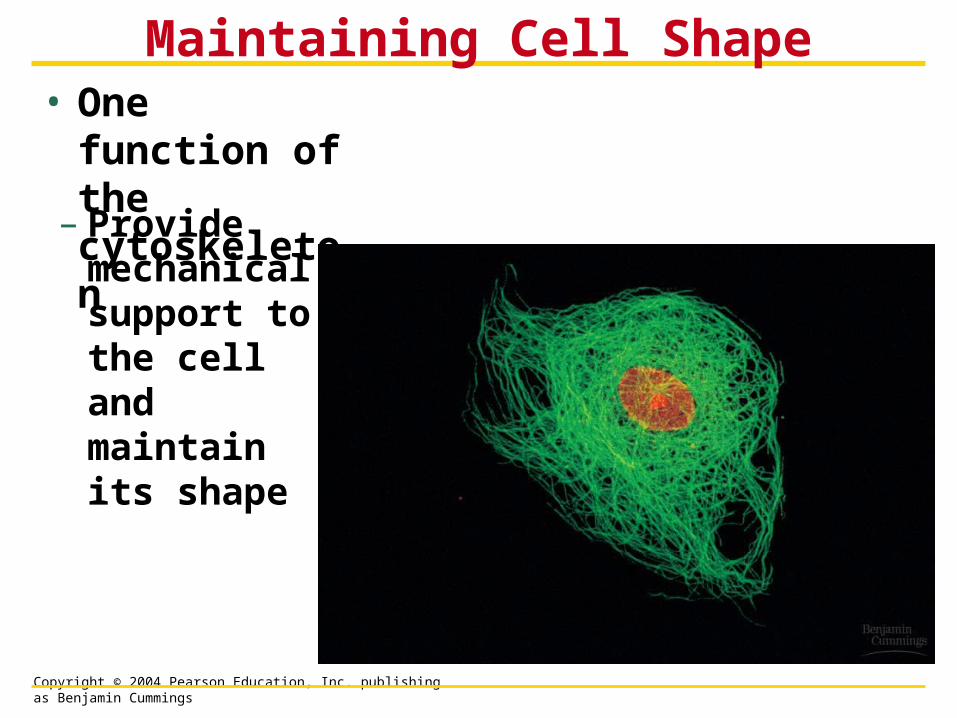
• One function of the cytoskeleton
Maintaining Cell Shape
– Provide mechanical support to the cell and maintain its shape

Copyright © 2004 Pearson Education, Inc. publishing as Benjamin Cummings
Figure4.9x

Copyright © 2004 Pearson Education, Inc. publishing as Benjamin Cummings
• The cytoskeleton can change the shape of a cell
– This allows cells like amoebae to move

Copyright © 2004 Pearson Education, Inc. publishing as Benjamin Cummings
• Cilia and flagella are motile appendages
Cilia and Flagella

Copyright © 2004 Pearson Education, Inc. publishing as Benjamin Cummings
• Flagella propel the cell in a whiplike motion
• Cilia move in a coordinated back-and-forth motion
Figure 4.20A, B

Copyright © 2004 Pearson Education, Inc. publishing as Benjamin Cummings
• Some cilia or flagella extend from nonmoving cells
– The human windpipe is lined with cilia
– Smoking damages the cilia

Copyright © 2004 Pearson Education, Inc. publishing as Benjamin Cummings
Cilia and Flagella
• Same structure and function
• 9 + 2 arrangement of microtubules
• Wrapped in plasma membrane

Copyright © 2004 Pearson Education, Inc. publishing as Benjamin Cummings
Mechanism of Movement• Dynein arms use ATP for energy to ‘walk’ up adjoining
microtubule, causing them to bend

Copyright © 2004 Pearson Education, Inc. publishing as Benjamin Cummings
• Most cells secrete materials that are external to the plasma membrane
• This extra cellular matrix
– Regulates
– Protects
– Supports
CELL SURFACES:PROTECTION, SUPPORT, AND CELL-CELL
INTERACTIONS

Copyright © 2004 Pearson Education, Inc. publishing as Benjamin Cummings
• Plant cells are encased by cell walls
Plant Cell Walls and Cell Junctions
Figure 4.21
– These provide support for the plant cells
Walls of two adjacentplant cells
Vacuole
Plasmodesmata(channels between cells)

Copyright © 2004 Pearson Education, Inc. publishing as Benjamin Cummings
• Animal cells lack cell walls
Animal Cell Surfaces and Cell Junctions
– They secrete a sticky covering called the extracellular matrix
– This layer helps hold cells together

Copyright © 2004 Pearson Education, Inc. publishing as Benjamin Cummings
• Animal cells connect by various types of junctions
– Tight junctions - leakproof
– Adhering junctions - hold cells together but allows movement between of materials between cells
– Communicating junctions - like plasmodesmata

Copyright © 2004 Pearson Education, Inc. publishing as Benjamin Cummings
Figure 4.22
Extracellular matrix
(a) Tight junctions
(b) Anchoring junctions
(c) Communicating junctions
Plasma membranesof adjacent cells
Extracellular matrix

Copyright © 2004 Pearson Education, Inc. publishing as Benjamin Cummings