chapter 3 the catalytic domain of leader peptidase inserts ... · protein translocation is an...
TRANSCRIPT

Chapter 3
The catalytic domain of leader peptidase inserts in a phosphatidylethanolamine dependent way in the outer
leaflet of the inner membrane of E. coli
based on publication (V)


membrane binding ofthe catalytic domain oJLep
Summary
Leader peptidase is an integral membrane protein of E. CON that catalyses the removal
of most signal peptides from translocated precursor proteins. Here, we show that when
the transmembrane anchors are removed in duo, the remaining catalytic domain can
bind to inner and outer membranes of E. coli. Furthermore, the purified catalytic
domain binds to inner membrane vesicles and vesicles composed of purified inner
membrane lipids with comparable efficiencies. It is shown that the interaction is
caused by penetration of a part of the catalytic domain between the lipids. Penetration
is mediated by phosphatidylethanolamine, the most abundant lipid in E. coli, and does
not seem to depend on electrostatic interactions. A mildly hydrophobic segment
around the catalytically important residue serine 90 is required for the interaction with
membranes.
Introduction
Protein translocation is an essential process in both prokaryotic and eukaryotic cells.
Approximately 20% of all proteins synthesized by the gram negative bacterium E. coli function
outside the cytosol (Cronan et al., 1987) and therefore must at least pass the cytosolic membrane.
Passage of the cytoplasmic membrane is most often catalysed by the combined action of a
proteinaceous secretion (sec) machinery (Arkowitz and Basiliana, 1994), anionic phospholipids
(De Vrije et al., 1988) and the membrane potential (Bakker and Randall, 1984).
For recognition by the export machinery proteins are usually synthesized as precursors
with amino terminal extensions called leader- or signal- sequences. Signal sequences contain a
hydrophobic core region which is preceeded by an amino terminal positively charged domain (von
Heijne, 1985). After translocation signal sequences are removed by the action of leader (signal)
peptidases which cleave 5 to 7 residues downstream of the hydrophobic core of the signal
sequence (von Heijne, 1985). Most signal sequences are removed by Lep (also called leader
peptidase or signal peptidase I) which is encoded by the k p B gene (Date and Wickner, 1981). It
recognizes a turn inducing residue (glycine or proline) at position -6 and small residues, preferably
alanine, at positions -3 and -1 with respect to the cleavage site (von Heijne, 1983; Fikes et al.,
1990).
Lep is comprised of an amino terminal part containing two transmembrane helices (HI
and H2) separated by a highly positively charged cytosolic loop (PI) and a carboxy terminal large

periplasmic domain (P2) (Fig. 1). Site directed mutagenesis studies suggested that the proteolytic
mechanism involves a serine at position 90 and a lysine at 145 and that all residues required for
cleavage are located in P2 (Black, 1993; Tschantz et al., 1333).
cc\ @ periplasm
P1 cytoplasm
Figure 1. Orientation of wild type Leader peptidase in the membrane and indication of the position of the engineered cleavage cassette (CC) behind the second transmembrane helix. The amino acid sequence of the region around H2 of a construct bearing a cleavage cassette is depicted. H2 (from residue 68 to 76) is underlined and the cleavage cassette between residues 76 and 77 is depicted in italics.
The substrates for Lep are membrane bound and may still be in the vicinity of the
translocation machinery at the moment of processing. It is therefore conceivable that Lep finds its
substrate by having affinity for components of the sec machinery. In this study this idea was tested
using constructs corresponding to the periplasmic domain. The association of this domain to
membranes with or without the translocation machinery was compared. It is found that the
presence of the sec machinery is not required for efficient binding. Interestingly the periplasmic
domain of Lep is able to penetrate into pure lipid bilayers and shows a profound specificity for the
lipid phosphatidylethanolamine. The results are discussed in relation to the mode of action of Lep.

membrane binding of the catalytic domain of Lep
Results
The periplasmic domain of Lep associates with membranes after removal of the membrane
anchors ' The catalytic domain of Lep is localised in the periplasm (Fig.1). To study possible interactions of 1 this domain with membranes in vivo, a mutant was required in which the catalytic P2 domain
could be separated from the membrane spanning segments H I and H2. Downstream of H2 a I , recognition sequence for cleavage by Lep was engineered (Fig.1) and the resulting construct was I named H2-CC. This construct was efficiently expressed and cleaved in vivo probably by the native ' population of Lep molecules in the membrane (Nilsson and von Heijne, 1991). When spheroplasts
were prepared the cleaved form was accessible to proteases (Nilsson and von Heijne, 1991, results
not shown). Periplasmic fractions of pulse labelled cells expressing H2-CC were isolated by
osmotic shock and screened by immunoprecipitations. While most of the periplasmic marker protein P-lactamase was recovered in the soluble periplasmic fraction (75?6%, n=3), the cleaved
domain of Lep was found predominantly in the pellet (84+7%, n=3) (results not shown). The
catalytic domain is therefore probably associated with membranes. To investigate more precisely
the localisation of the periplasmic domain, membrane fractions were prepared of cells expressing
H2-CC. Inner and outer membranes were separated on a sucrose gradient and the different
fractions analysed by SDS PAGE. Antibodies against Lep were used to stain blots made from the
different fractions (Fig.2 insert).
top 5 10 15 20 25 bottom fraction number
Figure 2. The P2 domain of k p fractionates both with the inner and outer membrane. The amount of k p and the cleaved P2 domain was determined in fractions which were collected from a sucrose gradient on which E. colt' membranes were separated. The amount of full lenth k p (circles) and cleaved form (squares) were plotted against the fraction number. The inner and outer membranes were found around fractions 8 and 20 respectively. The insen shows the result of the corresponding western blot treated with k p antibody.

Chapter 3
Two bands reacted with the antibody, one of 36 kDa coinciding with endogenous Lep was as
expected found mainly in the inner membranes around fraction 8 and to a lesser extent in the
outer membrane (Fig. 2, circles) as reported before (Zwizinski et al., 1981), while a band
corresponding to the cleaved off P2 domain was also found in the outer membranes around
fraction 20 (Fig. 2 squares). It is therefore concluded that the periplasmic domain of Lep has
affinity for membranes.
Binding of the periplasmic domain of Lep to membranes does not require a specific
proteinaceom component
The ability of the periplasmic domain of Lep to bind to membranes was confirmed by vesicle
binding experiments. For this purpose we made use of a purified, enzymatically active construct (A2-75) lacking the 2 membrane spanning segments (Kuo et al., 1993). This construct is
expressed in the cytoplasm and can be purified in large amounts. By means of ultracentrifugation experiments the binding of A2-75 to right side out inner membrane vesicles and outer
membranes was determined. In the absence of membranes A2-75 was quantitatively recovered in
the supernatant after ultracentrifugation (Fig.3 upper panel, lanes 1-3). Right side out inner
membrane vesicles (Fig. 3, upper panel lanes 4-6) contain many different proteins as judged by
Coomassie Brilliant Blue staining of gels, while outer membranes (Fig. 3, lower panel lanes 1-3)
showed a characteristic pattern with only two dominant bands. Both types of vesicles were pelleted efficiently. When A2-75 was incubated with vesicles prior to centrifugation part of the
molecules sedimented with the vesicles (Fig.3 upper panel lanes 7-9 and Fig. 3 lower panel lanes 4- 6). So A2-75 is apparently capable of binding to both inner and outer membranes while the
native population of Lep is found mostly in the inner membrane of E. coli. This suggests that the
periplasmic domain does not require specific inner membrane components for binding. To
investigate the possibility that the P2 domain recognises the lipid component of membranes the binding of 5 p g A2-75 to inner membranes and to single walled lipid vesicles made from purified
inner membrane lipids were compared. The same amount of lipid phosphorous was used for both
types of vesicles. The amount of bound protein was determined by laser scanning densitometry. Right side out inner membrane vesicles bound 1.7 + 0.3 pg of A2-75 while the lipid vesicles
bound 1.6 2 0.3 pg, this strongly suggests that the membrane binding of A2-75 is mediated by
lipids.

membrane binding of the catalytic domain of Lep
IM .. - - + + + + + + A2-75 + + + + + +
t p s t p s t p s
1 2 3 4 5 6 OM + + + + + + a - 7 5 - - - + + +
t p s t p s
Figure 3. Purified 62-75 associates both with inner and outer membrane vesicles. Samples with (upper panel, lanes 1-3 and 7-9; lower panel, lanes 4-6) or without A2-75 (upper panel, lanes 4-6; lower panel, lanes 1-3) were incubated in the absence (upper panel, lanes 1-3) or presence of membranes before ultracentrifugation. After centrifugation samples were split in pellet (p) and supernatant (s) fractions and compared to the total (t) amount before centrifugation. The upper panel shows incubations with inner membranes and the lower panel with outer membranes. Gels were stained with Coomassie Brilliant Blue.
Since membrane binding of the periplasmic domain of Lep does not seem to be caused
by proteinaceous interactions we looked for a general feature such as hydrophobicity that enables
proteins to interact with membranes. Figure 4 shows a hydrophobicity plot of Lep. Besides the two
transmembrane segments H1 and HZ a third hydrophobic segment H3 stands out. This region contains also the catalytic important serine 90 residue. A mutant lacking H3 (A2-98, Fig.4) was
purified and the binding of the two purified constructs to lipid vesicles were compared. Different
amounts of the constructs were added to the lipid vesicles and subjected to ultracentrifugation.
The amount of protein associated with vesicles was quantified and plotted against the amount of
added protein (Fig. 5).

Chapter 3
window number
P2
/ \\\ Lep ---TGASVFPPVLAIVLIVRSFIY DFILVE---
Figure 4. Hydrophobicity, domain structure and partial sequences of Lep and two truncated forms of Lep. The hydrophobicity of Lep was determined according Kyte and Doolittle using a window of 11 amino acids. The domain structure shows the relative positions of the hydrophobic parts. In the amino acid sequence the second transmembrane segment 012) and H3 are indicated. The catalytic important senne 90 is depicted in white.
Amount of protein added (ng) Figure 5. comparison of lipid vesicle binding by A2-75 and 112-98. Binding of A2-75 (closed squares) and A2-98 (open circles) were determined as described in materials and methods.

membrane binding of the catalytic domain of Lep
The sigmoidal binding curve of A2-75 might indicate that cooperativity is involved in binding of
A2-75 to the vesicles. In any case it is clear that A2-75 binds much more strongly to the lipid
vesicles than A2-98 indicating that the interaction of the periplasmic domain of Lep could be
mediated via the H3 region.
/ Interaction of 42-75 with membranes is caused by penetration between the lipids I The association of A2-75 with lipid vesicles can be caused by binding to the surface as well as by i / insertion of a part of the protein between the lipids. To gain insight into the nature and specificity 1 of the interaction of the periplasmic domain of Lep with lipids we made use of monolayer
experiments. In these experiments lipids are spread on top of a buffer solution in a teflon trough.
The lipids orient themselves with the head groups towards the buffer solution and their apolar acyl
chains away from the aqueous phase. They thereby resemble one half of the bilayer. Proteins can
be added to the aqueous phase and when they are able to insert between the lipid molecules this
will increase the surface pressure which can be measured online by means of a microbalance.
Lipids isolated from purified inner membranes ofE. coli were spread to an initial surface pressure of 22 mN/m (Fig.6A). When A2-98 was injected under these layers a small increase in surface
pressure was observed which stabilised in 30-40 minutes. A2-75 gave rise to a much larger
pressure increase indicating a more efficient interaction with lipids. Since proteins that only
interact with the lipid head group without penetration between the lipids do not give rise to a
pressure increase (Demel et al., 1973), the results show actual insertion between the
phospholipids in the monolayer.
The limiting surface pressure is the initial surface pressure at which a protein is just able
to insert into a monolayer, and therefore an important characteristic of insertion. In biological
membranes the packing densities of the lipids were found to correspond to surface pressures
between 31 and 35 mN/m (Demel et al., 1975). To investigate whether the constructs are able to
penetrate into membranes with physiological lipid packing densities, monolayers with different initial surface pressures were made. From figure 6B it is clear that A2-75 can insert in monolayers
with much higher initial pressures than A2-98. A2-75 is able to insert into monolayers with
initial pressures as high as 35mNIm and therefore this construct is expected to be capable of
penetrating between the lipids in biological membranes.

B time (min)
initial surface pressure (rnN/m)
Figure 6. Interaction of A2-75 and A2-98 with monolayers. (A) insertion profile of A2-75 and A2-98 into monolayers of E. coli inner membrane lipids which were spread until 22mN/m initial pressure. After injection of protein the changes in surface pressure were followed. (B) Ability of A2-75 and A2-98 to penetrate into monolayers as Function of the initial pressure of the monolayer.
membrane binding of the catalytic domain of Lep
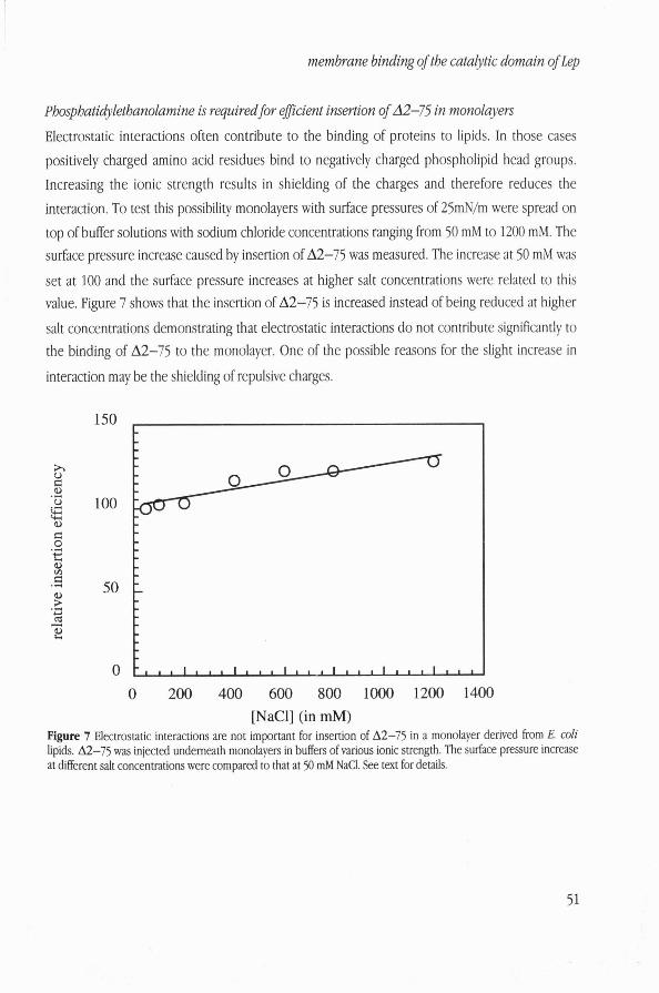
Phosphatidylethanolamine is required for efJicient insertion of A2-75 in monolayers
Electrostatic interactions often contribute to the binding of proteins to lipids. In those cases
positively charged amino acid residues bind to negatively charged phospholipid head groups.
Increasing the ionic strength results in shielding of the charges and therefore reduces the
interaction. To test this possibility monolayers with surface pressures of 25mN/m were spread on
top of buffer solutions with sodium chloride concentrations ranging from 50 mM to 1200 mM. The surface pressure increase caused by insertion of A2-75 was measured. The increase at 50 mM was
set at 100 and the surface pressure increases at higher salt concentrations were related to this value. Figure 7 shows that the insertion of A2-75 is increased instead of being reduced at higher
salt concentrations demonstrating that electrostatic interactions do not contribute significantly to the binding of A2-75 to the monolayer. One of the possible reasons for the slight increase in
interaction may be the shielding of repulsive charges.
150
e, e 0 .3
t: % fz .3 50 9 . 3 Y
cd - 2
0
1
- 1 1 1 1 1 1 1 1 1 1 1 1 ~ 1 ~ 1 1 ~ ~ 1 ~ ~ ~ 1 ~ ~ *
0 200 400 600 800 1000 1200 1400 [NaCl] (in rnM)
Figure 7 Electrostatic interactions are not important for insertion of A2-75 in a monolayer derived from E. coli lipids. A2-75 was injected underneath monolayers in buffers of various ionic strength. The surface pressure increase at different salt concentrations were compared to that at 50 mM NaCI. See text for details.

Chapter 3
Charge interactions are apparently not important for the insertion of the P2 domain into
monol~yers. To get more insight into the characteristics of interaction, insertion into monolayers
of pure lipids were compared.
initial surface pressure (mNIm)
18 20 22 24 26 28 30 32 initial surface pressure (rnNIm)
Figure 8. Specific interaction of A2-75 and A 2 4 6 with different lipid head group classes. (A) Surface pressure increase caused by insertion of A2-75 into monolayers of DOPE (triangles), DOPC (squares) and DOPG (circles) as function of the initial surface pressure. (B) Surface pressure increase caused by insertion of A2-98 into monolayers of DOPE (triangles), DOPC (squares) and DOPG (circles) as function of the initial surface pressure.

membrane binding of the catalytic domain of Lep
Phosphatidyl ethanolamine (PE) has a small zwitterionic headgroup and it accounts for
approximately 75 % of the phospholipids in the E.coli inner membrane (Raetz, 1978). The second
most abundant lipid is the negatively charged phosphatidyl glycerol (PG) which accounts for about 20% (Raetz, 1978). Insertion of A2-75 in dioleolyl phosphatidyl ethanolamine (DOPE) and
dioleolyl phosphatidyl glycerol (DOPG) monolayers were measured as function of the initial pressure. From figure 8A it is clear that A2-75 inserts best in DOPE monolayers. To see whether
this is only due to the zwitterionic character of DOPE also dioleolyphosphatidyl choline (DOPC)
was tested. The phosphatidyl choline head group is also zwitterionic but much bulkier than the
phosphatidyl ethanolamine head group. Insertion in DOPC is considerably less efficient and almost
comparable to insertion in DOPG. Therefore also other characteristics of the head group seems to
be important. Next the interaction of A2-98 with the same range of lipids was tested and while
interaction with all lipids was less than in the case of A2-75, it was striking that especially the
interaction with DOPE was vely much reduced. These results together suggest that the membrane
affinity of the periplasmic domain of Lep is mediated by the hydrophobic H3 segment and lipids
with the zwitterionic phosphatidyl ethanolamine head group.
Discussion
This study reports on the interaction of the catalytic domain of leader peptidase, one of the key
enzymes in preprotein translocation, with membranes. Evidence for membrane binding activity
was obtained using two approaches. Fractionation studies with the construct H2-CC from which
the membrane spanning regions are separated from the periplasmic domain in uiuo showed that
the periplasmic domain is associated both with inner and outer membranes indicating no affinity
for specific components of the inner membrane. This finding was confirmed by studies using purified A2-75 resembling the periplasmic domain of Lep. A2-75 binds to lipid vesicles and right
side out inner membrane vesicles equally well, again suggesting that the catalytic domain does not
bind to proteinaceous components of the preprotein translocation (sec) machinery but has a high
affinity for the membrane lipids.
The periplasmic domain not only has catalytic (Kuo et a1. ,1993; Tschantz et al., 1995) but
also membrane binding activity. It therefore seems that the function of H1 and H2 is limited to
assembly, in addition by firmly anchoring the protein to the cytoplasmic membrane, the P2
domain is prevented to move to the outer membrane. The lack of sequence conservation in HI, PI
Chapter 3
and the amino terminal half of the H2 segment between leader peptidases from different sources
(Dalbey and von Heijne, 1992) is consistent with this limited role in function. Alternatively the
membrane anchors could in principle also be involved in recognition of components of the sec
machinery although indications for such a recognition were not reported. Furthermore Lep does
not copurify with any of the known sec proteins and Lep is able to process precursors which do
not make use of a functional sec machinery. The binding to lipids was also demonstrated by efficient insertion of A2-75 into
monolayers derived from inner membrane lipids. Insertion does not seem to depend on
electrostatic interactions (Fig.7) Remarkably, while anionic phospholipids are important for the
insertion of a lot of proteins such as the signal sequence of prePhoE (Batenburg et al., 1988), and the translocation ATPase SecA (Breukink et al., 1992), A2-75 displayed best penetration in lipid
films made from the zwitterionic lipid phosphatidylethanolamine. The limiting surface pressure for
insertion into this type of lipids was very close to that of complete extracts of the E.coli inner
membrane which contain about 75% PE. This not only corroborates the relative unimportance of
electrostatic interactions, it also points to the important role PE can play because of its specific
structural properties. Insertion into monolayers of DOPC which is also zwitterionic was very much
reduced. This difference in insertion of proteins into PE compared to PC was also found in case of
the precursor to ferredoxin (van't Hof et al., 1993), SecA (Breukink el al., 1992) and prePhoE (van
Raalte et al., 1996). One of the possible reasons for efficient interactions with PE is the ability of PE
to form intermolecular hydrogen bridges. It is also very likely that the difference in insertion
efficiency between PC and PE is caused by the differences in size of the head groups. The PE head
group is smaller and could offer more space for proteins to insert. The size of the head group
relative to the diameter of the acyl chains is indeed so small that these lipids are unable to form
stable bilayers by themselves. This class of non-bilayer forming lipids is of great importance in
biological membranes and their relative amounts are strictly regulated (Rietveld et al., 1993).
Apparently their presence in membranes can give rise to specific packing conditions which are
essential for membrane functions as, for example, protein translocation (Rietveld et al., 1995).
Phosphatidyl ethanolamine was also shown to be important for specific interactions with
other proteins. It promotes the folding of a periplasmic loop of lactose permease (Bogdanov et al.,
1996), regulates the activity of glycero phosphate acyl transferase (Snider and Kennedy, 1977) and
was recently found in the crystals of cytochrome C oxidase (Tsukihara et al., 1996).

membrane binding of the catalytic domain of Lep
In the present study it is shown that the catalytic domain of Lep binds to lipids while it
was demonstrated before that activity of the full length Lep was enhanced by addition of
phospholipids to an in vitro assay (van Klompenburg et al., 1995). In that system all phospholipids
tested stimulated processing but anionic phospholipids were slightly more effective than
zwitterionic. This could indicate that anionic phospholipids act on the conformation of either the
signal peptide (Chupin et al., 1995) or the membrane anchors of Lep. It is also possible that
anionic phospholipids indeed have a special role in the catalysis of Lep. In this case it can be
envisaged that PE mediated insertion orients the catalytic site in such a way that anionic
phospholipids can get close to the active site and fulfill their function.
Considering the nature and specificity of the interaction between the periplasmic domain
of Lep and lipids it is most likely that membrane association is caused by interaction of a
hydrophobic segment within the periplasmic domain with the lipids. Deletion of H3 (residue 83 to
98) which is the most hydrophobic part within the periplasmic domain indeed diminished both
insertion into the lipid monolayers and association with the lipid vesicles.
The insertion into the lipid phase as described in this study has important implications for
the mode of action of Lep. There is compelling evidence that during translocation the amino
terminus of the signal peptide stays at the cytosolic side of the membrane (Kuhn, 1987; Shaw et
al., 1988), while the hydrophobic core is probably too short to span the hydrophobic part of the
bilayer. This means that in order to reach its substrate, the catalytic site of Lep must be at least very
close to the membrane. Insertion of the periplasmic domain into the lipid phase and possible
involvement of H3, which carries the catalytic serine 90 residue, implies that the active site of Lep
may actually be (partially) buried in the membrane.
Interestingly it was observed that lengthening the hydrophobic core of signal sequences
resulted in reduced processing of preproteins without much effect on translocation (Chou and
Kendall, 1990). In light of the foregoing this can be interpreted as moving the cleavage site of the
preprotein out of the membrane and therefore away from the catalytic site of leader peptidase.
Materials and methods Isolation andpurification ofproteins and lipids.
Two truncated forms of Lep were isolated essentially as described before(Kuo et al., 1993;
Tschantz et a l , 1995) but detergent was left out. The proteins were stored in 20 mM TrisMCI, pH
7.4. Total lipid extracts from the inner membrane were prepared by extraction (Bligh and Dyer,

Chapter 3
1959) and further purified by column chromatography on a sillica column (sillicagel 30-60 p m ,
Baker B.V.). After equilibration in chloroform lipids were eluted in a choloroform : methanol 1:l
mixture and converted to their sodium salts (Smaal et al., 1985)
Isolation of the periplasmicJi-action.
An overnight culture of MC1061 bearing a plasmid encoding H2-CC was diluted into fresh M9
medium supplemented with ampicillin (50 pg/ml), 0.2% fructose, thiamin and all amino acids
except for methionine. In early-exponential phase expression was induced with 0.2% arabinose
and cells were labelled with 50 pCi 35s methionine. After 2 minutes cells were harvested by
centrifugation in an Eppendorf centrifuge, washed and resuspended in 30mM Tris/HCI, pH7.5. An
equal volume of the same buffer with 40% sucrose and EDTA (final concentration: 0.1 mM) was
added and the mixture was incubated at room temperature for 10 minutes. Cells were pelleted and
the pellet was quickly resuspended in ice cold 0.5 mM MgC12 and incubated on ice for 10 minutes.
After centrifugation for 8 minutes the supernatant was separated from the pellet. Both fractions were immunoprecipitated with Lep and P-lactamase antibodies and analysed by SDS-PAGE
followed by autoradiography.
Vesicle isolation and preparation.
Inverted and right side out inner membrane vesicles (IIMVs and RSOs) and outer membranes
(OMVs) of E. coli strain MC1061 were isolated according to published procedures. Identity of the
fractions were confirmed by lipopolysaccharide (LPS) staining and immunodetection of OmpA,
Lep and SecY. Large unilamellar vesicles (LUVETs) were prepared by means of extrusion through a
polycarbonate filter (Nucleopore: 0.2 p m pore size) of a rehydrated (10mM Tris/HCl, 50 mM NaCI,
pH 7.5) total lipid extract from the inner membrane of MC1061.
Vesicle binding may.
LUVETs corresponding to 200 nmol lipid were incubated with the indicated amounts of proteins in
300pl lOmM Tris/HCl, 50 mM NaCI, pH 7.5 for 1 hour at room temperature. Inner membrane
vesicles and OMVs were incubated in 300pl50 mM triethanolamine/HAc, 250 mM sucrose, 1 mM
DTT, pH7.5 to maintain the same environment as during isolation. Vesicles were pelleted by
centrifugation for 30 minutes at 236 * 103 g at room temperature in a TL 100.3 rotor using a TL 100
ultracentrifuge (Beckmann Instruments Inc. Palo Alto, California, U.S.A.). Pelleting efficiencies of

membrane binding of the catalytic domain of Lep
the vesicles were calculated after phosphorus determination (Rouser et al., 1970) on supernatant
and pellet. The amount of bound protein was determined after SDS-PAGE and Coomassie Brilliant
Blue staining. The intensities of the bands were quantified by densitometry (Personal
Densitometer, Molecular Dynamics, Sunnyvale, CA, U.S.A.) and compared to calibration curves of
the same proteins which were run on the same gels. The amount of bound protein was corrected
for the pelleting efficiencies which were always above 80%.
Monolayer experiments.
The Wilhelmy plate method was used to measure protein induced changes in surface pressure of a
monomolecular layer of phospholipids at constant surface area. Surface pressures were measured
at 26 +- 1% using a Cahn 2000 microbalance while continuously stirring the subphase with a
magnetic bar. Unless stated otherwise, a subphase of 5 ml lOmM Tris/HCI, 50 mM NaCI, pH 7.5 was
placed in a teflon trough. The monomolecular lipid layers were spread from a
chloroform/methanol 3:l (by vol.) solution at the airbuffer interphase to give initial surface
pressures between 18 and 35mN/m. Lower initial surface pressures were not used since both
proteins gave rise to surface pressures of 18 mN/m in the absence of a lipid monolayer. In all
experiments saturating amounts of protein (for both proteins 4uglml) were added in the subphase
from a hole in the edge of the trough. The surface pressure was measured until a constant level
was reached.
