chapter 3 role of snares in membrane fusion files/article...3 role of snares in membrane fusion 15...
TRANSCRIPT

Chapter 3Role of SNAREs in Membrane Fusion
Bhanu P. Jena
Abstract Fusion between opposing cellular membranes is essential for numerous cellular activitiessuch as protein maturation, neurotransmission, hormone secretion, and enzyme release. The universalmolecular mechanism of membrane fusion involves Ca2+, and the assembly of a specialized set ofproteins present in the opposing membrane bilayers. For example in cell secretion, target membraneproteins at the cell plasma membrane SNAP-25 and syntaxin termed t-SNAREs, and secretory vesicle-associated protein VAMP or v-SNARE, are part of the conserved protein complex involved in fusion ofopposing membranes. In the presence of Ca2+, t-SNAREs and v-SNARE in opposing bilayers interactand self-assemble in a ring conformation, to form conducting channels. Such self-assembly of t-/v-SNARE ring occurs only when the respective SNAREs are in association with membrane. The size ofthe SNARE ring complex is dependent on the curvature of the opposing bilayers. Electron density mapand 3-D topography of the SNARE ring complex, suggests the formation of a leak-proof channel mea-suring 25 Å in ring thickness, and 42 Å in height. The mechanism of membrane-directed SNARE ringcomplex assembly, and the mathematical prediction of SNARE ring size, has been determined. X-raydiffraction measurements and simulation studies have further advanced that membrane-associated t-SNAREs and v-SNARE overcome repulsive forces to bring the opposing membranes close to within adistance of approximately 2.8 Å. Calcium is then able to bridge the closely apposed bilayers, leading tothe release of water from hydrated Ca2+ ions as well as the loosely coordinated water at phospholipidhead groups, leading to membrane destabilization and fusion.
3.1 Introduction
Membrane fusion is essential for numerous cellular activities, including hormone secretion, enzymerelease, and neurotransmission. In live cells, membrane fusion is mediated via a specialized set ofproteins present in opposing bilayers. In the past 2 decades, much progress has been made in ourunderstanding of membrane fusion in cells, beginning with the discovery of an N-ethylmaleimide-sensitive factor (NSF) [1] and SNARE proteins [2–4], and the determination of their participation inmembrane fusion [5–11]. VAMP and syntaxin are both integral membrane proteins, with the solu-ble SNAP-25 associating with syntaxin. Therefore the understanding of SNARE-induced membranefusion requires determining the atomic arrangement and interactions between membrane-associatedv- and t-SNARE proteins. Ideally, the atomic coordinates of membrane-associated SNARE complexusing x-ray crystallography would help to elucidate the chemistry of SNARE-induced membrane
B.P. Jena (B)Department of Physiology, Wayne State University School of Medicine, Detroit, MI 48201, USAe-mail: [email protected]
13T. Dittmar, K.S. Zänker (eds.), Cell Fusion in Health and Disease, Advances in Experimental Medicineand Biology 713, DOI 10.1007/978-94-007-0763-4_3, C© Springer Science+Business Media B.V. 2011

14 B.P. Jena
fusion in cells. So far such structural details at the atomic level of membrane-associated t-/v-SNARE complex has not been possible, primarily due to solubility problems of membrane-associatedSNAREs, compounded with the fact that v-SNARE and t-SNAREs need to reside in opposing mem-branes when they meet, to assemble in a physiologically relevant SNARE complex. The remainingoption, the use of nuclear magnetic resonance spectroscopy (NMR) has also been of little help, sincethe size of t-/v-SNARE ring complex is beyond the optimal limit for NMR studies. Regardless, high-resolution AFM force spectroscopy, and EM electron density map and 3-D topography of the SNAREring complex has enabled an understanding of the structure and assembly, and also the disassemblyof membrane-associated t-/v-SNARE complexes in physiological buffer solution [5–10, 12].
The structure and arrangement of SNAREs associated with lipid bilayers were first determinedusing AFM [5], almost a decade ago. Electrophysiological measurements of membrane conduc-tance and capacitance, enabled the determination of fusion of v-SNARE-reconstituted liposomeswith t-SNARE-reconstituted membrane. Results from these studies demonstrated that t-SNAREs andv-SNARE when present in opposing membrane interact and assemble in a circular array, and in pres-ence of calcium, form conducting channels [5]. The interaction of t-/v-SNARE proteins to form suchconducting channels is strictly dependent on the presence of t-SNAREs and v-SNARE in oppos-ing membranes. Simple addition of purified recombinant v-SNARE to a t-SNARE-reconstituted lipidmembrane, fails to form the SNARE ring complex, and is without influence on the electrical propertiesof the membrane [5]. However when v-SNARE vesicles are added to t-SNARE reconstituted mem-brane, SNAREs assemble in a ring conformation, and in the presence of calcium, establish continuitybetween the opposing membrane. The establishment of continuity between the opposing t-SNAREand v-SNARE reconstituted bilayers, is reflected in the increase in membrane capacitance and con-ductance. These results confirm that t- and v-SNAREs are required to reside in opposing membrane,similar to their presence in cells, to allow appropriate t-/v-SNARE interactions leading to membranefusion [5, 7]. Studies using SNARE-reconstituted liposomes and bilayers [9] further demonstrate alow fusion rate (τ=16 min) between t- and v-SNARE-reconstituted liposomes in the absence of Ca2+.Exposure of t-/v-SNARE liposomes to Ca2+ drives vesicle fusion on a near physiological relevanttime-scale (τ ∼10 s), demonstrating Ca2+ and SNAREs in combination to be the universal fusionmachinery in cells [9]. Native and synthetic vesicles exhibit a significant negative surface chargeprimarily due to the polar phosphate head groups, generating a repulsive force that prevent the aggre-gation and fusion of opposing vesicles. In cells, SNAREs provide direction and specificity, bringopposing bilayers closer to within a distance of 2–3 Å [9], enabling Ca2+ bridging and membranefusion. The bound Ca2+ then leads to the expulsion of water between the bilayers at the bridging site,leading to lipid mixing and membrane fusion. Hence SNAREs, besides bringing opposing bilayerscloser, dictate the site and size of the fusion area during cell secretion. The size of the t-/v-SNAREcomplex is dictated by the curvature of the opposing membranes [7], hence smaller the vesicle, thesmaller the channel formed.
A unique set of chemical and physical properties of the Ca2+ ion make it ideal for participating inthe membrane fusion reaction. Calcium ion exists in its hydrated state within cells. The properties ofhydrated calcium have been extensively studied using x-ray diffraction, neutron scattering, in com-bination with molecular dynamics simulations [13–16]. The molecular dynamic simulations includethree-body corrections compared with ab initio quantum mechanics/molecular mechanics molecu-lar dynamics simulations. First principle molecular dynamics has also been used to investigate thestructural, vibrational, and energetic properties of [Ca(H2O)n]2+ clusters, and the hydration shell ofthe calcium ion [13]. These studies demonstrate that hydrated calcium [Ca(H2O)n]2+ has more thanone shell around the Ca2+, with the first hydration shell having six water molecules in an octahe-dral arrangement [13]. In studies using light scattering and X-ray diffraction of SNARE-reconstitutedliposomes, it has been demonstrated that fusion proceeds only when Ca2+ ions are available betweenthe t- and v-SNARE-apposed proteoliposomes [8, 9]. Mixing of t- and v-SNARE proteoliposomes inthe absence of Ca2+ leads to a diffuse and asymmetric diffractogram in X-ray diffraction studies, atypical characteristic of short range ordering in a liquid system [15]. In contrast, when t-SNARE and

3 Role of SNAREs in Membrane Fusion 15
v-SNARE proteoliposomes in the presence of Ca2+ are mixed, it leads to a more structured diffrac-togram, with approximately a 12% increase in X-ray scattering intensity, suggesting an increase in thenumber of contacts between opposing bilayers, established presumably through calcium-phosphatebridges, as previously suggested [8, 9, 16]. The ordering effect of Ca2+ on inter-bilayer contactsobserved in X-ray studies [9] is in good agreement with light, AFM, and spectroscopic studies, sug-gesting close apposition of PO-lipid head groups in the presence of Ca2+, followed by formation ofCa2+-PO bridges between the adjacent bilayers [8, 9, 17]. X-ray diffraction studies show that theeffect of Ca2+ on bilayers orientation and inter-bilayer contacts is most prominent in the area of 3 Å,with additional appearance of a new peak at position 2.8 Å, both of which are within the ionic radiusof Ca2+ [9]. These studies further suggest that the ionic radius of Ca2+ may make it an ideal playerin the membrane fusion reaction. Hydrated calcium [Ca(H2O)n]2+ however, with a hydration shellhaving six water molecules and measuring ∼6 Å would be excluded from the t-/v-SNARE apposedinter-bilayer space, hence calcium has to be present in the buffer solution when t-SNARE vesicles andv-SNARE vesicles meet. Indeed, studies demonstrate that if t- and v-SNARE vesicles are allowed tomix in a calcium-free buffer, there is no fusion following post addition of calcium [8]. How does cal-cium work? Calcium bridging of apposing bilayers may lead to the release of water from the hydratedCa2+ ion, leading to bilayer destabilization and membrane fusion. Additionally, the binding of cal-cium to the phosphate head groups of the apposing bilayers may also displace the loosely coordinatedwater at the PO-lipid head groups, resulting in further dehydration, leading to destabilization of thelipid bilayer and membrane fusion. Recent studies in the laboratory [18], using molecular dynamicssimulations in the isobaric-isothermal ensemble to determine whether Ca2+ was capable of bridgingopposing phospholipid head groups in the early stages of the membrane fusion process, demonstrateindeed this to be the case. Furthermore, the distance between the oxygen atoms of the opposing PO-lipid head groups bridged by calcium was in agreement with the 2.8 Å distance previously determinedusing X-ray diffraction measurements. The hypothesis that there is loss of coordinated water bothfrom the hydrated calcium ion and oxygen of the phospholipid head groups in opposing bilayers,following calcium bridging, is further demonstrated from the study.
In presence of ATP, the highly stable, membrane-directed, and self-assembled t-/v-SNARE com-plex, can be disassembled by a soluble ATPase, the N-ethylmaleimide-sensitive factor (NSF). Carefulexamination of the partially disassembled t-/v-SNARE bundles within the complex using AFM,demonstrates a left-handed super coiling of SNAREs. These results demonstrate that t-/v-SNAREdisassembly requires the right-handed uncoiling of each SNARE bundle within the ring complex,demonstrating NSF to behave as a right-handed molecular motor [6]. Furthermore, recent studies inthe laboratory [19] using circular dichroism (CD) spectroscopy, we report for the first time that botht-SNAREs and v-SNARE and their complexes in buffered suspension, exhibit defined peaks at CDsignals of 208 and 222 nm wavelengths, consistent with a higher degree of helical secondary struc-ture. Surprisingly, when incorporated in lipid membrane, both SNAREs and their complexes exhibitreduced folding. NSF, in presence of ATP, disassembles the SNARE complex as reflected from theCD signals demonstrating elimination of a-helices within the structure. These results further demon-strate that NSF-ATP is sufficient for the disassembly of the t-/v-SNARE complex. These studies haveprovided a molecular understanding of SNARE-induced membrane fusion in cells. Findings from thestudies outlined above are described in this chapter.
3.2 Materials and Methods
3.2.1 Preparation of Lipid Bilayer
Lipid bilayers were prepared using brain phosphatidylethanolamine (PE) and phosphatidylcholine(PC), and dioleoylphosphatidylcholine (DOPC), and dioleylphosphatidylserine (DOPS), obtained

16 B.P. Jena
from Avanti Lipids, Alabaster, AL. A suspension of PE:PC in a ratio of 7:3, and at a concentration of10 mg/ml was prepared. 100 μl of the lipid suspension was dried under nitrogen gas and resuspendedin 50 μl of decane. To prepare membranes reconstituted with VAMP, 625 ng/ml VAMP-2 proteinstock was added to the lipid suspension and brushed onto a 200 μm hole in the bilayer preparationcup until a stable bilayer with a capacitance between 100 and 250 pF was formed.
3.2.2 Lipid Membrane on Mica Surface
To prepare lipid membrane on mica for AFM studies, freshly cleaved mica disks were placed in a fluidchamber. One hundred eighty microliters of bilayer bath solution containing 140 mM NaCl, 10 mMHEPES, and 1 mM CaCl2 was placed at the center of the cleaved mica disk. Twenty μl of PC:PSvesicles were added to the above bath solution. The mixture was then allowed to incubate for 60 minat RT, prior to washing (X10), using 100 μl of bath solution/wash. The lipid membrane on mica wasthen imaged before and following the addition of SNARE proteins and or v-SNARE reconstitutedvesicles. Ten microliters of t-SNAREs (10 μg/ml stock) and or v-SNAREs (5 μg/ml stock), wasadded to the lipid membrane. Similarly, 10 μl of v-SNARE reconstituted vesicles was added to eitherthe lipid membrane alone or lipid membrane containing t-SNAREs.
3.2.3 Atomic Force Microscopy
Atomic Force Microscopy was performed on mica and on lipid membrane. Lipid membrane alone orin the presence of SNAREs and or v-SNARE reconstituted vesicles on mica, were imaged using theNanoscope IIIa, an AFM from Digital Instruments, Santa Barbara, CA. Images were obtained bothin the “contact” and “tapping” mode in fluid. However, all images presented in this manuscript wereobtained in the “tapping” mode in fluid, using silicon nitride tips with a spring constant of 0.38 N.m–1,and an imaging force of <200 pN. Images were obtained at line frequencies of 2 Hz, with 512 lines perimage, and constant image gains. Topographical dimensions of SNARE complexes and lipid vesicleswere analyzed using the software nanoscopeIIIa4.43r8 supplied by Digital Instruments.
3.2.4 EPC9 Electrophysiological Lipid Bilayer Setup
Electrical measurements of the artificial lipid membrane were performed using a bilayer setup [20–22]. Current verses time traces were recorded using pulse software, an EPC9 amplifier and probefrom HEKA (Lambrecht, Germany). Briefly, membranes were formed while holding at 0 mV. Oncea bilayer was formed and demonstrated to be in the capacitance limits for a stable bilayer membraneaccording to the hole diameter, the voltage was switched to –60 mV. A baseline current was establishedbefore the addition of proteins or vesicles.
3.2.5 Preparation of Lipid Vesicles and SNARE Protein Reconstitutions
Purified recombinant SNAREs were reconstituted into lipid vesicles using mild sonication. Threehundred microliters of PC:PS, 100 μl ergosterol and 15 μl of nystatin (Sigma Chemical Company,St. Louis, MO.) were dried under nitrogen gas. The lipids were resuspended in 543 μl of 140 mMNaCl, 10 mM HEPES, and 1 mM CaCl2. The suspension was vortexed for 5 min, sonicated for 30 sand aliquoted into 100 μl samples (AVs). Twenty five μl of syntaxin 1A-1 and SNAP-25 (t-SNAREs)
3 Role of SNAREs in Membrane Fusion 17
at a concentration of 25 μg/ml was added to 100 μl of AVs. The t-SNARE vesicles were frozen andthawed 3 times and sonicated for 5 s before use. Bilayer bath solutions contained 140 mM NaCl and10 mM HEPES. KCl at a concentration of 300 mM was used as a control for testing vesicle fusion.
3.2.6 Circular Dichroism Spectroscopy
Overall secondary structural content of SNAREs and their complexes, both in suspension andmembrane-associated, were determined by CD spectroscopy using an Olis DSM 17 spectrometer.Data were acquired at 25◦C with a 0.01 cm path length quartz cuvette (Helma). Spectra were col-lected over a wavelength range of 185–260 nm using a 1-nm step spacing. In each experiment, 30scans were averaged per sample for enhanced signal to noise, and data were acquired on duplicateindependent samples to ensure reproducibility. SNAREs and their complexes, both in suspension andmembrane-associated, were analyzed for the following samples: v-SNARE, t-SNAREs, v-SNARE +t-SNAREs, v-SNARE + t-SNAREs + N-ethylmaleimide sensitive factor (NSF) and v-SNARE +t-SNAREs + NSF + 2.5 mM ATP. All samples had final protein concentrations of 10 μM in 5 mMsodium phosphate buffer at pH 7.5 and were baseline subtracted to eliminate buffer (or liposome inbuffer) signal. Data were analyzed using the GLOBALWORKS software (Olis), which incorporates asmoothing function and fit using the CONTINLL algorithm [19].
3.2.7 Wide-Angle X-Ray Diffraction
Ten microliter of a 10 mM lipid vesicle suspension was placed at the center of an X-ray polycarbonatefilm mounted on an aluminum sample holder and placed in a Rigaku RU2000 rotating anode X-raydiffractometer equipped with automatic data collection unit (DATASCAN) and processing software(JADE). Similarly, X-ray diffraction studies were also performed using t- and v-SNARE reconstitutedliposomes, both in the presence and absence of Ca2+. Samples were scanned with a rotating anode,using the nickel-filtered Cu Kα line (λ=1.5418 Å) operating at 40 kV and 150 mA. Diffraction pat-terns were recorded digitally with scan rate of 3◦/min. using a scintillation counter detector. Thescattered X-ray intensities were evaluated as a function of scattering angle 2θ and converted into Åunits, using the formula d (Å)=λ/2sinθ.
3.3 Discussion
3.3.1 V-SNARE and t-SNAREs Need to Reside in Opposing Membrane toAppropriately Interact and Establish Continuity Between Those Membranes
Purified recombinant t- and v-SNARE proteins, when applied to a lipid membrane, form globularcomplexes (Fig. 3.1a–d) ranging in size from 30 to 100 nm in diameter and 3 to 15 nm in height whenexamined using AFM. Section analysis of t-SNARE complexes (Fig. 3.1d) in lipid membrane, priorto (Fig. 3.1b), and following addition of v-SNARE (Fig. 3.1c), demonstrate changes only in the sizeof the complex. A 5% increase in diameter and 40% increase in height were seen following additionof v-SNARE to the t-SNARE complexes in the lipid membrane. Concomittant studies of conductancechanges in the bilayer following reconstitution of SNAREs into phospholipid membranes supportedthe AFM observations. Addition of t-SNAREs to v-SNARE reconstituted lipid membranes did notalter membrane current (Fig. 3.1e). Similarly, when t-SNAREs were added to the lipid membrane

18 B.P. Jena
Fig. 3.1 AFM micrographs and force plots of mica and lipid surface and of SNAREs on lipid membrane. AFMperformed on freshly cleaved mica (a, left), and on lipid membrane formed on the same mica surface (a, right), demon-strating differences in the force vs. distant curves. Note the curvilinear shape exhibited in the force vs. distant curvesof the lipid surface in contrast to mica. Three dimensional AFM micrographs of neuronal t-SNAREs deposited on thelipid membrane (b), and following the addition of v-SNARE (c). Section analysis of the SNARE complex in (b) and(c) is depicted in (d). Note the smaller curve belonging to the t-SNARE complex in (b), is markedly enlarged follow-ing addition of v-SNARE. Artificial bilayer lipid membranes are nonconducting either in the presence or absence ofSNAREs (e, f). Current verses time traces of bilayer membranes containing proteins involved in docking and fusion ofsynaptic vesicles while the membranes are held at –60 mV (current/reference voltage). (e) When t-SNAREs are addedto the planar lipid bilayer containing the synaptic vesicle protein, VAMP-2, no occurrence of current spike for fusionevent at the bilayer membrane is observed (n=7). (f) Similarly, no current spike is observed when t-SNAREs (syntaxin1A-1 and SNAP25) are added to the cis side of a bilayer chamber following with VAMP-2. Increasing the concentrationof t-SNAREs and VAMP-2 protein [5]
prior to addition of v-SNARE, no change in the baseline current of the bilayer membrane was demon-strated (Fig. 3.1f). In contrast, when t-SNAREs and v-SNARE in opposing bilayers were exposed toeach other, they interact and arrange in circular pattern, forming channel-like structures (Fig. 3.2a–d).These channels are conducting, since some vesicles are seen to have discharged their contents and aretherefore flattened (Fig. 3.2b), measuring only 10–15 nm in height as compared to the 40–60 nm sizeof filled vesicles (Fig. 3.2a). Since the t-/v-SNARE complex lies between the opposing bilayers, thedischarged vesicles clearly reveal t-/v-SNAREs forming a rosette pattern with a dimple or channel-like opening at the center (Fig. 3.2b–d). On the contrary, unfused v-SNARE vesicles associated withthe t-SNARE reconstituted lipid membrane, exhibit only the vesicle profile (Fig. 3.2a). These studiesdemonstrate that the t-/v-SNARE arrangement is in a circular array, having a channel-like opening atthe center of the complex.
In order to determine if the channel-like structures were capable of establishing continuity betweenthe opposing bilayers, changes in current across the bilayer were examined. T-SNARE vesicles con-taining the antifungal agent nystatin, and the cholesterol homologue ergosterol, where added to thecis side of the bilayer chamber containing v-SNARE in the bilayer membrane. Nystatin, in the pres-ence of ergosterol, forms a cation-conducting channel in lipid membranes [20–23]. When vesiclescontaining nystatin and ergosterol incorporate into an ergosterol-free membrane, a current spikecan be observed since the nystatin channel collapses as ergosterol diffuses into the lipid membrane

3 Role of SNAREs in Membrane Fusion 19
Fig. 3.2 Pore-like structures are formed when t-SNAREs and v-SNARE in opposing bilayers interact.(a) Unfused v-SNARE vesicles on t-SNARE reconstituted lipid membrane. (b) Dislodgement and/or fusion ofv-SNARE-reconstituted vesicles with a t-SNARE-reconstituted lipid membrane, exhibit formation of channel-like structures due to the interaction of v- and t-SNAREs in a circular array. The size of these channelsrange between 50 and 150 nm (b–d). Several 3D AFM amplitude images of SNAREs arranged in a circu-lar array (c) and some at higher resolution (d), illustrating a channel-like structure at the center is depicted.Scale bar is 100 nm. Recombinant t-SNAREs and v-SNARE in opposing bilayers drive membrane fusion.(e) When t-SNARE vesicles were exposed to v-SNARE reconstituted bilayers, vesicles fused. Vesicles con-taining nystatin/ergosterol and t-SNAREs were added to the cis side of the bilayer chamber. Fusion oft-SNARE containing vesicles with the membrane observed as current spikes that collapse as the nystatin spreads intothe bilayer membrane. To determine membrane stability, the transmembrane gradient of KCl was increased, allowinggradient driven fusion of nystatin-associated vesicles [5]
[20–22]. As a positive control, a KCl gradient was established to test the ability of vesicles to fuse atthe lipid membrane (410 mM cis: 150 mM trans). The KCl gradient provided a driving force for vesi-cle incorporation that was independent of the presence of SNARE proteins [21]. When t-SNAREvesicles were exposed to v-SNARE reconstituted bilayers, vesicles fused (Fig. 3.2e). Fusions of

20 B.P. Jena
t-SNARE containing vesicles with the membrane were observed as current spikes. To verify if thechannel-like structures were continuous across the membrane, capacitance and conductance mea-surements of the membrane were carried out (Fig. 3.3a). Phospholipid vesicles that come in contactwith the bilayer membrane do not readily fuse with the membrane. When v-SNARE-reconstitutedphospholipid vesicles were added to the cis compartment of the bilayer chamber, a small increase incapacitance and a simultaneous increase in conductance was observed with little or no further increaseover a 5 min period. The increase and no further change in conductance or capacitance is consistentwith vesicles making contact with the membrane but not fusing (Fig. 3.3b). These vesicles were fuso-genic because of a salt (KCl) gradient across the bilayer membrane, inducing fusion of vesicles withthe lipid membrane. When t-SNARE vesicles containing nystatin and ergosterol are added to the cisside of the bilayer chamber, an initial increase in capacitance and conductance occurred followed bya stepwise increase in both membrane capacitance and conductance (Fig. 3.3c) along with severalfusion events, observed as current spikes in separate recordings (Fig. 3.2e). The stepwise increase incapacitance demonstrates that the docked t-SNARE vesicles are continuous with the bilayer mem-brane. The simultaneous increase in membrane conductance is a reflection of the vesicle-associatednystatin channels that are conducting through SNARE-induced channels formed, allowing conduc-tance of ions from cis to the trans compartment of the bilayer membrane. SNARE induced fusionoccurrs at an average rate of four t-SNARE vesicle incorporations every 5 min into the v-SNAREreconstituted bilayer without osmotic pressure, compared to 6 vesicles using a KCl gradient (n=7).These studies demonstrate that when opposing bilayers meet, SNAREs arrange in a ring pattern resultsin the formation of a conducting channel [5].
3.3.2 Membrane Curvature Dictate the Size of the SNARE Ring Complex
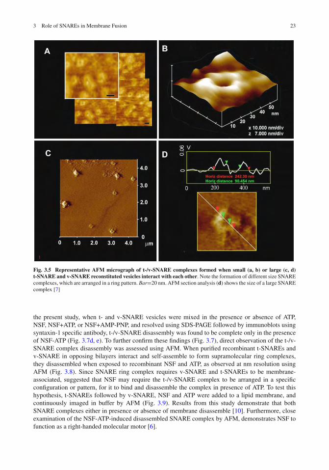
SNARE-ring complexes ranging in size from approximately 15 to 300 nm in diameter are formedwhen t-SNARE-reconstituted and v-SNARE-reconstituted lipid vesicles meet. Since vesicle curvaturewould dictate the contact area between opposing vesicles, this broad spectrum of SNARE complexesobserved, may be due to the interaction between SNARE-reconstituted vesicles of different size. Totest this hypothesis, t-SNARE- and v-SNARE-reconstituted liposomes (proteoliposomes) of distinctdiameters were used [7]. Lipid vesicles of different sizes used in the study were isolated using pub-lished extrusion method [9]. The size of each vesicle population was further assessed using the AFM(Fig. 3.4). AFM section analysis demonstrates the presence of small 40–50 nm-in diameter vesiclesisolated using a 50 nm extruder filter (Fig. 3.4a, b). Similarly, representative samples of large vesiclesmeasuring 150–200 and 800–1,000 nm were obtained using different size filters in the extruder. Suchlarge vesicles are shown in the AFM micrograph (Fig. 3.4c, d). Analysis of vesicle size using photoncorrelation spectroscopy, further confirmed the uniformity in the size of vesicles within each vesiclepopulation. The morphology and size of the SNARE complex formed by the interaction of t-SNARE-and v-SNARE-reconstituted vesicles of different diameter were examined using the AFM (Fig. 3.5).In each case, the t-SNARE and v-SNARE proteins in opposing proteoliposomes, interact and self-assemble in a circular pattern, forming channel-like structures. The interaction and arrangement ofSNAREs in a characteristic ring pattern were observed for all populations of proteoliposomes exam-ined (Fig. 3.5a–d). However, the size of the SNARE complex was determined to be dictated by thediameter of the proteoliposomes used (Fig. 3.5) [7]. When small (∼50 nm) t-SNARE- and v-SNARE-reconstituted vesicles were allowed to interact, SNARE-ring complexes of ∼20 nm in diameter weregenerated (Fig. 3.5a, b) [7]. With increase in the diameter of proteoliposomes, larger t-/v-SNAREcomplexes were formed (Fig. 3.5c, d). A strong linear relationship between size of the SNARE com-plex and vesicle diameter is demonstrated from these studies (Fig. 3.6) [7]. The experimental data fitwell with the high correlation coefficient, R2=0.9725 between vesicle diameter and SNARE-complexsize (Fig. 3.6).

3 Role of SNAREs in Membrane Fusion 21
Fig. 3.3 Opposing bilayers containing t- and v-SNAREs respectively, interact in a circular array to form con-ducting pores. (a) Schematic diagram of the bilayer-electrophysiology setup. (b) Lipid vesicle containing nystatinchannels (in red) and both vesicles and membrane bilayer without SNAREs, demonstrate no significant changes incapacitance and conductance. Initial increase in conductance and capacitance are due to vesicle-membrane attachment.To demonstrate membrane stability (both bilayer membrane and vesicles), the transmembrane gradient of KCl wasincreased to allow gradient driven fusion and a concomitance increase of conductance and capacitance. (c) When t-SNARE vesicles were added to a v-SNARE membrane support, the SNAREs in opposing bilayers arranged in a ringpattern, forming pores (as seen in the AFM micrograph on the extreme right) and there were seen stepwise increases incapacitance and conductance (–60 mV holding potential). Docking and fusion of the vesicle at the bilayer membrane,opens vesicle-associated nystatin channels and SNARE-induced pore formation, allowing conductance of ions from cisto the trans side of the bilayer membrane. Then further addition of KCl to induce gradient driven fusion, resulted inlittle or no further increase in conductance and capacitance, demonstrating docked vesicles have already fused [5]

22 B.P. Jena
Fig. 3.4 AFM micrograph of t-SNARE and v-SNARE reconstituted liposomes of different sizes. Note the∼40–50 nm vesicles (a, b), the ∼150 nm (c) and ∼800 nm vesicle (d) [7]
3.3.3 Disassembly of the SNARE Complex
Studies demonstrate that the soluble N-ethylmaleimide-sensitive factor (NSF) an ATPase, disassem-bles the t-/v-SNARE complex in presence of ATP [10]. This study was also the first conformationby direct physical observation, that NSF-ATP alone can lead to SNARE complex disassembly. In thisstudy, using purified recombinant NSF, and t- and v-SNARE-reconstituted liposomes, the disassemblyof the t-/v-SNARE complex was examined. Lipid vesicles ranging in size from 0.2–2 μm, were recon-stituted with either t-SNAREs or v-SNARE. Kinetics of association and dissociation of t-SNARE- andv-SNARE-reconstituted liposomes in solution, in the presence or absence of NSF, ATP, and AMP-PNP(the non-hydrolyzable ATP analogue), were monitored by right angle light scattering (Fig. 3.7a, b).Addition of NSF and ATP to the t/v-SNARE-vesicle mixture led to a rapid and significant increase inintensity of light scattering (Fig. 3.7a, b), suggesting rapid disassembly of the SNARE complex anddissociation of vesicles. Dissociation of t-/v-SNARE vesicles occurs on a logarithmic scale that canbe expressed by first order equation, with rate constant k=1.1 s–1 (Fig. 3.7b). To determine whetherNSF-induced dissociation of t- and v-SNARE vesicles is energy driven, experiments were performedin the presence and absence of ATP and AMP-PNP. No significant change with NSF alone, or inpresence of NSF-AMP-PNP, was observed (Fig. 3.7c). These results demonstrate that t-/v-SNAREdisassembly is an enzymatic and energy-driven process.
To further confirm the ability of NSF-ATP in the disassembly of the t-/v-SNARE complex,immunochemical studies were performed. It has been demonstrated that v-SNARE and t-SNAREsform an SDS-resistant complex [24]. NSF binds to SNAREs and forms a stable complex when lockedin the ATP-bound state (ATP-NSF). Thus, in the presence of ATP+EDTA, VAMP antibody has beendemonstrated to be able to immunoprecipitate this stable NSF-SNARE complex [24]. Therefore, in
3 Role of SNAREs in Membrane Fusion 23
Fig. 3.5 Representative AFM micrograph of t-/v-SNARE complexes formed when small (a, b) or large (c, d)t-SNARE and v-SNARE reconstituted vesicles interact with each other. Note the formation of different size SNAREcomplexes, which are arranged in a ring pattern. Bar=20 nm. AFM section analysis (d) shows the size of a large SNAREcomplex [7]
the present study, when t- and v-SNARE vesicles were mixed in the presence or absence of ATP,NSF, NSF+ATP, or NSF+AMP-PNP, and resolved using SDS-PAGE followed by immunoblots usingsyntaxin-1 specific antibody, t-/v-SNARE disassembly was found to be complete only in the presenceof NSF-ATP (Fig. 3.7d, e). To further confirm these findings (Fig. 3.7), direct observation of the t-/v-SNARE complex disassembly was assessed using AFM. When purified recombinant t-SNAREs andv-SNARE in opposing bilayers interact and self-assemble to form supramolecular ring complexes,they disassembled when exposed to recombinant NSF and ATP, as observed at nm resolution usingAFM (Fig. 3.8). Since SNARE ring complex requires v-SNARE and t-SNAREs to be membrane-associated, suggested that NSF may require the t-/v-SNARE complex to be arranged in a specificconfiguration or pattern, for it to bind and disassemble the complex in presence of ATP. To test thishypothesis, t-SNAREs followed by v-SNARE, NSF and ATP were added to a lipid membrane, andcontinuously imaged in buffer by AFM (Fig. 3.9). Results from this study demonstrate that bothSNARE complexes either in presence or absence of membrane disassemble [10]. Furthermore, closeexamination of the NSF-ATP-induced disassembled SNARE complex by AFM, demonstrates NSF tofunction as a right-handed molecular motor [6].

24 B.P. Jena
Fig. 3.6 SNARE complex isdirectly proportional tovesicle diameter. Schematicdiagram depicting theinteraction of t-SNARE andv-SNARE reconstitutedvesicles. At the extreme right,is a single t-/v-SNAREcomplex imaged by AFM (a).AFM images of vesiclesbefore and after their removalby the AFM cantilever tip,exposing the t-/v-SNAREcomplex (b). Interactingt-SNARE- andv-SNARE-vesicles imaged byAFM at low (<200 pN) andhigh forces (300–500 pN).Note, at low imaging forces,only the vesicle profile isimaged (left c). However athigher forces, the soft vesicleis flattened, allowing theSNARE complex to beimaged (right c). Plot ofvesicle diameter vs. size ofthe SNARE complex. Notethe high correlationcoefficient (R2=0.9725)between vesicle diameter andthe size of the SNAREcomplex (d) [7]

3 Role of SNAREs in Membrane Fusion 25
Fig. 3.7 NSF-ATP induced dissociation of t-SNARE and v-SNARE associated liposomes. (a) Real-time light scat-tering profiles of interacting t-SNARE and v-SNARE vesicles in solution in the presence and absence of NSF (depictedby arrow). In presence of ATP, NSF rapidly disassembles the SNARE complex and dissociates SNARE-vesicles repre-sented as a rapid increase in light scattering. No change in light scattering is observed when ATP is replaced with thenon-hydrolyzable analog AMP-PNP. (b) Kinetics of NSF-induced dissociation. The graph depicts first-order kineticsof vesicles dissociation elicited by NSF-ATP. (c) NSF requires ATP to dissociate vesicles. NSF in the presence of ATPdissociates vesicles (p<0.05, n=4, Student’s t-test). However, NSF alone or NSF in the presence of AMP-PNP had noeffect on the light scattering properties of SNARE-associated vesicle (p>0.05, n=4, Student’s t-test). (d) When t- andv-SNARE vesicles are mixed in the presence or absence of ATP, NSF, NSF+ATP, or NSF+AMP-PNP, and resolvedby SDS-PAGE followed by immunoblots using syntaxin-1 specific antibody, t-/v-SNARE disassembly was found to becomplete only in the presence of NSF-ATP (e). Densitometric scan of the bands reveals significant changes in SNAREcomplex and syntaxin-1 reactivity only when NSF and ATP were included in reaction mixture (p<0.05, n=3; andp<0.01, n=3, Student’s t-test) [10]

26 B.P. Jena
Fig. 3.8 AFM micrographs of NSF-ATP induced disassembly of the t-/v-SNARE ring complex. RepresentativeAFM micrograph of t-/v-SNARE complexes formed when large (top panel a) or small (bottom panel a) t-/v-SNAREring complexes are formed due to the interaction of large and small v-SNARE reconstituted vesicles interact with at-SNARE reconstituted lipid membrane. Bar = 250 nm. (b) Disassembly of large t-/v-SNARE complex. Bar = 250 nm.(c) High resolution of a t-/v-SNARE ring complex, and a disassembled one (d) [10]
3.3.4 CD Spectroscopy Confirm the Requirement of Membrane for Appropriatet-/v-SNARE Complex Assembly, and that NSF-ATP Alone Can MediatedSNARE Disassembly
The overall secondary structural content of full-length neuronal v-SNARE and t-SNAREs, andthe t-/v-SNARE complex, both in suspension and membrane-associated, were determined by CDspectroscopy using an Olis DSM 17 spectrometer [19]. Circular dichroism spectroscopy reveals that v-SNARE in buffered suspension (Fig. 3.10ai), when incorporated into liposomes (Fig. 3.10bi), exhibitreduced folding (Table 3.1). This loss of secondary structure following incorporation of full-lengthv-SNARE in membrane may be a result of self-association of the hydrophobic regions of the proteinin absence of membrane. When incorporated into liposomes, v-SNARE may freely unfold withoutthe artifactual induction of secondary structure, as reflective of the lack in CD signals at 208 and222 nm, distinct for a-helical content. The t-SNAREs (Fig. 3.10aii, bii), shows clearly defined peaks

3 Role of SNAREs in Membrane Fusion 27
Fig. 3.9 AFM micrographsof NSF-ATP induceddisassembly of thet-/v-SNARE complexformed when v-SNARE isadded to a t-SNAREreconstituted lipidmembrane. The left panela–d shows at low resolution,the sequential AFMmicrographs of one of tenrepresentative experiments,where v-SNARE is added to at-SNARE reconstituted lipidmembrane, followed by NSFand then ATP. Note thedramatic disassembly of theSNARE complexes in d. Theright panel shows at higherresolution, the disassembly ofone of such SNAREcomplexes [10]
at both these wavelengths, consistent with a higher degree of helical secondary structures formedboth in buffered suspension and in membrane, at ca. 66 and 20%, respectively (Table 3.1). Again,the membrane-associated SNARE exhibits less helical content than when in suspension. Similarly,there appears to be a dramatic difference in the CD signal observed in t-/v-SNARE complexesin suspension, and those complexes that are formed when membrane-associated SNAREs interact

28 B.P. Jena
Fig. 3.10 Circular dichroism data reflecting structural changes to SNAREs, both in suspension and in associ-ation with membrane. Structural changes, following the assembly and disassembly of the t-/v-SNARE complex isfurther shown. (a) CD spectra of purified full-length SNARE proteins in suspension and (b) in membrane-associated;their assembly and (NSF–ATP)-induced disassembly is demonstrated. (i) v-SNARE; (ii) t-SNAREs; (iii) t-/v-SNAREcomplex; (iv) t-/v-SNARE + NSF and (v) t-/v-SNARE + NSF + 2.5 mM ATP, is shown. CD spectra were recorded at25◦C in 5 mM sodium phosphate buffer (pH 7.5), at a protein concentration of 10 mM. In each experiment, 30 scanswere averaged per sample for enhanced signal to noise, and data were acquired on duplicate independent samples toensure reproducibility [19]
(Fig. 3.10aiii, biii). Interestingly, there is no increase of secondary structure upon complex for-mation. Rather, the CD spectra of the complexes are identical to a combination of individual spectra.Moreover, membrane associated t-/v-SNAREs are less folded than the purified SNARE complex.This data supports previous AFM results that lipid is required for proper arrangement of the SNAREproteins in membrane fusion. Addition of NSF to the t-/v-SNARE complex results in an increasein the unordered fraction (Fig. 3.10aiv, biv and Table 3.1), which may be attributed to an overalldisordered secondary structure of the NSF, and not necessarily unfolding of the t-/v-SNARE complex.In contrast, activation of NSF by the addition of ATP almost completely abolishes all α-helical contentwithin the multi-protein complex (Fig. 3.10av, bv). This direct observation of the helical unfoldingof the SNARE complex using CD spectroscopy under physiologically relevant conditions (i.e. inmembrane-associated SNAREs), confirms earlier AFM reports on NSF–ATP-induced t-/v-SNAREcomplex disassembly [10]. In further agreement with previously reported studies using the AFM, theconsequence of ATP addition to the t-/v-SNARE–NSF complex is disassembly, regardless of whether
Table 3.1 Secondary structural fit parameters of SNARE complex formation and dissociation [19]
Suspension (100 × fa) Membrane-associated (100 × f)
Proteinb α β O U Fitc α β O U Fitv-SNARE 4 36 18 43 0.19 0 30 32 38 0.21t-SNAREs 66 34 0 0 0.02 20 15 21 44 0.84v-/t-SNAREs 48 52 0 0 0.02 20 19 56 5 0.38v-/t-SNAREs+NSF 20 25 0 55 0.07 18 6 8 68 0.2v-/t-SNAREs+NSF+ATP 3 39 18 40 0.22 1 27 34 38 0.23
aAbbreviations used: f, fraction of residues is a given conformational class; α, α-helix; β, β-sheet; O, other (sumof turns, distorted helix, distorted sheet); U, unordered.bProtein constructs: v-SNARE (VAMP2); t-SNAREs (SNAP-25 + syntaxin 1A); NSF, N-ethylmaleimide SensitiveFactor. ATP, adenosine triphosphate.cFit: goodness of fit parameter expressed as Normalized Spectral Fit Standard Deviation (nm).

3 Role of SNAREs in Membrane Fusion 29
the t-/v-SNARE+NSF complex is membrane-associated or in buffered suspension. In earlier AFMstudies, 0.16–0.2 mg/ml of SNARE proteins were used, as opposed to the 800–1,000 mg/ml proteinconcentration required for the current CD studies. To determine if t-SNARE and v- SNARE interactdifferently at higher protein concentrations, both membrane-associated and in-suspension v- andt-SNARE complexes used in CD studies, were imaged using the AFM. In confirmation to previouslyreported AFM studies, results from the CD study demonstrated the formation of t-/v-SNARE ringcomplexes, only when t-SNARE-liposomes are exposed to v-SNARE-liposomes. Hence, higherSNARE protein concentrations are without influence on the membrane-directed self-assembly of theSNARE complex [19]. In summary, the CD results demonstrate that v-SNARE in suspension, whenincorporated into liposomes, exhibits reduced folding. Similarly, t-SNAREs which exhibit clearlydefined peaks at CD signals of 208 and 222 nm wavelengths, consistent with a higher degree of helicalsecondary structure in both the soluble and liposome-associated forms, exhibit reduced folding whenmembrane associated. ATP-induced activation of NSF bound to the t-/v-SNARE complex, resultsin disassembly of the SNARE complex, eliminating all α-helices within the structure. In addition,these studies are a further confirmation of earlier reports [10] that NSF–ATP is sufficient for thedisassembly of the t-/v-SNARE complex.
3.3.5 SNAREs Bring Opposing Bilayers Closer, Enabling Calcium Bridgingand Membrane Fusion
Diffraction patterns of non-reconstituted vesicles and t- and v-SNARE-reconstituted vesicles in theabsence and presence of 5 mM Ca2+ are shown for comparison in Fig. 3.11. To our knowledge, theseare the first recorded wide-angle diffractograms of unilamellar (single bilayer) vesicles in the 2–4 Ådiffraction range. They have broad pattern spanning 2θ ranges approximately 23–48◦ or d values of3.9–1.9 Å with sharp drop off intensity on either sides of the range. Relatively, broad feature of diffrac-togram indicate multitude of contacts between atoms of one vesicle as well between different vesiclesduring collision. However, two broad peaks are visible on the diffractogram, the stronger one at 3.1 Åand a weaker one at 1.9 Å. They indicate that the greatest number of contacts between them havethese two distances. Addition of Ca2+ or incorporation of SNAREs at the vesicles membrane or both,influence both peaks within the 2.1–3.3 Å intensity range (Fig. 3.11). However, the influence of Ca2+,SNAREs or both is more visible on peak positioned at 3.1 Å in form of an increased Imax of arbitraryunits and 2θ. This increase of Imax at the 3.1 Å can be explained in terms of increased vesicle pairingand/or a decrease in distance between apposed vesicles. Incorporation of t- and v-SNARE proteins atthe vesicle membrane allows for tight vesicle-vesicle interaction, demonstrated again as an Imax shifts
Fig. 3.11 Wide-angle X-raydiffraction patterns oninteracting lipid vesicles.Representative diffractionprofiles from one of fourseparate experiments usingplain and t- andv-SNARE-reconstituted lipidvesicles, both in the presenceor absence of 5 mM Ca2+ isshown. Note the shift in themajor peak to the right, whent-SNARE andv-SNARE-reconstitutedvesicles interact [9]

30 B.P. Jena
to 30.5◦ or 2.9 Å from 3.1 Å. Ca2+ and SNAREs work in manner that induces a much higher increaseof peak intensity with appearance of shoulders on both sides of the peak at 2.8 and 3.4 Å (Fig. 3.11).This indicates an increase in number of vesicle contact points at a constant distance between them.Vesicles containing either t- or v-SNAREs have little effect on the X-ray scattering patterns. Only asdiscussed above, when t-SNARE and v-SNARE reconstituted-vesicles were brought together, we diddetect change in the X-ray diffraction patterns. Since exposure of t-SNARE vesicle and v-SNAREvesicle mixture to Ca2+ results in maximum increase in a.u. and 2θ using X-ray diffraction, the effectof Ca2+ on fusion and aggregation of t-/v-SNARE vesicles were examined using light scattering, lightmicroscopy, AFM, fluorescent dequenching and electrical measurements of fusion [9].
In recent studies [12], using high-resolution electron microscopy, the electron density maps and3-D topography of the membrane-directed SNARE ring complex was determined at nm resolution(Fig. 3.12). Similar to the t-/v-SNARE ring complex formed when 50 nm v-SNARE liposomes meet
Fig. 3.12 The possible establishment of a leak-proof SNARE ring complex channel is demonstrated. (a) Size ofthe t-/v-SNARE ring complex is directly proportional to the size of the SNARE-associated vesicle (c). Different sizes ofv-SNARE-associated vesicles, when interact with t-SNARE-associated membrane (©), demonstrate the SNARE ringsize to be directly proportional to the vesicle size. When a 50 nm in diameter v-SNARE-reconstituted vesicle interactswith a t-SNARE-reconstituted membrane, an 11 nm in diameter t-/v-SNARE ring complex is formed. Similarly, thepresent study demonstrates that when a 50 nm in diameter v-SNARE-reconstituted vesicle, interacts with a 50 nm indiameter t-SNARE-reconstituted vesicle, a 8 nm in diameter t-/v-SNARE ring complex is established (�). Analogousto the 11 nm in diameter t-/v-SNARE ring complexes formed when 50 nm v-SNARE vesicles meet a t-SNARE-reconstituted planer membrane (b), approximately 11 nm in diameter t-/v-SNARE ring complexes are formed when50 nm in diameter synaptic vesicles meets a t-SNARE-reconstituted planer membrane (c, d) [12]

3 Role of SNAREs in Membrane Fusion 31
a t-SNARE-reconstituted planer membrane, SNARE rings are also formed when 50 nm in diame-ter isolated synaptic vesicles meet a t-SNARE-reconstituted planer lipid membrane. Furthermore, themathematical prediction of the SNARE ring complex size with reasonable accuracy, and the possi-ble mechanism of membrane-directed t-/v-SNARE ring complex assembly, was determined from thestudy. Using both lipososome-reconstituted recombinant t-/v-SNARE proteins, and native v-SNAREpresent in isolated synaptic vesicle membrane, the membrane-directed molecular assembly of theneuronal SNARE complex was determined for the first time and its size mathematically predicted.These results provide a new molecular understanding of the universal machinery and mechanism ofmembrane fusion in cells, having fundamental implications in human health and disease. The abovemention studies and their findings provide a molecular understanding of SNARE-induced membranefusion in cells.
Acknowledgments The author thanks the many students and collaborators who have participated in the various studiesdiscussed in this article. Support from the National Institutes of Health (USA), the National Science Foundation (USA),and Wayne State University, is greatly appreciated.
References
1. Malhotra V, Orci L, Glick BS et al (1988) Role of an N-ethylmaleimide-sensitive transport component in promotingfusion of transport vesicles with cisternae of the Golgi stack. Cell 54:221–227
2. Bennett MK, Calakos N, Scheller RH (1992) Syntaxin: a synaptic protein implicated in docking of synaptic vesiclesat presynaptic active zones. Science 257:255–259
3. Oyler GA, Higgins GA, Hart RA et al (1989) The identification of a novel synaptosomal-associated protein, SNAP-25, differentially expressed by neuronal subpopulations. J Cell Biol 109:3039–3052
4. Trimble WS, Cowan DM, Scheller RH (1988) VAMP-1: a synaptic vesicle-associated integral membrane protein.Proc Natl Acad Sci USA 85:4538–4542
5. Cho SJ, Kelly M, Rognlien KT et al (2002) SNAREs in opposing bilayers interact in a circular array to formconducting pores. Biophys J 83:2522–2527
6. Cho WJ, Jena BP (2007) N-ethylmaleimide-Sensitive Factor is a Right-Handed Molecular Motor. J BiomedNanotech 3:209–211
7. Cho WJ, Jeremic A, Jena BP (2005) Size of supramolecular SNARE complex: membrane-directed self-assembly.J Am Chem Soc 127:10156–10157
8. Jeremic A, Cho WJ, Jena BP (2004) Membrane fusion: what may transpire at the atomic level. J Biol Phys Chem4:139–142
9. Jeremic A, Kelly M, Cho JA et al (2004) Calcium drives fusion of SNARE-apposed bilayers. Cell Biol Int 28:19–3110. Jeremic A, Quinn AS, Cho WJ et al (2006) Energy-dependent disassembly of self-assembled SNARE complex:
observation at nanometer resolution using atomic force microscopy. J Am Chem Soc 128:26–2711. Weber T, Zemelman BV, McNew JA et al (1998) SNAREpins: minimal machinery for membrane fusion. Cell
92:759–77212. Cho WJ, Lee JS, Ren G et al (2011) Membrane-directed molecular assembly of the neuronal SNARE complex.
J Cell Mol Med 15:31–3713. Bako I, Hutter J, Palinkas G (2002) Car–Parrinello molecular dynamics simulation of the hydrated calcium ion.
J Chem Phys 117:9838–984314. Chialvo AA, Simonson JM (2003) The structure of CaCl2 aqueous solutions over a wide range of concentration.
Interpretation of diffraction experiments via molecular simulation. J Chem Phys 119:8052–806115. McIntosh TJ (2000) Short-range interactions between lipid bilayers measured by X-ray diffraction. Curr Opin
Struct Biol 10:481–48516. Portis A, Newton C, Pangborn W et al (1979) Studies on the mechanism of membrane fusion: evidence for
an intermembrane Ca2+-phospholipid complex, synergism with Mg2+, and inhibition by spectrin. Biochemistry18:780–790
17. Laroche G, Dufourc EJ, Dufourcq J et al (1991) Structure and dynamics of dimyristoylphosphatidic acid/calciumcomplexes by 2H NMR, infrared, spectroscopies and small-angle x-ray diffraction. Biochemistry 30:3105–3114
18. Potoff JJ, Issa Z, Manke CW, Jr et al (2008) Ca2+-dimethylphosphate complex formation: providing insight intoCa2+-mediated local dehydration and membrane fusion in cells. Cell Biol Int 32:361–366
19. Cook JD, Cho WJ, Stemmler TL et al (2008) Circular dichroism (CD) spectroscopy of the assembly anddisassembly of SNAREs: The proteins involved in membrane fusion in cells. Chem Phy Lett 462:6–9

32 B.P. Jena
20. Cohen FS, Niles WD (1993) Reconstituting channels into planar membranes: a conceptual framework and methodsfor fusing vesicles to planar bilayer phospholipid membranes. Methods Enzymol 220:50–68
21. Kelly ML, Woodbury DJ (1996) Ion channels from synaptic vesicle membrane fragments reconstituted into lipidbilayers. Biophys J 70:2593–2599
22. Woodbury DJ (1999) Nystatin/ergosterol method for reconstituting ion channels into planar lipid bilayers. MethodsEnzymol 294:319–339
23. Woodbury DJ, Miller C (1990) Nystatin-induced liposome fusion. A versatile approach to ion channel reconstitu-tion into planar bilayers. Biophys J 58:833–839
24. Jeong EH, Webster P, Khuong CQ et al (1998) The native membrane fusion machinery in cells. Cell Biol Int22:657–670