chapter 27 amino acids, peptides, and proteins slides copy
TRANSCRIPT

Amino Acids, Peptides, and Proteins
27
Ribbon model of human myoglobin

27.1 STRUCTURES OF THE α-AMINO ACIDSFigure 27.1 Chirality of the α-Amino Acids(a) Planar projection of an L-amino acid in unionized form. (b) !e α-carboxyl group and the α-amino group are ionized in aqueous solution at pH 7. (c)!e con"guration of the α-amino acids isolated from proteins is opposite to the con"guration of the reference compound D-glyceraldehyde. (d) Molecular model of L-serine, whose side chain is a CH2OH group.
C
CO2H
R
HNH2α C
CO2
R
HNH3α
(a) (b)
Cα
Cβ
N H
C OO
(c)α−Amino group
Side chain
(c)
α−Carboxyl group
CHO
HHO
CH2OH
CHO
OHH
CH2OH
mirror plane
CO2H
HNH2
CH2OH
CO2H
NH2H
CH2OH
D-glyceraldehyde
D-serineL-serine
L-glyceraldehyde
(d)

27.1 STRUCTURES OF THE α-AMINO ACIDSNonpolar (hydrophobic) side chains
CO2-
C H+NH3
CH3
Alanine (A) (Ala)
CO2-
C
CH
H+NH3
CH3
CO2-
C H+NH3
CH2
CHCH3
CH3
Valine (V) (Val)
CH3
Leucine (L) (Leu)
CO2-
C H
C
CH2
CH3
+NH3
CH3H
Isoleucine (Ile) (I)
CN+
H-O2CH
H
Proline (P) (Pro)
CO2-
C H+NH3
CH2
Phenylalanine (F) (Phe)
CO2-
C H+NH3
CH2
Tryptophan (W) (Trp)
NH
Methionine (M) (Met)
CO2-
C H
CH2
S
CH2
CH3
+NH3
Cysteine (C) (Cys)
CO2-
C H
CH2
SH
+NH3
Glycine (G) (Gly)
CO2-
C H
H
+NH3
CO2-
C H+NH3
CH2
Tyrosine (Y) (Tyr)
OH
CO2-
C H
C
CH3
+NH3
OHH
Threonine (Thr) (T)
CO2-
C H+NH3
CH2
C
Asparagine (N) (Asn)
O NH2
Glutamine (Q) (Gln)
CO2-
C H
CH2
CH2
C
+NH3
O NH2
CO2-
C H+NH3
CH2
OH
Serine (S) (Ser)
Polar, neutral (hydrophilic) side chains
CO2-
C H
CH2
CH2
CH2
+NH3
Lysine (Lys) (K)
CH2
NH3+
CO2-
C H
CH2
CH2
CH2
+NH3
Arginine (Arg) (R)
NH
CH2N NH2
CO2-
C H
CH2
+NH3
N
NH
Histidine (His) (H)
CO2-
C H
CH2
CH2
+NH3
Aspartate (Asp) (D)
CO2-
C H
CH2
CH2
CO2-
+NH3
Glutamate (Glu) (E)
Acidic Amino AcidsBasic Amino Acids
Figure 27.2 Structures of theα-Amino Acids at pH 7At pH the α-amino and α-carboxyl groups are both ionized. !e amino acids are classi"ed by their side chain polarities. !e amino acids have both on-letter and three-letter abbreviations.

27.2 ACID-BASE EQUILIBRIA OF α-AMINO ACIDSIonic Form of Amino Acids
CO2-
C H
R
+NH3
structure of a dipolar ion (zwitterion)
CO2-
C H
R
+NH3
zwitterion
+ OH-
CO2-
C H
R
NH2
conjugate base
+ HOH
CO2-
C H
R
+NH3
zwitterion
+ H3O+
CO2H
C H
R
NH2
conjugate acid
+ H2O
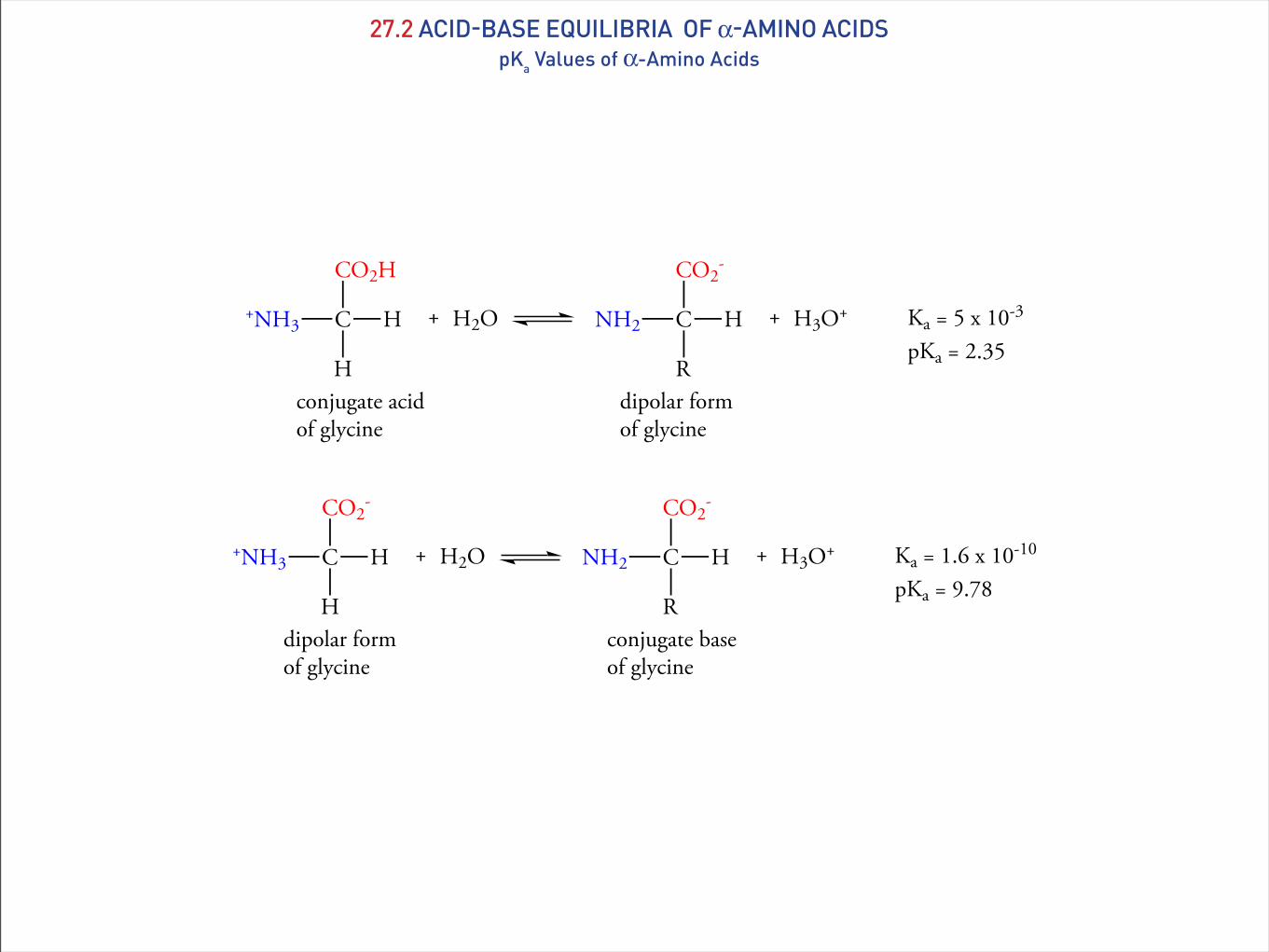
27.2 ACID-BASE EQUILIBRIA OF α-AMINO ACIDSpKa Values of α-Amino Acids
CO2H
C H
H
+NH3
conjugate acid of glycine
+ H2O
CO2-
C H
R
NH2
dipolar form of glycine
+ H3O+ Ka = 5 x 10⁻³
pKa = 2.35
CO2-
C H
H
+NH3
dipolar form of glycine
+ H2O
CO2-
C H
R
NH2
conjugate baseof glycine
+ H3O+ Ka = 1.6 x 10⁻¹⁰
pKa = 9.78

27.1pKa Values of Acidic and Basic Groups in α−Amino AcidsAmino Acid α−CO2H group α−NH3+ group Side chain
Glycine 2.35 9.78Alanine 2.35 9.87Valine 2.29 9.72Leucine 2.33 9.74Isoleucine 2.32 9.76Methionine 2.17 9.27Proline 1.95 10.64Phenylalanine 2.58 9.24Tryptophan 2.43 9.44Serine 2.19 9.44#reonine 2.09 9.10Cysteine 1.89 10.78 8.53Tyrosine 2.20 9.11 10.11Asparagine 2.02 8.80Glutamine 2.17 9.13Aspartate 1.99 10.00 3.96Glutamate 2.13 9.95 4.32Lysine 2.16 9.20 10.80Arginine 1.82 8.99 12.48Histidine 1.81 9.15 6.00
27.2 ACID-BASE EQUILIBRIA OF α-AMINO ACIDSpKa Values of α-Amino Acids

27.3 ISOIONIC POINT AND TITRATION OF α-AMINO ACIDSIsoionic Points of Amino Acids
Table 27.2Isoionic PointsAmino Acid pHI
Glycine 5.97Alanine 6.10Valine 5.96Leucine 5.98Isoleucine 6.02Methionine 5.74Proline 6.30Phenylalanine 5/48Tryptophan 5.89Serine 5.68!reonine 5.60Cysteine 5.07Tyrosine 5.66Asparagine 5.41Glutamine 5.65Aspartic acid 2.77Glutamic acid 3.22Lysine 9.74Arginine 10.76Histidine 7.59
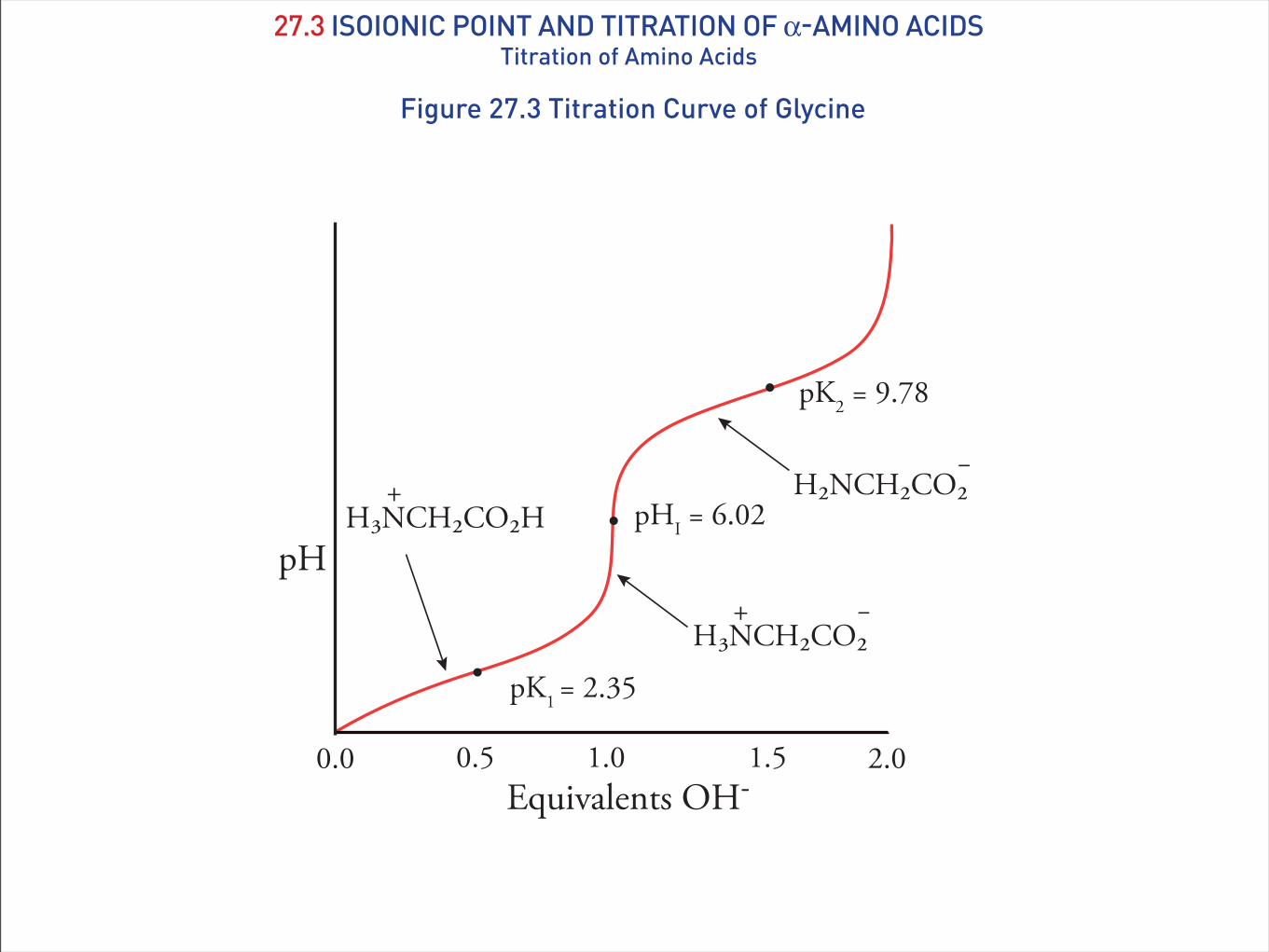
27.3 ISOIONIC POINT AND TITRATION OF α-AMINO ACIDSTitration of Amino Acids
Figure 27.3 Titration Curve of Glycine
pK1 = 2.35
pK2 = 9.78
pHI = 6.02
Equivalents OH-2.01.00.0 0.5 1.5
pHH3NCH2CO2H
+
H3NCH2CO2+ –
H2NCH2CO2–

27.4 SYNTHESIS OF α-AMINO ACIDSAmination of α-Halocarboxylic Acids
CH2 CO2HRBr2PBr3
NH3CH
NH2
R CO2HCH
Br
R CO2H
The Strecker Synthesis
NaCN
NH4Cl
H3O+
CH
NH3+
R CO2HCH
NH2
R CN
(an α-amino nitrile)
C HR
O
NH3H2OC HR
O
C HR
NH
+
CNC HR
NH
+ H CHR CN
NH2

27.4 SYNTHESIS OF α-AMINO ACIDSReductive Amination and Acetamidomalonate Synthesis
Reductive Amination
NH3C CO2HR
O
C CO2HR
NH
CHR CO2H
NH2
NH4Cl
H2
Pd
Acetamidomalonate Synthesis
CHNH CO2CH2CH3
CO2CH2CH3
CCH3
O
(diethyl acetamidomalonate)
C CO2CH2CH3
CO2CH2CH3
R
NHCCH3
OH3O+
heatC CO2H
CO2H
R
NH2 CH
R
NH2 CO2H
CHNH CO2CH2CH3
CO2CH2CH3
CCH3
O
(diethyl acetamidomalonate)
CH3CH2O-
CH3CH2OHCNH CO2CH2CH3
CO2CH2CH3
CCH3
OR Cl
C CO2CH2CH3
CO2CH2CH3
R
NHCCH3
O
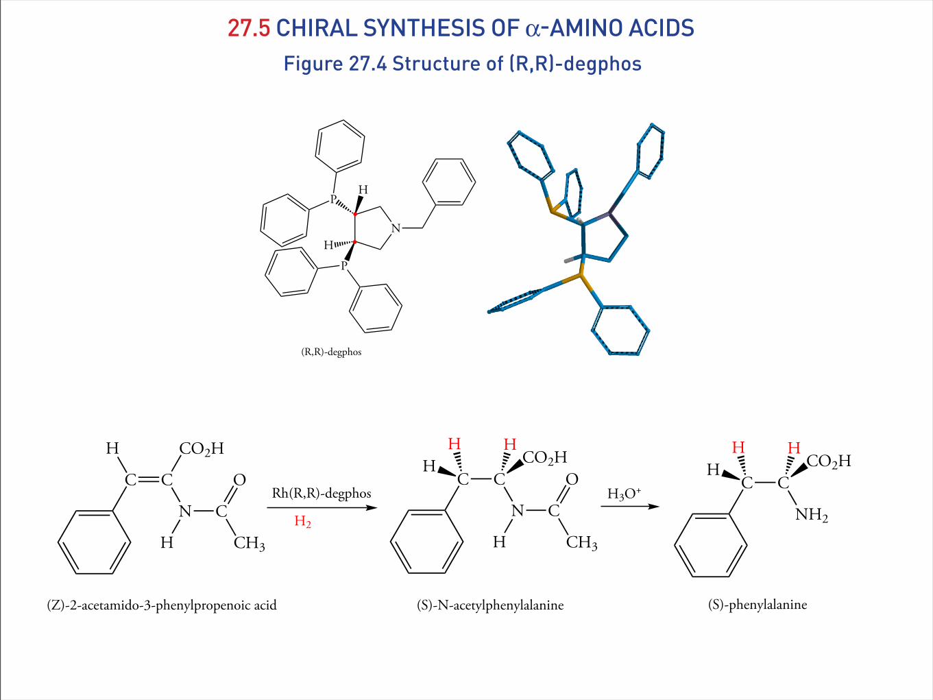
27.5 CHIRAL SYNTHESIS OF α-AMINO ACIDSFigure 27.4 Structure of (R,R)-degphos
N
PH
PH
(R,R)-degphos
C C
H CO2H
N C
H
O
CH3
Rh(R,R)-degphos
H2
C CH CO2H
N C
H
O
CH3
H H
(Z)-2-acetamido-3-phenylpropenoic acid (S)-N-acetylphenylalanine
H3O+ C CH CO2H
NH2
H H
(S)-phenylalanine

27.6 REACTIONS OF α-AMINO ACIDSEsterification of the α-Carboxyl Group
C HNH2
CH3
CO2CH2C6H5
alanine benzyl ester
H2 / PdC HNH2
CH3
CO2H
+ C6H5CH3
alanine
C HNH3
CH3
CO2H
+ CH3CH2OHHCl (g)
C HNH3
CH3
CO2CH2CH3
alanine alanine ethyl ester
C HNH3
CH(CH3)2
CO2H
+ C6H5CH2OHHCl (g)
C HNH3
CH(CH3)2
CO2CH2C6H5
valine valine benzyl ester

27.6 REACTIONS OF α-AMINO ACIDSAcetylation of the α-Amino Group
OCO
O
(CH3)3C C O
O
C(CH3)3
di-tert-butyldicarbonate (Boc)2O
CNH3
CH3
CO2
alanine
H NHCO
O
(CH3)3C
N-tert-butoxycarbonyl alanine (Boc alanine)
(Boc)2OC H
CH3
CO2H
CNH2
CH3
CO2
alanine
H + ClCO
O
C6H5CH2pyridine
NHCO
O
C6H5CH2
N-benzyloxycarbonyl alanine (Cbz alanine)
benzyl chloroformate
C H
CH3
CO2H

27.6 REACTIONS OF α-AMINO ACIDSAcetylation of the α-Amino Group
NHCO
O
C6H5CH2
N-benzyloxycarbonyl alanine (Cbz alanine)
C H
CH3
NH C H
CH3
CO2H
a carbamic acid
C H
CH3
CO2H
NH2 + CO2CHO
OCO2H
NHCO
O
C6H5CH2
N-benzyloxycarbonyl alanine (Cbz alanine)
C H
CH3
NH C H
CH3
CO2H
C H
CH3
CO2H
NH2 + C6H5CH3CHO
OCO2HH2
Pd
CO2
NHCO
O
(CH3)3C
N-tert-butoxycarbonyl alanine (Boc alanine)
C H
CH3
CO2HCF3CO2H
NHCHO
O
C H
CH3
CO2H
+ C(CH3)2CH2

27.7 PEPTIDESPeptide Nomenclature
Figure 27.4 Peptide Nomenclature(a) Structure of glycylalanine. !e N-terminal α-amino group and the C-terminal α-carboxyl group and are ionized in aqueous solution at pH 7. !ree-letter and one-letter abbreviations for the amino acids are commonly used. (b) Structure of alanylglycine. (c) An aminoacyl group consists of the —NH—CHR—CO— group of each amino acid in the peptide.
H
C C
H
+NH3
N-terminal residue
NH
O
C C
H
CH3
O
OC-terminal residue
glycylalanine(Gly-Ala or GA)
peptide bond
H
C C
CH3
+NH3
N-terminal residue
NH
O
C C
H
H
O
OC-terminal residue
alanylglycine(Ala-Gly or Ag)
H
C C
R1
+NH3 NH
O
C C
H
R2
NH
O
aminoacyl group
C
R3
α1 α2 α3
(a) (b)
(c)

27.8 OVERVIEW OF PEPTIDE SYNTHESIS
CHNH2 C
R1
OH
OPC
CHNH2 C
R2
O
O
PC
CHNH2 C
R1
OH
OPN
CHNH C
R2
OH
O
PN
CHNH C
R1
OH
O
PN CHNH2 C
R2
O
O
PC+ CHHN C
R1
NH
O
PN CH C O
O
R2
PC

27.8 OVERVIEW OF PEPTIDE SYNTHESISProtecting the Carboxyl Group
C HNH3
R
CO2H
+ C6H5CH2OHHCl (g)
C HNH3
R
CO2CH2C6H5
benzyl ester
C HNH2
R
CO2CH2C6H5
amino acid (or peptide) benzyl ester
H2 / PdC HNH2
R
CO2H
+ C6H5CH3
deprotected amino acid (or peptide)

27.8 OVERVIEW OF PEPTIDE SYNTHESISProtecting the Amino Group
CNH3
R
CO2
H NHCO
O
(CH3)3C
N-tert-butoxycarbonyl amino acid (a Boc-amino acid)
(Boc)2OC H
R
CO2H
NHCO
O
(CH3)3C
N-tert-butoxycarbonyl amino acid (Boc-amino acid)
C H
R
CO2BzCF3CO2H
NHCHO
O
C H
R
CO2Bz
+ C(CH3)2CH2
unstable carbamate derivative ofan amino acid
NH C H
R
CO2Bz
+ CO2

27.8 OVERVIEW OF PEPTIDE SYNTHESISPeptide Bond Synthesis
N C N
dicyclohexylcarbodiimide (DCCI)
C
O
HN
HN
dicyclohexylurea
CHNH C
R1
OH
O
PN CHNH2 C
R2
O
O
PC+ CHHN C
R1
NH
O
PN CH C O
O
R2
PCDCCI
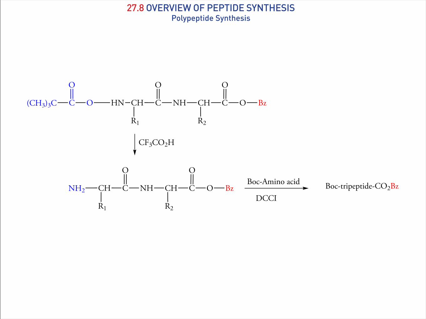
27.8 OVERVIEW OF PEPTIDE SYNTHESISPolypeptide Synthesis
CHHN C
R1
NH
O
O CH C O
O
R2
BzC(CH3)3C
O
CF3CO2H
CHNH2 C
R1
NH
O
CH C O
O
R2
BzBoc-Amino acid
DCCIBoc-tripeptide-CO2Bz
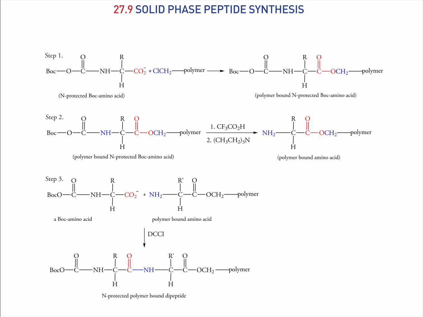
27.9 SOLID PHASE PEPTIDE SYNTHESIS
NHCO
O
Boc
(N-protected Boc-amino acid)
C CO2
H
R
+ polymerClCH2 NHCO
O
Boc C C
H
R
OCH2
O
polymer
(polymer bound N-protected Boc-amino acid)
Step 1.
Step 2.
NHCO
O
Boc C C
H
R
OCH2
O
polymer
(polymer bound N-protected Boc-amino acid)
1. CF3CO2H
2. (CH3CH2)3NNH2 C C
H
R
OCH2
O
polymer
(polymer bound amino acid)
NH2 C C
H
R'
OCH2
O
polymer
polymer bound amino acid
+NHCBocO
O
a Boc-amino acid
C CO2
H
R
DCCI
NHCBocO
O
C C
H
R O
NH C C
H
R'
OCH2
O
polymer
N-protected polymer bound dipeptide
Step 3.
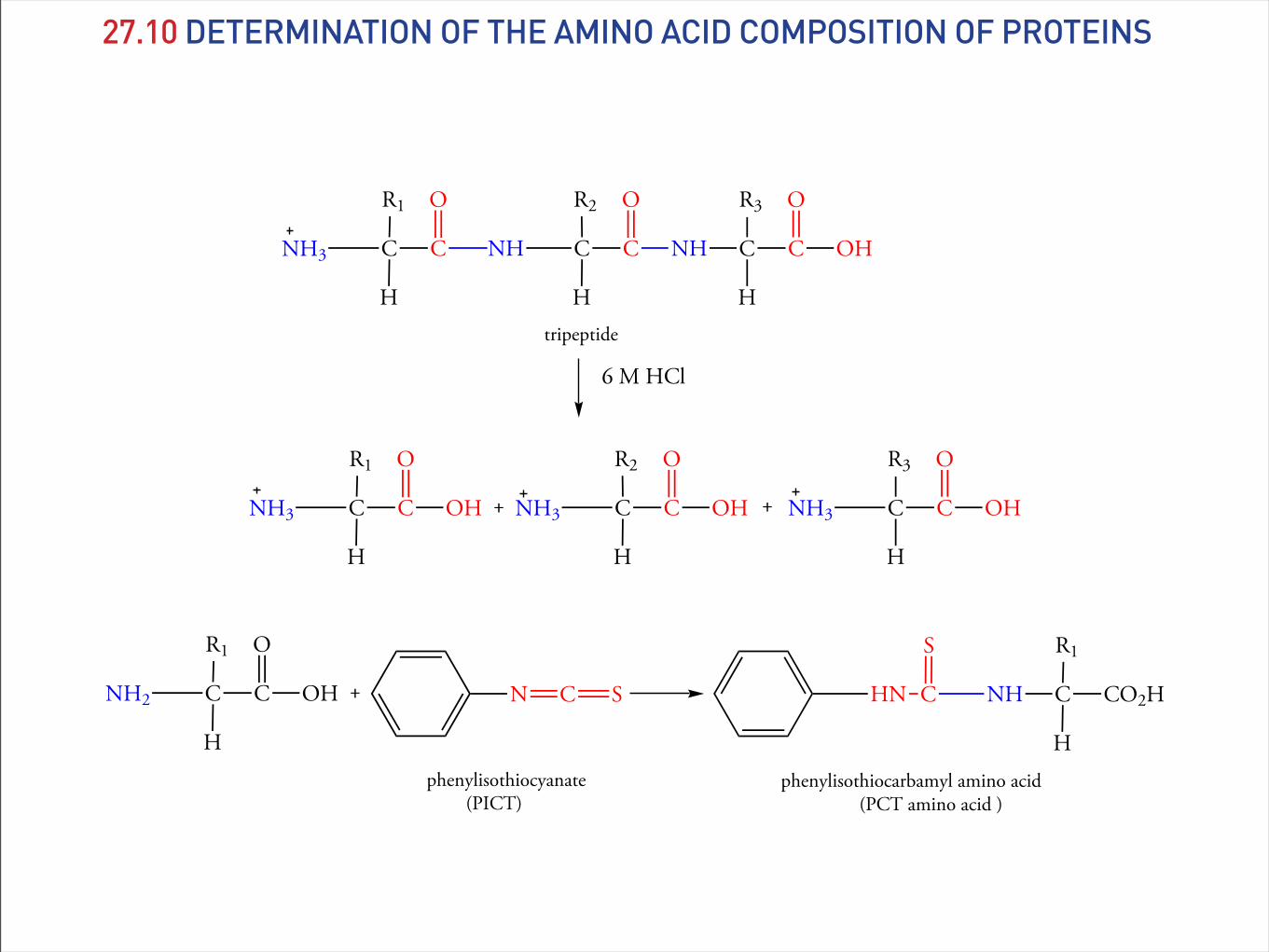
27.10 DETERMINATION OF THE AMINO ACID COMPOSITION OF PROTEINS
NH3 C C
H
R1 O
NH3 C C
H
R2
NH3
O
C C
R3
H
OH
O
NH3 C C
H
R1 O
NH C C
H
R2
NH
O
tripeptide
C C
R3
H
OH
O
6 M HCl
OHOH + +
NH2 C C
H
R1 O
OH + N C S NHCHN
S
C CO2H
H
R1
phenylisothiocyanate (PICT)
phenylisothiocarbamyl amino acid (PCT amino acid )

27.10 DETERMINATION OF THE AMINO ACID COMPOSITION OF PROTEINS
Figure 27.5 HPLC Separation of PCT Amino Acids
Abs
orba
nce,
254
nm
Min5 10 15
DE S G H
RH TP
YV
M
C
I
LF
K

Table 27.4Amino Acid Composition of Human LysozymeAmino Acid
Number of Amino Acids
Per Cent Composition
Ala 5 4.1Arg 1 0.8Asn 4 3.3Asp 12 9.8Cys 8 6.5Gln 7 4.9Glu 8 6.5Gly 6 9.8His 2 11.4Ile 12 9.8Leu 14 11.4Lys 12 9.8Met 2 1.6Phe 4 3.3Pro 2 1.6Ser 8 6.5!r 7 5.7Trp 3 2.4Tyr 4 3.3Val 2 1.6
27.10 DETERMINATION OF THE AMINO ACID COMPOSITION OF PROTEINS

Figure 27.6 Edman DegradationFirst, the peptide is converted to its N-terminal PCT derivative by treatment with phenylisothiocyanate. Next, the PCT protein is treated with tri!uoroacetic acid, then with water to give the phenylthiohydanto-in derivative. "e N-terminal amino acid is released in this step. "e other peptide bonds are not a#ected.
NH2 C C
H
CH3 O
NH C C
H
H
NH
O
C C
R3
H
protein
O
Ala Gly
PICT/base
NHCHN
S
C
H
CH3
C6H5 NH C C
H
H
NH
O
C C
R3
H
protein
O
Gly
C
O
N-terminal PCT derivative of the protein
N-terminal PCT group
1. CF3CO2H2. H+, H2O
NH3 C C
H
H
NH
O
C C
R3
H
protein
O
Gly
+N
NH
S
OCH3
phenylthiohydantoin derivative of alanine
27.11 DETERMINATION OF THE AMINO ACID SEQUENCE OF PROTEINSThe Edman Degradation

27.11 DETERMINATION OF THE AMINO ACID SEQUENCE OF PROTEINSBlocking Cystine Residues
CH2SH
CNH C
H
CH2SH
NH
O
C
H
NHC
O
+
cysteine residues
I CH2CH2CO2-
iodoacetate
CH2SCH2CH2CO2-
CNH C
H
SCH2CH2CO2-
NH
O
C
H
NHC
O
+
S-Carboxymethylcysteine residues
CH2
CNH C
H
cystine residue
SS
CH2
NH
O
C
H
NHCNH
O
2 HSCH2CH2OH
(β-mercatpethanol)CH2SH
CNH C
H
CH2SH
NH
O
C
H
NHC
O
+
2 cysteine residues
+SCH2CH2OH
SCH2CH2OH

27.11 DETERMINATION OF THE AMINO ACID SEQUENCE OF PROTEINSPeptide Cleavage at Methionine Residues
-Ala-Phe-Arg-Gly-Lys-Met-Val-Leu----H3N CO2-
BrCN
-Val-Leu----CO2--Ala-Phe-Arg-Gly-Lys NH
O
O
C-terminal homoserine lactone (HSL)
+ NH3NH3

27.11 DETERMINATION OF THE AMINO ACID SEQUENCE OF PROTEINSEnzymatic Cleavage of Polypeptide Chains
(H3N—)Ala—Arg—Phe—Gly—Lys—Trp—Val(—CO2H)Trypsin
(H3N—)Ala—Arg(—CO2H) (H3N—)Phe—Gly—Lys(—CO2H)+ (H3N—)Trp—Val(—CO2H)+
(H3N—)Ala—Arg—Phe—Gly—Lys—Trp—Val(—CO2H)
Chymotrypsin
+ (H3N—)Val(—CO2H)+(H3N—)Ala—Arg—Phe(—CO2H) (H3N—)Gly—Lys—Trp(—CO2H)
ValAla—Arg—Phe Gly—Lys—Trp
Ala—Arg Phe—Gly—Lys Trp—Val

27.11 DETERMINATION OF THE AMINO ACID SEQUENCE OF PROTEINSPrimary Structures and Evolutionary Relationships
Figure 27.7 Evolutionary Family Tree for Cytcochrome c
Bacteria
Yeast
Candidakrusei
Neurosporacrasa
Baker’s yeast
Fruit !ySilk moth
Hornwormmoth
Insects
Plants
WheatMung bean Pumpkin
Tomato
Sun!ower
Paci"clamprey
CarpTuna
Bonito
Snapping turtleDuck
Chicken, TurkeyPenguin
Human, chimpanzeeMonkey
Zebra,Horse
Cow, pig, sheepDog
BullfrogGrey whale

CC
NC
O
H
αα' C
CN
C
O
H
αα'
contributing structure 1 contributing structure 2
CC
NC
O
H
αα'
resonance hybrid
δ
δ
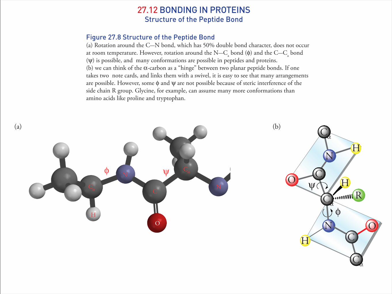
27.12 BONDING IN PROTEINSStructure of the Peptide Bond
27.12 BONDING IN PROTEINSStructure of the Peptide Bond
Figure 27.8 Structure of the Peptide Bond(a) Rotation around the C—N bond, which has 50% double bond character, does not occur at room temperature. However, rotation around the N—Ca bond (φ) and the C—Ca bond (ψ) is possible, and many conformations are possible in peptides and proteins. (b) we can think of the α-carbon as a “hinge” between two planar peptide bonds. If one takes two note cards, and links them with a swivel, it is easy to see that many arrangements are possible. However, some φ and ψ are not possible because of steric interference of the side chain R group. Glycine, for example, can assume many more conformations than amino acids like proline and tryptophan.
(a)
O
Cα
N
H
C
O
N
Cα
ψφ
Cα
ONH
HCα
N
HR
ψ
φ
O
C
C
Cα
(b)

Figure 27.8 Dimensions of an α-Helix!e distance between amino acid residues in an α-helix is 0.15 nm. !e distance required for one turn of the helix, its pitch, is 5.4 nm.
27.12 BONDING IN PROTEINSThe α-Helix
0.15 nm Rise per aminoacid residue
Pitch: distancefor one turn
0.54 nm

Figure 27.9 Ribbon Diagram of an α-HelixHydrogen bonds in an α-helix are approximately parallel to long axis of the helix. !ey from between carbonyl oxygen and amide hydrogens separated by 3.6 residues. Side chains radiate outward from the helix. !e α-helix is right-handed.
27.12 BONDING IN PROTEINSThe α-Helix
Lys
Asp
Gln
Met
Leu
Ala
Ala
H-bond
H-bond

27.12 BONDING IN PROTEINSβ-Pleated Sheets
Figure 27.10 Hydrogen Bonding In Parallel β-Pleated Sheet
CR
C O
N
C
CO
N H
CR
H
HRH
H
CR
C O
N
C
CO
N H
CR
H
HRH
H
CR
C O
N
C
CO
N H
CR
H
HRH
H

27.12 BONDING IN PROTEINSDisulfide Bonds
C
CO2
HNH3
CH2SH
2 [O]
SCH2 S CH2
CCNH3 H
CO2
HNH3
CO2
cysteine cystine
Figure 27.12 Conformation of an s-trans Disulfide Bond
s-trans disul!de bond

27.13 PROTEIN STRUCTURE
Table 27.5Examples of Proteins Having Quatenary StructureProtein Molecular
WeightNumber ofSubunits
Function
alcohol dehydrogenase
80,000 4 enzymatic reaction in fermentation
aldolase 150,000 4 enzymatic reaction in glycolysisfumarase 194,000 4 enzymatic reaction in citric acid cyclehemoglobin 65,000 4 oxygen transport in bloodinsulin 11,500 2 hormone that regulates metabolism of glucose
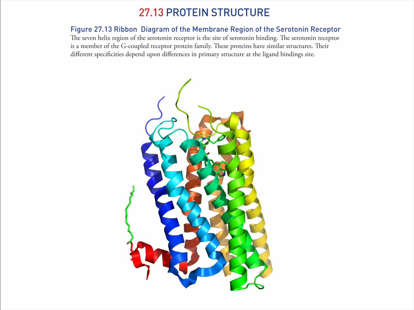
Figure 27.13 Ribbon Diagram of the Membrane Region of the Serotonin Receptor!e seven helix region of the serotonin receptor is the site of serotonin binding. !e serotonin receptor is a member of the G-coupled receptor protein family. !ese proteins have similar structures. !eir di"erent speci#cities depend upon di"erences in primary structure at the ligand bindings site.
27.13 PROTEIN STRUCTURE
27.13 PROTEIN STRUCTURE
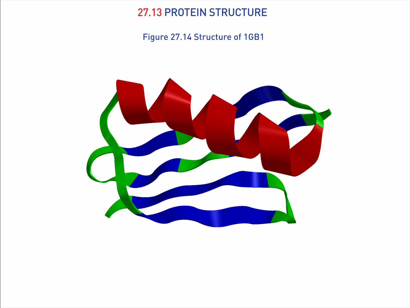
Figure 27.14 Structure of 1GB1
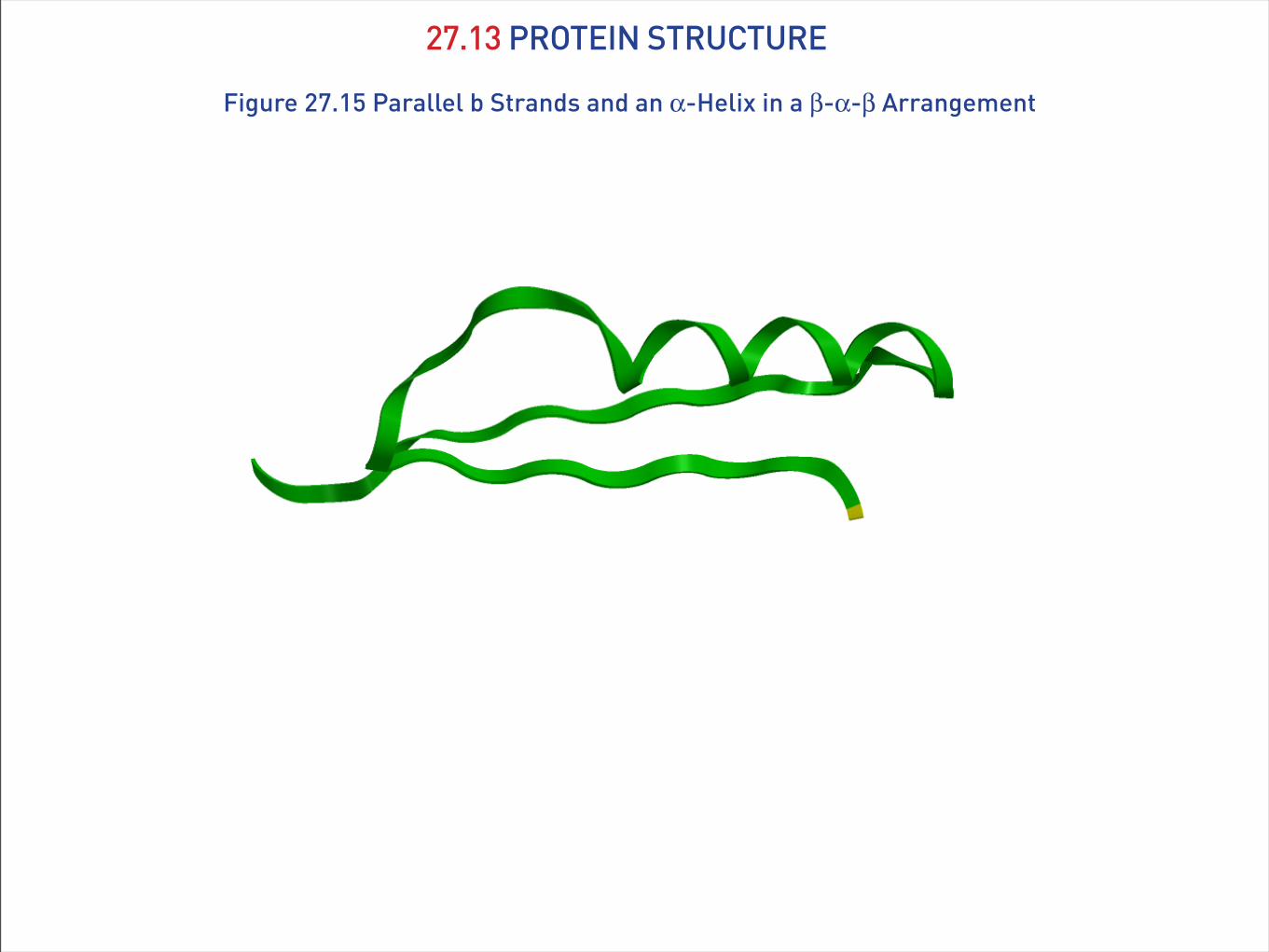
Figure 27.15 Parallel b Strands and an α-Helix in a β-α-β Arrangement
27.13 PROTEIN STRUCTURE

Figure 27.16 Tertiary Structure of Triose Phosphate Isomerase!e α-helices are shown in red, β-pleated sheets are blue, and less structured “loops” are shown in green.
27.13 PROTEIN STRUCTURE

27.14 OXYGEN STORAGE AND TRANSPORT: MYOGLOBIN AND HEMOGLOBINMyoglobin
Figure 27.18 Heme
N
N N
N
OOO O
FeII
heme

27.14 OXYGEN STORAGE AND TRANSPORT: MYOGLOBIN AND HEMOGLOBINMyoglobin
Figure 27.17 Structure of Oxymyoglobin.(a) !e α-helices are shown in red, and less structured “loops” are shown in green. (b) Structure of the heme group bound to myoglobin via a bond from a nitrogen on histidine 93 and the FeII ion. Oxygen binds on the opposite side of the histidine.
Heme
O2(a)
His 93
O2FeII
(b)

27.14 OXYGEN STORAGE AND TRANSPORT: MYOGLOBIN AND HEMOGLOBINHemoglobin
Figure 27.19 Structure of Deoxyhemoglobin.!e α and β subunits of hemoglobin interact cooperatively, and when one heme binds O2, the each of the others rapidly bind O2.
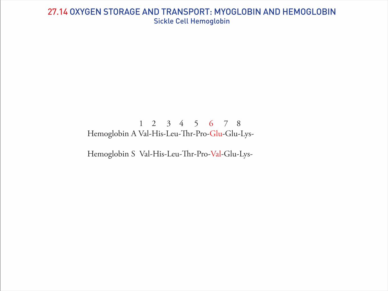
27.14 OXYGEN STORAGE AND TRANSPORT: MYOGLOBIN AND HEMOGLOBINSickle Cell Hemoglobin
1 2 3 4 5 6 7 8Hemoglobin A Val-His-Leu-!r-Pro-Glu-Glu-Lys-
Hemoglobin S Val-His-Leu-!r-Pro-Val-Glu-Lys-

27.14 OXYGEN STORAGE AND TRANSPORT: MYOGLOBIN AND HEMOGLOBINSickle Cell Hemoglobin
Figure 27.20 Structure of Deoxyhemoglobin Dimer.!e β subunits of hemoglobin interact by van der Waals contact between the isopropyl side chains at residue 6 of sickle cell hemoglobin (HbS). Since each HbS has two β subunits on opposite sides of the tetramer, a "brous polymer forms. HbS polymerizes when HbS releases O2, which disorts the red blood cells into the shape of a sickle.