chapter 26 - psau.edu.sa...(suprarenal) gland abdominal aorta inferior vena cava rectum right kidney...
TRANSCRIPT

993
CHAPTER 26
As body cells carry out metabolic activities, they consume oxygen and nutrients and produce waste products such as carbon dioxide, urea, and uric acid. Wastes must be eliminated from the body because they can be toxic to cells if they accumulate. While the respiratory system rids the body of carbon dioxide, the urinary system disposes of most other wastes. The urinary system performs this function by removing wastes from the blood and excreting them into urine. Disposal of wastes through
the release of urine is not the only purpose of the urinary system. The urinary system also helps regulate blood composition, pH, volume, and pressure; maintains blood osmolarity; and produces hormones.
Q Did you ever wonder how diuretics work and why they are used?
The Urinary System
The Urinary System and HomeostasisThe urinary system contributes to homeostasis by excreting wastes; altering blood composition, pH, volume, and pressure; maintaining blood osmolarity; and producing hormones.

994 CHAPTER 26 The Urinary System
wastes and excrete them into a fluid called urine. Once formed, urine passes through the ureters and is stored in the urinary bladder until it is excreted from the body through the urethra. Nephrology (nef-ROL-ō-jē; nephr- = kidney; -ology = study of) is the scientific study of the anatomy, physiology, and pathology of the kidneys. The branch of medicine that deals with the male and female urinary sys-tems and the male reproductive system is called urology (ū-ROL-ō-jē; uro- = urine). A physician who specializes in this branch of medicine is called a urologist (ū-ROL-ō-jist).
Functions of the KidneysThe kidneys do the major work of the urinary system. The other parts of the system are mainly passageways and storage areas. Functions of the kidneys include the following:
26.1 Overview of the Urinary System
OBJECTIVE
• Describe the major structures of the urinary system and the functions they perform.
Components of the Urinary SystemThe urinary system consists of two kidneys, two ureters, one urinary bladder, and one urethra (Figure 26.1). The kidneys filter blood of
FIGURE 26.1 Organs of the urinary system in a female.
Urine formed by the kidneys passes first into the ureters, then to the urinary bladder for storage, and finally through the urethra for elimination from the body.
Functions of the Urinary System1. Kidneys regulate blood volume and
composition; help regulate blood pressure, pH, and glucose levels; produce two hormones (calcitriol and erythropoietin); and excrete wastes in urine.
2. Ureters transport urine from kidneys to urinary bladder.
3. Urinary bladder stores urine and expels it into urethra.
4. Urethra discharges urine from body.
Diaphragm
Esophagus
Left adrenal (suprarenal) gland
Abdominal aortaInferior vena cava
Rectum
Right kidney
Right renal artery
Right ureterLeft ureter
Left kidney
Left renal vein
Urinary bladder
Urethra
Left ovary
Uterus
(a) Anterior view of urinary system
MEDIAL
(b) Anterior view of right kidney
Ureter
Renal vein
Kidney
Renal artery
Suprarenal arteriesInferior vena cava
Adrenal (suprarenal) gland
Q Which organs constitute the urinary system?
Diss
ectio
n Sh
awn
Mill
er, P
hoto
grap
h M
ark
Nie
lsen

26.2 Anatomy of the Kidneys 995
• Excretion of wastes. By forming urine, the kidneys help excrete wastes from the body. Some wastes excreted in urine result from metabolic reactions. These include urea and ammonia from the deamination of amino acids; creatinine from the breakdown of creatine phosphate; uric acid from the catabolism of nucleic acids; and urobilin from the breakdown of hemoglobin. Urea, ammonia, creatinine, uric acid, and urobilin are collectively known as nitrogenous wastes because they are waste products that contain nitrogen. Other wastes excreted in the urine are for-eign substances that have entered the body, such as drugs and environmental toxins.
• Regulation of blood ionic composition. The kidneys help regu-late the blood levels of several ions, most importantly sodium ions (Na+), potassium ions (K+), calcium ions (Ca2+), chloride ions (Cl−), and phosphate ions (HPO4
2−). The kidneys accomplish this task by adjusting the amounts of these ions that are excreted into the urine.
• Regulation of blood pH. The kidneys excrete a variable amount of hydrogen ions (H+) into the urine and conserve bicarbonate ions (HCO3
−), which are an important buff er of H+ in the blood. Both of these activities help regulate blood pH.
• Regulation of blood volume. The kidneys adjust blood volume by conserving or eliminating water in the urine. An increase in blood volume increases blood pressure; a decrease in blood volume de-creases blood pressure.
• Regulation of blood pressure. The kidneys also help regulate blood pressure by secreting the enzyme renin, which activates the renin–angiotensin–aldosterone pathway (see Figure 18.15). Increased renin causes an increase in blood pressure.
• Maintenance of blood osmolarity. By separately regulating loss of water and loss of solutes in the urine, the kidneys maintain a rela-tively constant blood osmolarity close to 300 milliosmoles per liter (mOsm/liter).*
• Production of hormones. The kidneys produce two hormones. Calcitriol, the active form of vitamin D, helps regulate calcium homeostasis (see Figure 18.13), and erythropoietin stimulates the production of red blood cells (see Figure 19.5).
• Regulation of blood glucose level. Like the liver, the kidneys can use the amino acid glutamine in gluconeogenesis, the synthesis of new glucose molecules. They can then release glucose into the blood to help maintain a normal blood glucose level.
As is evident from the functions listed, urine contains more than just waste products. It also contains water and other substances, such ions, that have important roles in the body, but are in excess of
the body’s needs. You will learn more about the composition of urine in Section 26.8.
Checkpoint
1. Explain the role of each organ of the urinary system.
2. What are examples of wastes that may be present in urine?
26.2 Anatomy of the Kidneys
OBJECTIVES
• Describe the external and internal gross anatomical features of the kidneys.
• Trace the path of blood flow through the kidneys.
The paired kidneys are reddish, kidney bean–shaped organs located just above the waist between the peritoneum and the posterior wall of the abdomen. Because their position is posterior to the perito-neum of the abdominal cavity, the organs are said to be retroperito-neal (re′-trō-per-i-tō-NE-al; retro- = behind) (Figure 26.2). The kidneys are located between the levels of the last thoracic and third lumbar vertebrae, a position where they are partially protected by ribs 11 and 12. If these lower ribs are fractured, they can puncture the kidneys and cause significant, even life-threatening damage. The right kidney is slightly lower than the left (see Figure 26.1) because the liver occupies considerable space on the right side superior to the kidney.
External Anatomy of the KidneysA typical adult kidney is 10–12 cm (4–5 in.) long, 5–7 cm (2–3 in.) wide, and 3 cm (1 in.) thick—about the size of a bar of bath soap—and has a mass of 135–150 g (4.5–5 oz). The concave medial border of each kidney faces the vertebral column (see Figure 26.1). Near the center of the concave border is an indentation called the renal hilum (RE-nal HĪ-lum; renal = kidney) (see Figure 26.3), through which the ureter emerges from the kidney along with blood vessels, lymphatic vessels, and nerves.
Three layers of tissue surround each kidney (Figure 26.2). The deep layer, the renal capsule, is a smooth, transparent sheet of dense irregular connective tissue that is continuous with the outer coat of the ureter. It serves as a barrier against trauma and helps maintain the shape of the kidney. The middle layer, the adipose cap-sule, is a mass of fatty tissue surrounding the renal capsule. It also protects the kidney from trauma and holds it firmly in place within the abdominal cavity. The superficial layer, the renal fascia (FASH-ē-a), is another thin layer of dense irregular connective tissue that anchors the kidney to the surrounding structures and to the abdomi-nal wall. On the anterior surface of the kidneys, the renal fascia is deep to the peritoneum.
*The osmolarity of a solution is a measure of the total number of dissolved particles per liter of solution. The particles may be molecules, ions, or a mixture of both. To calculate osmolarity, multiply molarity (see Section 2.4) by the number of particles per molecule, once the molecule dissolves. A similar term, osmolality, is the number of particles of solute per kilogram of water. Because it is easier to measure volumes of solutions than to determine the mass of water they contain, osmolarity is used more commonly than osmolality. Most body fluids and solutions used clinically are dilute, in which case there is less than a 1% diff erence between the two measures.

996 CHAPTER 26 The Urinary System
Liver
ANTERIOR
(a) Inferior view of transverse section of abdomen (L2)
Quadratuslumborummuscle
Rib
Right kidney
LAYERS
LAYERS
SpleenRenal capsule
Left kidneyAdipose capsule
Renal fascia
Renal arteryand vein
Peritoneum
Inferior vena cava
Abdominalaorta
Largeintestine
Transverseplane
Pancreas
Body ofL2
Stomach
Renal hilum
View
Lung
Liver
Adrenal(suprarenal) gland
Peritoneum
Renal fascia
Adipose capsule
Renal capsule
Large intestine
ANTERIORHip bone
Quadratus lumborummuscle
Right kidney
Rib 12
Diaphragm
SUPERIOR
(b) Sagittal section through the right kidney
Parasagittalplane
The kidneys are surrounded by a renal capsule, adipose capsule, and renal fascia.
Q Why are the kidneys said to be retroperitoneal?
FIGURE 26.2 Position and coverings of the kidneys.

26.2 Anatomy of the Kidneys 997
Q What structures pass through the renal hilum?
Clinical Connection
Nephroptosis (Floating Kidney)Nephroptosis (nef′-rōp-TO- -sis; -ptosis = falling), or floating kidney, is an inferior displacement or dropping of the kidney. It occurs when the kidney slips from its normal position because it is not securely held in place by adjacent organs or its covering of fat. Nephroptosis develops most oft en in very thin people whose adipose capsule or renal fascia is deficient. It is dangerous because the ureter may kink and block urine flow. The resulting backup of urine puts pressure on the kidney, which damages the tissue. Twisting of the ureter also causes pain. Nephroptosis is very common; about one in four people has some degree of weakening of the fibrous bands that hold the kidney in place. It is 10 times more common in females than males.
Renal artery
Renal pelvis
MEDIAL
Ureter
Renal vein
Renal cortex
Renal capsule
Major calyx
Minor calyx
Renal medulla
SUPERIOR
Renal cortex
Renal medulla
Renal column
Renal pyramid
Renal sinus
Renal papilla
Renal capsule
Renal hilum
Nephron PATH OF URINE DRAINAGE:
Minor calyx
Major calyx
Renal pelvisRenal vein
Renal artery
Ureter
Urinary bladder
(a) Anterior view of dissection of right kidney
Collecting duct
Papillary duct
Renal lobe
The two main regions of the kidney are the superficial, light red region called the renal cortex and the deep, dark red region called the renal medulla.
Internal Anatomy of the KidneysA frontal section through the kidney reveals two distinct regions: a superficial, light red region called the renal cortex (cortex = rind or bark) and a deep, darker reddish-brown inner region called the renal medulla (medulla = inner portion) (Figure 26.3). The renal medulla consists of several cone-shaped renal pyramids. The base (wider end) of each pyramid faces the renal cortex, and its apex (narrower
(b) Posterior view of dissection of left kidney
FIGURE 26.3 Internal anatomy of the kidneys.
Mark Nielsen and Shawn Miller

998 CHAPTER 26 The Urinary System
the calyces it becomes urine because no further reabsorption can oc-cur. The reason for this is that the simple epithelium of the nephron and ducts becomes transitional epithelium in the calyces. From the major calyces, urine drains into a single large cavity called the renal pelvis (pelv- = basin) and then out through the ureter to the urinary bladder.
The hilum expands into a cavity within the kidney called the renal sinus, which contains part of the renal pelvis, the calyces, and branches of the renal blood vessels and nerves. Adipose tissue helps stabilize the position of these structures in the renal sinus.
Blood and Nerve Supply of the KidneysBecause the kidneys remove wastes from the blood and regulate its volume and ionic composition, it is not surprising that they are abun-dantly supplied with blood vessels. Although the kidneys constitute less than 0.5% of total body mass, they receive 20–25% of the resting cardiac output via the right and left renal arteries (Figure 26.4). In
end), called a renal papilla, points toward the renal hilum. The renal cortex is the smooth-textured area extending from the renal capsule to the bases of the renal pyramids and into the spaces between them. It is divided into an outer cortical zone and an inner juxtamedullary zone (juks′-ta-MED-ū-la-rē). Those portions of the renal cortex that extend between renal pyramids are called renal columns.
Together, the renal cortex and renal pyramids of the renal medulla constitute the parenchyma (pa-RENG-kī-ma) or functional portion of the kidney. Within the parenchyma are the functional units of the kid-ney—about 1 million microscopic structures called nephrons. Filtrate (filtered fluid) formed by the nephrons drains into large papillary ducts (PAP-i-lar′-ē), which extend through the renal papillae of the pyramids. The papillary ducts drain into cuplike structures called minor and major calyces (KA- -li-sēz = cups; singular is calyx, pro-nounced KA- -liks). Each kidney has 8 to 18 minor calyces and 2 or 3 major calyces. A minor calyx receives filtrate from the papillary ducts of one renal papilla and delivers it to a major calyx. Once the filtrate enters
Renal capsule
Renal cortex
Renal pyramidin renal medulla
Frontalplane
(a) Frontal section of right kidney
Blood supply of nephron
Cortical radiatearteryArcuate arteryInterlobararterySegmental artery
Renal artery
Renal vein
Interlobar vein
Arcuate vein
Cortical radiate vein
Efferentarteriole
Peritubularcapillary
Afferentarteriole
Glomerulus
Cortical radiate vein
Vasa recta
(b) Path of blood flow
Renal vein
Interlobar veins
Arcuate veins
Cortical radiate veins
Peritubular capillaries
Peritubular venules
Efferent arterioles
Glomerular capillaries
Afferent arterioles
Cortical radiate arteries
Arcuate arteries
Interlobar arteries
Segmental arteries
Renal artery
The renal arteries deliver 20–25% of the resting cardiac output to the kidneys.
FIGURE 26.4 Blood supply of the kidneys.
Q What volume of blood enters the renal arteries per minute?

26.3 The Nephron 999
26.3 The Nephron
OBJECTIVES
• Describe the parts of a nephron.
• Explain the histology of a nephron and collecting duct.
Parts of a NephronNephrons (NEF-rons) are the functional units of the kidneys. Each nephron consists of two parts: a renal corpuscle (KOR-pus-el = tiny body), where blood plasma is filtered, and a renal tubule into which the filtered fluid (glomerular filtrate) passes (Figure 26.5). Closely associated with a nephron is its blood supply, which was just described. The two components of a renal corpuscle are the glomerulus (capillary network) and the glomerular capsule or Bowman’s capsule, a double-walled epithelial cup that surrounds the glomerular capillaries. Blood plasma is filtered in the glomerular capsule, and then the filtered fluid passes into the renal tubule, which has three main sections. In the order that fluid passes through them, the renal tubule consists of a (1) proximal convoluted tubule (PCT) (kon′-vō-LOOT-ed), (2) nephron loop (loop of Henle), and (3) distal convoluted tubule (DCT). Proximal denotes the part of the tubule attached to the glomerular capsule, and distal denotes the part that is further away. Convoluted means the tubule is tightly coiled rather than straight. The renal corpuscle and both convoluted tubules lie within the renal cortex; the nephron loop extends into the renal medulla, makes a hairpin turn, and then returns to the renal cortex.
The distal convoluted tubules of several nephrons empty into a single collecting duct (CD). Collecting ducts then unite and converge into several hundred large papillary ducts, which drain into the minor calyces. The collecting ducts and papillary ducts extend from the renal cortex through the renal medulla to the renal pelvis. So one kidney has about 1 million nephrons, but a much smaller number of collecting ducts and even fewer papillary ducts.
In a nephron, the nephron loop connects the proximal and distal convoluted tubules. The first part of the nephron loop begins at the point where the proximal convoluted tubule takes its final turn down-ward. It begins in the renal cortex and extends downward into the re-nal medulla, where it is called the descending limb of the nephron loop (Figure 26.5). It then makes that hairpin turn and returns to the renal cortex where it terminates at the distal convoluted tubule and is known as the ascending limb of the nephron loop. About 80–85% of the nephrons are cortical nephrons (KOR-ti-kul). Their renal corpus-cles lie in the outer portion of the renal cortex, and they have short nephron loops that lie mainly in the cortex and penetrate only into the outer region of the renal medulla (Figure 26.5b). The short nephron loops receive their blood supply from peritubular capillaries that arise from eff erent arterioles. The other 15–20% of the nephrons are juxta-medullary nephrons (juks′-ta-MED-ū-lar′-e; juxta- = near to). Their renal corpuscles lie deep in the cortex, close to the medulla, and they have a long nephron loop that extends into the deepest region of the medulla (Figure 26.5c). Long nephron loops receive their blood supply from peritubular capillaries and from the vasa recta that arise from
adults, renal blood flow, the blood flow through both kidneys, is about 1200 mL per minute.
Within the kidney, the renal artery divides into several segmen-tal arteries (seg-MEN-tal), which supply diff erent segments (areas) of the kidney. Each segmental artery gives off several branches that en-ter the parenchyma and pass through the renal columns between the renal lobes as the interlobar arteries (in′-ter-LO- -bar). A renal lobe consists of a renal pyramid, some of the renal column on either side of the renal pyramid, and the renal cortex at the base of the renal pyra-mid (see Figure 26.3a). At the bases of the renal pyramids, the inter-lobar arteries arch between the renal medulla and cortex; here they are known as the arcuate arteries (AR-kū-at = shaped like a bow). Divisions of the arcuate arteries produce a series of cortical radiate (interlobular) arteries (KOR-ti-kal RA- -dē-at). These arteries radiate outward and enter the renal cortex. Here, they give off branches called aff erent arterioles (AF-er-ent; af- = toward; -ferrent = to carry).
Each nephron receives one aff erent arteriole, which divides into a tangled, ball-shaped capillary network called the glomerulus (glō-MER-ū-lus = little ball; plural is glomeruli). The glomerular capillaries then reunite to form an eff erent arteriole (EF-er-ent; ef- = out) that carries blood out of the glomerulus. Glomerular capillaries are unique among capillaries in the body because they are positioned between two arterioles, rather than between an arteriole and a venule. Because they are capillary networks and they also play an important role in urine formation, the glomeruli are considered part of both the cardiovascular and the urinary systems.
The eff erent arterioles divide to form the peritubular capillaries (per-i-TOOB-ū-lar; peri- = around), which surround tubular parts of the nephron in the renal cortex. Extending from some eff erent arteri-oles are long, loop-shaped capillaries called vasa recta (VA- -sa REK-ta; vasa = vessels; recta = straight) that supply tubular portions of the nephron in the renal medulla (see Figure 26.4a).
The peritubular capillaries eventually reunite to form cortical ra-diate (interlobular) veins, which also receive blood from the vasa recta. Then the blood drains through the arcuate veins to the interlo-bar veins running between the renal pyramids. Blood leaves the kidney through a single renal vein that exits at the renal hilum and carries venous blood to the inferior vena cava.
Many renal nerves originate in the renal ganglion and pass through the renal plexus into the kidneys along with the renal arteries. Renal nerves are part of the sympathetic division of the autonomic nervous system. Most are vasomotor nerves that regulate the flow of blood through the kidney by causing vasodilation or vasoconstriction of renal arterioles.
Checkpoint
3. Describe the location of the kidneys. Why are they said to be retroperitoneal?
4. Identify the three layers that surround the kidney from internal to external.
5. Describe the components of the renal cortex and renal medulla.
6. Trace a drop of blood into a renal artery, through the kidney, and out a renal vein.
7. Which branch of the autonomic nervous system innervates renal blood vessels?

1000 CHAPTER 26 The Urinary System
layer consists of modified simple squamous epithelial cells called podocytes (POD-ō-sıts; podo- = foot; -cytes = cells). The many footlike projections of these cells (pedicels) wrap around the single layer of endothelial cells of the glomerular capillaries and form the inner wall of the capsule. The parietal layer of the glomerular capsule consists of simple squamous epithelium and forms the outer wall of the capsule. Fluid filtered from the glomerular capillaries enters the capsular space, the space between the two layers of the glomerular capsule, which is continuous with the lumen of the renal tubule. Think of the relationship between the glomerulus and glomerular capsule in the following way; the glomerulus is a fist punched into a limp balloon (the glomerular capsule) until the fist is covered by two layers of balloon (the layer of the balloon touching the fist is the visceral layer and the layer not against the fist is the parietal layer) with a space in between (the inside of the balloon), the capsular space.
Renal Tubule and Collecting Duct Table 26.1 illustrates the histology of the cells that form the renal tubule and collecting duct. In the proximal convoluted tubule, the cells are simple cuboidal epithelial cells with a prominent brush border of microvilli
eff erent arterioles. In addition, the ascending limb of the nephron loop of juxtamedullary nephrons consists of two portions: a thin ascending limb followed by a thick ascending limb (Figure 26.5c). The lumen of the thin ascending limb is the same as in other areas of the renal tubule; it is only the epithelium that is thinner. Nephrons with long nephron loops enable the kidneys to excrete very dilute or very concentrated urine (described in Section 26.7).
Histology of the Nephron and Collecting DuctA single layer of epithelial cells forms the entire wall of the glomeru-lar capsule, renal tubule, and ducts (Figure 26.6). However, each part has distinctive histological features that reflect its particular func-tions. We will discuss them in the order that fluid flows through them: glomerular capsule, renal tubule, and collecting duct.
Glomerular Capsule The glomerular (Bowman’s) capsule consists of visceral and parietal layers (Figure 26.6a). The visceral
Nephrons are the functional units of the kidneys.
FIGURE 26.5 The structure of nephrons and associated blood vessels. Note that the collecting duct and papillary duct are not part of a nephron.
Kidney
Minor calyx
Renal cortex
Renal medulla
Renal papilla
(a) Components of a nephron
Glomerular capsuleRenal corpuscle:
Glomerulus
Descending limb of the nephron loopNephron loop:
Ascending limb of the nephron loop
Proximal convolutedtubule
Distal convolutedtubule

26.3 The Nephron 1001
Minor calyx
Renal cortex
Renal medulla
Renal papilla
Kidney
(b) Cortical nephron and vascular supply
Papillary duct
Renal papilla
Minor calyx
Urine
Renal capsule
Renal corpuscle: Glomerular (Bowman’s) capsule
Glomerulus
Efferent arteriole
Distal convoluted tubule
Afferent arteriole
Cortical radiate artery
Cortical radiate vein
Arcuate vein
Arcuate artery
Corticomedullary junction
Nephron loop:
Descending limb
Collecting duct
Ascending limb
Renal cortex
Renal medulla
Proximal convoluted tubule
Peritubular capillary
Distal convoluted tubule(drains into collecting duct)
FLOW OF FLUID THROUGH ACORTICAL NEPHRON
Glomerular (Bowman’s) capsule
Proximal convoluted tubule
Descending limb of thenephron loop
Ascending limb of thenephron loop
Figure 26.5 Continues

1002 CHAPTER 26 The Urinary System
Q What are the basic differences between cortical and juxtamedullary nephrons?
Renal capsule
Renal corpuscle: Glomerular (Bowman’s) capsule
Glomerulus
Efferent arteriole
Distal convoluted tubule
Afferent arteriole
Cortical radiate artery
Cortical radiate vein
Arcuate vein
Arcuate artery
Corticomedullary junction
Nephron loop:
Descending limb
Thick ascending limb
Thin ascending limb
Collecting duct
Papillary duct
Renal papilla
Minor calyx
Urine
(c) Juxtamedullary nephron and vascular supply
FLOW OF FLUID THROUGH AJUXTAMEDULLARY NEPHRON
Glomerular (Bowman’s) capsule
Proximal convoluted tubule
Descending limb of thenephron loop
Thin ascending limb of thenephron loop
Distal convoluted tubule(drains into collecting duct)
Thick ascending limb of thenephron loop
Proximal convoluted tubule
Peritubular capillary
Renal cortex
Renal medulla
Minor calyx
Renal cortex
Renal medulla
Renal papilla
Kidney
FIGURE 26.5 Continued

26.3 The Nephron 1003
Q Is the photomicrograph in (b) from a section through the renal cortex or renal medulla? How can you tell?
A renal corpuscle consists of a glomerulus and a glomerular (Bowman’s) capsule.
FIGURE 26.6 Histology of a renal corpuscle.
Proximalconvolutedtubule
Glomerulus
Glomerularcapsule
(c) Renal corpuscle150xSEMProf. P. Motta/Science Source
Parietal layer of glomerular (Bowman’s) capsule
Mesangial cell
Capsular space
Proximal convoluted tubule
Podocyte of visceral layer of glomerular (Bowman’s) capsule
Afferent arteriole
Juxtaglomerular cell
Macula densa
Mesangial cell
Efferent arteriole
Ascending limbof the nephron loop
Endothelium of glomerulus
(a) Renal corpuscle (internal view)
Pedicel
Renal corpuscle(external view)
1380xLM
Parietal layer
Glomerulus
Podocytesof visceral layer ofglomerular capsule
Capsularspace
Simple squamousepithelium
Visceral layer
Juxtaglomerular cell
Ascending limb ofnephron loop
Macula densa cell
Efferent arteriole
Proximal convoluted tubule
(b) Renal corpuscle
Glomerular (Bowman’s)capsule:
Afferent arteriole
Dennis Strete

1004 CHAPTER 26 The Urinary System
(JGA). As you will see later, the JGA helps regulate blood pressure within the kidneys. The distal convoluted tubule (DCT) begins a short distance past the macula densa. In the last part of the DCT and con-tinuing into the collecting ducts, two diff erent types of cells are present. Most are principal cells, which have receptors for both antidiuretic hormone (ADH) and aldosterone, two hormones that regulate their functions. A smaller number are intercalated cells (in-TER-ka-la-ted), which play a role in the homeostasis of blood pH. The collecting ducts drain into large papillary ducts, which are lined by simple columnar epithelium.
The number of nephrons is constant from birth. Any increase in kidney size is due solely to the growth of individual nephrons. If nephrons are injured or become diseased, new ones do not form. Signs of kidney dysfunction usually do not become apparent until function declines to less than 25% of normal because the remaining functional nephrons adapt to handle a larger-than-normal load. Surgical removal of one kidney, for example, stimulates hypertrophy
on their apical surface (surface facing the lumen). These microvilli, like those of the small intestine, increase the surface area for reabsorption and secretion. The descending limb of the nephron loop and the first part of the ascending limb of the nephron loop (the thin ascending limb) are composed of simple squamous epithelium. (Recall that cortical or short-loop nephrons lack the thin ascending limb.) The thick ascending limb of the nephron loop is composed of simple cuboidal to low columnar epithelium.
In each nephron, the final part of the ascending limb of the nephron loop makes contact with the aff erent arteriole serving that renal corpuscle (Figure 26.6b). Because the columnar tubule cells in this region are crowded together, they are known as the macula densa (MAK-ū-la DEN-sa; macula = spot; densa = dense). Alongside the macula densa, the wall of the aff erent arteriole (and sometimes the eff erent arteriole) contains modified smooth muscle fibers called juxtaglomerular cells (JG) ( juks′-ta-glō-MER-ū-lar). Together with the macula densa, they constitute the juxtaglomerular apparatus
TABLE 26.1 Histological Features of the Renal Tubule and Collecting Duct
REGION AND HISTOLOGY DESCRIPTION
Proximal convoluted tubule (PCT) Microvilli
Apical surface
Mitochondrion Simple cuboidal epithelial cells with prominent brush borders of microvilli.
Nephron loop: descending limb and thin ascending limb
Simple squamous epithelial cells.
Nephron loop: thick ascending limb Simple cuboidal to low columnar epithelial cells.
Most of distal convoluted tubule (DCT)
Simple cuboidal epithelial cells.
Last part of DCT and all of collecting duct (CD)
Intercalatedcell
Principalcell
Simple cuboidal epithelium consisting of principal cells and intercalated cells.

26.4 Overview of Renal Physiology 1005
To produce urine, nephrons and collecting ducts perform three basic processes—glomerular filtration, tubular reabsorption, and tubular secretion (Figure 26.7):
1 Glomerular filtration. In the first step of urine production, water and most solutes in blood plasma move across the wall of glomerular capillaries, where they are filtered and move into the glomerular capsule and then into the renal tubule.
2 Tubular reabsorption. As filtered fluid flows through the renal tubules and through the collecting ducts, tubule cells reabsorb about 99% of the filtered water and many useful solutes. The water and solutes return to the blood as it flows through the peritubular capillaries and vasa recta. Note that the term reabsorption refers to the return of substances to the bloodstream. The term absorption, by contrast, means entry of new substances into the body, as occurs in the gastrointestinal tract.
3 Tubular secretion. As filtered fluid flows through the renal tubules and collecting ducts, the renal tubule and duct cells secrete other materials, such as wastes, drugs, and excess ions, into the fluid. Notice that tubular secretion removes a substance from the blood.
Solutes and the fluid that drain into the minor and major calyces and renal pelvis constitute urine and are excreted. The rate of urinary
(enlargement) of the remaining kidney, which eventually is able to fil-ter blood at 80% of the rate of two normal kidneys.
Checkpoint
8. What are the two main parts of a nephron?
9. What are the components of the renal tubule?
10. Where is the juxtaglomerular apparatus (JGA) located, and what is its structure?
26.4 Overview of Renal Physiology
OBJECTIVE
• Identify the three basic functions performed by nephrons and collecting ducts, and indicate where each occurs.
Q When cells of the renal tubules secrete the drug penicillin, is the drug being added to or removed from the bloodstream?
Glomerular filtration occurs in the renal corpuscle. Tubular reabsorption and tubular secretion occur all along the renal tubule and collecting duct.
FIGURE 26.7 Relationship of a nephron’s structure to its three basic functions: glomerular filtration, tubular reabsorption, and tubular secretion. Excreted substances remain in the urine and subsequently leave the body. For any substance S, excretion rate of S = filtration rate of S − reabsorption rate of S + secretion rate of S.
Renal corpuscle Renal tubule and collecting duct
Peritubularcapillaries
3
Urine (containssecreted substances)
Blood(containsreabsorbedsubstances)
2
1
Afferentarteriole
Efferent arteriole
GlomerulusGlomerular capsule
Tubular secretion:All along the renal tubule and collecting duct, substances such as wastes, drugs, and excess ions get secreted from the peritubular capillaries into the renal tubule. These substances ultimately make their way into the urine.
Tubular reabsorption:All along the renal tubule and collecting duct, water, ions, and other substances get reabsorbed from the renal tubule lumen into the peritubular capillaries and ultimately into the blood.
Glomerular filtration:In the glomerulus, blood plasma and dissolved substances (smaller than most proteins) get filtered into the glomerular capsule.
1 2 3
Glomerular filtrate in renal tubule

1006 CHAPTER 26 The Urinary System
The fluid that enters the capsular space is called the glomerular filtrate. The fraction of blood plasma in the aff erent arterioles of the kidneys that becomes glomerular filtrate is the filtration frac-tion. Although a filtration fraction of 0.16–0.20 (16–20%) is typical, the value varies considerably in both health and disease. On average, the daily volume of glomerular filtrate in adults is 150 liters in females and 180 liters in males. More than 99% of the glomerular filtrate returns to the bloodstream via tubular reabsorption, so only 1–2 liters (about 1–2 qt) is excreted as urine.
The Filtration MembraneTogether, the glomerular capillaries and the podocytes, which com-pletely encircle the capillaries, form a leaky barrier known as the filtration (endothelial–capsular) membrane. This sandwichlike assembly permits filtration of water and small solutes but prevents filtration of most plasma proteins and blood cells. Substances filtered from the blood cross three filtration barriers—a glomerular endothe-lial cell, the basement membrane, and a filtration slit formed by a podocyte (Figure 26.8):
1 Glomerular endothelial cells are quite leaky because they have large fenestrations (fen′-es-TRA- -shuns) (pores) that measure 0.07–0.1 𝜇m in diameter. This size permits all solutes in blood plasma to exit glomerular capillaries but prevents filtration of blood cells. Located among the glomerular capillaries and in the cleft between aff erent and eff erent arterioles are mesangial cells (mes-AN-jē-al; mes- = in the middle; -angi- = blood vessel) (see Figure 26.6a). These contractile cells help regulate glomerular filtration.
excretion of any solute is equal to its rate of glomerular filtration, plus its rate of secretion, minus its rate of reabsorption.
By filtering, reabsorbing, and secreting, nephrons help maintain homeostasis of the blood’s volume and composition. The situation is somewhat analogous to a recycling center: Garbage trucks dump gar-bage into an input hopper, where the smaller garbage passes onto a conveyor belt (glomerular filtration of plasma). As the conveyor belt carries the garbage along, workers remove useful items, such as alu-minum cans, plastics, and glass containers (reabsorption). Other workers place additional garbage left at the center and larger items onto the conveyor belt (secretion). At the end of the belt, all remaining garbage falls into a truck for transport to the landfill (excretion of wastes in urine).
Checkpoint
11. How do tubular reabsorption and tubular secretion diff er?
26.5 Glomerular Filtration
OBJECTIVES
• Describe the filtration membrane.
• Discuss the pressures that promote and oppose glomerular filtration.
Q Which part of the filtration membrane prevents red blood cells from entering the capsular space?
During glomerular filtration, water and solutes pass from blood plasma into the capsular space.
FIGURE 26.8 The filtration membrane. The size of the endothelial fenestrations and filtration slits have been exaggerated for emphasis.
Details of filtration membrane
Filtration slit
Pedicel
Fenestration (pore) of glomerular endothelial cell: prevents filtration of blood cells but allows all components of blood plasma to pass through
Basement membrane of glomerulus: prevents filtration of larger proteins
Slit membrane between pedicels: prevents filtration of medium-sized proteins
Podocyte of visceral layer of glomerular (Bowman’s) capsule
1
2
3
Podocyte
Filtration membrane
5000xSEMThomas Deerinck, NCMIR / Science Source Images

26.5 Glomerular Filtration 1007
1. Glomerular capillaries present a large surface area for filtration be-cause they are long and extensive. Mesangial cells regulate how much surface area is available. When mesangial cells are relaxed, surface area is maximal, and glomerular filtration is very high. Con-traction of mesangial cells reduces the available surface area, and glomerular filtration decreases.
2. The filtration membrane is thin and porous. Despite having several layers, the thickness of the filtration membrane is only 0.1 mm. Glomerular capillaries also are about 50 times leakier than blood capillaries in most other tissues, mainly because of their large fenestrations.
3. Glomerular capillary blood pressure is high. Because the eff erent arteriole is smaller in diameter than the aff erent arteriole, resis-tance to the outflow of blood from the glomerulus is high. As a re-sult, blood pressure in glomerular capillaries is considerably higher than in blood capillaries elsewhere in the body.
Net Filtration PressureGlomerular filtration depends on three main pressures. One pressure promotes filtration and two pressures oppose filtration (Figure 26.9):
2 The basement membrane, a layer of acellular material between the endothelium and the podocytes, consists of minute collagen fibers and negatively charged glycoproteins. The pores within the basement membrane allow water and most small solutes to pass through. However, the negative charges of the glycoproteins repel plasma proteins, most of which are anionic; the repulsion hinders filtration of these proteins.
3 Extending from each podocyte are thousands of footlike processes termed pedicels (PED-i-sels = little feet) that wrap around glomerular capillaries. The spaces between pedicels are the filtration slits. A thin membrane, the slit membrane, extends across each filtration slit; it permits the passage of molecules having a diameter smaller than 0.006–0.007 𝜇m, including water, glucose, vitamins, amino acids, very small plasma proteins, ammonia, urea, and ions. Less than 1% of albumin, the most plentiful plasma protein, passes the slit membrane because, with a diameter of 0.007 𝜇m, it is slightly too big to get through.
The principle of filtration—the use of pressure to force fluids and sol-utes through a membrane—is the same in glomerular capillaries as in blood capillaries elsewhere in the body (see Starling’s law of the capillaries, Section 21.2). However, the volume of fluid filtered by the renal corpuscle is much larger than in other blood capillaries of the body for three reasons:
Q Suppose a tumor is pressing on and obstructing the right ureter. What effect might this have on CHP and thus on NFP in the right kidney? Would the left kidney also be affected?
Glomerular blood hydrostatic pressure promotes filtration, whereas capsular hydrostatic pressure and blood colloid osmotic pressure oppose filtration.
FIGURE 26.9 The pressures that drive glomerular filtration. Taken together, these pressures determine net filtration pressure (NFP).
Net filtration pressure (NFP) = GBHP – CHP – BCOP = 55 mmHg – 15 mmHg – 30 mmHg = 10 mmHg
Proximal convoluted tubule
Blood colloidosmotic pressure(BCOP) = 30 mmHg
Capsular hydrostaticpressure (CHP) = 15 mmHg
Glomerular bloodhydrostatic pressure(GBHP) = 55 mmHg
Capsularspace
Renal corpuscle (internal view)
Glomerular(Bowman’s)capsule
Efferent arteriole
Afferent arteriole
1 21
3

1008 CHAPTER 26 The Urinary System
GFR is directly related to the pressures that determine net filtra-tion pressure; any change in net filtration pressure will aff ect GFR. Severe blood loss, for example, reduces mean arterial blood pres-sure and decreases the glomerular blood hydrostatic pressure. Filtration ceases if glomerular blood hydrostatic pressure drops to 45 mmHg because the opposing pressures add up to 45 mmHg. Amazingly, when systemic blood pressure rises above normal, net filtration pressure and GFR increase very little. GFR is nearly con-stant when the mean arterial blood pressure is anywhere between 80 and 180 mmHg.
The mechanisms that regulate glomerular filtration rate operate in two main ways: (1) by adjusting blood flow into and out of the glomerulus and (2) by altering the glomerular capillary surface area available for filtration. GFR increases when blood flow into the glomerular capillaries increases. Coordinated control of the diameter of both aff erent and eff erent arterioles regulates glomerular blood flow. Constriction of the aff erent arteriole decreases blood flow into the glomerulus; dilation of the aff erent arteriole increases it. Three mechanisms control GFR: renal autoregulation, neural regulation, and hormonal regulation.
Renal Autoregulation of GFR The kidneys themselves help maintain a constant renal blood flow and GFR despite normal, everyday changes in blood pressure, like those that occur during exercise. This capability is called renal autoregulation (aw′-tō-reg′-ū-LA- -shun) and consists of two mechanisms—the myogenic mechanism and tubuloglomerular feedback. Working together, they can maintain nearly constant GFR over a wide range of systemic blood pressures.
The myogenic mechanism (mĪ-ō-JEN-ik; myo- = muscle; -genic = producing) occurs when stretching triggers contraction of smooth muscle cells in the walls of aff erent arterioles. As blood pressure rises, GFR also rises because renal blood flow increases. However, the ele-vated blood pressure stretches the walls of the aff erent arterioles. In response, smooth muscle fibers in the wall of the aff erent arteriole contract, which narrows the arteriole’s lumen. As a result, renal blood flow decreases, thus reducing GFR to its previous level. Conversely, when arterial blood pressure drops, the smooth muscle cells are stretched less and thus relax. The aff erent arterioles dilate, renal blood flow increases, and GFR increases. The myogenic mechanism normalizes renal blood flow and GFR within seconds aft er a change in blood pressure.
The second contributor to renal autoregulation, tubuloglomer-ular feedback (too′-bū-lō-glō-MER-ū-lar), is so named because part of the renal tubules—the macula densa—provides feedback to the glomerulus (Figure 26.10). When GFR is above normal due to elev-ated systemic blood pressure, filtered fluid flows more rapidly along the renal tubules. As a result, the proximal convoluted tubule and nephron loop have less time to reabsorb Na+, Cl−, and water. Macula densa cells are thought to detect the increased delivery of Na+, Cl−, and water and to inhibit release of nitric oxide (NO) from cells in the juxtaglomerular apparatus (JGA). Because NO causes vasodilation, aff erent arterioles constrict when the level of NO declines. As a result,
1 Glomerular blood hydrostatic pressure (GBHP) is the blood pressure in glomerular capillaries. Generally, GBHP is about 55 mmHg. It promotes filtration by forcing water and solutes in blood plasma through the filtration membrane.
2 Capsular hydrostatic pressure (CHP) is the hydrostatic pressure exerted against the filtration membrane by fluid already in the capsular space and renal tubule. CHP opposes filtration and represents a “back pressure” of about 15 mmHg.
3 Blood colloid osmotic pressure (BCOP), which is due to the presence of proteins such as albumin, globulins, and fibrinogen in blood plasma, also opposes filtration. The average BCOP in glomerular capillaries is 30 mmHg.
Net filtration pressure (NFP), the total pressure that promotes fil-tration, is determined as follows:
Net filtration pressure (NFP) = GBHP − CHP − BCOP
By substituting the values just given, normal NFP may be calculated:
NFP = 55 mmHg − 15 mmHg −30 mmHg= 10 mmHg
Thus, a pressure of only 10 mmHg causes a normal amount of blood plasma (minus plasma proteins) to filter from the glomerulus into the capsular space.
Clinical Connection
Loss of Plasma Proteins in Urine Causes EdemaIn some kidney diseases, glomerular capillaries are damaged and become so permeable that plasma proteins enter glomerular filtrate. As a result, the filtrate exerts a colloid osmotic pressure that draws water out of the blood. In this situation, the NFP increases, which means more fluid is fil-tered. At the same time, blood colloid osmotic pressure decreases because plasma proteins are being lost in the urine. Because more fluid filters out of blood capillaries into tissues throughout the body than returns via reab-sorption, blood volume decreases and interstitial fluid volume increases. Thus, loss of plasma proteins in urine causes edema, an abnormally high volume of interstitial fluid.
Glomerular Filtration RateThe amount of filtrate formed in all renal corpuscles of both kidneys each minute is the glomerular filtration rate (GFR). In adults, the GFR averages 125 mL/min in males and 105 mL/min in females. Homeostasis of body fluids requires that the kidneys maintain a rela-tively constant GFR. If the GFR is too high, needed substances may pass so quickly through the renal tubules that some are not reab-sorbed and are lost in the urine. If the GFR is too low, nearly all the filtrate may be reabsorbed and certain waste products may not be adequately excreted.

26.5 Glomerular Filtration 1009
less blood flows into the glomerular capillaries, and GFR decreases. When blood pressure falls, causing GFR to be lower than normal, the opposite sequence of events occurs, although to a lesser degree. Tubuloglomerular feedback operates more slowly than the myogenic mechanism.
Neural Regulation of GFR Like most blood vessels of the body, those of the kidneys are supplied by sympathetic ANS fibers that release norepinephrine. Norepinephrine causes vasoconstriction through the activation of α1 receptors, which are particularly plenti-ful in the smooth muscle fibers of aff erent arterioles. At rest, sympathetic stimulation is moderately low, the aff erent and eff erent arterioles are dilated, and renal autoregulation of GFR prevails. With moderate sympathetic stimulation, both aff erent and eff erent arterioles constrict to the same degree. Blood flow into and out of the glomerulus is restricted to the same extent, which decreases GFR only slightly. With greater sympathetic stimulation, however, as occurs during exercise or hemorrhage, vasoconstriction of the aff erent arterioles predominates. As a result, blood flow into glomerular capillaries is greatly decreased, and GFR drops. This lowering of renal blood flow has two consequences: (1) It reduces urine output, which helps conserve blood volume. (2) It permits greater blood flow to other body tissues.
Hormonal Regulation of GFR Two hormones contribute to regulation of GFR. Angiotensin II reduces GFR; atrial natriuretic peptide (ANP) increases GFR. Angiotensin II (an′-jē-ō-TEN-sin) is a very potent vasoconstrictor that narrows both aff erent and eff erent arterioles and reduces renal blood flow, thereby decreasing GFR. Cells in the atria of the heart secrete atrial natriuretic peptide (ANP) (na′-trē-ū-RET-ik). Stretching of the atria, as occurs when blood volume increases, stimulates secretion of ANP. By causing relaxation of the glomerular mesangial cells, ANP increases the capillary surface area available for filtration. Glomerular filtration rate rises as the surface area increases.
Table 26.2 summarizes the regulation of glomerular filtration rate.
Checkpoint
12. If the urinary excretion rate of a drug such as penicillin is greater than the rate at which it is filtered at the glomerulus, how else is it getting into the urine?
13. What is the major chemical diff erence between blood plasma and glomerular filtrate?
14. Why is there much greater filtration through glomerular capillaries than through capillaries elsewhere in the body?
15. Write the equation for the calculation of net filtration pressure (NFP), and explain the meaning of each term.
16. How is glomerular filtration rate regulated?
STIMULUS
CONTROLLED CONDITION
Glomerular filtration rate (GFR)
RESPONSE
Decrease in GFR
RECEPTORS
CONTROL CENTER
Juxtaglomerular apparatus (JGA)
Afferent arteriole
EFFECTORS
Disrupts homeostasis by increasing
Return to homeostasis when the response brings GFR back to normal
Constricts, which decreases blood flow through glomerulus
–
Output
Input
Decreased secretion of nitric oxide
Macula densa cells of JGA
Detect increased delivery of Na+, Cl–, and water
Macula densa cells of the juxtaglomerular apparatus (JGA) provide neg-ative feedback regulation of the glomerular filtration rate.
FIGURE 26.10 Tubuloglomerular feedback.
Q Why is this process termed autoregulation?

1010 CHAPTER 26 The Urinary System
passes through the proximal convoluted tubule, cells located more distally fine-tune the reabsorption processes to maintain homeo-static balances of water and selected ions. Most small proteins and peptides that pass through the filter also are reabsorbed, usually via pinocytosis. To appreciate the magnitude of tubular reabsorption, look at Table 26.3 and compare the amounts of substances that are filtered, reabsorbed, and secreted in urine.
The third function of nephrons and collecting ducts is tubular se-cretion, the transfer of materials from the blood and tubule cells into glomerular filtrate. Secreted substances include hydrogen ions (H+), K+, ammonium ions (NH4
+), creatinine, and certain drugs such as penicillin. Tubular secretion has two important outcomes: (1) The secretion of H+ helps control blood pH. (2) The secretion of other substances helps eliminate them from the body in urine.
As a result of tubular secretion, certain substances pass from blood into urine and may be detected by a urinalysis (see Section 26.8). It is especially important to test athletes for the presence of performance-enhancing drugs such as anabolic steroids, plasma ex-panders, erythropoietin, hCG, hGH, and amphetamines. Urine tests can also be used to detect the presence of alcohol or illegal drugs such as marijuana, cocaine, and heroin.
Reabsorption Routes A substance being reabsorbed from the fluid in the tubule lumen can take one of two routes before entering a peritubular capillary: It can move between adjacent tubule cells or through an individual tubule cell (Figure 26.11). Along the renal tubule, tight junctions surround and join neighboring cells to one another, much like the plastic rings that hold a six-pack of soda cans together. The apical membrane (the tops of the soda cans) contacts the tubular fluid, and the basolateral membrane (the bottoms and sides of the soda cans) contacts interstitial fluid at the base and sides of the cell.
26.6 Tubular Reabsorption and Tubular Secretion
OBJECTIVES
• Outline the routes and mechanisms of tubular reabsorption and secretion.
• Describe how specific segments of the renal tubule and collecting duct reabsorb water and solutes.
• Explain how specific segments of the renal tubule and collecting duct secrete solutes into the urine.
Principles of Tubular Reabsorption and SecretionThe volume of fluid entering the proximal convoluted tubules in just half an hour is greater than the total blood plasma volume because the normal rate of glomerular filtration is so high. Obviously some of this fluid must be returned somehow to the bloodstream. Reabsorp-tion—the return of most of the filtered water and many of the filtered solutes to the bloodstream—is the second basic function of the nephron and collecting duct. Normally, about 99% of the filtered water is reabsorbed. Epithelial cells all along the renal tubule and duct carry out reabsorption, but proximal convoluted tubule cells make the largest contribution. Solutes that are reabsorbed by both active and passive processes include glucose, amino acids, urea, and ions such as Na+ (sodium), K+ (potassium), Ca2+ (calcium), Cl− (chlo-ride), HCO3
− (bicarbonate), and HPO42− (phosphate). Once fluid
TABLE 26.2 Regulation of Glomerular Filtration Rate (GFR)
TYPE OF REGULATION MAJOR STIMULUS MECHANISM AND SITE OF ACTION EFFECT ON GFR
Renal autoregulation
Myogenic mechanism Increased stretching of smooth muscle fibers in afferent arteriole walls due to increased blood pressure.
Stretched smooth muscle fibers contract, thereby narrowing lumen of afferent arterioles.
Decrease.
Tubuloglomerular feedback Rapid delivery of Na+ and Cl− to the macula densa due to high systemic blood pressure.
Decreased release of nitric oxide (NO) by juxtaglomerular apparatus causes constriction of afferent arterioles.
Decrease.
Neural regulation Increase in activity level of renal sympathetic nerves releases norepinephrine.
Constriction of afferent arterioles through activation of α1 receptors and increased release of renin.
Decrease.
Hormone regulation
Angiotensin II Decreased blood volume or blood pressure stimulates production of angiotensin II.
Constriction of afferent and efferent arterioles.
Decrease.
Atrial natriuretic peptide (ANP)
Stretching of atria of heart stimulates secretion of ANP.
Relaxation of mesangial cells in glomerulus increases capillary surface area available for filtration.
Increase.

26.6 Tubular Reabsorption and Tubular Secretion 1011
Fluid can leak between the cells in a passive process known as paracellular reabsorption (par′-a-SEL-ū-lar; para- = beside). Even though the epithelial cells are connected by tight junctions, the tight junctions between cells in the proximal convoluted tubules are “leaky” and permit some reabsorbed substances to pass between cells into peritubular capillaries. In some parts of the renal tubule, the paracellular route is thought to account for up to 50% of the reabsorp-tion of certain ions and the water that accompanies them via osmosis. In transcellular reabsorption (trans′-SEL-ū-lar; trans- = across), a substance passes from the fluid in the tubular lumen through the api-cal membrane of a tubule cell, across the cytosol, and out into inter-stitial fluid through the basolateral membrane.
Transport Mechanisms When renal cells transport solutes out of or into tubular fluid, they move specific substances in one direction only. Not surprisingly, diff erent types of transport proteins are present in the apical and basolateral membranes. The tight junctions form a barrier that prevents mixing of proteins in the apical and basolateral membrane compartments. Reabsorption of Na+ by the renal tubules is especially important because of the large number of sodium ions that pass through the glomerular filters.
Cells lining the renal tubules, like other cells throughout the body, have a low concentration of Na+ in their cytosol due to the ac-tivity of sodium–potassium pumps (Na+–K+ ATPases). These pumps are located in the basolateral membranes and eject Na+ from the re-nal tubule cells (Figure 26.11). The absence of sodium–potassium pumps in the apical membrane ensures that reabsorption of Na+ is a one-way process. Most sodium ions that cross the apical membrane will be pumped into interstitial fluid at the base and sides of the cell. The amount of ATP used by sodium–potassium pumps in the renal tubules is about 6% of the total ATP consumption of the body at rest. This may not sound like much, but it is about the same amount of energy used by the diaphragm as it contracts during quiet breathing.
Active transport
Diffusion
Sodium–potassium pump (Na+/K+ ATPase)
Key:
Tight junction Interstitialfluid
Apicalmembrane
Basolateralmembrane
Peritubular capillary
Na+
Na+
Na+
Na+Na+
Na+
Na+
Fluid intubulelumen
ADP
ATP
Tubulecell
Paracellularreabsorption
Transcellularreabsorption
In paracellular reabsorption, water and solutes in tubular fluid return to the bloodstream by moving between tubule cells; in transcellular reab-sorption, solutes and water in tubular fluid return to the bloodstream by passing through a tubule cell.
FIGURE 26.11 Reabsorption routes: paracellular reabsorption and transcellular reabsorption.
Q What is the main function of the tight junctions between tubule cells?
TABLE 26.3 Substances Filtered, Reabsorbed, and Secreted per Day
SUBSTANCEFILTERED* (ENTERS GLOMERULAR CAPSULE)
REABSORBED (RETURNED TO BLOOD)
SECRETED (TO BECOME URINE)
Water 180 liters 178–178.5 liters 1.5–2 liters
Proteins 2.0 g 1.9 g 0.1 g
Sodium ions (Na+) 579 g 575 g 4 g
Chloride ions (Cl−) 640 g 633.7 g 6.3 g
Bicarbonate ions (HCO3−) 275 g 274.97 g 0.03 g
Glucose 162 g 162 g 0 g
Urea 54 g 24 g 30 g†
Potassium ions (K+) 29.6 g 29.6 g 2.0 g‡
Uric acid 8.5 g 7.7 g 0.8 g
Creatinine 1.6 g 0 g 1.6 g
*Assuming GFR is 180 liters per day.†In addition to being filtered and reabsorbed, urea is secreted.‡Aft er virtually all filtered K+ is reabsorbed in the convoluted tubules and nephron loop, a variable amount of K+ is secreted by principal cells in the collecting duct.

1012 CHAPTER 26 The Urinary System
Reabsorption and Secretion in the Proximal Convoluted TubuleThe largest amount of solute and water reabsorption from filtered fluid occurs in the proximal convoluted tubules, which reabsorb 65% of the filtered water, Na+, and K+; 100% of most filtered organic sol-utes such as glucose and amino acids; 50% of the filtered Cl−; 80–90% of the filtered HCO3
−; 50% of the filtered urea; and a variable amount of the filtered Ca2+, Mg2+, and HPO4
2− (phosphate). In addition, proxi-mal convoluted tubules secrete a variable amount of H+, ammonium ions (NH4
+), and urea.Most solute reabsorption in the proximal convoluted tubule
(PCT) involves Na+. Na+ transport occurs via symport and antiport mechanisms in the proximal convoluted tubule. Normally, filtered glucose, amino acids, lactic acid, water-soluble vitamins, and other nutrients are not lost in the urine. Rather, they are completely reab-sorbed in the first half of the proximal convoluted tubule by several types of Na+ symporters located in the apical membrane. Figure 26.12 depicts the operation of one such symporter, the Na+–glucose symporter in the apical membrane of a cell in the PCT. Two Na+ and a molecule of glucose attach to the symporter protein, which carries them from the tubular fluid into the tubule cell. The glucose mole-cules then exit the basolateral membrane via facilitated diff usion and they diff use into peritubular capillaries. Other Na+ symporters in the PCT reclaim filtered HPO4
2− (phosphate) and SO42− (sulfate) ions, all
amino acids, and lactic acid in a similar way.In another secondary active transport process, the Na+–H+ anti-
porters carry filtered Na+ down its concentration gradient into a PCT cell as H+ is moved from the cytosol into the lumen (Figure 26.13a), causing Na+ to be reabsorbed into blood and H+ to be secreted into tubular fluid. PCT cells produce the H+ needed to keep the antiporters running in the following: way: Carbon dioxide (CO2) diff uses from per-itubular blood or tubular fluid or is produced by metabolic reactions within the cells. As also occurs in red blood cells (see Figure 23.24), the enzyme carbonic anhydrase (CA) (an-HĪ-dras) catalyzes the reac-tion of CO2 with water (H2O) to form carbonic acid (H2CO3), which then dissociates into H+ and HCO3
−:
CO2 + H2O H2CO3 H+ + HCO3−
Most of the HCO3− in filtered fluid is reabsorbed in proximal con-
voluted tubules, thereby safeguarding the body’s supply of an impor-tant buff er (Figure 26.13b). Aft er H+ is secreted into the fluid within the lumen of the proximal convoluted tubule, it reacts with filtered HCO3
− to form H2CO3, which readily dissociates into CO2 and H2O. Car-bon dioxide then diff uses into the tubule cells and joins with H2O to form H2CO3, which dissociates into H+ and HCO3
−. As the level of HCO3
− rises in the cytosol, it exits via facilitated diff usion transporters in the basolateral membrane and diff uses into the blood with Na+. Thus, for every H+ secreted into the tubular fluid of the proximal con-voluted tubule, one HCO3
− and one Na+ are reabsorbed.Solute reabsorption in proximal convoluted tubules promotes
osmosis of water. Each reabsorbed solute increases the osmolarity, first inside the tubule cell, then in interstitial fluid, and finally in the
Carbonicanhydrase
As we noted in Chapter 3, transport of materials across mem-branes may be either active or passive. Recall that in primary active transport the energy derived from hydrolysis of ATP is used to “pump” a substance across a membrane; the sodium–potassium pump is one such pump. In secondary active transport the energy stored in an ion’s electrochemical gradient, rather than hydrolysis of ATP, drives another substance across a membrane. Secondary active transport couples movement of an ion down its electrochemical gradient to the “uphill” movement of a second substance against its electrochemical gradient. Symporters are membrane proteins that move two or more substances in the same direction across a membrane. Antiporters move two or more substances in opposite directions across a mem-brane. Each type of transporter has an upper limit on how fast it can work, just as an escalator has a limit on how many people it can carry from one level to another in a given period. This limit, called the transport maximum (Tm), is measured in mg/min.
Solute reabsorption drives water reabsorption because all water reabsorption occurs via osmosis. About 90% of the reabsorption of water filtered by the kidneys occurs along with the reabsorption of solutes such as Na+, Cl−, and glucose. Water reabsorbed with solutes in tubular fluid is termed obligatory water reabsorption (ob-LIG-a-tor′-ē) because the water is “obliged” to follow the solutes when they are reabsorbed. This type of water reabsorption occurs in the proxi-mal convoluted tubule and the descending limb of the nephron loop because these segments of the nephron are always permeable to water. Reabsorption of the final 10% of the water, a total of 10–20 liters per day, is termed facultative water reabsorption (FAK-ul-ta′-tiv). The word facultative means “capable of adapting to a need.” Facultative water reabsorption is regulated by antidiuretic hormone and occurs mainly in the collecting ducts.
Clinical Connection
GlucosuriaWhen the blood concentration of glucose is above 200 mg/mL, the renal symporters cannot work fast enough to reabsorb all the glucose that enters the glomerular filtrate. As a result, some glucose remains in the urine, a condition called glucosuria (gloo′-kō-SOO-rē-a). The most common cause of glucosuria is diabetes mellitus, in which the blood glucose level may rise far above normal because insulin activity is deficient. Excessive glucose in the glomerular filtrate inhibits water reabsorption by kidney tubules. This leads to increased urinary output (polyuria), decreased blood volume, and dehydration.
Now that we have discussed the principles of renal transport, we will follow the filtered fluid from the proximal convoluted tubule, into the nephron loop, on to the distal convoluted tubule, and through the collecting ducts. In each segment, we will examine where and how specific substances are reabsorbed and secreted. The filtered fluid be-comes tubular fluid once it enters the proximal convoluted tubule. The composition of tubular fluid changes as it flows along the nephron tubule and through the collecting duct due to reabsorption and secre-tion. The fluid that drains from papillary ducts into the renal pelvis is urine.

26.6 Tubular Reabsorption and Tubular Secretion 1013
blood. Water thus moves rapidly from the tubular fluid, via both the paracellular and transcellular routes, into the peritubular capillaries and restores osmotic balance (Figure 26.14). In other words, reab-sorption of the solutes creates an osmotic gradient that promotes the reabsorption of water via osmosis. Cells lining the proximal convo-luted tubule and the descending limb of the nephron loop are espe-cially permeable to water because they have many molecules of aquaporin-1 (ak-kwa-PO- R-in). This integral protein in the plasma membrane is a water channel that greatly increases the rate of water movement across the apical and basolateral membranes.
As water leaves the tubular fluid, the concentrations of the re-maining filtered solutes increase. In the second half of the PCT, elec-trochemical gradients for Cl−, K+, Ca2+, Mg2+, and urea promote their passive diff usion into peritubular capillaries via both paracellular and transcellular routes. Among these ions, Cl− is present in the highest
Peritubular capillaryFluidin tubulelumen
Na+ Na+
Proximalconvolutedtubule cell
Glucose
ADNa+
Glucose
Tight junction
2 Na+
Glucose
Interstitialfluid
ATP
Na+–glucose symporter
Glucose facilitated diffusion transporter
Sodium–potassium pump
Brush border (microvilli)
Diffusion
Key:
P
Normally, all filtered glucose is reabsorbed in the PCT.
FIGURE 26.12 Reabsorption of glucose by Na+–glucose symporters in cells of the proximal convoluted tubule (PCT).
Q How does filtered glucose enter and leave a PCT cell?
Na+–H+ antiporters promote transcellular reabsorption of Na+ and secretion of H+.
FIGURE 26.13 Actions of Na+–H+ antiporters in proximal convoluted tubule cells. (a) Reabsorption of sodium ions (Na+) and secretion of hydrogen ions (H+) via secondary active transport through the apical membrane. (b) Reabsorption of bicarbonate ions (HCO3
−) via facilitated diff usion through the basolateral membrane. CO2 = carbon dioxide; H2CO3 = carbonic acid; CA = carbonic anhydrase.
Q Which step in Na+ movement in part (a) is promoted by the electrochemical gradient?
Sodium–potassium pump
Key:
Na+–H+ antiporter
HCO3– facilitated diffusion transporter
Diffusion
(b) HCO3– reabsorption
H2CO3
CO2
HCO3–
CO2
H2O
H2CO3
Fluidin tubulelumen
CA
HCO3–
H2O
Na+Na+
H+H+
ADP
Peritubularcapillary
HCO3–
Na+
ATP
(a) Na+ reabsorption and H+ secretion
H2CO3
HCO3–
H2O
Interstitialfluid
CO2
CA
HCO3–
Metabolic reactions
Peritubular capillaryFluidin tubulelumen
Proximal convolutedtubule cell
CO2
ADP
Na+
Na+
Na+Na+
H+
H+
CO2
HCO3–
Na+ATP
1014 CHAPTER 26 The Urinary System
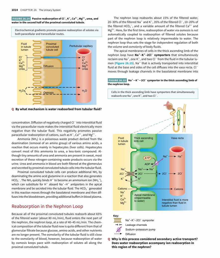
The nephron loop reabsorbs about 15% of the filtered water, 20–30% of the filtered Na+ and K+, 35% of the filtered Cl−, 10–20% of the filtered HCO3
−, and a variable amount of the filtered Ca2+ and Mg2+. Here, for the first time, reabsorption of water via osmosis is not automatically coupled to reabsorption of filtered solutes because part of the nephron loop is relatively impermeable to water. The nephron loop thus sets the stage for independent regulation of both the volume and osmolarity of body fluids.
The apical membranes of cells in the thick ascending limb of the nephron loop have Na+–K+–2Cl− symporters that simultaneously reclaim one Na+, one K+, and two Cl− from the fluid in the tubular lu-men (Figure 26.15). Na+ that is actively transported into interstitial fluid at the base and sides of the cell diff uses into the vasa recta. Cl− moves through leakage channels in the basolateral membrane into
concentration. Diff usion of negatively charged Cl− into interstitial fluid via the paracellular route makes the interstitial fluid electrically more negative than the tubular fluid. This negativity promotes passive paracellular reabsorption of cations, such as K+, Ca2+, and Mg2+.
Ammonia (NH3) is a poisonous waste product derived from the deamination (removal of an amino group) of various amino acids, a reaction that occurs mainly in hepatocytes (liver cells). Hepatocytes convert most of this ammonia to urea, a less-toxic compound. Al-though tiny amounts of urea and ammonia are present in sweat, most excretion of these nitrogen-containing waste products occurs via the urine. Urea and ammonia in blood are both filtered at the glomerulus and secreted by proximal convoluted tubule cells into the tubular fluid.
Proximal convoluted tubule cells can produce additional NH3 by deaminating the amino acid glutamine in a reaction that also generates HCO3
−. The NH3 quickly binds H+ to become an ammonium ion (NH4+),
which can substitute for H+ aboard Na+–H+ antiporters in the apical membrane and be secreted into the tubular fluid. The HCO3
− generated in this reaction moves through the basolateral membrane and then dif-fuses into the bloodstream, providing additional buff ers in blood plasma.
Reabsorption in the Nephron LoopBecause all of the proximal convoluted tubules reabsorb about 65% of the filtered water (about 80 mL/min), fluid enters the next part of the nephron, the nephron loop, at a rate of 40–45 mL/min. The chem-ical composition of the tubular fluid now is quite diff erent from that of glomerular filtrate because glucose, amino acids, and other nutrients are no longer present. The osmolarity of the tubular fluid is still close to the osmolarity of blood, however, because reabsorption of water by osmosis keeps pace with reabsorption of solutes all along the proximal convoluted tubule.
K+
H2O H2O
Peritubular capillaryFluidin tubulelumen
Proximalconvolutedtubule cell
Mg2+Ca2+
Diffusion
Osmosis
Cl–
Ca2+
Urea
K+
Mg2+
Urea
Cl–
Electrochemical gradients promote passive reabsorption of solutes via both paracellular and transcellular routes.
FIGURE 26.14 Passive reabsorption of Cl−, K+, Ca2+, Mg2+, urea, and water in the second half of the proximal convoluted tubule.
Q By what mechanism is water reabsorbed from tubular fluid?
Cells in the thick ascending limb have symporters that simultaneously reabsorb one Na+, one K+, and two Cl−.
FIGURE 26.15 Na+−K+−2Cl− symporter in the thick ascending limb of the nephron loop.
Q Why is this process considered secondary active transport? Does water reabsorption accompany ion reabsorption in this region of the nephron?
2Cl–
Vasa rectaThick ascendinglimb cell
K+
K+
Ca2+
Mg2+
Na+
2Cl–
Interstitial fluid is morenegative than fluid intubule lumen
Cations
2Cl–
Fluidin tubulelumen
2Cl–
K+
Na+
Na+
Apical membrane(impermeableto water)
Cations:
ADP
Na+
Na+
Na+
Na+–K+– 2Cl– symporter
Leakage channels
Sodium–potassium pump
Diffusion
Key:
ATP

26.6 Tubular Reabsorption and Tubular Secretion 1015
the bloodstream. To adjust for varying dietary intake of potassium and to maintain a stable level of K+ in body fluids, principal cells secrete a variable amount of K+ (Figure 26.16). Because the basolat-eral sodium–potassium pumps continually bring K+ into principal cells, the intracellular concentration of K+ remains high. K+ leakage channels are present in both the apical and basolateral membranes. Thus, some K+ diff uses down its concentration gradient into the tubu-lar fluid, where the K+ concentration is very low. This secretion mech-anism is the main source of K+ excreted in the urine.
Homeostatic Regulation of Tubular Reabsorption and Tubular SecretionFive hormones aff ect the extent of Na+, Ca2+, and water reabsorption as well as K+ secretion by the renal tubules. These hormones include angiotensin II, aldosterone, antidiuretic hormone, atrial natriuretic peptide, and parathyroid hormone.
interstitial fluid and then into the vasa recta. Because many K+ leak-age channels are present in the apical membrane, most K+ brought in by the symporters moves down its concentration gradient back into the tubular fluid. Thus, the main eff ect of the Na+−K+−2Cl− sym-porters is reabsorption of Na+ and Cl−.
The movement of positively charged K+ into the tubular fluid through the apical membrane channels leaves the interstitial fluid and blood with more negative charges relative to fluid in the ascending limb of the nephron loop. This relative negativity promotes reabsorp-tion of cations—Na+, K+, Ca2+, and Mg2+—via the paracellular route.
Although about 15% of the filtered water is reabsorbed in the descending limb of the nephron loop, little or no water is reabsorbed in the ascending limb. In this segment of the tubule, the apical mem-branes are virtually impermeable to water. Because ions but not water molecules are reabsorbed, the osmolarity of the tubular fluid decreas-es progressively as fluid flows toward the end of the ascending limb.
Reabsorption in the Early Distal Convoluted TubuleFluid enters the distal convoluted tubules at a rate of about 25 mL/min because 80% of the filtered water has now been reabsorbed. The early or initial part of the distal convoluted tubule (DCT) reabsorbs about 10–15% of the filtered water, 5% of the filtered Na+, and 5% of the filtered Cl−. Reabsorption of Na+ and Cl− occurs by means of Na+–Cl− symporters in the apical membranes. Sodium–potassium pumps and Cl− leakage channels in the basolateral membranes then permit reabsorption of Na+ and Cl− into the peritubular capillaries. The early DCT also is a major site where parathyroid hormone (PTH) stimulates reabsorption of Ca2+. The amount of Ca2+ reabsorption in the early DCT varies depending on the body’s needs.
Reabsorption and Secretion in the Late Distal Convoluted Tubule and Collecting DuctBy the time fluid reaches the end of the distal convoluted tubule, 90–95% of the filtered solutes and water have returned to the bloodstream. Recall that two diff erent types of cells—principal cells and intercalated cells—are present at the late or terminal part of the distal convoluted tubule and throughout the collecting duct. The principal cells reabsorb Na+ and secrete K+. These cells also have receptors for aldosterone and antidiuretic hormone (ADH). The intercalated cells reabsorb HCO3
− and secrete H+, thereby playing a role in blood pH regulation. In addition, the intercalated cells reabsorb K+. In the late distal convoluted tubules and collecting ducts, the amount of water and solute reabsorption and the amount of solute secretion vary depending on the body’s needs.
In contrast to earlier segments of the nephron, Na+ passes through the apical membrane of principal cells via Na+ leakage channels rather than by means of symporters or antiporters (Figure 26.16). The con-centration of Na+ in the cytosol remains low, as usual, because the sodium–potassium pumps actively transport Na+ across the basolat-eral membranes. Then Na+ passively diff uses into the peritubular cap-illaries from the interstitial spaces around the tubule cells.
Normally, transcellular and paracellular reabsorption in the prox-imal convoluted tubule and nephron loop return most filtered K+ to
In the apical membrane of principal cells, Na+ leakage channels allow entry of Na+ while K+ leakage channels allow exit of K+ into the tubular fluid.
FIGURE 26.16 Reabsorption of Na+ and secretion of K+ by principal cells in the last part of the distal convoluted tubule and in the collecting duct.
Diffusion
Sodium–potassium pump
Key:
Leakage channels
Peritubular capillaryFluidin tubulelumen
Principalcell
Na+
K+
Na+
Interstitialfluid
Na+
Na+
K+K+
ADP
ATP
Q Which hormone stimulates reabsorption and secretion by principal cells, and how does this hormone exert its effect?

1016 CHAPTER 26 The Urinary System
Renin–Angiotensin–Aldosterone System When blood volume and blood pressure decrease, the walls of the aff erent arterioles are stretched less, and the juxtaglomerular cells secrete the enzyme renin (RE--nin) into the blood. Sympathetic stimulation also directly stimulates release of renin from juxtaglomerular cells. Renin clips off a 10–amino acid peptide called angiotensin I (an′-jē-ō-TEN-sin) from angiotensinogen, which is synthesized by hepatocytes (see Figure 18.16). By clipping off two more amino acids, angiotensin-converting enzyme (ACE) converts angiotensin I to angiotensin II, which is the active form of the hormone.
Angiotensin II aff ects renal physiology in three main ways:
1. It decreases the glomerular filtration rate by causing vasoconstric-tion of the aff erent arterioles.
2. It enhances reabsorption of Na+ and water in the proximal convo-luted tubule by stimulating the activity of Na+–H+ antiporters.
3. It stimulates the adrenal cortex to release aldosterone (al-DOS-ter-ōn), a hormone that in turn stimulates the principal cells in the collecting ducts to reabsorb more Na+ and secrete more K+. The osmotic consequence of reabsorbing more Na+ is that more water is reabsorbed, which causes an increase in blood volume and blood pressure.
Antidiuretic Hormone Antidiuretic hormone (ADH) or vasopressin is released by the posterior pituitary. It regulates facultative water reabsorption by increasing the water permeability of principal cells in the last part of the distal convoluted tubule and throughout the collecting duct. In the absence of ADH, the apical membranes of principal cells have a very low permeability to water. Within principal cells are tiny vesicles containing many copies of a water channel protein known as aquaporin-2.* ADH stimulates insertion of the aquaporin-2–containing vesicles into the apical membranes via exocytosis. As a result, the water permeability of the principal cell’s apical membrane increases, and water molecules move more rapidly from the tubular fluid into the cells. Because the basolateral membranes are always relatively permeable to water, water molecules then move rapidly into the blood. This results in an increase in blood volume and blood pressure. When the ADH level declines, the aquaporin-2 channels are removed from the apical membrane via endocytosis, and water permeability of the principal cells decreases.
A negative feedback system involving ADH regulates facultative water reabsorption (Figure 26.17). When the osmolarity or osmotic pressure of plasma and interstitial fluid increases—that is, when water concentration decreases—by as little as 1%, osmoreceptors in the hypothalamus detect the change. Their nerve impulses stimulate secretion of more ADH into the blood, and the principal cells become more permeable to water. As facultative water reabsorption increases, plasma osmolarity decreases to normal. A second powerful stimulus for ADH secretion is a decrease in blood volume, as occurs in hemor-rhaging or severe dehydration. In the pathological absence of ADH activity, a condition known as diabetes insipidus, a person may excrete up to 20 liters of very dilute urine daily.
*ADH does not govern the previously mentioned water channel (aquaporin-1).
Most water reabsorption (90%) is obligatory; 10% is facultative.
FIGURE 26.17 Negative feedback regulation of facultative water reabsorption by ADH.
Q In addition to ADH, which other hormones contribute to the regulation of water reabsorption?
ADH which other hormones cont
STIMULUS
CONTROLLED CONDITION
Osmolarity of plasma and interstitial fluid
RESPONSE
Decrease in plasma osmolarity
RECEPTORS
CONTROL CENTER
Hypothalamus and posterior pituitary
Principal cells
EFFECTORS
Disrupts homeostasis by increasing
Return to homeostasis when the response brings plasma osmolarity back to normal
Become more permeable to water, which increases facultative water absorption
–
Output
Input
Increased releaseof ADH
Nerve impulses
Osmoreceptors in hypothalamus
ADH
The degree of facultative water reabsorption caused by ADH in the late distal tubule and collecting duct depends on whether the body is normally hydrated, dehydrated, or overhydrated.
• Normal hydration. Under conditions of normal body hydration (ad-equate water intake), enough ADH is present in the blood to cause

26.6 Tubular Reabsorption and Tubular Secretion 1017
decrease from just below 19% to as low as 0%. As a result, more than 1% of filtered water remains unreabsorbed in the late distal tubule and collecting duct, which corresponds to a urine output above the normal 1.5–2 L/day. The urine produced under these conditions is hypoosmotic (dilute) compared to blood because it contains more water than normal. In the case of severe overhydration, no ADH is present in the blood, and the amount of water reabsorbed in the late distal tubule and collecting duct is 0%. This means that the to-tal amount of filtered water that is reabsorbed in the renal tubule and collecting duct is 80%: 65% in the proximal tubule + 15% in the nephron loop + 0% in the late distal tubule and collecting duct. The remaining 20% of filtered water (about 36 L/day) is excreted in urine. Hence, the kidneys produce a large volume of dilute urine when the body is overhydrated.
Atrial Natriuretic Peptide A large increase in blood volume promotes release of atrial natriuretic peptide (ANP) from the heart. Although the importance of ANP in normal regulation of tubular function is unclear, it can inhibit reabsorption of Na+ and water in the proximal convoluted tubule and collecting duct. ANP also suppresses the secretion of aldosterone and ADH. These eff ects increase the excretion of Na+ in urine (natriuresis) and increase urine output (diuresis), which decreases blood volume and blood pressure.
Parathyroid Hormone Although the hormones mentioned thus far involve regulation of water loss as urine, the kidney tubules also respond to a hormone that regulates ionic composition. For example, a lower than normal level of Ca2+ in the blood stimulates the parathyroid glands to release parathyroid hormone (PTH). PTH in turn stimulates cells in the early distal convoluted tubules to reabsorb more Ca2+ into the blood. PTH also inhibits HPO4
2− (phosphate) reabsorption in proximal convoluted tubules, thereby promoting phosphate excretion.
Table 26.4 summarizes hormonal regulation of tubular reab-sorption and tubular secretion.
reabsorption of 19% of the filtered water in the late distal tubule and the collecting duct. This means that the total amount of filtered water reabsorbed in the renal tubule and collecting duct is 99%: 65% in the proximal tubule + 15% in the nephron loop + 19% in the late distal tubule and collecting duct. The remaining 1% of the filtered water (about 1.5–2 L/day) is excreted in urine. Therefore, when the body is normally hydrated, the kidneys produce about 1.5–2 L of urine on a daily basis and the urine is slightly hyperosmotic (slightly concentrated) compared to blood.
• Dehydration. When the body is dehydrated, the concentration of ADH in the blood increases. This in turn causes an increase in the amount of filtered water that is reabsorbed in the late distal tubule and collecting duct. Depending on how much the blood ADH level increases, the amount of filtered water that is reabsorbed in the late distal tubule and collecting duct can increase from just above 19% to as high as 19.8%. As a result, less than 1% of filtered water remains unreabsorbed in the late distal tubule and collecting duct, which corresponds to a urine output below the normal 1.5–2 L/day. The urine produced under these circumstances is very hyper-osmotic (highly concentrated) compared to blood because it con-tains less water than normal. In the case of severe dehydration, the amount of filtered water that is reabsorbed in the late distal tubule and collecting duct reaches a maximum limit of 19.8%. This means that the total amount of filtered water reabsorbed in the renal tubule and collecting duct is 99.8%: 65% in the proximal tubule + 15% in the nephron loop + 19.8% in the late distal tubule and col-lecting duct. The remaining 0.2% of the filtered water (about 400 mL/day) is excreted in urine. Thus, the kidneys produce a small volume of highly concentrated urine when the body is dehydrated.
• Overhydration. When the body is overhydrated (too much water in-take), the concentration of ADH in the blood decreases. This in turn causes a decrease in the amount of filtered water that is reabsorbed in the late distal tubule and collecting duct. Depending on how much the blood ADH level decreases, the amount of filtered water that is reabsorbed in the late distal tubule and collecting duct can
TABLE 26.4 Hormonal Regulation of Tubular Reabsorption and Tubular Secretion
HORMONE MAJOR STIMULI THAT TRIGGER RELEASE MECHANISM AND SITE OF ACTION EFFECTS
Angiotensin II Low blood volume or low blood pressure stimulates renin-induced production of angiotensin II.
Stimulates activity of Na+–H+ antiporters in proximal tubule cells.
Increases reabsorption of Na+ and water, which increases blood volume and blood pressure.
Aldosterone Increased angiotensin II level and increased level of plasma K+ promote release of aldosterone by adrenal cortex.
Enhances activity of sodium–potassium pumps in basolateral membrane and Na+ channels in apical membrane of principal cells in collecting duct.
Increases secretion of K+ and reabsorption of Na+; increases reabsorption of water, which increases blood volume and blood pressure.
Antidiuretic hormone (ADH)
Increased osmolarity of extracellular fluid or decreased blood volume promotes release of ADH from posterior pituitary gland.
Stimulates insertion of water channel proteins (aquaporin-2) into apical membranes of principal cells.
Increases facultative reabsorption of water, which decreases osmolarity of body fluids.
Atrial natriuretic peptide (ANP)
Stretching of atria of heart stimulates ANP secretion.
Suppresses reabsorption of Na+ and water in proximal tubule and collecting duct; inhibits secretion of aldosterone and ADH.
Increases excretion of Na+ in urine (natriuresis); increases urine output (diuresis) and thus decreases blood volume and blood pressure.
Parathyroid hormone (PTH)
Decreased level of plasma Ca2+ promotes release of PTH from parathyroid glands.
Stimulates opening of Ca2+ channels in apical membranes of early distal tubule cells.
Increases reabsorption of Ca2+.

1018 CHAPTER 26 The Urinary System
2. Cells lining the thick ascending limb of the loop have symport-ers that actively reabsorb Na+, K+, and Cl− from the tubular fluid (see Figure 26.15). The ions pass from the tubular fluid into thick ascending limb cells, then into interstitial fluid, and finally some diff use into the blood inside the vasa recta.
3. Although solutes are being reabsorbed in the thick ascending limb, the water permeability of this portion of the nephron is always quite low, so water cannot follow by osmosis. As solutes—but not water molecules—are leaving the tubular fluid, its osmolarity drops to about 150 mOsm/liter. The fluid entering the distal convoluted tubule is thus more dilute than plasma.
4. While the fluid continues flowing along the distal convoluted tubule, additional solutes but only a few water molecules are reab-sorbed. The early distal convoluted tubule cells are not very perme-able to water and are not regulated by ADH.
5. Finally, the principal cells of the late distal convoluted tubules and collecting ducts are impermeable to water when the ADH level is very low. Thus, tubular fluid becomes progressively more dilute as
Checkpoint
17. Diagram the reabsorption of substances via the transcellular and paracellular routes. Label the apical membrane and the basolateral membrane. Where are the sodium–potassium pumps located?
18. Describe two mechanisms in the PCT, one in the nephron loop, one in the DCT, and one in the collecting duct for reabsorption of Na+. What other solutes are reabsorbed or secreted with Na+ in each mechanism?
19. How do intercalated cells secrete hydrogen ions?
20. Graph the percentages of filtered water and filtered Na+ that are reabsorbed in the PCT, nephron loop, DCT, and collecting duct. Indicate which hormones, if any, regulate reabsorption in each segment.
26.7 Production of Dilute and Concentrated Urine
OBJECTIVE
• Describe how the renal tubule and collecting ducts produce dilute and concentrated urine.
Even though your fluid intake can be highly variable, the total volume of fluid in your body normally remains stable. Homeostasis of body fluid volume depends in large part on the ability of the kidneys to regulate the rate of water loss in urine. Normally functioning kidneys produce a large volume of dilute urine when fluid intake is high, and a small volume of concentrated urine when fluid intake is low or fluid loss is large. ADH con-trols whether dilute urine or concentrated urine is formed. In the absence of ADH, urine is very dilute. However, a high level of ADH stimulates reab-sorption of more water into blood, producing a concentrated urine.
Formation of Dilute UrineGlomerular filtrate has the same ratio of water and solute particles as blood; its osmolarity is about 300 mOsm/liter. As previously noted, fluid leaving the proximal convoluted tubule is still isotonic to plasma. When dilute urine is being formed (Figure 26.18), the osmolarity of the fluid in the tubular lumen increases as it flows down the descending limb of the nephron loop, decreases as it flows up the ascending limb, and decreases still more as it flows through the rest of the nephron and collecting duct. These changes in osmolarity result from the fol-lowing conditions along the path of tubular fluid:
1. Because the osmolarity of the interstitial fluid of the renal medulla becomes progressively greater, more and more water is reabsorbed by osmosis as tubular fluid flows along the descending limb toward the tip of the nephron loop. (The source of this medullary osmotic gradient is explained shortly.) As a result, the fluid remaining in the lumen becomes progressively more concentrated.
When the ADH level is low, urine is dilute and has an osmolarity less than the osmolarity of blood.
FIGURE 26.18 Formation of dilute urine. Numbers indicate osmolarity in milliosmoles per liter (mOsm/liter). Heavy brown lines in the ascending limb of the nephron loop and in the distal convoluted tubule indicate impermeability to water; heavy blue lines indicate the last part of the distal convoluted tubule and the collecting duct, which are impermeable to water in the absence of ADH; light blue areas around the nephron represent interstitial fluid.
Q Which portions of the renal tubule and collecting duct reabsorb more solutes than water to produce dilute urine?
Glomerular (Bowman’s) capsuleAfferentarteriole
Efferentarteriole
Nephron loop
Papillaryduct
Collectingduct
Glomerulus
Proximalconvolutedtubule
Diluteurine
Interstitialfluid inrenalmedulla
Interstitialfluid inrenalcortex
100
90
65900
550750
350300
Distal convolutedtubule
150
550 350 80
300
70
750
350
550
750
350
550
300
65
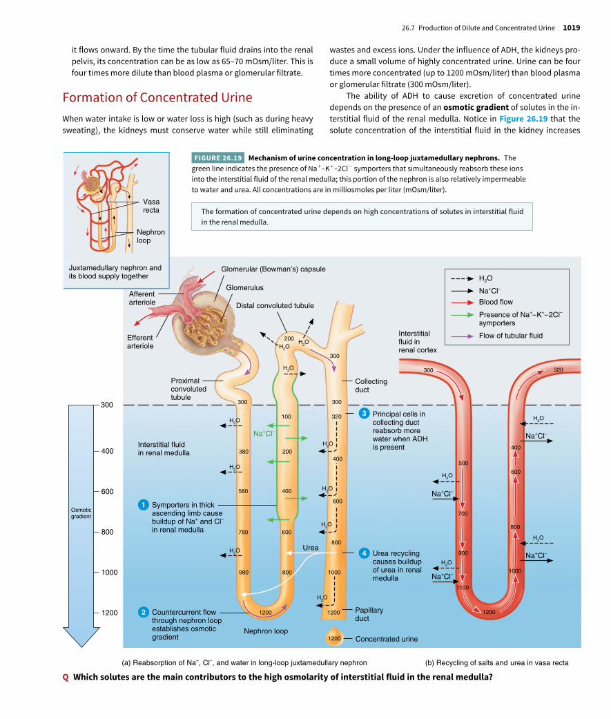
26.7 Production of Dilute and Concentrated Urine 1019
wastes and excess ions. Under the influence of ADH, the kidneys pro-duce a small volume of highly concentrated urine. Urine can be four times more concentrated (up to 1200 mOsm/liter) than blood plasma or glomerular filtrate (300 mOsm/liter).
The ability of ADH to cause excretion of concentrated urine depends on the presence of an osmotic gradient of solutes in the in-terstitial fluid of the renal medulla. Notice in Figure 26.19 that the solute concentration of the interstitial fluid in the kidney increases
it flows onward. By the time the tubular fluid drains into the renal pelvis, its concentration can be as low as 65–70 mOsm/liter. This is four times more dilute than blood plasma or glomerular filtrate.
Formation of Concentrated UrineWhen water intake is low or water loss is high (such as during heavy sweating), the kidneys must conserve water while still eliminating
Q Which solutes are the main contributors to the high osmolarity of interstitial fluid in the renal medulla?(b) Recycling of salts and urea in vasa recta(a) Reabsorption of Na+, Cl–, and water in long-loop juxtamedullary nephron
Glomerular (Bowman’s) capsule
Afferentarteriole
Efferentarteriole
Glomerulus
Distal convoluted tubule
Proximalconvolutedtubule
Symporters in thickascending limb causebuildup of Na+ and Cl–
in renal medulla
Interstitial fluidin renal medulla
300
1200
1000
800
Osmoticgradient
600
400
Na+Cl–
H2OH2O
H2O
200
1200
980
600780
400580
200380
300
100H2O
H2O
H2O
Nephron loop1200 Concentrated urine
300
300
320
400
600
800
1000
1200
800
H2O
H2O
H2O
H2O
Urea
Papillaryduct
Urea recyclingcauses buildupof urea in renal medulla
Collectingduct
Countercurrent flowthrough nephron loopestablishes osmoticgradient
Principal cells incollecting ductreabsorb morewater when ADHis present
H2O
300
500
700
900
1100
1200
400
800
1000
600
Na+Cl–
Na+Cl–
Na+Cl–
Na+Cl–
Na+Cl–
Blood flow
Flow of tubular fluid
Presence of Na+–K+–2Cl–
symportersInterstitialfluid inrenal cortex
320
Juxtamedullary nephron andits blood supply together
Vasarecta
Nephronloop
H2O
H2O
H2O
H2O
1
4
3
2
The formation of concentrated urine depends on high concentrations of solutes in interstitial fluid in the renal medulla.
FIGURE 26.19 Mechanism of urine concentration in long-loop juxtamedullary nephrons. The green line indicates the presence of Na+–K+–2Cl− symporters that simultaneously reabsorb these ions into the interstitial fluid of the renal medulla; this portion of the nephron is also relatively impermeable to water and urea. All concentrations are in milliosmoles per liter (mOsm/liter).

1020 CHAPTER 26 The Urinary System
but its symporters reabsorb Na+ and Cl− from the tubular fluid into the interstitial fluid of the renal medulla, so the osmolarity of the tubular fluid progressively decreases as it flows through the ascending limb. At the junction of the medulla and cortex, the osmolarity of the tubular fluid has fallen to about 100 mOsm/liter. Overall, tubular fluid becomes progressively more concentrated as it flows along the descending limb and progressively more dilute as it moves along the ascending limb.
3 Cells in the collecting ducts reabsorb more water and urea. When ADH increases the water permeability of the principal cells, water quickly moves via osmosis out of the collecting duct tubular fluid, into the interstitial fluid of the inner medulla, and then into the vasa recta. With loss of water, the urea left behind in the tubular fluid of the collecting duct becomes increasingly concentrated. Because duct cells deep in the medulla are permeable to it, urea diff uses from the fluid in the duct into the interstitial fluid of the medulla.
4 Urea recycling causes a buildup of urea in the renal medulla. As urea accumulates in the interstitial fluid, some of it diff uses into the tubular fluid in the descending and thin ascending limbs of the long nephron loops, which also are permeable to urea (Figure 26.19a). However, while the fluid flows through the thick ascending limb, distal convoluted tubule, and cortical portion of the collecting duct, urea remains in the lumen because cells in these segments are impermeable to it. As fluid flows along the collecting ducts, water reabsorption continues via osmosis because ADH is present. This water reabsorption further increases the concentration of urea in the tubular fluid, more urea diff uses into the interstitial fluid of the inner renal medulla, and the cycle repeats. The constant transfer of urea between segments of the renal tubule and the interstitial fluid of the medulla is termed urea recycling. In this way, reabsorption of water from the tubular fluid of the ducts promotes the buildup of urea in the interstitial fluid of the renal medulla, which in turn promotes water reabsorption. The solutes left behind in the lumen thus become very concentrated, and a small volume of concentrated urine is excreted.
Countercurrent Exchange Countercurrent exchange is the process by which solutes and water are passively exchanged between the blood of the vasa recta and interstitial fluid of the renal medulla as a result of countercurrent flow. Note in Figure 26.19b that the vasa recta also consists of descending and ascending limbs that are parallel to each other and to the nephron loop. Just as tubular fluid flows in opposite directions in the nephron loop, blood flows in opposite directions in the ascending and descending parts of the vasa recta. Since countercurrent flow between the descending and ascending limbs of the vasa recta allows for exchange of solutes and water between the blood and interstitial fluid of the renal medulla, the vasa recta is said to function as a countercurrent exchanger.
Blood entering the vasa recta has an osmolarity of about 300 mOsm/liter. As it flows along the descending part into the renal medulla, where the interstitial fluid becomes increasingly concentrated, Na+, Cl−, and urea diff use from interstitial fluid into the blood and water diff uses from the blood into the interstitial fluid. But aft er its osmolar-ity increases, the blood flows into the ascending part of the vasa recta. Here blood flows through a region where the interstitial fluid becomes
from about 300 mOsm/liter in the renal cortex to about 1200 mOsm/liter deep in the renal medulla. The three major solutes that con tri bute to this high osmolarity are Na+, Cl−, and urea. Two main factors con-tribute to building and maintaining this osmotic gradient: (1) diff er-ences in solute and water permeability and reabsorption in diff erent sections of the long nephron loops and the collecting ducts, and (2) the countercurrent flow of fluid through tube-shaped structures in the renal medulla. Countercurrent flow refers to the flow of fluid in op-posite directions. This occurs when fluid flowing in one tube runs counter (opposite) to fluid flowing in a nearby parallel tube. Examples of countercurrent flow include the flow of tubular fluid through the descending and ascending limbs of the nephron loop and the flow of blood through the ascending and descending parts of the vasa recta. Two types of countercurrent mechanisms exist in the kidneys: coun-tercurrent multiplication and countercurrent exchange.
Countercurrent Multiplication Countercurrent multi-plication is the process by which a progressively increasing osmotic gradient is formed in the interstitial fluid of the renal medulla as a result of countercurrent flow. Countercurrent multiplication involves the long nephron loops of juxtamedullary nephrons. Note in Figure 26.19a that the descending limb of the nephron loop carries tubular fluid from the renal cortex deep into the medulla, and the ascending limb carries it in the opposite direction. Since countercurrent flow through the descending and ascending limbs of the long nephron loop establishes the osmotic gradient in the renal medulla, the long nephron loop is said to function as a countercurrent multiplier. The kidneys use this osmotic gradient to excrete concentrated urine.
Production of concentrated urine by the kidneys occurs in the fol-lowing way (Figure 26.19):
1 Symporters in thick ascending limb cells of the nephron loop cause a buildup of Na+ and Cl− in the renal medulla. In the thick ascending limb of the nephron loop, the Na+–K+–2Cl− symporters reabsorb Na+ and Cl− from the tubular fluid (Figure 26.19a). Water is not reabsorbed in this segment, however, because the cells are impermeable to water. As a result, there is a buildup of Na+ and Cl− ions in the interstitial fluid of the medulla.
2 Countercurrent flow through the descending and ascending limbs of the nephron loop establishes an osmotic gradient in the renal medulla. Since tubular fluid constantly moves from the descending limb to the thick ascending limb of the nephron loop, the thick ascending limb is constantly reabsorbing Na+ and Cl−. Consequently, the reabsorbed Na+ and Cl− become increasingly concentrated in the interstitial fluid of the medulla, which results in the formation of an osmotic gradient that ranges from 300 mOsm/liter in the outer medulla to 1200 mOsm/liter deep in the inner medulla. The descending limb of the nephron loop is very permeable to water but impermeable to solutes except urea. Because the osmolarity of the interstitial fluid outside the descending limb is higher than the tubular fluid within it, water moves out of the descending limb via osmosis. This causes the osmolarity of the tubular fluid to increase. As the fluid continues along the descending limb, its osmolarity increases even more: At the hairpin turn of the loop, the osmolarity can be as high as 1200 mOsm/liter in juxtamedullary nephrons. As you have already learned, the ascending limb of the loop is impermeable to water,

26.7 Production of Dilute and Concentrated Urine 1021
Q In which segments of the nephron and collecting duct does secretion occur?
EARLY DISTAL CONVOLUTED TUBULE
Reabsorption (into blood) of:
Water 10–15% (osmosis)
Na+ 5% (symporters)
CI– 5% (symporters)
Ca2+ variable (stimulated by parathyroid hormone)
LATE DISTAL CONVOLUTED TUBULEAND COLLECTING DUCT
Reabsorption (into blood) of:
Water 5–9% (insertion of water channels stimulated by ADH)Na+ 1–4% (sodium–potassium pumps and sodium channels stimulated by aldosterone)
Urea variable (recycling to nephron loop)
Secretion (into urine) of:
K+ variable amount to adjust for dietary intake (leakage channels)
Tubular fluid leaving the collecting ductis dilute when ADH level is low andconcentrated when ADH level is high.
RENAL CORPUSCLE
Glomerular filtration rate: 105–125 mL/min of fluid that is isotonic to blood
Filtered substances: water and all solutes present in blood (except proteins) including ions, glucose, amino acids, creatinine, uric acid
H+ variable amounts to maintain acid–base homeostasis (H+ pumps)
PROXIMAL CONVOLUTED TUBULE
Reabsorption (into blood) of filtered:
Water 65% (osmosis)
Na+ 65% (sodium–potassium pumps, symporters, antiporters)
K+ 65% (diffusion)
Glucose 100% (symporters and facilitated diffusion)
Amino acids 100% (symporters and facilitated diffusion)
CI– 50% (diffusion)
HCO3– 80–90% (facilitated
diffusion)
Urea 50% (diffusion)
Secretion (into urine) of:
H+ variable (antiporters)
Ca2+, Mg2+ variable (diffusion)
NH4+ variable, increases in
acidosis (antiporters)
Urea variable (diffusion)
Creatinine small amount
At end of PCT, tubular fluid is stillisotonic to blood (300 mOsm/liter).
NEPHRON LOOP
Reabsorption (into blood) of:
Water 15% (osmosis in descending limb)
Na+ 20–30% (symporters in ascending limb)
K+ 20–30% (symporters in ascending limb)
CI– 35% (symporters in ascending limb)
HCO3– 10–20% (facilitated
diffusion)
Secretion (into urine) of:
Urea variable (recycling from collecting duct)
At end of nephron loop, tubular fluid ishypotonic (100–150 mOsm/liter).
Ca2+, Mg2+ variable (diffusion)
Urine
HCO3– variable amount, depends
on H+ secretion (antiporters)
Filtration occurs in the renal corpuscle; reabsorption occurs all along the renal tubule and collecting ducts.
FIGURE 26.20 Summary of filtration, reabsorption, and secretion in the nephron and collecting duct.
increasingly less concentrated. As a result Na+, Cl−, and urea diff use from the blood back into interstitial fluid, and water diff uses from in-terstitial fluid back into the vasa recta. The osmolarity of blood leav-ing the vasa recta is only slightly higher than the osmolarity of blood entering the vasa recta. Thus, the vasa recta provides oxygen and nu-trients to the renal medulla without washing out or diminishing the
osmotic gradient. The long nephron loop establishes the osmotic gra-dient in the renal medulla by countercurrent multiplication, but the vasa recta maintains the osmotic gradient in the renal medulla by countercurrent exchange.
Figure 26.20 summarizes the processes of filtration, reabsorption, and secretion in each segment of the nephron and collecting duct.

1022 CHAPTER 26 The Urinary System
tubules and decreases urine volume. By contrast, when blood osmo-larity decreases—for example, aft er drinking a large volume of water—secretion of ADH is inhibited and a larger volume of urine is excreted.
Water accounts for about 95% of the total volume of urine. The remaining 5% consists of electrolytes, solutes derived from cellular metabolism, and exogenous substances such as drugs. Normal urine is virtually protein-free. Typical solutes normally present in urine in-clude filtered and secreted electrolytes that are not reabsorbed, urea (from breakdown of proteins), creatinine (from breakdown of creatine phosphate in muscle fibers), uric acid (from breakdown of nucleic acids), urobilinogen (from breakdown of hemoglobin), and small quantities of other substances, such as fatty acids, pigments, en-zymes, and hormones.
If disease alters body metabolism or kidney function, traces of substances not normally present may appear in the urine, or normal constituents may appear in abnormal amounts. Table 26.6 lists sev-eral abnormal constituents in urine that may be detected as part of a urinalysis. Normal values of urine components and the clinical impli-cations of deviations from normal are listed in Appendix D.
Blood TestsTwo blood-screening tests can provide information about kidney function. One is the blood urea nitrogen (BUN) test, which measures
Clinical Connection
DiureticsDiuretics (dī-ū-RET-iks) are substances that slow renal reabsorption of water and thereby cause diuresis, an elevated urine flow rate, which in turn reduces blood volume. Diuretic drugs oft en are prescribed to treat hypertension (high blood pressure) because lowering blood volume usu-ally reduces blood pressure. Naturally occurring diuretics include caff eine in coff ee, tea, and sodas, which inhibits Na+ reabsorption, and alcohol in beer, wine, and mixed drinks, which inhibits secretion of ADH. Most diuretic drugs act by interfering with a mechanism for reabsorption of filtered Na+. For example, loop diuretics, such as furosemide (Lasix®), selectively inhibit the Na+–K+–2Cl− symporters in the thick ascending limb of the nephron loop (see Figure 26.15). The thiazide diuretics, such as chlorothiazide (Diuril®), act in the distal convoluted tubule, where they promote loss of Na+ and Cl− in the urine by inhibiting Na+–Cl− symporters.
Checkpoint
21. How do symporters in the ascending limb of the nephron loop and principal cells in the collecting duct contribute to the formation of concentrated urine?
22. How does ADH regulate facultative water reabsorption?
23. What is the countercurrent mechanism? Why is it important?
26.8 Evaluation of Kidney Function
OBJECTIVES
• Define urinalysis and describe its importance.
• Define renal plasma clearance and describe its importance.
Routine assessment of kidney function involves evaluating both the quantity and quality of urine and the levels of wastes in the blood.
UrinalysisAn analysis of the volume and physical, chemical, and microscopic properties of urine, called a urinalysis (ū-ri-NAL-i-sis), reveals much about the state of the body. Table 26.5 summarizes the major char-acteristics of normal urine. The volume of urine eliminated per day in a normal adult is 1–2 liters (about 1–2 qt). Fluid intake, blood pres-sure, blood osmolarity, diet, body temperature, diuretics, mental state, and general health influence urine volume. For example, low blood pressure triggers the renin–angiotensin–aldosterone pathway. Aldosterone increases reabsorption of water and salts in the renal
TABLE 26.5 Characteristics of Normal Urine
CHARACTERISTIC DESCRIPTION
Volume One to two liters in 24 hours; varies considerably.
Color Yellow or amber; varies with urine concentration and diet. Color due to urochrome (pigment produced from breakdown of bile) and urobilin (from breakdown of hemoglobin). Concentrated urine is darker in color. Color affected by diet (reddish from beets), medications, and certain diseases. Kidney stones may produce blood in urine.
Turbidity Transparent when freshly voided; becomes turbid (cloudy) on standing.
Odor Mildly aromatic; becomes ammonia-like on standing. Some people inherit ability to form methylmercaptan from digested asparagus, which gives characteristic odor. Urine of diabetics has fruity odor due to presence of ketone bodies.
pH Ranges between 4.6 and 8.0; average 6.0; varies considerably with diet. High-protein diets increase acidity; vegetarian diets increase alkalinity.
Specific gravity (density)
Specific gravity (density) is ratio of weight of volume of substance to weight of equal volume of distilled water. In urine, 1.001–1.035. The higher the concentration of solutes, the higher the specific gravity.

26.8 Evaluation of Kidney Function 1023
of a substance in the urine; low clearance indicates ineff icient excre-tion. For example, the clearance of glucose normally is zero because it is completely reabsorbed (see Table 26.3); therefore, glucose is not excreted at all. Knowing a drug’s clearance is essential for determin-ing the correct dosage. If clearance is high (one example is penicillin), then the dosage must also be high, and the drug must be given sev-eral times a day to maintain an adequate therapeutic level in the blood.
The following equation is used to calculate clearance:
Renal plasma clearance of substance S = U × V
P) )where U and P are the concentrations of the substance in urine and plasma, respectively (both expressed in the same units, such as mg/mL), and V is the urine flow rate in mL/min.
The clearance of a solute depends on the three basic processes of a nephron: glomerular filtration, tubular reabsorption, and tubu-lar secretion. Consider a substance that is filtered but neither reab-sorbed nor secreted. Its clearance equals the glomerular filtration rate because all molecules that pass the filtration membrane appear in the urine. This is the situation for the plant polysaccharide inulin (IN-ū-lin); it easily passes the filter, it is not reabsorbed, and it is not secreted. (Do not confuse inulin with the hormone insulin, which is produced by the pancreas.) Typically, the clearance of inulin is about 125 mL/min, which equals the GFR. Clinically, the clearance of inulin
the blood nitrogen that is part of the urea resulting from catabolism and deamination of amino acids. When glomerular filtration rate decreases severely, as may occur with renal disease or obstruction of the urinary tract, BUN rises steeply. One strategy in treating such patients is to minimize their protein intake, thereby reducing the rate of urea production.
Another test oft en used to evaluate kidney function is measure-ment of plasma creatinine (krē-AT-i-nin), which results from catabo-lism of creatine phosphate in skeletal muscle. Normally, the blood creatinine level remains steady because the rate of creatinine excre-tion in the urine equals its discharge from muscle. A creatinine level above 1.5 mg/dL (135 mmol/liter) usually is an indication of poor renal function. Normal values for selected blood tests are listed in Appendix C along with situations that may cause the values to increase or decrease.
Renal Plasma ClearanceEven more useful than BUN and blood creatinine values in the diag-nosis of kidney problems is an evaluation of how eff ectively the kid-neys are removing a given substance from blood plasma. Renal plasma clearance is the volume of blood that is “cleaned” or cleared of a substance per unit of time, usually expressed in units of milliliters per minute. High renal plasma clearance indicates eff icient excretion
TABLE 26.6 Summary of Abnormal Constituents in Urine
ABNORMAL CONSTITUENT COMMENTS
Albumin Normal constituent of plasma; usually appears in only very small amounts in urine because it is too large to pass through capillary fenestrations. Presence of excessive albumin in urine—albuminuria (al′-bū-mi-NOO-rē-a)—indicates increase in permeability of filtration membranes due to injury or disease, increased blood pressure, or irritation of kidney cells by substances such as bacterial toxins, ether, or heavy metals.
Glucose Presence of glucose in urine—glucosuria (gloo-kō-SOO-rē-a)—usually indicates diabetes mellitus. Occasionally caused by stress, which can cause excessive epinephrine secretion. Epinephrine stimulates breakdown of glycogen and liberation of glucose from liver.
Red blood cells (erythrocytes) Presence of red blood cells in urine—hematuria (hēm-a-TOO-rē-a)—generally indicates pathological condition. One cause is acute inflammation of urinary organs due to disease or irritation from kidney stones. Other causes: tumors, trauma, kidney disease, contamination of sample by menstrual blood.
Ketone bodies High levels of ketone bodies in urine—ketonuria (kē-tō-NOO-rē-a)—may indicate diabetes mellitus, anorexia, starvation, or too little carbohydrate in diet.
Bilirubin When red blood cells are destroyed by macrophages, the globin portion of hemoglobin is split off and heme is converted to biliverdin. Most biliverdin is converted to bilirubin, which gives bile its major pigmentation. Above-normal level of bilirubin in urine is called bilirubinuria (bil′-ē-roo-bi-NOO-rē-a).
Urobilinogen Presence of urobilinogen (breakdown product of hemoglobin) in urine is called urobilinogenuria (ū′-rō-bi-lin′-ō-je-NOO-rē-a). Trace amounts are normal, but elevated urobilinogen may be due to hemolytic or pernicious anemia, infectious hepatitis, biliary obstruction, jaundice, cirrhosis, congestive heart failure, or infectious mononucleosis.
Casts Casts are tiny masses of material that have hardened and assumed shape of lumen of tubule in which they formed, from which they are flushed when filtrate builds up behind them. Casts are named after cells or substances that compose them or based on appearance (for example, white blood cell casts, red blood cell casts, and epithelial cell casts that contain cells from walls of tubules).
Microbes Number and type of bacteria vary with specific urinary tract infections. One of the most common is E. coli. Most common fungus is yeast Candida albicans, cause of vaginitis. Most frequent protozoan is Trichomonas vaginalis, cause of vaginitis in females and urethritis in males.

1024 CHAPTER 26 The Urinary System
Aft er PAH is administered intravenously, it is filtered and secreted in a single pass through the kidneys. Thus, the clearance of PAH is used to measure renal plasma flow, the amount of plasma that passes through the kidneys in one minute. Typically, the renal plasma flow is 650 mL per minute, which is about 55% of the renal blood flow (1200 mL per minute).
Checkpoint
24. What are the characteristics of normal urine?
25. What chemical substances normally are present in urine?
26. How may kidney function be evaluated?
27. Why are the renal plasma clearances of glucose, urea, and creatinine diff erent? How does each clearance compare to glomerular filtration rate?
26.9 Urine Transportation, Storage, and Elimination
OBJECTIVE
• Describe the anatomy, histology, and physiology of the ureters, urinary bladder, and urethra.
From collecting ducts, urine drains into the minor calyces, which join to become major calyces that unite to form the renal pelvis (see Fig-ure 26.3). From the renal pelvis, urine first drains into the ureters and then into the urinary bladder. Urine is then discharged from the body through the single urethra (see Figure 26.1).
UretersEach of the two ureters (Ū-rē-ters) transports urine from the renal pelvis of one kidney to the urinary bladder. Peristaltic contractions of the muscular walls of the ureters push urine toward the urinary blad-der, but hydrostatic pressure and gravity also contribute. Peristaltic waves that pass from the renal pelvis to the urinary bladder vary in frequency from one to five per minute, depending on how fast urine is being formed.
The ureters are 25–30 cm (10–12 in.) long and are thick-walled, narrow tubes that vary in diameter from 1 mm to 10 mm along their course between the renal pelvis and the urinary bladder. Like the kid-neys, the ureters are retroperitoneal. At the base of the urinary blad-der, the ureters curve medially and pass obliquely through the wall of the posterior aspect of the urinary bladder (Figure 26.21).
Even though there is no anatomical valve at the opening of each ureter into the urinary bladder, a physiological one is quite eff ective. As the urinary bladder fills with urine, pressure within it compresses the oblique openings into the ureters and prevents the backflow of urine. When this physiological valve is not operating properly, it is
can be used to determine the GFR. The clearance of inulin is obtained in the following way: Inulin is administered intravenously and then the concentrations of inulin in plasma and urine are measured along with the urine flow rate. Although using the clearance of inulin is an accurate method for determining the GFR, it has its drawbacks: Inulin is not produced by the body and it must be infused continuously while clearance measurements are being determined. Measuring the creatinine clearance is an easier way to assess the GFR because creatinine is a substance that is naturally produced by the body as an end product of muscle metabolism. Once creatinine is filtered, it is not reabsorbed, and is secreted only to a very small extent. Because there is a small amount of creatinine secretion, the creatinine clear-ance is only a close estimate of the GFR and is not as accurate as using the inulin clearance. The creatinine clearance is normally about 120–140 mL/min.
Clinical Connection
DialysisIf a person’s kidneys are so impaired by disease or injury that he or she is unable to function adequately, then blood must be cleansed artificially by dialysis (dī-AL-i-sis; dialyo = to separate), the separation of large sol-utes from smaller ones by diff usion through a selectively permeable mem-brane. One method of dialysis is hemodialysis (hē-mō-dī-AL-i-sis; hemo- = blood), which directly filters the patient’s blood by removing wastes and excess electrolytes and fluid and then returning the cleansed blood to the patient. Blood removed from the body is delivered to a hemodialyzer (arti-ficial kidney). Inside the hemodialyzer, blood flows through a dialysis mem-brane, which contains pores large enough to permit the diff usion of small solutes. A special solution, called the dialysate (dī-AL-i-sāt), is pumped into the hemodialyzer so that it surrounds the dialysis membrane. The dialysate is specially formulated to maintain diff usion gradients that remove wastes from the blood (such as urea, creatinine, uric acid, excess phosphate, po-tassium, and sulfate ions) and add needed substances (such as glucose and bicarbonate ions) to it. The cleansed blood is passed through an air embo-lus detector to remove air and then returned to the body. An anticoagulant (heparin) is added to prevent blood from clotting in the hemodialyzer. As a rule, most people on hemodialysis require about 6–12 hours a week, typi-cally divided into three sessions.
Another method of dialysis, called peritoneal dialysis (per′-i-tō-NE--al), uses the peritoneum of the abdominal cavity as the dialysis membrane to filter the blood. The peritoneum has a large surface area and numerous blood vessels, and is a very eff ective filter. A catheter is inserted into the peritoneal cavity and connected to a bag of dialysate. The fluid flows into the peritoneal cavity by gravity and is left there for suff icient time to permit wastes and excess electrolytes and fluids to diff use into the dialysate. Then the dialysate is drained out into a bag, discarded, and replaced with fresh dialysate.
Each cycle is called an exchange. One variation of peritoneal dialy-sis, called continuous ambulatory peritoneal dialysis (CAPD), can be performed at home. Usually, the dialysate is drained and replenished four times a day and once at night during sleep. Between exchanges the person can move about freely with the dialysate in the peritoneal cavity.
The clearance of the organic anion para-aminohippuric acid (PAH) (par′-a-a-mē′-nō-hi-PYOOR-ik) is also of clinical importance.

26.9 Urine Transportation, Storage, and Elimination 1025
Urinary BladderThe urinary bladder is a hollow, distensible muscular organ situated in the pelvic cavity posterior to the pubic symphysis. In males, it is directly anterior to the rectum; in females, it is anterior to the vagina and inferior to the uterus (see Figure 26.22). Folds of the peritoneum hold the urinary bladder in position. When slightly distended due to the accumulation of urine, the urinary bladder is spherical. When it is empty, it collapses. As urine volume increases, it becomes pear-shaped and rises into the abdominal cavity. Urinary bladder capacity averages 700–800 mL. It is smaller in females because the uterus occupies the space just superior to the urinary bladder.
Anatomy and Histology of the Urinary Bladder In the floor of the urinary bladder is a small triangular area called the trigone (TRĪ-gōn = triangle). The two posterior corners of the trigone contain the two ureteral openings; the opening into the urethra, the internal urethral orifice (OR-i-fis), lies in the anterior corner (see Figure 26.21). Because its mucosa is firmly bound to the muscularis, the trigone has a smooth appearance.
Three coats make up the wall of the urinary bladder. The deepest is the mucosa, a mucous membrane composed of transitional epithelium and an underlying lamina propria similar to that of the ureters. The transitional epithelium permits stretching. Rugae (the folds in the mucosa) are also present to permit expansion of the urinary bladder. Surrounding the mucosa is the intermediate muscularis,
possible for microbes to travel up the ureters from the urinary bladder to infect one or both kidneys.
Three layers of tissue form the wall of the ureters. The deepest coat, the mucosa, is a mucous membrane with transitional epithe-lium (see Table 4.1) and an underlying lamina propria of areolar connective tissue with considerable collagen, elastic fibers, and lym-phatic tissue. Transitional epithelium is able to stretch—a marked advantage for any organ that must accommodate a variable volume of fluid. Mucus secreted by the goblet cells of the mucosa prevents the cells from coming in contact with urine, the solute concentration and pH of which may diff er drastically from the cytosol of cells that form the wall of the ureters.
Throughout most of the length of the ureters, the intermediate coat, the muscularis, is composed of inner longitudinal and outer cir-cular layers of smooth muscle fibers. This arrangement is opposite to that of the gastrointestinal tract, which contains inner circular and outer longitudinal layers. The muscularis of the distal third of the ureters also contains an outer layer of longitudinal muscle fibers. Thus, the muscularis in the distal third of the ureter is inner longitudinal, middle circular, and outer longitudinal. Peristalsis is the major function of the muscularis.
The superficial coat of the ureters is the adventitia, a layer of areolar connective tissue containing blood vessels, lymphatic vessels, and nerves that serve the muscularis and mucosa. The adventitia blends in with surrounding connective tissue and anchors the ureters in place.
Q What is a lack of voluntary control over micturition called?
Rugae of mucosa (allowexpansion of urinarybladder as it fills)
Peritoneum (holds urinarybladder in place)
Frontalplane
Trigone (triangular area)
Internal urethralsphincter (involuntarilycontrols openingand closing of urethra)
Detrusor muscle(contracts to push urineinto urethra)
Ureters(transport urine
from kidneysto urinary bladder)
Ureteral openings
Anterior view of frontal section
External urethralsphincter in deep musclesof perineum (voluntarily controlsopening and closing of urethra)
External urethralorifice (opening of urethrato outside)
Internal urethral orifice(opening into urethra)
Hip bone (pubis)
Urethra(passageway fordischarging urinefrom body)
Urine is stored in the urinary bladder before being expelled by micturition.
FIGURE 26.21 Ureters, urinary bladder, and urethra in a female.
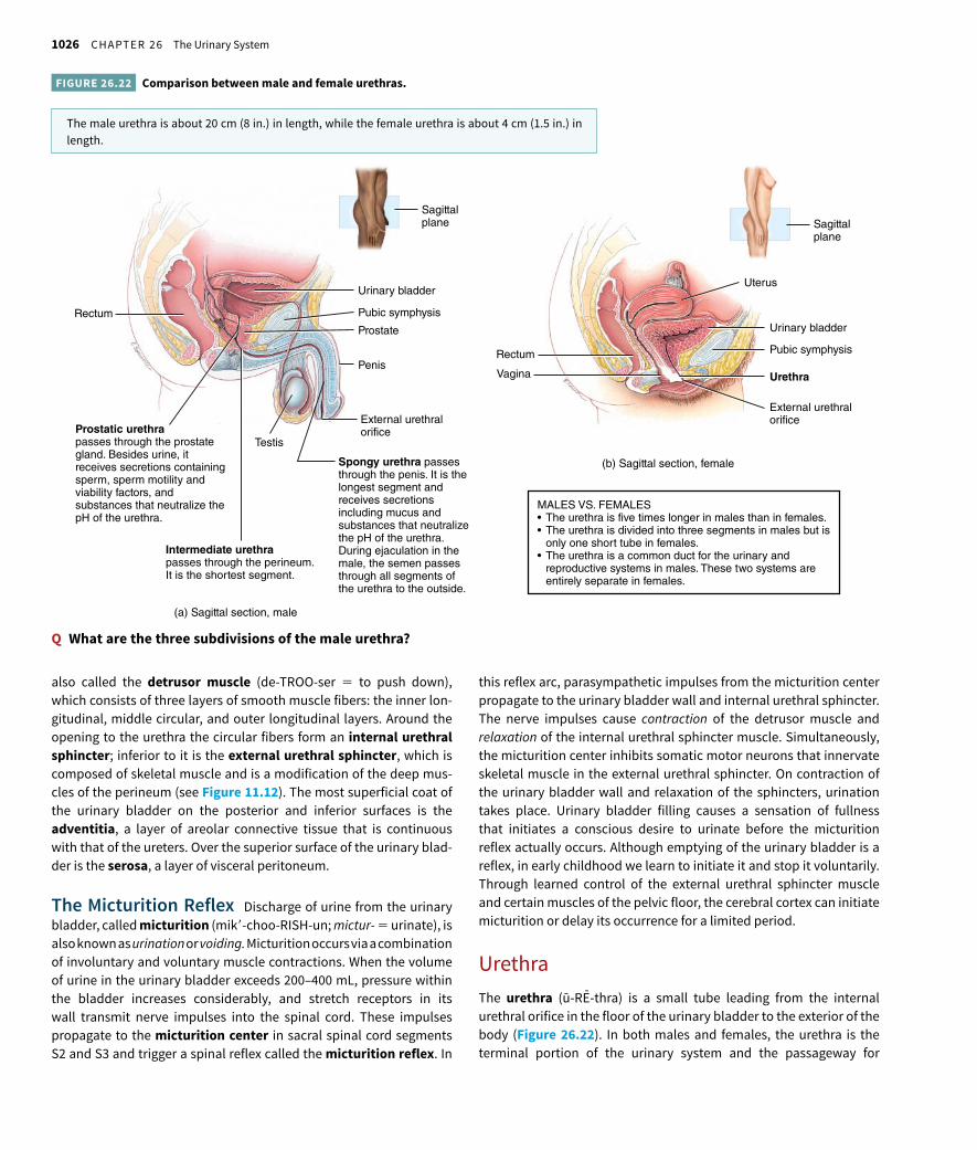
1026 CHAPTER 26 The Urinary System
this reflex arc, parasympathetic impulses from the micturition center propagate to the urinary bladder wall and internal urethral sphincter. The nerve impulses cause contraction of the detrusor muscle and relaxation of the internal urethral sphincter muscle. Simultaneously, the micturition center inhibits somatic motor neurons that innervate skeletal muscle in the external urethral sphincter. On contraction of the urinary bladder wall and relaxation of the sphincters, urination takes place. Urinary bladder filling causes a sensation of fullness that initiates a conscious desire to urinate before the micturition reflex actually occurs. Although emptying of the urinary bladder is a reflex, in early childhood we learn to initiate it and stop it voluntarily. Through learned control of the external urethral sphincter muscle and certain muscles of the pelvic floor, the cerebral cortex can initiate micturition or delay its occurrence for a limited period.
UrethraThe urethra (ū-RE--thra) is a small tube leading from the internal urethral orifice in the floor of the urinary bladder to the exterior of the body (Figure 26.22). In both males and females, the urethra is the terminal portion of the urinary system and the passageway for
also called the detrusor muscle (de-TROO-ser = to push down), which consists of three layers of smooth muscle fibers: the inner lon-gitudinal, middle circular, and outer longitudinal layers. Around the opening to the urethra the circular fibers form an internal urethral sphincter; inferior to it is the external urethral sphincter, which is composed of skeletal muscle and is a modification of the deep mus-cles of the perineum (see Figure 11.12). The most superficial coat of the urinary bladder on the posterior and inferior surfaces is the adventitia, a layer of areolar connective tissue that is continuous with that of the ureters. Over the superior surface of the urinary blad-der is the serosa, a layer of visceral peritoneum.
The Micturition Reflex Discharge of urine from the urinary bladder, called micturition (mik′-choo-RISH-un; mictur- = urinate), is also known as urination or voiding. Micturition occurs via a combination of involuntary and voluntary muscle contractions. When the volume of urine in the urinary bladder exceeds 200–400 mL, pressure within the bladder increases considerably, and stretch receptors in its wall transmit nerve impulses into the spinal cord. These impulses propagate to the micturition center in sacral spinal cord segments S2 and S3 and trigger a spinal reflex called the micturition reflex. In
Q What are the three subdivisions of the male urethra?
Rectum
Vagina
Urinary bladder
Pubic symphysis
Urethra
External urethralorifice
Uterus
Sagittalplane
Rectum
Urinary bladder
Pubic symphysis
Penis
Testis
Prostate
External urethral orifice
(a) Sagittal section, male
(b) Sagittal section, female
Sagittalplane
Intermediate urethrapasses through the perineum. It is the shortest segment.
Spongy urethra passesthrough the penis. It is thelongest segment andreceives secretionsincluding mucus andsubstances that neutralizethe pH of the urethra.During ejaculation in themale, the semen passesthrough all segments ofthe urethra to the outside.
Prostatic urethrapasses through the prostate gland. Besides urine, it receives secretions containing sperm, sperm motility and viability factors, and substances that neutralize the pH of the urethra.
MALES VS. FEMALES
only one short tube in females.
reproductive systems in males. These two systems are entirely separate in females.
The male urethra is about 20 cm (8 in.) in length, while the female urethra is about 4 cm (1.5 in.) in length.
FIGURE 26.22 Comparison between male and female urethras.

26.9 Urine Transportation, Storage, and Elimination 1027
and a plexus of veins). Near the urinary bladder, the mucosa contains transitional epithelium that is continuous with that of the urinary bladder; near the external urethral orifice, the epithelium is non-keratinized stratified squamous epithelium. Between these areas, the mucosa contains stratified columnar or pseudostratified colum-nar epithelium. The muscularis consists of circularly arranged smooth muscle fibers and is continuous with that of the urinary bladder.
Clinical Connection
Urinary IncontinenceA lack of voluntary control over micturition is called urinary incon-tinence (in-KON-ti-nens). In infants and children under 2–3 years old, incontinence is normal because neurons to the external urethral sphincter muscle are not completely developed; voiding occurs whenever the uri-nary bladder is suff iciently distended to stimulate the micturition reflex. Urinary incontinence also occurs in adults. There are four types of urinary incontinence—stress, urge, overflow, and functional. Stress incontinence is the most common type of incontinence in young and middle-aged fe-males, and results from weakness of the deep muscles of the pelvic floor. As a result, any physical stress that increases abdominal pressure, such as coughing, sneezing, laughing, exercising, straining, lift ing heavy ob-jects, and pregnancy, causes leakage of urine from the urinary bladder. Urge incontinence is most common in older people and is characterized by an abrupt and intense urge to urinate followed by an involuntary loss of urine. It may be caused by irritation of the urinary bladder wall by in-fection or kidney stones, stroke, multiple sclerosis, spinal cord injury, or anxiety. Overflow incontinence refers to the involuntary leakage of small amounts of urine caused by some type of blockage or weak contractions of the musculature of the urinary bladder. When urine flow is blocked (for example, from an enlarged prostate or kidney stones) or when the urinary bladder muscles can no longer contract, the urinary bladder becomes overfilled and the pressure inside increases until small amounts of urine dribble out. Functional incontinence is urine loss resulting from the in-ability to get to a toilet facility in time as a result of conditions such as stroke, severe arthritis, or Alzheimer’s disease. Choosing the right treat-ment option depends on correct diagnosis of the type of incontinence. Treatments include Kegel exercises (see Clinical Connection: Injury of Levator Ani and Urinary Stress Incontinence in Chapter 11), urinary bladder training, medication, and possibly even surgery.
A summary of the organs of the urinary system is presented in Table 26.7.
Checkpoint
28. What forces help propel urine from the renal pelvis to the urinary bladder?
29. What is micturition? How does the micturition reflex occur?
30. How do the location, length, and histology of the urethra com-pare in males and females?
discharging urine from the body. In males, it discharges semen (fluid that contains sperm) as well.
In males, the urethra also extends from the internal urethral ori-fice to the exterior, but its length and passage through the body are considerably diff erent than in females (Figure 26.22a). The male urethra first passes through the prostate, then through the deep mus-cles of the perineum, and finally through the penis, a distance of about 20 cm (8 in.).
The male urethra, which also consists of a deep mucosa and a superficial muscularis, is subdivided into three anatomical regions: (1) The prostatic urethra passes through the prostate. (2) The inter-mediate (membranous) urethra, the shortest portion, passes through the deep muscles of the perineum. (3) The spongy urethra, the longest portion, passes through the penis. The epithelium of the prostatic urethra is continuous with that of the urinary bladder and consists of transitional epithelium that becomes stratified columnar or pseudostratified columnar epithelium more distally. The mucosa of the intermediate urethra contains stratified columnar or pseu-dostratified columnar epithelium. The epithelium of the spongy ure-thra is stratified columnar or pseudostratified columnar epithelium, except near the external urethral orifice. There it is nonkeratinized stratified squamous epithelium. The lamina propria of the male ure-thra is areolar connective tissue with elastic fibers and a plexus of veins.
The muscularis of the prostatic urethra is composed of mostly circular smooth muscle fibers superficial to the lamina propria; these circular fibers help form the internal urethral sphincter of the urinary bladder. The muscularis of the intermediate (membranous) urethra consists of circularly arranged skeletal muscle fibers of the deep mus-cles of the perineum that help form the external urethral sphincter of the urinary bladder.
Several glands and other structures associated with reproduc-tion deliver their contents into the male urethra (see Figure 28.9). The prostatic urethra contains the openings of (1) ducts that transport se-cretions from the prostate and (2) the seminal vesicles and ductus (vas) deferens, which deliver sperm into the urethra and provide se-cretions that both neutralize the acidity of the female reproductive tract and contribute to sperm motility and viability. The openings of the ducts of the bulbourethral glands (bul′-bō-ū-RE--thral) or Cowper’s glands empty into the spongy urethra. They deliver an alka-line substance prior to ejaculation that neutralizes the acidity of the urethra. The glands also secrete mucus, which lubricates the end of the penis during sexual arousal. Throughout the urethra, but espe-cially in the spongy urethra, the openings of the ducts of urethral glands or Littré glands (LE--trē) discharge mucus during sexual arousal and ejaculation.
In females, the urethra lies directly posterior to the pubic sym-physis; is directed obliquely, inferiorly, and anteriorly; and has a length of 4 cm (1.5 in.) (Figure 26.22b). The opening of the urethra to the exterior, the external urethral orifice, is located between the clitoris and the vaginal opening (see Figure 28.11a). The wall of the female urethra consists of a deep mucosa and a superficial muscula-ris. The mucosa is a mucous membrane composed of epithelium and lamina propria (areolar connective tissue with elastic fibers

1028 CHAPTER 26 The Urinary System
less toxic ones, such as ammonia into urea. These functions of the liver are described in Chapters 24 and 25.
• Lungs. With each exhalation, the lungs excrete CO2, and expel heat and a little water vapor.
• Sweat (sudoriferous) glands. Especially during exercise, sweat glands in the skin help eliminate excess heat, water, and CO2, plus small quantities of salts and urea as well.
• Gastrointestinal tract. Through defecation, the gastrointestinal tract excretes solid, undigested foods; wastes; some CO2; water; salts; and heat.
Checkpoint
31. What roles do the liver and lungs play in the elimination of wastes?
26.11 Development of the Urinary System
OBJECTIVE
• Describe the development of the urinary system.
Starting in the third week of fetal development, a portion of the meso-derm along the posterior aspect of the embryo, the intermediate mesoderm, diff erentiates into the kidneys. The intermediate
26.10 Waste Management in Other Body Systems
OBJECTIVE
• Describe the ways that body wastes are handled.
As we have seen, just one of the many functions of the urinary system is to help rid the body of some kinds of waste materials. Besides the kidneys, several other tissues, organs, and processes contribute to the temporary confinement of wastes, the transport of waste materi-als for disposal, the recycling of materials, and the excretion of excess or toxic substances in the body. These waste management systems include the following:
• Body buff ers. Buff ers in body fluids bind excess hydrogen ions (H+), thereby preventing an increase in the acidity of body fluids. Buff ers, like wastebaskets, have a limited capacity; eventually the H+, like the paper in a wastebasket, must be eliminated from the body by excretion.
• Blood. The bloodstream provides pickup and delivery services for the transport of wastes, in much the same way that garbage trucks and sewer lines serve a community.
• Liver. The liver is the primary site for metabolic recycling, as oc-curs, for example, in the conversion of amino acids into glucose or of glucose into fatty acids. The liver also converts toxic substances into
TABLE 26.7 Summary of Urinary System Organs
STRUCTURE LOCATION DESCRIPTION FUNCTION
Kidneys Posterior abdomen between last thoracic and third lumbar vertebrae posterior to peritoneum (retroperitoneal). Lie against ribs 11 and 12.
Solid, reddish, bean-shaped organs. Internal structure: three tubular systems (arteries, veins, urinary tubes).
Regulate blood volume and composition, help regulate blood pressure, synthesize glucose, release erythropoietin, participate in vitamin D synthesis, excrete wastes in urine.
Ureters Posterior to peritoneum (retroperitoneal); descend from kidney to urinary bladder along anterior surface of psoas major muscle and cross back of pelvis to reach inferoposterior surface of urinary bladder anterior to sacrum.
Thick, muscular walled tubes with three structural layers: mucosa of transitional epithelium, muscularis with circular and longitudinal layers of smooth muscle, adventitia of areolar connective tissue.
Transport tubes that move urine from kidneys to urinary bladder.
Urinary bladder In pelvic cavity anterior to sacrum and rectum in males and sacrum, rectum, and vagina in females and posterior to pubis in both sexes. In males, superior surface covered with parietal peritoneum; in females, uterus covers superior aspect.
Hollow, distensible, muscular organ with variable shape depending on how much urine it contains. Three basic layers: inner mucosa of transitional epithelium, middle smooth muscle coat (detrusor muscle), outer adventitia or serosa over superior aspect in males.
Storage organ that temporarily stores urine until convenient to discharge from body.
Urethra Exits urinary bladder in both sexes. In females, runs through perineal floor of pelvis to exit between labia minora. In males, passes through prostate, then perineal floor of pelvis, and then penis to exit at its tip.
Thin-walled tubes with three structural layers: inner mucosa that consists of transitional, stratified columnar, and stratified squamous epithelium; thin middle layer of circular smooth muscle; thin connective tissue exterior.
Drainage tube that transports stored urine from body.

26.11 Development of the Urinary System 1029
mesonephric duct near the cloaca. The metanephros (met-a-NEF-rōs; meta- = aft er), or ultimate kidney, develops from the ureteric bud and metanephric mesoderm. The ureteric bud forms the collecting ducts, calyces, renal pelvis, and ureter. The metanephric mesoderm (met′-a-NEF-rik) forms the nephrons of the kidneys. By the third month, the fetal kidneys begin excreting urine into the surrounding amniotic fluid; indeed, fetal urine makes up most of the amniotic fluid.
During development, the cloaca divides into a urogenital sinus, into which urinary and genital ducts empty, and a rectum that discharges into the anal canal. The urinary bladder develops from the urogenital sinus. In females, the urethra develops as a result of lengthening of the short duct that extends from the urinary bladder to the urogenital sinus. In males, the urethra is considerably longer and more complicated, but it is also derived from the urogenital sinus.
Although the metanephric kidneys form in the pelvis, they as-cend to their ultimate destination in the abdomen. As they do so, they receive renal blood vessels. Although the inferior blood vessels usually degenerate as superior ones appear, sometimes the inferior vessels
mesoderm is located in paired elevations called urogenital ridges (ū-rō-JEN-i-tal). Three pairs of kidneys form within the intermediate mesoderm in succession: the pronephros, the mesonephros, and the metanephros (Figure 26.23). Only the last pair remains as the func-tional kidneys of the newborn.
The first kidney to form, the pronephros (prō-NEF-rōs; pro- = before; -nephros = kidney), is the most superior of the three and has an associated pronephric duct. This duct empties into the cloaca (klō-A- -ka), the expanded terminal part of the hindgut, which func-tions as a common outlet for the urinary, digestive, and reproductive ducts. The pronephros begins to degenerate during the fourth week and is completely gone by the sixth week.
The second kidney, the mesonephros (mez′-ō-NEF-rōs; meso- = middle), replaces the pronephros. The retained portion of the pro-nephric duct, which connects to the mesonephros, develops into the mesonephric duct. The mesonephros begins to degenerate by the sixth week and is almost gone by the eighth week.
At about the fift h week, a mesodermal outgrowth, called a ureteric bud (ū-rē-TER-ik), develops from the distal portion of the
Q When do the kidneys begin to develop?
(e) Anterior view 8-week embryo
Degeneratingpronephros
Yolk sac
Degeneratingpronephros
HindgutCloacalmembraneCloaca
Metanephros:Ureteric bud
Metanephric mesoderm
AllantoisMesonephros
Urogenitalridges
Mesonephricduct
(a) Fifth week
Mesonephros
Gut
Allantois
Urogenital sinus
Mesonephric duct
Rectum
Urinary bladder
Genital tubercle
Urogenitalsinus
Rectum
Anus
Urinarybladder
Urogenitalsinus
Rectum
Ureter
Urinarybladder
Kidney
Gonad
Gonad
Metanephros
(b) Sixth week
(c) Seventh week (d) Eighth week
Kidney
Ureter
Allantois
Three pairs of kidneys form within intermediate mesoderm in succession: pronephros, meso-nephros, and metanephros.
FIGURE 26.23 Development of the urinary system.
1030 CHAPTER 26 The Urinary System
function seem to be linked to a progressive reduction in blood supply to the kidneys as an individual gets older; for example, blood vessels such as the glomeruli become damaged or decrease in number. The mass of the two kidneys decreases from an average of nearly 300 g in 20-year-olds to less than 200 g by age 80, a decrease of about one-third. Likewise, renal blood flow and filtration rate decline by 50% between ages 40 and 70. By age 80, about 40% of glomeruli are not functioning and thus filtration, reabsorption, and secretion decrease. Kidney diseases that become more common with age include acute and chronic kidney inflammations and renal calculi (kidney stones). Because the sensation of thirst diminishes with age, older individuals also are susceptible to dehydration. Urinary bladder changes that occur with aging include a reduction in size and capacity and weaken-ing of the muscles. Urinary tract infections are more common among the elderly, as are polyuria (excessive urine production), nocturia (excessive urination at night), increased frequency of urination, dysuria (painful urination), urinary retention or incontinence, and hematuria (blood in the urine).
Checkpoint
34. To what extent do kidney mass and filtration rate decrease with age?
To appreciate the many ways that the urinary system contributes to homeostasis of other body systems, examine Focus on Homeosta-sis: Contributions of the Urinary System. Next, in Chapter 27, we will see how the kidneys and lungs contribute to maintenance of homeo-stasis of body fluid volume, electrolyte levels in body fluids, and acid–base balance.
do not degenerate. Consequently, some individuals (about 30%) develop multiple renal vessels.
In a condition called unilateral renal agenesis (ā-JEN-e-sis; a- = without; -genesis = production; unilateral = one side) only one kid-ney develops (usually the right) due to the absence of a ureteric bud. The condition occurs once in every 1000 newborn infants and usually aff ects males more than females. Other kidney abnormalities that oc-cur during development are malrotated kidneys (the hilum faces an-teriorly, posteriorly, or laterally instead of medially); ectopic kidney (one or both kidneys may be in an abnormal position, usually in-ferior); and horseshoe kidney (the fusion of the two kidneys, usually inferiorly, into a single U-shaped kidney).
Checkpoint
32. Which type of embryonic tissue develops into nephrons?
33. Which tissue gives rise to collecting ducts, calyces, renal pelves, and ureters?
26.12 Aging and the Urinary System
OBJECTIVE
• Describe the eff ects of aging on the urinary system.
With aging, the kidneys shrink in size, have a decreased blood flow, and filter less blood. These age-related changes in kidney size and
Disorders: Homeostatic Imbalances
Renal CalculiThe crystals of salts present in urine occasionally precipitate and solidify into insoluble stones called renal calculi (KAL-kū-lī = peb-bles) or kidney stones. They commonly contain crystals of calcium oxalate, uric acid, or calcium phosphate. Conditions leading to cal-culus formation include the ingestion of excessive calcium, low water intake, abnormally alkaline or acidic urine, and overactivity of the parathyroid glands. When a stone lodges in a narrow passage, such as a ureter, the pain can be intense. Shock-wave lithotripsy (LITH-ō-trip′-sē; litho- = stone) is a procedure that uses high-energy shock waves to disintegrate kidney stones and off ers an alternative to surgical removal. Once the kidney stone is located using x-rays, a device called a lithotripter delivers brief, high-intensity sound waves through a water- or gel-filled cushion placed under the back. Over a
period of 30 to 60 minutes, 1000 or more shock waves pulverize the stone, creating fragments that are small enough to wash out in the urine.
Urinary Tract InfectionsThe term urinary tract infection (UTI) is used to describe either an infection of a part of the urinary system or the presence of large num-bers of microbes in urine. UTIs are more common in females due to the shorter length of the urethra. Symptoms include painful or burn-ing urination, urgent and frequent urination, low back pain, and bed-wetting. UTIs include urethritis (ū-rē-THRĪ-tis), inflammation of the urethra; cystitis (sis-TĪ-tis), inflammation of the urinary bladder; and pyelonephritis (pī-e-lō-ne-FRĪ-tis), inflammation of the kidneys. If pye-lonephritis becomes chronic, scar tissue can form in the kidneys and severely impair their function. Drinking cranberry juice can prevent the attachment of E. coli bacteria to the lining of the urinary bladder so that they are more readily flushed away during urination.

CONTRIBUTIONS OF
FOCUS on HOMEOSTASIS
THE URINARY SYSTEM
SKELETALSYSTEM
Kidneys help adjust levels of blood calcium and phosphates, needed forbuilding extracellular bone matrix
RESPIRATORYSYSTEM
Kidneys and lungs cooperate inadjusting pH of body fluids
DIGESTIVESYSTEM
Kidneys help synthesize calcitriol, the active form of vitamin D, which is needed for absorption of dietary calcium
REPRODUCTIVESYSTEMS
In males, portion of urethra that extends through prostate and penis is passageway for semen as well as urine
MUSCULARSYSTEM
Kidneys help adjust level of blood calcium, needed for contraction of muscle
ENDOCRINESYSTEM
Kidneys participate in synthesis of calcitriol, the active form of vitamin DKidneys release erythropoietin, the hormone that stimulates production ofred blood cells
FOR ALL BODY SYSTEMSKidneys regulate volume, composition, and pH of body fluids by removing wastes and excess substances from blood and excreting them in urineUreters transport urine from kidneys to urinary bladder, which stores urine until it is eliminated through urethra
INTEGUMENTARYSYSTEM
Kidneys and skin both contribute to synthesis of calcitriol, the active formof vitamin D
CARDIOVASCULARSYSTEM
By increasing or decreasing their reabsorption of water filtered fromblood, kidneys help adjust blood volume and blood pressureRenin released by juxtaglomerular cells in kidneys raises blood pressureSome bilirubin from hemoglobin breakdown is converted to a yellowpigment (urobilin), which is excreted in urine
NERVOUSSYSTEM
Kidneys perform gluconeogenesis, which provides glucose for ATP production in neurons, especially during fasting or starvation
LYMPHATIC SYSTEMand IMMUNITY
By increasing or decreasing their reabsorption of water filtered from blood, kidneys help adjust volume of interstitial fluid and lymph; urine flushes microbes out of urethra
1031

1032 CHAPTER 26 The Urinary System
Chronic renal failure (CRF) refers to a progressive and usually irreversible decline in glomerular filtration rate (GFR). CRF may result from chronic glomerulonephritis, pyelonephritis, polycystic kidney disease, or traumatic loss of kidney tissue. CRF develops in three stages. In the first stage, diminished renal reserve, nephrons are de-stroyed until about 75% of the functioning nephrons are lost. At this stage, a person may have no signs or symptoms because the remain-ing nephrons enlarge and take over the function of those that have been lost. Once 75% of the nephrons are lost, the person enters the second stage, called renal insufficiency, characterized by a decrease in GFR and increased blood levels of nitrogen-containing wastes and creatinine. Also, the kidneys cannot eff ectively concentrate or dilute the urine. The final stage, called end-stage renal failure, occurs when about 90% of the nephrons have been lost. At this stage, GFR dimin-ishes to 10–15% of normal, oliguria is present, and blood levels of nitrogen-containing wastes and creatinine increase further. People with end-stage renal failure need dialysis therapy and are possible candidates for a kidney transplant operation.
Polycystic Kidney DiseasePolycystic kidney disease (PKD) (pol′-ē-SIS-tik) is one of the most common inherited disorders. In PKD, the kidney tubules become rid-dled with hundreds or thousands of cysts (fluid-filled cavities). In addition, inappropriate apoptosis (programmed cell death) of cells in noncystic tubules leads to progressive impairment of renal function and eventually to end-stage renal failure.
People with PKD also may have cysts and apoptosis in the liver, pancreas, spleen, and gonads; increased risk of cerebral aneurysms; heart valve defects; and diverticula in the colon. Typically, symptoms are not noticed until adulthood, when patients may have back pain, urinary tract infections, blood in the urine, hypertension, and large abdominal masses. Using drugs to restore normal blood pressure, re-stricting protein and salt in the diet, and controlling urinary tract in-fections may slow progression to renal failure.
Urinary Bladder CancerEach year, nearly 12,000 Americans die from urinary bladder cancer. It generally strikes people over 50 years of age and is three times more likely to develop in males than females. The disease is typically pain-less as it develops, but in most cases blood in the urine is a primary sign of the disease. Less oft en, people experience painful and/or fre-quent urination.
As long as the disease is identified early and treated promptly, the prognosis is favorable. Fortunately, about 75% of urinary bladder cancers are confined to the epithelium of the urinary bladder and are easily removed by surgery. The lesions tend to be low-grade, meaning that they have only a small potential for metastasis.
Urinary bladder cancer is frequently the result of a carcinogen. About half of all cases occur in people who smoke or have at some time smoked cigarettes. The cancer also tends to develop in people who are exposed to chemicals called aromatic amines. Workers in the leather, dye, rubber, and aluminum industries, as well as painters, are oft en exposed to these chemicals.
Glomerular DiseasesA variety of conditions may damage the kidney glomeruli, either directly or indirectly because of disease elsewhere in the body. Typi-cally, the filtration membrane sustains damage, and its permeability increases.
Glomerulonephritis (glō-mer′-ū-lō-ne-FRĪ-tis) is an inflamma-tion of the kidney that involves the glomeruli. One of the most common causes is an allergic reaction to the toxins produced by streptococcal bacteria that have recently infected another part of the body, espe-cially the throat. The glomeruli become so inflamed, swollen, and engorged with blood that the filtration membranes allow blood cells and plasma proteins to enter the filtrate. As a result, the urine contains many erythrocytes (hematuria) and a lot of protein. The glomeruli may be permanently damaged, leading to chronic renal failure.
Nephrotic syndrome (nef-ROT-ik) is a condition characterized by proteinuria (prō-tēn-OO-rē-a), protein in the urine, and hyperlipi-demia (hī′-per-lip-i-DE--mē-a), high blood levels of cholesterol, phos-pholipids, and triglycerides. The proteinuria is due to an increased permeability of the filtration membrane, which permits proteins, es-pecially albumin, to escape from blood into urine. Loss of albumin results in hypoalbuminemia (hī′-pō-al-bū-mi-NE--mē-a), low blood al-bumin level, once liver production of albumin fails to meet increased urinary losses. Edema, usually seen around the eyes, ankles, feet, and abdomen, occurs in nephrotic syndrome because loss of albumin from the blood decreases blood colloid osmotic pressure. Nephrotic syndrome is associated with several glomerular diseases of unknown cause, as well as with systemic disorders such as diabetes mellitus, systemic lupus erythematosus (SLE), a variety of cancers, and AIDS.
Renal FailureRenal failure is a decrease or cessation of glomerular filtration. In acute renal failure (ARF), the kidneys abruptly stop working entirely (or almost entirely). The main feature of ARF is the suppression of urine flow, usually characterized either by oliguria (ol′-i-GŪ-rē-a), daily urine output between 50 mL and 250 mL, or by anuria (an-Ū-rē-a), daily urine output less than 50 mL. Causes include low blood volume (for example, due to hemorrhage), decreased cardiac output, damaged renal tubules, kidney stones, the dyes used to visualize blood vessels in angiograms, nonsteroidal anti-inflammatory drugs, and some antibiotic drugs. It is also common in people who suff er a devastating illness or overwhelming traumatic injury; in such cases it may be related to a more general organ failure known as multiple organ dysfunction syndrome (MODS).
Renal failure causes a multitude of problems. There is edema due to salt and water retention and metabolic acidosis due to an inability of the kidneys to excrete acidic substances. In the blood, urea builds up due to impaired renal excretion of metabolic waste products and potassium level rises, which can lead to cardiac arrest. Oft en, there is anemia because the kidneys no longer produce enough erythropoie-tin for adequate red blood cell production. Because the kidneys are no longer able to convert vitamin D to calcitriol, which is needed for adequate calcium absorption from the small intestine, osteomalacia also may occur.

Chapter Review 1033
sive drugs for the rest of his or her life to avoid rejection of the “foreign” organ.
CystoscopyCystoscopy (sis-TOS-kō-pē; cysto- = bladder; -scopy = to examine) is a very important procedure for direct examination of the mucosa of the urethra and urinary bladder and prostate in males. In the procedure, a cystoscope (a flexible narrow tube with a light) is inserted into the ure-thra to examine the structures through which it passes. With special attachments, tissue samples can be removed for examination (biopsy) and small stones can be removed. Cystoscopy is useful for evaluating urinary bladder problems such as cancer and infections. It can also evaluate the degree of obstruction resulting from an enlarged prostate.
Kidney TransplantA kidney transplant is the transfer of a kidney from a donor to a recipient whose kidneys no longer function. In the procedure, the donor kidney is placed in the pelvis of the recipient through an abdominal incision. The renal artery and vein of the transplanted kidney are attached to a nearby artery or vein in the pelvis of the recipient and the ureter of the transplanted kidney is then attached to the urinary bladder. During a kidney transplant, the patient receives only one donor kidney, since only one kidney is needed to maintain suff icient renal function. The nonfunctioning diseased kidneys are usually left in place. As with all organ transplants, kidney transplant recipients must be ever vigilant for signs of infection or organ rejection. The transplant recipient will take immunosuppres-
Medical Terminology
Azotemia (az-ō-TE--mē-a; azot- = nitrogen; -emia = condition of blood) Presence of urea or other nitrogen-containing substances in the blood.
Cystocele (SIS-tō-sēl; cysto- = bladder; -cele = hernia or rupture) Hernia of the urinary bladder.
Diabetic kidney disease A disorder caused by diabetes mellitus in which glomeruli are damaged. The result is the leakage of proteins into the urine and a reduction in the ability of the kidney to remove water and waste.
Dysuria (dis-Ū-rē-a; dys- = painful; -uria = urine) Painful urination.
Enuresis (en′-ū-RE--sis = to void urine) Involuntary voiding of urine aft er the age at which voluntary control has typically been attained.
Hydronephrosis (hī′-drō-ne-FRO- -sis; hydro- = water; -nephros- = kid-ney; -osis = condition) Swelling of the kidney due to dilation of the renal pelvis and calyces as a result of an obstruction to the flow of urine. It may be due to a congenital abnormality, a narrowing of the ureter, a kidney stone, or an enlarged prostate.
Intravenous pyelogram (IVP) (in′-tra-VE--nus PĪ-e-lō-gram′; intra- = within; -veno- = vein; pyelo- = pelvis of kidney; -gram = record) Radio-graph (x-ray) of the kidneys, ureters, and urinary bladder aft er venous in-jection of a radiopaque contrast medium.
Nephropathy (ne-FROP-a-thē; nephro- = kidney; -pathos = suff ering) Any disease of the kidneys. Types include analgesic (from long-term and
excessive use of drugs such as ibuprofen), lead (from ingestion of lead-based paint), and solvent (from carbon tetrachloride and other solvents).Nocturnal enuresis (nokt-Ū-rē-a en′-ū-RE--sis) Discharge of urine during sleep, resulting in bed-wetting; occurs in about 15% of 5-year-old children and generally resolves spontaneously, aff licting only about 1% of adults. It may have a genetic basis, as bed-wetting occurs more oft en in identical twins than in fraternal twins and more oft en in children whose parents or siblings were bed-wetters. Possible causes include smaller than normal bladder ca-pacity, failure to awaken in response to a full bladder, and above-normal pro-duction of urine at night. Also referred to as nocturia (nok-too-rē-a).Polyuria (pol′-ē-Ū-rē-a; poly- = too much) Excessive urine formation. It may occur in conditions such as diabetes mellitus and glomerulonephritis.Stricture (STRIK-chur) Narrowing of the lumen of a canal or hollow organ, as may occur in the ureter, urethra, or any other tubular structure in the body.Uremia (ū-RE--mē-a; -emia = condition of blood) Toxic levels of urea in the blood resulting from severe malfunction of the kidneys.Urinary retention A failure to completely or normally void urine; may be due to an obstruction in the urethra or neck of the urinary bladder, to nervous contraction of the urethra, or to lack of urge to urinate. In men, an enlarged prostate may constrict the urethra and cause urinary retention. If urinary retention is prolonged, a catheter (slender rubber drainage tube) must be placed into the urethra to drain the urine.
26.2 Anatomy of the Kidneys1. The kidneys are retroperitoneal organs attached to the posterior abdomi-nal wall.
2. Three layers of tissue surround the kidneys; renal capsule, adipose capsule, and renal fascia.
3. Internally, the kidneys consist of a renal cortex, a renal medulla, renal pyramids, renal papillae, renal columns, major and minor calyees, and a renal pelvis.
4. Blood flows into the kidney through the renal artery and successively into segmental, interlobar, arcuate, and cortical radiate arteries; aff erent arte-
Review
26.1 Overview of the Urinary System1. The organs of the urinary system are the kidneys, ureters, urinary bladder, and urethra.2. The kidneys excrete wastes; alter blood ionic composition, blood volume, blood pressure, and blood pH; maintain blood osmolarity; produce the hor-mones calcitriol and erythropoietin; and perform gluconeogenesis.3. The ureters convey urine from the kidneys to the urinary bladder; the urinary bladder stores urine; and the urethra allows urine to pass from the urinary bladder to the outside environment.
Chapter Review

1034 CHAPTER 26 The Urinary System
a substance that can be reabsorbed per unit time is called the transport maximum (Tm).
4. About 90% of water reabsorption is obligatory; it occurs via osmosis, to-gether with reabsorption of solutes, and is not hormonally regulated. The remaining 10% is facultative water reabsorption, which varies according to body needs and is regulated by antidiuretic hormone (ADH).
5. Sodium ions are reabsorbed throughout the basolateral membrane via primary active transport.
6. In the proximal convoluted tubule, Na+ ions are reabsorbed through the apical membranes via Na+–glucose symporters and Na+–H+ antiporters; water is reabsorbed via osmosis; Cl−, K+, Ca2+, Mg2+, and urea are reabsorbed via passive diff usion; and NH3 and NH4
+ are secreted.
7. The nephron loop reabsorbs 20–30% of the filtered Na+, K+, Ca2+, and HCO3
−; 35% of the filtered Cl−; and 15% of the filtered water.
8. The distal convoluted tubule reabsorbs sodium and chloride ions via Na+–Cl− symporters.
9. In the collecting duct, principal cells reabsorb Na+ and secrete K+; interca-lated cells reabsorb K+ and HCO3
− and secrete H+.
10. Angiotensin II, aldosterone, antidiuretic hormone, atrial natriuretic pep-tide, and parathyroid hormone regulate solute and water reabsorption, as summarized in Table 26.4.
26.7 Production of Dilute and Concentrated Urine1. In the absence of ADH, the kidneys produce dilute urine; renal tubules absorb more solutes than water.
2. In the presence of ADH, the kidneys produce concentrated urine; large amounts of water are reabsorbed from the tubular fluid into interstitial fluid, increasing solute concentration of the urine.
3. The countercurrent multiplier establishes an osmotic gradient in the inter-stitial fluid of the renal medulla that enables production of concentrated urine when ADH is present.
26.8 Evaluation of Kidney Function1. A urinalysis is an analysis of the volume and physical, chemical, and microscopic properties of a urine sample. Table 26.5 summarizes the princi-pal physical characteristics of normal urine.
2. Chemically, normal urine contains about 95% water and 5% solutes. The solutes normally include urea, creatinine, uric acid, urobilinogen, and various ions.
3. Table 26.6 lists several abnormal components that can be detected in a urinalysis, including albumin, glucose, red and white blood cells, ketone bod-ies, bilirubin, excessive urobilinogen, casts, and microbes.
4. Renal clearance refers to the ability of the kidneys to clear (remove) a specific substance from blood.
26.9 Urine Transportation, Storage, and Elimination1. The ureters are retroperitoneal and consist of a mucosa, muscularis, and adventitia. They transport urine from the renal pelvis to the urinary bladder, primarily via peristalsis.
2. The urinary bladder is located in the pelvic cavity posterior to the pubic symphysis; its function is to store urine before micturition.
3. The urinary bladder consists of a mucosa with rugae, a muscularis (detrusor muscle), and an adventitia (serosa over the superior surface).
4. The micturition reflex discharges urine from the urinary bladder via para-sympathetic impulses that cause contraction of the detrusor muscle and relaxation of the internal urethral sphincter muscle and via inhibition of impulses in somatic motor neurons to the external urethral sphincter.
rioles; glomerular capillaries; eff erent arterioles; peritubular capillaries and vasa recta; and cortical radiate, arcuate, and interlobar veins before flowing out of the kidney through the renal vein.
5. Vasomotor nerves from the sympathetic division of the autonomic ner-vous system supply kidney blood vessels; they help regulate the flow of blood through the kidney.
26.3 The Nephron1. The nephron is the functional unit of the kidneys. A nephron consists of a renal corpuscle (glomerulus and glomerular capsule) and a renal tubule.
2. A renal tubule consists of a proximal convoluted tubule, a nephron loop, and a distal convoluted tubule, which drains into a collecting duct (shared by several nephrons). The nephron loop consists of a descending limb and an ascending limb.
3. A cortical nephron has a short loop that dips only into the superficial region of the renal medulla; a juxtamedullary nephron has a long nephron loop that stretches through the renal medulla almost to the renal papilla.
4. The wall of the entire glomerular capsule, renal tubule, and ducts consists of a single layer of epithelial cells. The epithelium has distinctive histological features in diff erent parts of the tubule. Table 26.1 summarizes the histologi-cal features of the renal tubule and collecting duct.
5. The juxtaglomerular apparatus (JGA) consists of the juxtaglomerular cells of an aff erent arteriole and the macula densa of the final portion of the as-cending limb of the nephron loop.
26.4 Overview of Renal Physiology1. Nephrons perform three basic tasks: glomerular filtration, tubular secre-tion, and tubular reabsorption.
26.5 Glomerular Filtration1. Fluid that is filtered by glomeruli enters the capsular space and is called glomerular filtrate.
2. The filtration membrane consists of the glomerular endothelium, basement membrane, and filtration slits between pedicels of podocytes.
3. Most substances in blood plasma easily pass through the glomerular filter. However, blood cells and most proteins normally are not filtered.
4. Glomerular filtrate amounts to up to 180 liters of fluid per day. This large amount of fluid is filtered because the filter is porous and thin, the glomerular capillaries are long, and the capillary blood pressure is high.
5. Glomerular blood hydrostatic pressure (GBHP) promotes filtration; cap-sular hydrostatic pressure (CHP) and blood colloid osmotic pressure (BCOP) oppose filtration. Net filtration pressure (NFP) = GBHP − CHP − BCOP. NFP is about 10 mmHg.
6. Glomerular filtration rate (GFR) is the amount of filtrate formed in both kid-neys per minute; it is normally 105–125 mL/min.
7. Glomerular filtration rate depends on renal autoregulation, neural regula-tion, and hormonal regulation. Table 26.2 summarizes regulation of GFR.
26.6 Tubular Reabsorption and Tubular Secretion1. Tubular reabsorption is a selective process that reclaims materials from tubular fluid and returns them to the bloodstream. Reabsorbed substances include water, glucose, amino acids, urea, and ions, such as sodium, chloride, potassium, bicarbonate, and phosphate (Table 26.3).
2. Some substances not needed by the body are removed from the blood and discharged into the urine via tubular secretion. Included are ions (K+, H+, and NH4
+), urea, creatinine, and certain drugs.
3. Reabsorption routes include both paracellular (between tubule cells) and transcellular (across tubule cells) routes. The maximum amount of

Answers to Figure Questions 1035
26.11 Development of the Urinary System1. The kidneys develop from intermediate mesoderm.
2. The kidneys develop in the following sequence: pronephros, mesonephros, and metanephros. Only the metanephros remains and develops into a func-tional kidney.
26.12 Aging and the Urinary System1. With aging, the kidneys shrink in size, have a decreased blood flow, and filter less blood.
2. Common problems related to aging include urinary tract infections, in-creased frequency of urination, urinary retention or incontinence, and renal calculi.
5. The urethra is a tube leading from the floor of the urinary bladder to the exterior. Its anatomy and histology diff er in females and males. In both sexes, the urethra functions to discharge urine from the body; in males, it discharges semen as well.
26.10 Waste Management in Other Body Systems1. Besides the kidneys, several other tissues, organs, and processes temporar-ily confine wastes, transport waste materials for disposal, recycle materials, and excrete excess or toxic substances.
2. Buff ers bind excess H+, the blood transports wastes, the liver converts toxic substances into less toxic ones, the lungs exhale CO2, sweat glands help eliminate excess heat, and the gastrointestinal tract eliminates solid wastes.
3. Bruce is experiencing sudden, rhythmic waves of pain in his groin area. He has noticed that, although he is consuming fluids, his urine output has de-creased. From what condition is Bruce suff ering? How is it treated? How can he prevent future episodes?
1. Imagine the discovery of a new toxin that blocks renal tubule reabsorption but does not aff ect filtration. Predict the short-term eff ects of this toxin.
2. For each of the following urinalysis results, indicate whether you should be concerned or not and why: (a) dark yellow urine that is turbid; (b) ammonia-like odor of the urine; (c) presence of excessive albumin; (d) presence of epi-thelial cell casts; (e) pH of 5.5; (f) hematuria.
Critical Thinking Questions
Answers to Figure Questions26.1 The kidneys, ureters, urinary bladder, and urethra are the components of the urinary system.
26.2 The kidneys are retroperitoneal because they are posterior to the peri-toneum.
26.3 Blood vessels, lymphatic vessels, nerves, and a ureter pass through the renal hilum.
26.4 About 1200 mL of blood enters the renal arteries each minute.
26.5 Cortical nephrons have glomeruli in the superficial renal cortex, and their short nephron loops penetrate only into the superficial renal medulla. Jux-tamedullary nephrons have glomeruli deep in the renal cortex, and their long nephron loops extend through the renal medulla nearly to the renal papilla.
26.6 This section must pass through the renal cortex because there are no renal corpuscles in the renal medulla.
26.7 Secreted penicillin is being removed from the bloodstream.
26.8 Endothelial fenestrations (pores) in glomerular capillaries are too small for red blood cells to pass through.
26.9 Obstruction of the right ureter would increase CHP and thus decrease NFP in the right kidney; the obstruction would have no eff ect on the left kidney.
26.10 Auto- means self; tubuloglomerular feedback is an example of autoreg-ulation because it takes place entirely within the kidneys.
26.11 The tight junctions between tubule cells form a barrier that prevents diff usion of transporter, channel, and pump proteins between the apical and basolateral membranes.
26.12 Glucose enters a PCT cell via a Na+–glucose symporter in the apical mem-brane and leaves via facilitated diff usion through the basolateral membrane.
26.13 The electrochemical gradient promotes movement of Na+ into the tubule cell through the apical membrane antiporters.
26.14 Reabsorption of the solutes creates an osmotic gradient that promotes the reabsorption of water via osmosis.
26.15 This is considered secondary active transport because the symporter uses the energy stored in the concentration gradient of Na+ between extra-cellular fluid and the cytosol. No water is reabsorbed here because the thick ascending limb of the nephron loop is virtually impermeable to water.
26.16 In principal cells, aldosterone stimulates secretion of K+ and reab-sorption of Na+ by increasing the activity of sodium–potassium pumps and number of leakage channels for Na+ and K+.
26.17 Aldosterone and atrial natriuretic peptide influence renal water reab-sorption along with ADH.
26.18 Dilute urine is produced when the thick ascending limb of the nephron loop, the distal convoluted tubule, and the collecting duct reabsorb more solutes than water.
26.19 The high osmolarity of interstitial fluid in the renal medulla is due mainly to Na+, Cl−, and urea.
26.20 Secretion occurs in the proximal convoluted tubule, the nephron loop, and the collecting duct.
26.21 Lack of voluntary control over micturition is termed urinary inconti-nence.
26.22 The three subdivisions of the male urethra are the prostatic urethra, membranous urethra, and spongy urethra.
26.23 The kidneys start to form during the third week of development.