chapter 26
TRANSCRIPT

Chapter 26 The
Urinary System

Copyright © John Wiley & Sons, Inc. All rights reserved.
The Urinary System The urinary systems consists of the kidneys, ureters,
bladder, and urethra, along with its associated nerves and
blood vessels.
The system maintains homeostasis by:
Regulating blood volume, pressure, pH, and
concentration (osmolarity) of electrolytes (Na+, K+,
Ca 2+, Cl-, HPO4-3, Mg2+, HCO3
-)
Reabsorbing glucose and excreting wastes
Releasing certain hormones like renin and EPO

Copyright © John Wiley & Sons, Inc. All rights reserved.
The kidneys are bean-shaped organs located just above the
waist between the peritoneum and the posterior wall of
the abdomen (in the retroperitoneal space).
They are partially protected
by the eleventh and
twelfth pairs of ribs.
Because of the position
of the liver, the right
kidney is slightly
lower than the left.
Renal Anatomy

Copyright © John Wiley & Sons, Inc. All rights reserved.
A ureter (approximately 25 cm long)
originates near an indented area of each
kidney called the hilum and travels to
the base of the bladder.
From the bladder, the
urethra (4 cm in length in
women and 24 cm in length
in men) allows urine to
be excreted.
Renal Anatomy

Copyright © John Wiley & Sons, Inc. All rights reserved.
Renal Anatomy A frontal section through the kidney reveals two distinct
regions of internal anatomy, the cortex and medulla.
The main function of the cortex
is filtration to form urine.
The main function of
the medulla is to
collect and
excrete urine.

Copyright © John Wiley & Sons, Inc. All rights reserved.
The renal pyramids consists of 8 to 18 conical subdivisions
within the medulla that contain the kidney’s
secreting apparatus and tubules.
Renal columns are
composed of lines of
blood vessels and
fibrous material
which allows the
cortex to be better anchored.
Renal Anatomy

Copyright © John Wiley & Sons, Inc. All rights reserved.
The renal papilla is the location where the medullary pyramids empty urine into cuplike structures called minor and major calyces. 8-18 minor calyces and 2-3 major calyces receive urine from the papilla of one renal pyramid.
Renal Anatomy

Copyright © John Wiley & Sons, Inc. All rights reserved.
From the major calyces, urine drains into a single large
cavity called the renal pelvis and then out through
the ureter.
The hilum expands into
a cavity within the
kidney called the renal
sinus, which contains part
of the renal pelvis, the calyces,
and branches of the renal blood
vessels and nerves.
Renal Anatomy

Copyright © John Wiley & Sons, Inc. All rights reserved.
The ureters transport urine from the renal pelvis of the
kidneys to the bladder using peristaltic waves,
hydrostatic pressure and gravity to move the urine.
There is no anatomical
valve at the
opening of the
ureter into
the bladder.
Renal Anatomy

Copyright © John Wiley & Sons, Inc. All rights reserved.
The urethra is a small tube leading from the internal
urethral orifice in the bladder floor to the exterior.
In males, it
is also used
to discharge
semen.
Renal Anatomy

Copyright © John Wiley & Sons, Inc. All rights reserved.
Renal Blood Flow The renal artery and renal vein pass into the substance of
the kidney (the parenchyma) at the hilum.
Arterial blood enters via the renal artery and exits the
renal vein.

Copyright © John Wiley & Sons, Inc. All rights reserved.
Blood follows the path depicted in the
graphic to the right:
Renal Blood Flow

Copyright © John Wiley & Sons, Inc. All rights reserved.
The Nephron The nephron is the functional unit of the kidney.
It is a microscopic
structure composed
of blood vessels and
tubules that collect
the filtrate which
will ultimately
become urine.

Copyright © John Wiley & Sons, Inc. All rights reserved.
The Nephron Each nephron receives one afferent arteriole, which
divides into a tangled, ball-shaped capillary network
called the glomerulus.
The glomerular
capillaries then
reunite to form an
efferent arteriole that
carries blood out of
the glomerulus.

Copyright © John Wiley & Sons, Inc. All rights reserved.
The Nephron Glomerular capillaries are unique among capillaries in
the body because they are positioned between two
arterioles, rather than between an arteriole and a venule.
There are venules in the kidney, but they come later.
The Renal Corpuscle consists of two structures:
The glomerular capillaries
The glomerular capsule (Bowman’s capsule) – a double-
walled epithelial cup that surrounds the glomerular
capillaries.

Copyright © John Wiley & Sons, Inc. All rights reserved.
Bowman’s capsule consists of visceral and parietal layers.
The visceral layer is made of modified simple squamous
epithelial cells called podocytes. The many foot-like
projections of these cells (pedicels) wrap around the
single layer of endothelial cells of the glomerular
capillaries and form the inner wall of the capsule.
The parietal layer of the glomerular capsule is a simple
squamous epithelium and forms the outer wall of the
capsule.
The Nephron

Copyright © John Wiley & Sons, Inc. All rights reserved.
The epithelium of the visceral
and parietal layers of the
renal corpuscle form
festrations (pores)
which act as a
filtration (dialysis)
membrane.
The Nephron

Copyright © John Wiley & Sons, Inc. All rights reserved.
Fluid filtered from the glomerular capillaries enters
Bowman’s space, (the space between the two layers of the
glomerular capsule), which is the lumen of the urinary
tube.
The Nephron

Copyright © John Wiley & Sons, Inc. All rights reserved.
Blood plasma is filtered through the glomerular capillaries
into the glomerular capsule.
Filtered fluid passes into
the renal tubule, which
has three main sections:
◦the proximal convoluted
tubule (PCT)
◦the loop of Henle
◦the distal convoluted
tubule (DCT)
The Nephron

Copyright © John Wiley & Sons, Inc. All rights reserved.
The distal convoluted tubules of several nephrons empty
into a single collecting duct.
Collecting ducts
unite and converge
into several hundred
large papillary ducts
which drain into the
minor calyces, major calyces,
renal pelvis, and ureters.
The Nephron

Copyright © John Wiley & Sons, Inc. All rights reserved.
The Nephron The first part of the loop of Henle (the descending limb)
dips into the renal medulla. It then makes a hairpin turn and returns to the renal cortex as the ascending limb. The descending limb of the loop of Henle is composed of
a simple squamous epithelium. The ascending limb of the loop may be either “thin”
(composed of a simple squamous epithelium) or “thick” (composed of simple cuboidal to low columnar cells). ◦ Some nephrons contain both thick and thin ascending
limbs.

Copyright © John Wiley & Sons, Inc. All rights reserved.
The Nephron Based on the length of the loop of Henle and the presence
of thin segments in the ascending limb, nephrons can be
sorted into two populations:
cortical and juxtamedullary.
Nephrons with long loops of
Henle enable the kidneys to
create a concentration gradient
in the renal medulla and to
excrete very dilute or very
concentrated urine.

Copyright © John Wiley & Sons, Inc. All rights reserved.
The Nephron Cortical nephrons make up about 80–85% of the 1 million
microscopic nephrons that comprise each kidney.
Their renal corpuscles are located in the outer portion of
the cortex, with short loops of Henle that penetrate only
a small way into the medulla.
The ascending limbs of their loops of Henle consist of
only a thick segment, lacking any thin portions.
Nephrons with short loops receive their blood supply
from peritubular capillaries that arise from efferent
arterioles.

Copyright © John Wiley & Sons, Inc. All rights reserved.
The Nephron

Copyright © John Wiley & Sons, Inc. All rights reserved.
The Nephron The other 15–20% of the nephrons are juxtamedullary
nephrons .
Their renal corpuscles lie deep in the cortex, close to
the medulla, and they have long loops of Henle that
extend into the deepest region of the medulla.
The ascending limbs of their loops of Henle consist of
both thin and thick segments.
Nephrons with long loops receive their blood supply
from the vasa recta that arise from peritubular
capillaries before becoming peritubular venules.

Copyright © John Wiley & Sons, Inc. All rights reserved.
Juxtamedullary Nephron

Copyright © John Wiley & Sons, Inc. All rights reserved.
In each nephron, the final part of the ascending limb of
the loop of Henle makes contact with the afferent
arteriole serving that renal corpuscle. Because the
columnar tubule cells in this region are crowded together,
they are known as the macula densa.
Alongside the macula densa, the wall of the afferent
arteriole contains modified smooth muscle fibers called
juxtaglomerular (JG) cells.
◦ Together with the macula densa, they constitute the
juxtaglomerular apparatus (JGA).
The Nephron

Copyright © John Wiley & Sons, Inc. All rights reserved.
The Nephron As we will shortly see, the JGA helps regulate blood
pressure within the kidneys.

Copyright © John Wiley & Sons, Inc. All rights reserved.
Renal Physiology The 3 basic functions performed by nephrons and
collecting ducts are:
Glomerular filtration - pressure forces filtration of
waste-laden blood in the glomerulus. The glomerular
filtration rate (GFR) is the amount of filtrate formed in
all the renal corpuscles of both kidneys each minute.
Tubular reabsorption – the process of returning
important substances from the filtrate back to the body.
Tubular secretion – the movement of waste materials
from the body to the filtrate.

Copyright © John Wiley & Sons, Inc. All rights reserved.
Glomerular Filtration Glomerular filtration is the formation of a protein-free
filtrate (ultrafiltrate) of plasma across the glomerular
membrane.
Only a portion of the blood plasma delivered to the
kidney via the renal artery is filtered.
Plasma which escapes filtration, along with its protein
and cellular elements, exits the renal corpuscle via the
efferent arteriole, perfuses the tubular capillary beds,
and is eventually collected in the renal venous system.

Copyright © John Wiley & Sons, Inc. All rights reserved.
Glomerular Filtration In the average adult, the body’s entire extracellular fluid
volume is filtered about 12 times per day.
Since we cannot afford to lose that amount of fluid, the vast majority must be reclaimed, with just a small portion being excreted in the urine.

Copyright © John Wiley & Sons, Inc. All rights reserved.
The daily composition of plasma, filtrate, and urine are compared.
Glomerular Filtration Total
Amount in Plasma
Amount in 180 L of filtrate (/day)
Amount returned to
blood/d (Reabsorbed)
Amount in Urine (/day)
Water (passive) 3 L 180 L 178-179 L 1-2 L
Protein (active) 200 g 2 g 1.9 g 0.1 g
Glucose (active) 3 g 162 g 162 g 0 g
Urea (passive) 1 g 54 g 24 g (about 1/2)
30 g (about 1/2)
Creatinine 0.03 g 1.6 g 0 g (all filtered)
1.6 g (none reabsorbed)

Copyright © John Wiley & Sons, Inc. All rights reserved.
Glomerular Filtration Filtration is controlled by 2 opposing hydrostatic forces
and 2 opposing osmotic forces at the glomerular
membrane called Starling forces.
Blood hydrostatic pressure (55mmHg) is the main force
that “pushes” water and solutes through the filtration
membrane (promotes filtration).
Capsular hydrostatic pressure (15 mmHg) is exerted
against the filtration membrane by fluid in the capsular
space (opposes filtration).

Copyright © John Wiley & Sons, Inc. All rights reserved.
Glomerular Filtration Starling forces continued
Blood osmotic (oncotic) pressure (30 mmHg) is the
pressure of plasma proteins “pulling” on water (opposes
filtration).
Normally very little protein escapes through the
filtration membrane making capsular oncotic pressure a
negligible force except in certain disease states.
Net Filtration = Blood Hydrostatic Pressure – Blood
Osmotic Pressure – Capsular Hydrostatic Pressure
Net Filtration = 55-30-15 = 10 mmHg

Copyright © John Wiley & Sons, Inc. All rights reserved.
Glomerular Filtration
Normally, 3 Starling forces are at work in glomerular filtration
Copyright © John Wiley & Sons, Inc. All rights reserved.
Regulation of the GFR is critical to maintaining
homeostasis and is regulated by an assortment of local and
systemic mechanisms:
Renal autoregulation occurs when the kidneys
themselves regulate GFR.
Neural regulation occurs when the ANS regulates renal
blood flow and GFR.
Hormonal regulation involves angiotensin II and atrial
natriuretic peptide (ANP).
Glomerular Filtration
Copyright © John Wiley & Sons, Inc. All rights reserved.
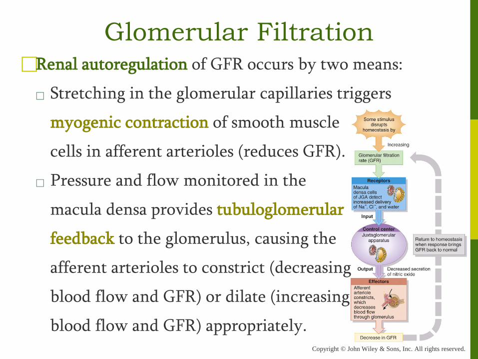
Renal autoregulation of GFR occurs by two means:
Stretching in the glomerular capillaries triggers
myogenic contraction of smooth muscle
cells in afferent arterioles (reduces GFR).
Pressure and flow monitored in the
macula densa provides tubuloglomerular
feedback to the glomerulus, causing the
afferent arterioles to constrict (decreasing
blood flow and GFR) or dilate (increasing
blood flow and GFR) appropriately.
Glomerular Filtration

Copyright © John Wiley & Sons, Inc. All rights reserved.
Neural regulation of GFR is possible because the renal
blood vessels are supplied by sympathetic ANS fibers that
release norepinephrine causing vasoconstriction.
Sympathetic input to
the kidneys is most
important with extreme
drops of B.P. (as occurs
with hemorrhage).
Glomerular Filtration

Copyright © John Wiley & Sons, Inc. All rights reserved.
Two hormones contribute to regulation of GFR
Angiotensin II is a potent vasoconstrictor of both
afferent and efferent arterioles (reduces GFR).
A sudden large increase in BP stretches the cardiac atria
and releases atrial
natriuretic peptide (ANP).
◦ ANP causes the
glomerulus to relax,
increasing the surface
area for filtration.
Glomerular Filtration

Copyright © John Wiley & Sons, Inc. All rights reserved.
Filtration slit Pedicel of podocyte
Fenestration (pore) of glomerular endothelial cell
Basal lamina
Lumen of glomerulus
(b) Filtration membrane
TEM 78,000x
(a) Details of filtration membrane
Filtration slit Pedicel
Fenestration (pore) of glomerular endothelial cell: prevents filtration of blood cells but allows all components of blood plasma to pass through
Podocyte of visceral layer of glomerular (Bowman’s) capsule
1
Filtration slit Pedicel of podocyte
Fenestration (pore) of glomerular endothelial cell
Basal lamina
Lumen of glomerulus
(b) Filtration membrane
TEM 78,000x
(a) Details of filtration membrane
Filtration slit Pedicel
Fenestration (pore) of glomerular endothelial cell: prevents filtration of blood cells but allows all components of blood plasma to pass through
Basal lamina of glomerulus: prevents filtration of larger proteins
Podocyte of visceral layer of glomerular (Bowman’s) capsule
1
2
Filtration slit Pedicel of podocyte
Fenestration (pore) of glomerular endothelial cell
Basal lamina
Lumen of glomerulus
(b) Filtration membrane
TEM 78,000x
(a) Details of filtration membrane
Filtration slit Pedicel
Fenestration (pore) of glomerular endothelial cell: prevents filtration of blood cells but allows all components of blood plasma to pass through
Basal lamina of glomerulus: prevents filtration of larger proteins
Slit membrane between pedicels: prevents filtration of medium-sized proteins
Podocyte of visceral layer of glomerular (Bowman’s) capsule
1
2
3
The Filtration Membrane

Copyright © John Wiley & Sons, Inc. All rights reserved.
Glomerular Filtration (Interactions Animation)
Renal Filtration
You must be connected to the internet to run this animation

Copyright © John Wiley & Sons, Inc. All rights reserved.
NET FILTRATION PRESSURE (NFP) =GBHP – CHP – BCOP = 55 mmHg 15 mmHg 30 mmHg = 10 mmHg
GLOMERULAR BLOOD HYDROSTATIC PRESSURE (GBHP) = 55 mmHg
Capsular space
Glomerular (Bowman's) capsule
Efferent arteriole
Afferent arteriole
1
Proximal convoluted tubule
NET FILTRATION PRESSURE (NFP) =GBHP – CHP – BCOP = 55 mmHg 15 mmHg 30 mmHg = 10 mmHg
CAPSULAR HYDROSTATIC PRESSURE (CHP) = 15 mmHg
GLOMERULAR BLOOD HYDROSTATIC PRESSURE (GBHP) = 55 mmHg
Capsular space
Glomerular (Bowman's) capsule
Efferent arteriole
Afferent arteriole
1 2
Proximal convoluted tubule
NET FILTRATION PRESSURE (NFP) =GBHP – CHP – BCOP = 55 mmHg 15 mmHg 30 mmHg = 10 mmHg
BLOOD COLLOID OSMOTIC PRESSURE (BCOP) = 30 mmHg
CAPSULAR HYDROSTATIC PRESSURE (CHP) = 15 mmHg
GLOMERULAR BLOOD HYDROSTATIC PRESSURE (GBHP) = 55 mmHg
Capsular space
Glomerular (Bowman's) capsule
Efferent arteriole
Afferent arteriole
1 2
3
Proximal convoluted tubule
Pressures That Drive Glomerular Filtration

Copyright © John Wiley & Sons, Inc. All rights reserved.
Tubular Reabsorption Tubular reabsorption is the process of returning important
substances (“good stuff”) from the filtrate back into the
renal interstitium, then into the renal blood vessels... and
ultimately back into the body.

Copyright © John Wiley & Sons, Inc. All rights reserved.
Tubular Reabsorption The “good stuff” is glucose, electrolytes, vitamins, water,
amino acids, and any small proteins that might have
inadvertently escaped from the blood into the filtrate.
Ninety nine percent of the glomerular filtrate is
reabsorbed (most of it before the end of the PCT)!
To appreciate the magnitude of tubular reabsorption,
look once again at the table in the next slide and
compare the amounts of substances that are filtered,
reabsorbed, and excreted in urine.

Copyright © John Wiley & Sons, Inc. All rights reserved.
Tubular Reabsorption Total
Amount in Plasma
Amount in 180 L of filtrate (/day)
Amount returned to
blood/d (Reabsorbed)
Amount in Urine (/day)
Water (passive) 3 L 180 L 178-179 L 1-2 L
Protein (active) 200 g 2 g 1.9 g 0.1 g
Glucose (active) 3 g 162 g 162 g 0 g
Urea (passive) 1 g 54 g 24 g (about 1/2)
30 g (about 1/2)
Creatinine 0.03 g 1.6 g 0 g (all filtered)
1.6 g (none reabsorbed)

Copyright © John Wiley & Sons, Inc. All rights reserved.
Reabsorption into the interstitium has two routes:
Paracellular reabsorption is a passive process that occurs
between adjacent tubule
cells (tight junctions do
not completely seal off
interstitial fluid from
tubule fluid.)
Transcellular reabsorption
is movement through an
individual cell.
Tubular Reabsorption

Copyright © John Wiley & Sons, Inc. All rights reserved.
It is a tremendous feat to reabsorb all of the nutrients and
fluid we must to survive, while still filtering out,
concentrating and excreting toxic substance.
To accomplish this, the kidney establishes a
countercurrent flow between the filtrate in the limbs of
the Loops of Henle and the blood in the peritubular
capillaries and Vasa Recta.
◦ Two types of countercurrent mechanisms exist in the
kidneys: countercurrent multiplication and
countercurrent exchange.
Tubular Reabsorption

Copyright © John Wiley & Sons, Inc. All rights reserved.
Countercurrent multiplication is the process by which a
progressively increasing osmotic gradient is formed in the
interstitial fluid of the renal medulla as a result of
countercurrent flow.
Countercurrent exchange is the process by which solutes
and water are passively exchanged between the blood of
the vasa recta and interstitial fluid of the renal medulla as
a result of countercurrent flow.
This provides oxygen and nutrients to the renal medulla
without washing out or diminishing the gradient.
Tubular Reabsorption

Copyright © John Wiley & Sons, Inc. All rights reserved.
Both mechanisms contribute to reabsorption of fluid and
electrolytes and the formation of concentrated urine.
Tubular Reabsorption

Copyright © John Wiley & Sons, Inc. All rights reserved.
Tubular Reabsorption Reabsorption of fluids, ions, and other substances occurs
by active and passive means.
A variety of symporters and antiporters actively
transport Na+ , Cl– , Ca2+, H+, HCO3– , glucose, HPO4
2– ,
SO42– , NH4
+, urea, all amino acids, and lactic acid.
Reabsorption of water can be obligatory or facultative,
but it always moves by osmosis down its concentration
gradient depending on the permeability of the tubule
cells (which varies between the PCT, the different
portions of the loop of Henle, DCT, and collecting ducts).

Copyright © John Wiley & Sons, Inc. All rights reserved.
Tubular Reabsorption Obligatory reabsorption of water occurs when it is
obliged to follow the solutes as they are reabsorbed (to
maintain the osmotic gradient).
Facultative reabsorption describes variable water
reabsorption, adapted to specific needs.
It is regulated by the effects
of ADH and aldosterone on
the principal cells of the renal
tubules and collecting ducts.

Copyright © John Wiley & Sons, Inc. All rights reserved.
This graphic depicts the formation of a dilute urine, mostly
through obligatory
reabsorption of water.
Compare this process to
the one depicted on the
next slide where urine is
concentrated by the action
of ADH on the DCT and
collecting ducts of
juxtamedullary nephrons.
Tubular Reabsorption

Copyright © John Wiley & Sons, Inc. All rights reserved.
Urine can be
up to 4 times
more
concentrated
than blood
plasma.
Tubular Reabsorption

Copyright © John Wiley & Sons, Inc. All rights reserved. (b) Recycling of salts and urea in the vasa recta (a) Reabsorption of Na+CI– and water in a long-loop juxtamedullary nephron
Glomerular (Bowman’s) capsule
Afferent arteriole
Efferent arteriole
Glomerulus
Distal convoluted tubule
Proximal convoluted tubule
Symporters in thick ascending limb cause buildup of Na+ and Cl–
Interstitial fluid in renal medulla
300
1200
1000
800
Osmotic gradient
600
400
H 2 O H 2 O
H 2 O
200
1200
980
600 780
400 580
200 380
300
100
Loop of Henle 1200 Concentrated urine
300
300
320
400
600
800
1000
1200
800
H2O
Urea
Papillary duct
Collecting duct
300
500
700
900
1100
1200
400
800
1000
600
Na+CI– Blood flow
Flow of tubular fluid
Presense of Na+-K+-2CI–
symporters Interstitial fluid in renal cortex
320
Juxtamedullary nephron and its blood supply together
Vasa recta
Loop of Henle
H2O
H2O
H2O
H2O
H2O
H2O
H2O
1
H2O
H2O
Na+CI–
Na+CI–
H2O
Na+CI–
H2O
Na+CI–
(b) Recycling of salts and urea in the vasa recta (a) Reabsorption of Na+CI– and water in a long-loop juxtamedullary nephron
Glomerular (Bowman’s) capsule
Afferent arteriole
Efferent arteriole
Glomerulus
Distal convoluted tubule
Proximal convoluted tubule
Symporters in thick ascending limb cause buildup of Na+ and Cl–
Interstitial fluid in renal medulla
300
1200
1000
800
Osmotic gradient
600
400
H 2 O H 2 O
H 2 O
200
1200
980
600 780
400 580
200 380
300
100
Loop of Henle 1200 Concentrated urine
300
300
320
400
600
800
1000
1200
800
H2O
Urea
Papillary duct
Collecting duct
Countercurrent flow through loop of Henle establishes an osmotic gradient
300
500
700
900
1100
1200
400
800
1000
600
Na+CI– Blood flow
Flow of tubular fluid
Presense of Na+-K+-2CI–
symporters Interstitial fluid in renal cortex
320
Juxtamedullary nephron and its blood supply together
Vasa recta
Loop of Henle
H2O
H2O
H2O
H2O
H2O
H2O
H2O
1
2
H2O
H2O
Na+CI–
Na+CI–
H2O
Na+CI–
H2O
Na+CI–
(b) Recycling of salts and urea in the vasa recta (a) Reabsorption of Na+CI– and water in a long-loop juxtamedullary nephron
Glomerular (Bowman’s) capsule
Afferent arteriole
Efferent arteriole
Glomerulus
Distal convoluted tubule
Proximal convoluted tubule
Symporters in thick ascending limb cause buildup of Na+ and Cl–
Interstitial fluid in renal medulla
300
1200
1000
800
Osmotic gradient
600
400
H 2 O H 2 O
H 2 O
200
1200
980
600 780
400 580
200 380
300
100
Loop of Henle 1200 Concentrated urine
300
300
320
400
600
800
1000
1200
800
H2O
Urea
Papillary duct
Collecting duct
Countercurrent flow through loop of Henle establishes an osmotic gradient
Principal cells in collecting duct reabsorb more water when ADH is present
300
500
700
900
1100
1200
400
800
1000
600
Na+CI– Blood flow
Flow of tubular fluid
Presense of Na+-K+-2CI–
symporters Interstitial fluid in renal cortex
320
Juxtamedullary nephron and its blood supply together
Vasa recta
Loop of Henle
H2O
H2O
H2O
H2O
H2O
H2O
H2O
1
2
3
H2O
H2O
Na+CI–
Na+CI–
H2O
Na+CI–
H2O
Na+CI–
(b) Recycling of salts and urea in the vasa recta (a) Reabsorption of Na+CI– and water in a long-loop juxtamedullary nephron
Glomerular (Bowman’s) capsule
Afferent arteriole
Efferent arteriole
Glomerulus
Distal convoluted tubule
Proximal convoluted tubule
Symporters in thick ascending limb cause buildup of Na+ and Cl–
Interstitial fluid in renal medulla
300
1200
1000
800
Osmotic gradient
600
400
H 2 O H 2 O
H 2 O
200
1200
980
600 780
400 580
200 380
300
100
Loop of Henle 1200 Concentrated urine
300
300
320
400
600
800
1000
1200
800
H2O
Urea
Papillary duct
Urea recycling causes buildup of urea in the renal medulla
Collecting duct
Countercurrent flow through loop of Henle establishes an osmotic gradient
Principal cells in collecting duct reabsorb more water when ADH is present
300
500
700
900
1100
1200
400
800
1000
600
Na+CI– Blood flow
Flow of tubular fluid
Presense of Na+-K+-2CI–
symporters Interstitial fluid in renal cortex
320
Juxtamedullary nephron and its blood supply together
Vasa recta
Loop of Henle
H2O
H2O
H2O
H2O
H2O
H2O
H2O
1
2
3
4
H2O
H2O
Na+CI–
Na+CI–
H2O
Na+CI–
H2O
Na+CI–

Copyright © John Wiley & Sons, Inc. All rights reserved.
Tubular Reabsorption (Interactions Animation)
Water Homeostasis
You must be connected to the internet to run this animation

Copyright © John Wiley & Sons, Inc. All rights reserved.
Tubular Reabsorption If higher than normal amounts of a substance are present
in the filtrate, then the renal threshold for reabsorption of
that substance may be surpassed.
When that happens, the substance cannot be reabsorbed
fast enough, and it will be excreted in the urine.
For example, the renal [reabsorption] threshold of
glucose is 180-200mg/dl. When this level is exceeded (as
in diabetes mellitus), the glucose is said to “spill” into
the urine (meaning a substance which is not normally
present in urine begins to appear).

Copyright © John Wiley & Sons, Inc. All rights reserved.
Tubular Secretion Tubular secretion is the movement of substances from the
capillaries which surround the nephron into the filtrate.
It occurs at a site other than the filtration membrane (in
the proximal convoluted tubule, distal convoluted
tubule and collecting ducts) by active transport.
The process of tubular secretion controls pH.
Hydrogen and ammonium ions are secreted to decrease
the acidity in the body, and bicarbonate is conserved.
◦ Secreted substances include H+, K+, NH4+, and some
drugs; the amount often depends on body needs.

Copyright © John Wiley & Sons, Inc. All rights reserved.
Tubular Secretion Maintaining the body’s proper pH requires cooperation
mainly between the lungs and the kidneys.
The lungs eliminate CO2.
◦ Provides a rapid response (minutes)
The kidneys eliminate H+ and NH4+ ions and conserve
bicarbonate.
◦ This is a slower response (hours-days).
The alimentary canal (digestive), and integumentary
system (skin) provide minor contributions.
Copyright © John Wiley & Sons, Inc. All rights reserved.
Five hormones affect the extent of Na+, Cl–, Ca2+, and
water reabsorption as well as K+ secretion by the renal
tubules. These hormones, all of which are key to
maintaining homeostasis of not only renal blood flow
and B.P., but systemic blood flow and B.P., are:
angiotensin II
antidiuretic hormone (ADH)
aldosterone
atrial natriuretic peptide (ANP)
parathyroid hormone (PTH)
Hormones and Homeostasis

Copyright © John Wiley & Sons, Inc. All rights reserved.
We have already mentioned the effect of ANP on GFR.
ADH is released by the posterior pituitary in response to
low blood flow in this part of the brain.
ADH affects facultative water reabsorption by
increasing the water permeability of principal cells in
the last part of the distal convoluted tubule and
throughout the collecting duct.
◦ In the absence of ADH, the apical
membranes of principal cells
are almost impermeable to water.
Hormones and Homeostasis

Copyright © John Wiley & Sons, Inc. All rights reserved.
Secretion of the hormones angiotensin II and aldosterone
are tied to one another: When blood volume and blood
pressure decrease or the sympathetic NS is stimulated, the
walls of the
afferent arterioles
are stretched
less, and the
cells of the JGA
secrete renin.
Hormones and Homeostasis
Copyright © John Wiley & Sons, Inc. All rights reserved.
Renin clips off a 10-amino-acid peptide called
angiotensin I from angiotensinogen, which is synthesized
by hepatocytes. By clipping off two more amino acids,
angiotensin converting enzyme (ACE) converts
angiotensin I to angiotensin II, which is the active form
of the hormone. Angiotensin II has 3 main effects:
1. Vasoconstriction decreases GFR.
2. It increases blood volume by increasing reabsorption
of water and electrolytes in the PCT.
3. It stimulates the adrenal cortex to release aldosterone.
Hormones and Homeostasis
Copyright © John Wiley & Sons, Inc. All rights reserved.
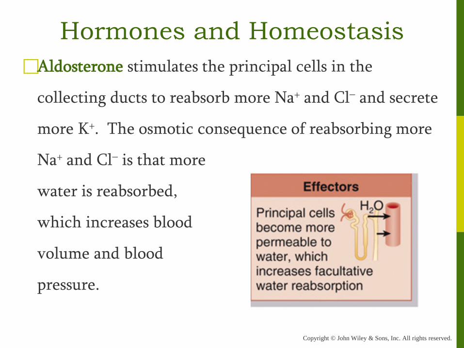
Aldosterone stimulates the principal cells in the
collecting ducts to reabsorb more Na+ and Cl– and secrete
more K+. The osmotic consequence of reabsorbing more
Na+ and Cl– is that more
water is reabsorbed,
which increases blood
volume and blood
pressure.
Hormones and Homeostasis
Copyright © John Wiley & Sons, Inc. All rights reserved.
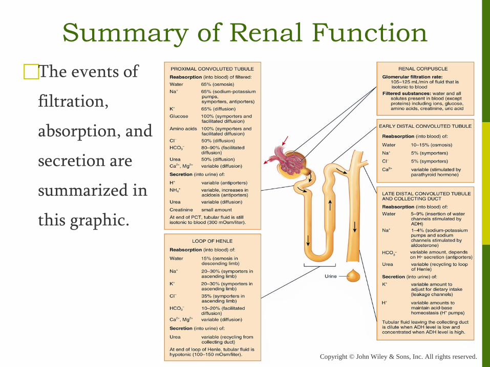
The events of
filtration,
absorption, and
secretion are
summarized in
this graphic.
Summary of Renal Function

Copyright © John Wiley & Sons, Inc. All rights reserved.
Increases in GFR usually result in an increase in urine
production. However, as we have seen, the mechanisms
which control electrolyte and
water reabsorption in the
various parts of the nephron
and collecting ducts are subject
to many complex controls.
Normal urine output (UOP)
is 1-2 L/d.
Urine

Copyright © John Wiley & Sons, Inc. All rights reserved.
A urinalysis analyzes the physical, chemical and
microscopic properties of urine.
Water accounts for 95% of total urine volume.
The solutes normally present in urine are filtered and
secreted substances that are not reabsorbed.
If disease alters metabolism or kidney function, traces of
substances normally not present or normal constituents
in abnormal amounts may appear (bacteria, albumin
protein, glucose, white blood cells, red blood cells to
name a few).
Urine

Copyright © John Wiley & Sons, Inc. All rights reserved.
In addition to a urinalysis, two blood tests are commonly
done clinically to assess the adequacy of renal function.
Blood urea nitrogen (BUN) measures nitrogen wastes in
blood from catabolism and deamination of amino acids.
Creatinine levels appear in the blood as a result of
catabolism of creatine phosphate in skeletal muscle.
◦ The serum creatinine test measures the amount of
creatinine in the blood, which increases in states of
renal dysfunction.
Urine