chapter 2 anatomy of digestive system of dairy …agitsolution.com/cbse/ebooks/11th class/animal...
TRANSCRIPT

Animal Nutrition and Reproduction 25 (Student Handbook for Class-XI)
cHAPTeR 2 Anatomy of Digestive System of Dairy Animals:
digestion, absorption and utilization of different nutrients
oBJecTiVe1. to study the different organs and their role in digestion and absorption in ruminants.
iNTRoDucTioNRuminants are the mammals that digests forages by softening it within its first compartment of the stomach, principally through bacterial actions, then regurgitating the semi-digested mass, now known as cud, and chewing it again. the process of rechewing the cud to further break down plant matter and stimulate digestion is called “ruminating”. There are about 150 species of ruminants which include both domestic and wild species. ruminating mammals including cattle, goat, sheep, giraffes, yak, deer, camels, llamas, antelope.
ANAToMy oF DigeSTiVe SySTeMthe cow’s digestive tract consists of the mouth, esophagus, a complex four-compartment stomach, small intestine and large intestine. the stomach includes the rumen or paunch, reticulum or “honeycomb,” the omasum or “manyplies,” and the abomasum or “true stomach.”
Fig: Anatomy of the adult digestive tract.

26 Animal Nutrition and Reproduction (Student Handbook for Class-XI)
The Rumen
the rumen (on the left side of the animal) is the largest of four compartments and is divided into several sacs. It can hold 25 gallons or more of material, depending on the size of the cow. Because of its size, the rumen acts as a storage or holding vat for feed. It is also a fermentation vat. A microbial population in the rumen digests or ferments feed eaten by the animal. Conditions within the rumen favor the growth of microbes. the rumen absorbs most of the volatile fatty acids produced from fermentation of feedstuffs by rumen microbes. Absorption of volatile fatty acids and some other products of digestion is enhanced by a good blood supply to the walls of the rumen. tiny projections called papillae increase the surface area and the absorption capacity of the rumen.
Fig.: The interior surface of the rumen forms numerous papillae that vary in shape and size from short and pointed to long and foliate.
The Reticulum
the reticulum is a pouch-like structure in the forward area of the body cavity. the tissues are arranged in a network resembling a honeycomb. A small fold of tissue lies between the reticulum and the rumen, but the two are not actually separate compartments. Collectively they are called the rumino-reticulum. Heavy or dense feed and metal objects eaten by the cow generally drop into this compartment. the reticulum lies close to the heart. nails and other sharp objects may work into the tissue and cause “hardware disease.” If not prevented by a magnet or corrected by surgery, infection may occur and the animal may die.
Animal Nutrition and Reproduction 27 (Student Handbook for Class-XI)
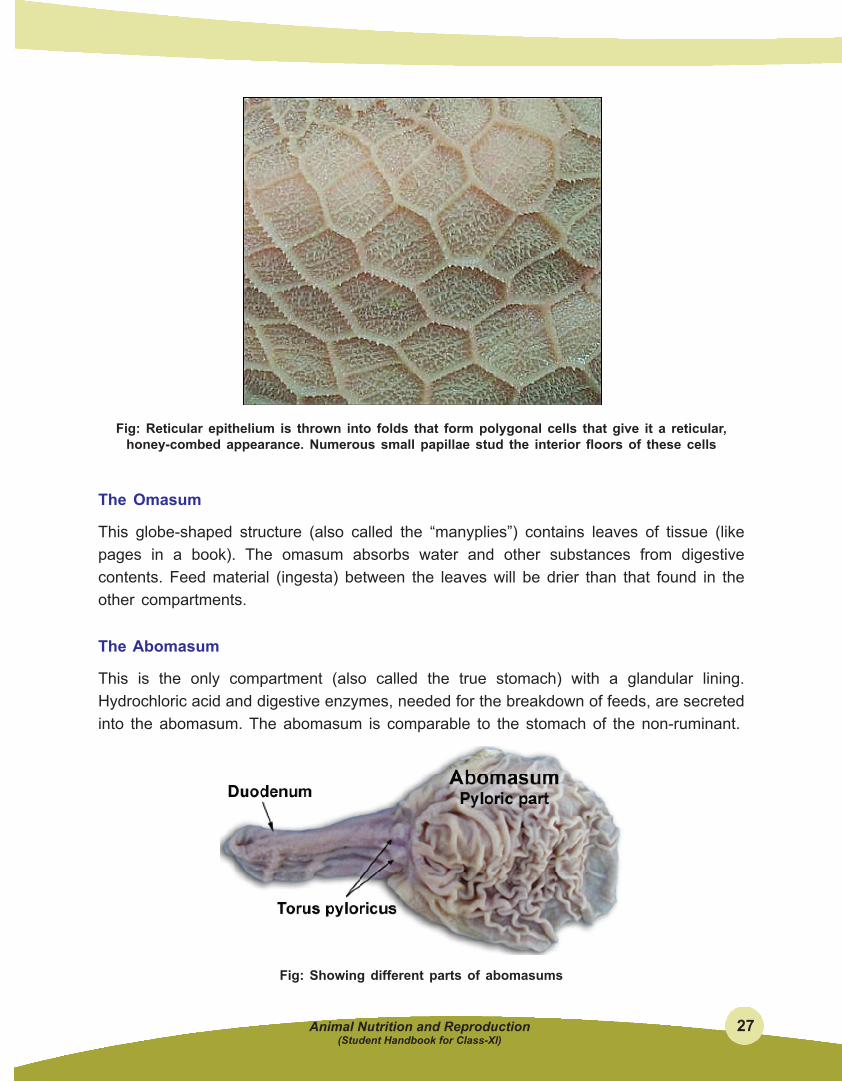
Fig: Reticular epithelium is thrown into folds that form polygonal cells that give it a reticular, honey-combed appearance. Numerous small papillae stud the interior floors of these cells
The omasum
This globe-shaped structure (also called the “manyplies”) contains leaves of tissue (like pages in a book). the omasum absorbs water and other substances from digestive contents. Feed material (ingesta) between the leaves will be drier than that found in the other compartments.
The Abomasum
this is the only compartment (also called the true stomach) with a glandular lining. Hydrochloric acid and digestive enzymes, needed for the breakdown of feeds, are secreted into the abomasum. the abomasum is comparable to the stomach of the non-ruminant.
Fig: Showing different parts of abomasums

28 Animal Nutrition and Reproduction (Student Handbook for Class-XI)
The Small intestine
the small intestine measures about 20 times the length of the animal. It is composed of three sections: the duodenum, jejunum, and ileum. the small intestine receives the secretions of the pancreas and the gallbladder, which aid digestion. Most of the digestive process is completed here, and many nutrients are absorbed through the villi (small finger-like projections) into the blood and lymphatic systems.
cecum
the cecum is the large area located at the junction of the small and large intestine, where some previously undigested fiber may be broken down. The exact significance of the cecum has not been established.
large intestine
this is the last segment of the tract through which undigested feedstuffs pass. some bacterial digestion of undigested feed occurs, but absorption of water is the primary digestive activity occurring in the large intestine.
The anatomic features described above are exemplified by cattle, sheep and goats. Certain other animals are also generally called ruminants, but have slightly different forestomach anatomy. Camelids (camels, llamas, alpacas, vicunas) have a reticulum with areas of gland-like cells, and an omasum that is tubular and almost indistinct. these animals are occasionally referred to as pseudoruminants or as having “three stomachs” rather than four.
calf Digestive System
At birth and during the first few weeks of life, the rumen, reticulum, and omasum are undeveloped. In contrast to the mature cow, in the calf, the abomasum is the largest compartment of the stomach. At this stage of life, the rumen is nonfunctional and some feeds digested by the adult cannot be used by the calf. during suckling milk, milk bypasses the rumen via the esophageal groove and passes directly into the abomasum. Reflex action closes the groove to form a tube-like structure which prevents milk or milk replacer from entering the rumen. When milk is consumed very rapidly, some may overflow into the rumen.
As long as the calf remains on milk, the rumen remains undeveloped. When calves begin consuming grain and forage, a microbial population becomes established in the rumen and reticulum. end products of microbial fermentation are responsible for the development of the rumen. this occurs as early as 3 weeks of age with most feeding programs. Cud

Animal Nutrition and Reproduction 29 (Student Handbook for Class-XI)
inoculation is not necessary to initiate rumen development. If grain feeding with or without forage is started during the first few weeks of life, the rumen will become larger and heavier with papillae development, and will begin functioning like the adult’s when the calf is about 3 months of age.
Digestion
Fig: Showing different parts of ruminant stomach including esophagus and small intestine.
the rumen is a fermentation vat par excellance, providing an anaerobic environment, constant temperature and pH, and good mixing. Well-masticated substrates are delivered through the esophagus on a regular schedule, and fermentation products are either absorbed in the rumen itself or flow out for further digestion and absorption downstream.
Feed, water and saliva are delivered to the reticulorumen through the esophageal orifice. Heavy objects (grain, rocks, nails) fall into the reticulum, while lighter material (grass, hay) enters the rumen proper. Added to this mixture are voluminous quantities of gas produced during fermentation.
Ruminants produce prodigious quantities of saliva. Adult cows are in the range of 100 to 150 liters of saliva per day. Aside from its normal lubricating qualities, saliva serves at least two very important functions in the ruminant:
l provision of fluid for the fermentation vat
l alkaline buffering - saliva is rich in bicarbonate, which buffers the large quanitity of acid produced in the rumen and is probably critical for maintainance of rumen pH.
All these materials within the rumen partition into three primary zones based on their specific gravity. gas rises to fill the upper regions, grain and fluid-saturated roughage (“yesterday’s hay”) sink to the bottom, and newly arrived roughage floats in a middle layer.
The rate of flow of solid material through the rumen is quite slow and dependent on its size and density. Water flows through the rumen rapidly and appears to be critical in flushing particulate matter downstream.
As fermentation proceeds, feedstuffs are reduced to smaller and smaller sizes and microbes

30 Animal Nutrition and Reproduction (Student Handbook for Class-XI)
constantly proliferate. Ruminal contractions constantly flush lighter solids back into the rumen. the smaller and more dense material tends to be pushed into the reticulum and cranial sac of the rumen, from which it is ejected with microbe-laden liquid through the reticulo-omasal orifice into the omasum.
the function of the omasum is rather poorly understood. It may function to absorb residual volatile fatty acids and bicarbonate. The tendency is for fluid to pass rapidly through the omasal canal, but for particulate matter to be retained between omasal leaves. Periodic contractions of the omasum knocks flakes of material out of the leaves for passage into the abomasum.
the abomasum is a true, glandular stomach which secretes acid and otherwise functions very similarly to the stomach of a monogastric. One fascinating specialization of this organ relates to its need to process large masses of bacteria. In contrast to the stomach of non-ruminants, the abomasum secretes lysozyme, an enzyme that efficiently breaks down bacterial cell walls.
The processes described above apply to adult ruminants. For the first month or so of life, the ruminant is functionally a monogastric. the forestomach are formed, but are not yet fully developed. If milk is introduced into such a rumen, it basically rots rather than being fermented. To avoid this problem in such young ruminants, suckling causes a reflex closure of muscular folds that form a channel from the esophageal orifice toward the omasum (the esophageal groove), shunting milk away from the rumen and straight toward the stomach where it can be curdled by rennin and eventually digested enzymatically.
Digestion of Different Nutrient
Digestion of energy feeds in the rumen
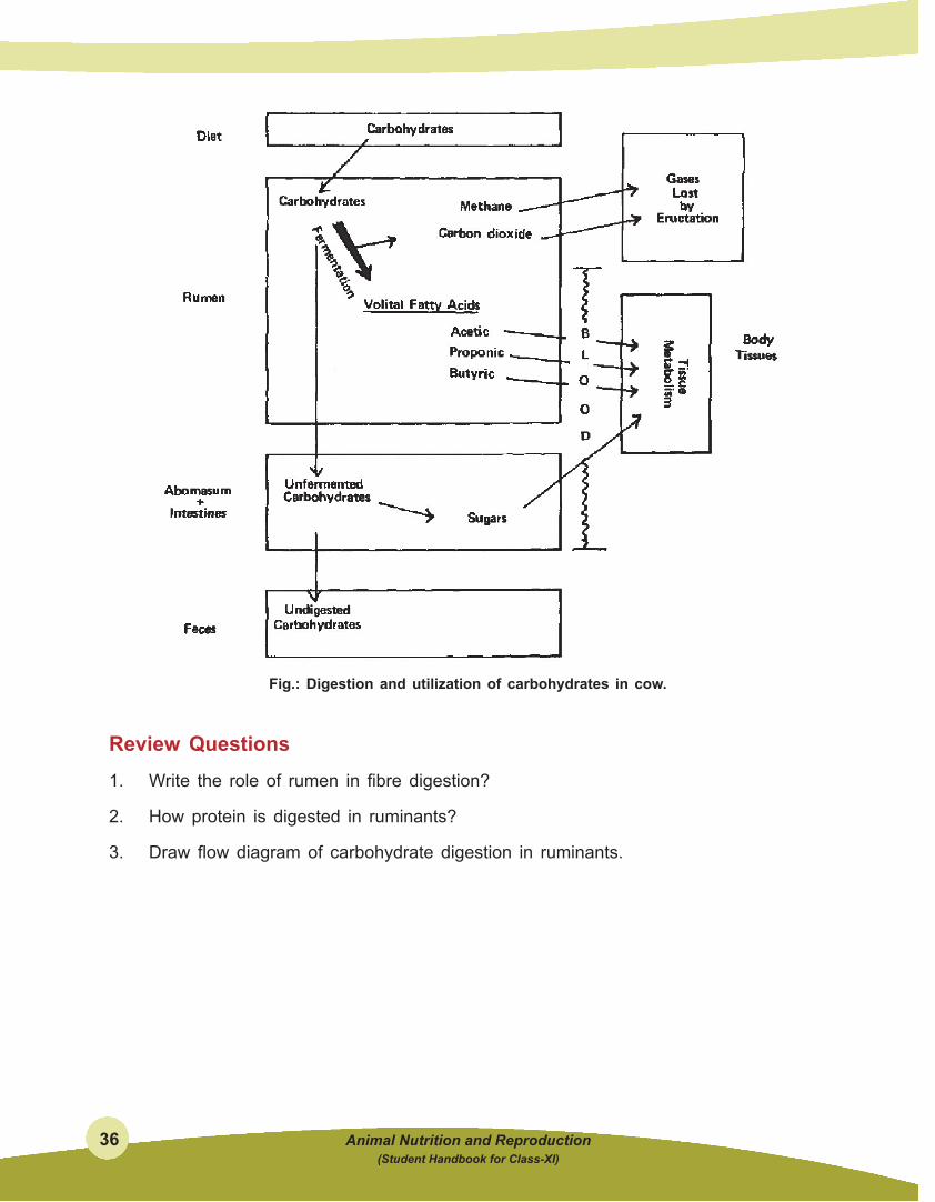
Simple and complex carbohydrates (fiber) are digested by rumen microbes and converted into volatile fatty acids. the volatile fatty acids, which consist mainly of acetic, propionic, and butyric acids, are the primary energy source for ruminants. When large amounts of forage are fed, the formation of acetic acid predominates (60 to 70 percent of total) with lesser amounts of propionic (15 to 20 percent) and butyric (5 to 15 percent) acids

Animal Nutrition and Reproduction 31 (Student Handbook for Class-XI)
occurring. However, when grain feeding is increased or when finely ground forages are fed, the proportion of acetic acid may decrease to 40 percent, while the amount of propionic acid may increase to 40 percent. Approximately 30 to 50 percent of the cellulose and hemicellulose is digested in the rumen by the microbial population. sixty percent or more of the starch is degraded, depending on the amount fed and how fast ingested materials move through the rumen. Most sugars are 100 percent digested within the rumen.
the volatile fatty acids are absorbed from the rumen into the blood stream and transported to body tissues, including the udder, where they are used as sources of energy for maintenance, growth, reproduction, and milk production. the cow derives 50 to 70 percent of its energy from the volatile fatty acids produced in the rumen.
Fig: Microbial digestion of feed carbohydrate in the rumen.
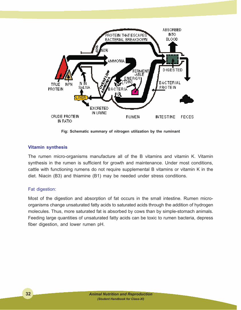
Protein and nonprotein nitrogen utilization in the rumen.
some of the protein consumed by the cow is escapes breakdown in the rumen. Protein undergoing fermentation is converted to ammonia, organic acids, amino acids, and other products. Approximately 40 to 75 percent of the natural protein in feed is broken down. the extent of breakdown depends on many factors including solubility of the protein, resistance to breakdown, rate of feed passage through the rumen, and others. Many rumen micro-organisms require ammonia (breakdown product of protein) for growth and synthesis of microbial protein. Ammonia also may be provided from NPN sources such as urea, ammonium salts, nitrates, and other compounds. rumen microbes convert the ammonia and organic acids into amino acids that are assembled into microbial protein. excess ammonia is mostly absorbed from the rumen into the blood stream, but small amounts may pass into the lower digestive tract and be absorbed. Feed protein (that escapes breakdown in the rumen) and microbial protein pass to the abomasum and small intestine for digestion and absorption.
32 Animal Nutrition and Reproduction (Student Handbook for Class-XI)
Fig: Schematic summary of nitrogen utilization by the ruminant
Vitamin synthesis
the rumen micro-organisms manufacture all of the B vitamins and vitamin K. Vitamin synthesis in the rumen is sufficient for growth and maintenance. Under most conditions, cattle with functioning rumens do not require supplemental B vitamins or vitamin K in the diet. niacin (B3) and thiamine (B1) may be needed under stress conditions.
Fat digestion:
Most of the digestion and absorption of fat occurs in the small intestine. Rumen micro-organisms change unsaturated fatty acids to saturated acids through the addition of hydrogen molecules. thus, more saturated fat is absorbed by cows than by simple-stomach animals. Feeding large quantities of unsaturated fatty acids can be toxic to rumen bacteria, depress fiber digestion, and lower rumen pH.

Animal Nutrition and Reproduction 33 (Student Handbook for Class-XI)
Fig: Digestion utilization of fat by cow
Nutrient Absorption and utilization in RuminantVolatile fatty acids (VFA) are produced in large amounts through ruminal fermentation and are of paramount importance in that they provide greater than 70% of the ruminant’s energy supply. Virtually all of the acetic, proprionic and butyric acids formed in the rumen are absorbed across the ruminal epithelium, from which they are carried by ruminal veins to the portal vein and hence through the liver. Continuous removal of VFA from the rumen is important not only for distribution, but to prevent excessive and damaging drops in pH of rumen fluid. The rumen is lined with stratified squamous epithelium similar to skin, which is generally not noted for efficient absorption. Nonetheless, this squamous epithelium has a structure which functions similarly to the columnar epithelium in the small gut and performs efficient absorption of VFA, as well as lactic acid, electrolytes and water. Recall also, that the epithelial surface is expanded greatly by formation of well-vascularized papillae. It is of considerable practical importance that the size and length of ruminal papillae respond to concentrations of VFA in the rumen. Animals that have been on a high

34 Animal Nutrition and Reproduction (Student Handbook for Class-XI)
plane of nutrition, with abundant VFA production, have long, luxuriant papillae well suited to promote absorption. In contrast, animals which have been under nutritional deprivation have small, blunted papillae, and require time on a high quality diet to allow for development of their papillae and absorptive capacity. All the VFA appear to be absorbed by the same mechanism, which is diffusion through the epithelium, down a concentration gradient. As they pass through the epithelium, the different VFA undergo different degrees of metabolism. Acetate and proprionate pass through the epithelium largely unchanged, but almost all of the butyric acid is metabolized in the epithelium to beta-hydroxybutyric acid, a type of ketone body.
The three major VFA absorbed from the rumen have somewhat distinctive metabolic fates:
l Acetic acid is utilized minimally in the liver, and is oxidized throughout most of the body to generate ATP. Another important use of acetate is as the major source of acetyl CoA for synthesis of lipids.
l Proprionic acid is almost completely removed from portal blood by the liver. Within the liver, proprionate serves as a major substrate for gluconeogenesis, which is absolutely critical to the ruminant because almost no glucose reaches the small intestine for absorption.
l Butyric acid, most of which comes out of the rumen as the ketone beta-hydroxybutyric acid, is oxidized in many tissues for energy production.

Animal Nutrition and Reproduction 35 (Student Handbook for Class-XI)
THe DigeSTioN AND uTiliZATioN oF PRoTeiN AND cARBoHyDRATeS ARe SHowN Below:
Fig.: Digestion and utilization of protein in cow.
36 Animal Nutrition and Reproduction (Student Handbook for Class-XI)
Fig.: Digestion and utilization of carbohydrates in cow.
Review Questions1. Write the role of rumen in fibre digestion?
2. How protein is digested in ruminants?
3. Draw flow diagram of carbohydrate digestion in ruminants.