chapter 1shodhganga.inflibnet.ac.in/bitstream/10603/23659/8/08_chapter 1.pdf · since there is no...
TRANSCRIPT

Chapter 1
REVIEW OF LITERA TURE
1. Review of Literature
1.1 History of malaria
It is speculated that malaria originated in Africa and accompanied human migration to
the Mediterranean shores, India and South East Asia. The origin of the name malaria
stems from the archaic association between the disease and the bad air of marshy areas
around Rome, "mal aria" in Italian meaning "bad air" (Bruce-Chwatt, 1985). Writings from
the vedic period (1500 to 800 B.C.) describe the enlargement of the spleen in persons
having autumnal fever, suggesting that at that time malaria existed in India (Sherman,
1998). Since there is no mention in any of the Hippocratic writings about severe,
malignant tertian fever, it is assumed that at that time P. falciparum infections were rare
or non existent.
By the 12th century, malaria has spread reaching European countries like Spain, Polland
and Russia. There are no records of malaria in the New World before European
explorers landed on its shores, so it is assumed that they brought P. malarie and P.
vivax to the Americas, P. fa/ciparum was introduced later by the importation of African
slaves (Bruce-Chwatt, 1988). Thus, by early 1800's malaria was worldwide in its
geographical distribution.
The first recorded treatment dates back to 1600, when the bitter bark of the Cinchona
tree in Peru was used by the native Peruvian Indians. By 1649, the bark was available in
England, as "Jesuits powder," so that those suffering from "agues" might benefit from
the chemical substance quinine, which it contained (Nobel Prize Foundation, 2006).
The discoveries of Alphonse Laveran in 1880 when he identified the causative agent for
human malaria to be a blood parasite, and Sir Ronald Ross in 1897 when he
demonstrated that mosquitoes were the vectors of maiaria, were two Nobel prize
discoveries in malaria, which marked the beginning of a new era for the control of this
deadly disease (Gibson, 1998). In 1885, Golgi identified the asexual development and
reproduction by multiple fission of P. vivax and P. malariae and showed the correlation
between the beginning of the fever and rupture of the infected erythrocytes.
4
Today malaria is known as an infection transmitted by mosquitoes of the genus
Anopheles. There are four species of Plasmodium, which can cause malaria in humans
i.e. P. fa/ciparum, P. vivax, P. ma/ariae, and P. o vale, which belong to the phylum
Protozoa. The spectrum of clinical illness ranges from cyclic fevers with rigors and chills,
to anemia, hypoglycemia, convulsions, hepatic dysfunctions and bleeding abnormalities
(White, 1998).
1.2 Current situation of malaria in the world
Malaria is considered the world's most important tropical parasitic disease.
Approximately 300 - 500 million clinical cases are recorded every year in the world.
Each year malaria kills 1-million people worldwide, 80% of which are living in Sub-
Saharan Africa. The large majority of the victims are children under the age of 5, as
indicated in the first joint World Malaria Report 2005 of the WHO (World Health
Organization) and UNICEF (United Nations Children's Fund). Around 107 countries or
territories in the world are affected by malaria, with almost half of them located in Africa
south of the Sahara and around 3.2 billion people at risk of acquiring the infection. It has
been estimated that malaria kills 3.000 children every day only in Africa (Phillips, 2001).
Many factors contribute to the successful transmission of the infection in endemic areas.
The host (human) immunity, the specificities of each parasite species, the anopheline
longevity and its avidity for human are intrinsic factors that have the greatest impact on
the malaria burden. In addition, the increasing development of resistance of
Plasmodium parasites to the available drugs, and of the mosquitos to the insecticides,
makes the task of controlling malaria in actual times much more challenging. Among the
extrinsic factors, climate changes (mainly rainfall), economic conditions (poverty),
political commitment and effectiveness of prevention efforts are the most important
determinants.
5
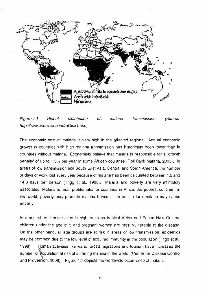
Figure 1. 1 Global distribution
http://www. wpro. who. int/rdtllink 1. asp)
of malaria transmission (Source:
The economic cost of malaria is very high in the affected regions. Annual economic
growth in countries with high malaria transmission has historically been lower than in
countries without malaria. Economists believe that malaria is responsible for a 'growth
penalty' of up to 1.3% per year in some African countries (Roll Back Malaria, 2005). In
areas of low transmission like South East Asia, Central and South America; the number
of days of work lost every year because of malaria has been calculated between 1 .5 and
14.3 days per person (Trigg et aI., 1998). Malaria and poverty are very intimately
associated. Malaria is most problematic for countries in Africa, the poorest continent in
the world; poverty may promote malaria transmission and in turn malaria may cause
poverty.
In areas where transmission is high, such as tropical Africa and Papua New Guinea,
children under the age of 5 and pregnant women are most vulnerable to the disease.
On the other hand, all age groups are at risk in areas of low transmission; epidemics
may be common due to the low level of acquired immunity in the population (Trigg et aI.,
1998). ~,uman activities like wars, forced migrations and tourism have increased the
number of'fPulation at risk of suffering malaria in the world. (Center for Disease Control
and Preven'~i:On, 2006). Figure 1.1 depicts the worldwide occurrence of malaria.
6

Effective control of malaria depends on three essential elements. The reduction of the
population of Anopheline mosquitoes that can transmit the parasite, the early diagnosis
and prompt treatment of malarial disease in all areas where people are at risk and an
adequate epidemiologic surveillance, which allows forecasting of epidemics in specific
regions. The development of effective drugs and/or a vaccine against human malaria
parasites, as well as, the implementation of selective vector control strategies are the
actual priorities of governments and institutions around the world. At present, no
vaccine is in use against malaria, however some of the experimental vaccines are under
different stages of trials using parasite proteins as protective antigens (Portfolio Malaria
Vaccine Candidates, 2005). In recent years, several international agencies have been
created to initiate new programs to combat malaria. Roll Back Malaria was started in
1998, mainly to control malaria in children and pregnant women in Africa. In 1999 two
new international programs were started i.e. MMV- Medicines for Malaria Venture and
MVI- Malaria Vaccine Initiative. These two programs are funding several research
projects involving the discovery of new drugs and the development of an effective
vaccine.
1.3 Life cycle of malaria parasites
The life cycle of Plasmodium is complex, involving human and mosquito hosts, and
many developmental stages (Figure 1.2). Sporozoites, thought to be less than 100 on
each occasion, are transmitted to humans by the bite of infected female Anopheles
mosquitoes. Within 30 to 45 minutes these sporozoites enter the host's hepatocytes,
where they develop into exo-erythrocytic schizonts during the next 6 to 15 days,
depending on the species. At the end of the pre-erythrocytic cycle, 30,000 to 40,000
merozoites are released into the blood circulation and invade erythrocytes in a process
that takes approximately 30 seconds (Fujioka and Aikawa, 2002). The complete
process of invasion can be observed in Figure 1.3
Plasmodium vivax, Plasmodium ovale and Plasmodium cynomolgi have a dormant
stage, named hypnozoite (Krotoski et aI., 1982) that may remain in the liver for weeks to
many years before the development of preerythrocytic schizogony. This results in
relapses of malaria infection.
7
Within the erythrocyte, the parasite develops over a period of 2 (P. fa/ciparum, P. vivax
and P. ovale) or 3 days (P. malariae). Within the parasitophorous vacuole, the parasite
modifies its host cell in several ways to enhance its survival. The parasite matures from
an initial ring form to a trophozoite, which, following mitotic division, develops into the
schizont stage containing up to 32 merozoites. Following rupture of the cell, the
merozoites are released which then immediately invade additional erythrocytes.
After invading red blood cells, eventually some of the merozoites differentiate into sexual
forms: the male and female gametocytes. When a female Anopheles mosquito takes a
blood meal from an infected host, the gametocytes that are ingested with the blood meal
mature to male and female gametes in the mosquito mid-gut. After fertilization, the
resulting zygote matures within 24 hours into the motile ookinete, which burrows through
the midgut wall to encyst on the basal lamina, the extracellular matrix layer separating
the haemocoel from the midgut. Within the developing oocyst, there are many mitotic
divisions resulting in oocysts full of sporozoites. Between 7 and 15 days post infection,
depending on the Plasmodium species and the ambient temperature, a single oocyst
forms more than 10,000 sporozoites. These motile sporozoites migrate into the salivary
glands and accumulate in the acinar cells of the salivary glands. When an infected
mosquito bites a susceptible vertebrate host, the Plasmodium life cycle begins again,
with the inoculation of sporozoite stages into the blood stream of the infected host.
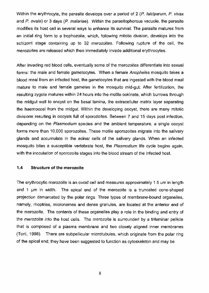
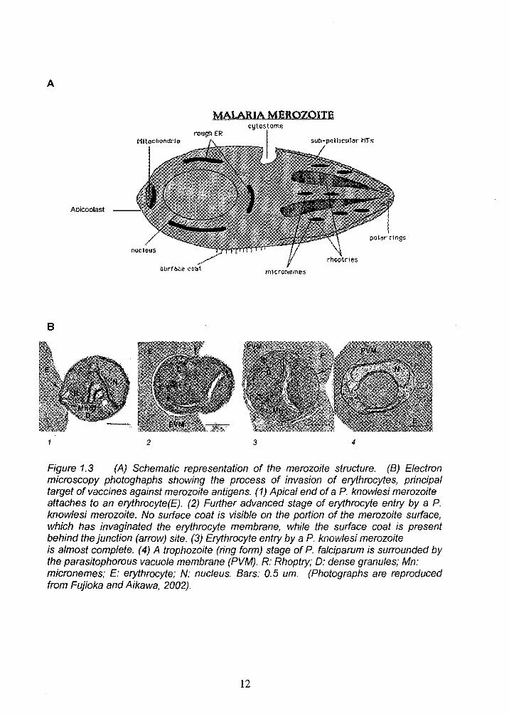
1.4 Structure of the merozoite
The erythrocytic merozoite is an ovoid cell and measures approximately 1.5 um in length
and 1 IJm in width. The apical end of the merozoite is a truncated cone-shaped
projection demarcated by the polar rings. Three types of membrane-bound organelles,
namely, rhoptries, micronemes and dense granules, are located at the anterior end of
the merozoite. The contents of these organelles playa role in the binding and entry of
the merozoite into the host cells. The merozoite is surrounded by a trilaminar pellicle
that is composed of a plasma membrane and two closely aligned inner membranes
(Torii, 1998). There are subpellicular microtubules, which originate from the polar ring
of the apical end; they have been suggested to function as cytoskeleton and may be
8
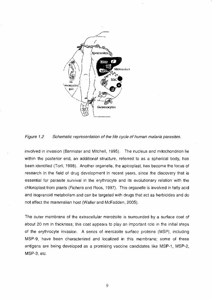
Figure 1.2 Schematic representation of the life cycle of human malaria parasites.
involved in invasion (Bannister and Mitchell, 1995). The nucleus and mitochondrion lie
within the posterior end, an additional structure, referred to as a spherical body, has
been identified (Torii, 1998). Another organelle, the apicoplast, has become the focus of
research in the field of drug development in recent years, since the discovery that is
essential for parasite survival in the erythrocyte and its evolutionary relation with the
chloroplast from plants (Fichera and Roos, 1997). This organelle is involved in fatty acid
and isoprenoid metabolism and can be targeted with drugs that act as herbicides and do
not affect the mammalian host (Waller and McFadden, 2005).
The outer membrane of the extracellular merozoite is surrounded by a surface coat of
about 20 nm in thickness; this coat appears to play an important role in the initial steps
of the erythrocyte invasion. A series of merozoite surface proteins (MSP), including
MSP-9, have been characterized and localized in this membrane; some of these
antigens are being developed as a promising vaccine candidates like MSP-1, MSP-2,
MSP-3, etc.
9
1.5 Rationale of malaria vaccines against merozoite surface proteins
Several evidences support the development of a vaccine against erythrocytic stages of
malaria parasites; 1) Following repeated attacks of malaria, a majority of infected
individuals in endemic areas are able to control the parasite replication to levels that do
not result in clinical disease, 2) hyperimmune globulin prepared from the sera of
individuals chronically infected with malaria can eliminate circulating parasites from P.
falciparum-infected individuals, 3) maternal antibodies passively transfer to the fetus
may provided a window of protection against clinical malaria (Ballou et aI., 2004; Good,
2005)
In contrast to the requirement of vaccines for smallpox, polio or many of the other
infectious diseases for which vaccines are available, the requirement for a vaccine
against malaria is not universal. One of the main target groups comprises infants,
children and pregnant women resident in malaria endemic areas. Another target group
is individuals who have never been, or are infrequently exposed to malaria (non-
immunes). These non-immunes include tourists, business, and military personnel from
countries without malaria who travel to countries where malaria is transmitted; in
addition, there is increasing number of residents from malaria endemic countries, living
in malaria-free areas, who should be considered as non-immune individuals (Hoffman
and Miller, 1996).
A malaria vaccine based on asexual blood-stage antigens is expected to control the
magnitude of the asexual parasitaemia and also to decrease the incidence of severe
disease. Such "anti-morbidity" vaccine would be of great benefit for the communities
living in highly endemic areas, in particular, pregnant women and infants of these
communities (Richie and Saul, 2002). It is believed that the principal immune
mechanism induced by these vaccines is the generation of protective memory B-cell
response that, at the time of infection, will generate antibodies that will neutralize
important merozoite antigens, preventing in this way the process of invasion of
erythrocytes. However, there is growing evidence from murine models of malaria that
cell-mediated mechanisms may be critical to the acquisition of acquired immunity to
malaria (Ballou et aI., 2004).
10
1.6 Merozoite Surface Proteins
The proteins located on the surface of the merozoite are considered potential targets of
the protective immune response; for that reason their identification and characterization
is important for the development of an effective malaria vaccine. The first protein of this
kind was identified in 1981 (Holder and Freeman, 1981) and since that time, a total of 10
proteins have been categorized as Merozoite Surface Proteins (MSPs); numbered
according to the order in which they have been characterized. In recent times, the
information generated from the complete genome sequences of P. fa/ciparum and P.
yoeJii (Carlton et aI., 2002; Gardner et aI., 2002), as well as the development of
transfection techniques has facilitate the characterization of new MSPs, not only for their
potential as a vaccine candidates but also to better understand their role in the biology of
Plasmodium parasites.
1.6.1 Merozoite surface protein-9 (MSP-9)
The first description of MSP-9 was done by Stahl et al. (1986), who reported the isolation
of a group of E. coli clones expressing P. fa/ciparum antigens that reacted with human
immune sera. One of the antigens that they identified was described as a protein of 102
kDa, predominantly present in schizonts, which partial DNA sequence revealed blocks of
hydrophilic aminoacids at the C-terminal. For this reason, the new protein was
designated as acidic basic repeat antigen (ABRA) (Recently named PfMSP-9).
Simultaneusly, Lyon et al. (1986), identified an antigen of 101 kDa molecular weight
(p1 01) along with 14 more antigens from P. fa/ciparum blood-stages, that were isolated
from immune clusters of merozoites (ICM), formed in the presence of inhibitory human
immune sera. The authors considered this method as a useful approach to identify
exposed targets of protective immunity against malaria.
11
A
MAlARIA MEROZOITE
Aoicoolast
B
2 3 4
Figure 1.3 (A) Schematic representation of the merozoite structure. (B) Electron microscopy photoghaphs showing the process of invasion of erythrocytes, principal target of vaccines against merozoite antigens. (1) Apical end of a P. knowlesi merozoite attaches to an erythrocyte (E). (2) Further advanced stage of erythrocyte entry by a P. knowlesi merozoite. No surface coat is visible on the portion of the merozoite surface, which has invaginated the erythrocyte membrane, while the surface coat is present behind the junction (arrow) site. (3) Erythrocyte entry by a P. knowlesi merozoite is almost complete. (4) A trophozoite (ring form) stage of P. falciparum is surrounded by the parasitophorous vacuole membrane (PVM). R: Rhoptry; 0: dense granules; Mn: micronemes; E: erythrocyte; N: nucleus. Bars: 0.5 um. (Photographs are reproduced from Fujioka and Aikawa, 2002).
12

Subsequently, Chulay et al. (1987) characterized the monoclonal antibody 3D5 (3D5
mAb) that was produced from mice immunized with the ICM, which specifically
recognized the antigen of 101 kDa reported by Lyon et al. (1986). With the use of 3D5
mAb it was possible to localize p101 antigen at the surface of schizonts and free
merozoites as well as in the parasitophorous vacuole. This antigen was detected as
secreted in the media when schizonts rupture occurred in normal culture media but not
when the rupture occurred in the presence of immune sera. In addition, the affinity-
purified p101 antigen was recognized in western blot by 3D5 antibodies dissociated from
the ICM. Using [3H]lIe-labeled parasites, they also showed that p101 antigen was
synthesized late in the parasite cycle, when mature throphozoites and early schizonts
predominated.
In 1988, Weber et al. (1988) reported the complete DNA sequence of ABRA and
described the full-length protein as having 743 aminoacids, which included two
tandemly repeated regions, one near the amino terminus containing eight hexapeptide
repeats of sequence TVNDEDED, and the second near the carboxyl terminus containing
primarily KE and KEE repeats. The protein was defined as hydrophilic and highly
charged with a calculated isoelectric point of 5.6. Nine potential sites for N-
glycosylation were identified in the sequence of ABRA, however no experimental
evidence was obtained. In addition, there were no evidences of hydrophobic motifs that
could suggest that ABRA was a structural membrane protein.
The immunogenic properties of ABRA has been partially studied. Sharma et al. (1998)
used synthetic peptides (12 - 18 residues) covering the most hydrophilic regions of
ABRA as well as the repetitive sequences, to study the presence of B and T-cell
epitopes in the sequence of this protein. They found that all the peptides were
recognized by specific antibodies present in the sera of 50 individuals with acute P.
fa/ciparum malaria, with peptide AB-3 (395 - 409 aa) being the most frequently
recognized. In addition, high Iymphoproliferative response was observed against AB-
1(19 - 30 aa) and AB-3 peptides after in vitro stimulation of PBMC from 11 of these
individuals. Six of the peptides induced a strong antibody response in rabbits when they
were used as immunogen in the absence of any carrier, indicating the presence of both
B-cell determinants and T-helper-cell epitopes in these six constructs. The antibodies
13
generated against peptides AB-1 and AB-5 (518 - 531 aa) inhibited the merozoite
invasion of human erythrocytes in vitro up to approximately 90%.
The presence of Band T cell epitopes in ABRA was further confirmed by Kushwaha et
al. (2001), who used recombinant proteins representing different fragments of ABRA
expressed in Escherichia coli, and purified by affinity chromatography. Immunogenicity
studies in mice and rabbits showed that the N-terminal portion was the most
immunogenic region of ABRA, IgG1 predominance was found in mice immunized with
the constructs designated ABRA(P) (aa 24 - 507) and ABRA (N) (aa 24 - 369). In this
study, the antibodies against the middle and C-terminal region of ABRA were able to
prevent the parasite growth in 82% and 93%, respectively. The recombinant proteins
were also recognized by the sera from P. falciparum-infected patients, with ABRA(P)
showing the highest frequency of reactivity (73%). T-cell proliferation experiments in
mice immunized with these constructs revealed that the T-cell epitopes were localized in
the middle portion of the protein. Pimtanothai et al. (2000) using computer algorithms,
predicted T-cell epitopes in the sequence of ABRA and showed that the synthetic
peptide ABRA 14 (487 - 506 aa) induced significant T-cell proliferation in splenocytes
from immunized mice and Th-1 associated cytokine production.
A study by Bonnefoy et al. (1994) described the used of a P. falciparum protein fraction,
containing ABRA and other blood-stage antigens in the range of 90 - 110 kDa, to
immunize S. sciureus monkeys that were subsequently challenged with P. falciparum
Uganda Palo Alto FUP/SP strain. Three monkeys out of five resisted the dose of virulent
parasites, however no correlation was found between the antibody response to ABRA
and the state of protection in these monkeys.
The identification of the homologues of ABRA from P. vivax, P. cynomolgi and P.
knowlesi (Barnwell et aI., 1999; Vargas-Serrato et aI., 2002), allowed the delineation of
features common to this group of proteins and the definition of a new family named
Merozoite Surface Protein - 9. Recently the MSP-9 homologue from P. coatneyi was
also described (Vargas-Serrato et aI., 2003). One of the main features of this family is
the conservation of the N-terminal region, including four cysteine residues in all these
proteins and the presence of tandem repeats at the C-terminal region. However the
sequence of these tandem repeats is not conserved among the MSP-9 proteins and only
14
PfMSP-9 (ABRA) contain the repeats KE/KEE that confer the character of acidic-basic
repeat antigen. In addition, P. vivax MSP-9 and the simian homologues do not contain
the block of repeat sequences present in the middle region of PfMSP-9. P. vivax MSP-9
is the largest protein of this family, composed of 979 aa, followed by P. cynomolgi (818
aa), P. coatneyi (764 aa), P. falciparum (743 aa) and P. knowlesi (707 aa). These
proteins are predicted to assume an alpha-helical conformation at the N-terminus and to
have a combination of alpha helix and random coils at the C-terminus. There is no
indication of GPI lipid anchor associated with these proteins. The level of identity
between PfMSP-9 and P. vivax, P. cynomolgi and P. knowlesi MSP-9 proteins is
estimated in 33-34%; antisera against the recombinant N-terminal fragment of P.
cynomolgi MSP-9 and P. knowlesi MSP-9 cross-react with the native P. vivax MSP-9 but
not with P. falciparum MSP-9 native protein. In a similar way, antisera against
recombinant P. vivax MSP-9 N-terminal fragment recognize P. cynomolgi and P.
knowlesi homologues but not P. falciparum MSP-9.
Oliveira-Ferreira et al. (2004) have shown that the N-terminal region of PvMSP-9 as well
as the second block of tandem repeats are immunogenic in mice, inducing high levels of
antibody titers, with predominance of IgG1, IgG2a and IgG2b isotypes. Moreover the
immunization with N-terminal PvMSP-9 induced IFNy and IL-5 by splenocytes re-
estimulated in vitro.
Another important characteristic of this family of proteins is the presence of species-
specific repeat sequences in the C-terminal region. Alignment of all the seven members
of this family shows that there is a direct correlation between the length of the repeats
and the total number of aa for each protein; for example P. vivax and P. cynomolgi being
the biggest proteins of these family (979 aa and 818 aa respectively), contain two
regions with repeat sequences as long as 14 aa which are repeated 4 to 6 times, while
in P. yoe/ii (678 aa), the repeat sequences are maximum 9 aa and are repeated only
three times. Although the structure of the repeats is different for each MSP-9 homolog,
all these sequences are characterized by a high content of glutamic acid that gives a
total percentage for the whole protein ranging from 11 % to 18%. The function of these
repeats in MSP-9 proteins is not known but is possible that they are merely related with
the mechanisms of evolution of these proteins.
15
1.6.1.1 Predicted functions for MSP·9 proteins in Plasmodium parasites
1.6.1.1.1 Binding to erythocytes
Being located in the surface of merozoites, is logical to think that MSP-9 proteins may
interact with erythrocyte proteins at the time of invasion. There are examples of other
surface antigens interacting with structural proteins of erythrocytes like MSP-1, ring-
infected erythrocyte surface antigen (RESA), etc. (Foley et aI., 1991; Herrera et aI.,
1993). In addition, it has been demonstrated that the erythrocyte protein Band 3 is
subjected to proteolytic cleavage in parasite infected erythrocytes, which induced
conformational changes that probably leads to cytoadherence (Winograd and Sherman,
1989). It is also possible that parasite proteases are involved in this process.
The hypothesis that PfMSP-9 binds to human erythrocyte proteins was addressed by
Kushwaha et al. (2002). Using E. coli expressed recombinant fragments of PfMSP-9,
they demonstrate that the cysteine-rich N-proximal region of this protein binds in a
specific dose-dependant manner to Band 3 protein. The binding of purified native
PfMSP-9 to band 3 protein, confirm the observations with the recombinant proteins.
They also observed the conservation of 528 bp region containing the four cysteines in
the N-terminal region among fifteen field isolates of P. falciparum, confirming the
importance of this region for the function of MSP-9 proteins. Simultaneously, Curtidor et
al. (2001) screened the complete sequence of PfMSP-9 to identified specific motifs that
bind to human erythrocytes. They were able to define a region in PfMSP-9
encompassed between residues 121 and 240 with high binding specific activity to
erythrocyte membranes. They concluded that the specific binding was independent of
the charge of the peptides and mainly dependent on hydrophobic interactions, since
30% of the critical residues were hydrophobic. In addition, peptides 2148 (121-140 aa)
and 2149 (141-160 aa) were able inhibit the erythrocyte invasion by merozoites,
suggesting that they were able to compete with native ABRA for the binding and that this
event is important for erythrocyte invasion.
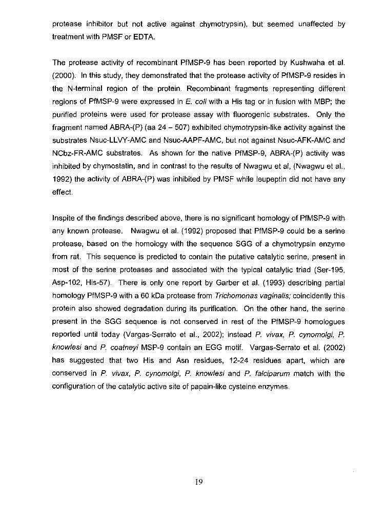
Recently, the interaction of PfMSP-9 with Band 3 protein has been studied in more detail
by Li et al. (2004). They demonstrate that the N-terminal portion of PfMSP-9 interacts
specifically with a domain denominated 5ABC, present in a non-glycosylated exofacial
16
region of Band 3 protein, which they previously reported as an important host receptor
for the sialic acid-independent invasion pathway. They also showed that SABC domain
interacts with MSP-142 protein through its 19 kDa C-terminal domain (Goel et ai., 2003).
They were able to detect the interaction PfMSP-9/SABC through yeast-two hybrid
system, using recombinant constructs representing multiple regions of PfMSP-9 as well
as Band 3 protein. The interaction was confirmed with a binding assay in solution, using
purified GST fusion proteins expressed in E. coli. As suggested by the results of
Curtidor et al (Curtidor et aI., 2001), this study demonstrates the importance of PfMSP-
9/8and 3 interaction for the erythrocyte invasion. Because SABC domain has been
shown to bind MSP-142 also (Goel et aI., 2003), they studied the interaction of MSP-9
and MSP-142 finding that, indeed, MSP-9 recombinant proteins were able to co-
precipitate native MSP-142 from parasite culture supernatant and viceversa. These
results allowed them to propose a model for the simultaneous interaction of P.
falciparum MSP-9 and MSP-1 with the erythrocyte Band 3 protein (Figure 1.4), in which
the co-ligand complex MSP-142 - MSP-9 interacts with the SABC domain of the same
Band 3 molecule or with different SABC domains within the Band 3 dimer/tetramer in the
erythrocyte membrane. The existence of MSP-1 42/MSP-9 in a complex is consistent
with the previous evidences of MSP-1 processing fragments associated with other
proteins like MSP-636 and MSP-722 in the surface of the merozoites (Pachebat et aI.,
2001; Trucco et aI., 2001). A recent study by Kariuki et al. (200S) confirms these results.
Recently, a network of protein interactions from P. falciparum blood-stages have been
described by LaCount (200S), using a high-throughput yeast two-hybrid assay. The
interactions were defined by computer analyses of the network connectivity, presence of
common protein domains, the coexpression of the genes encoding interacting proteins
and Gene Ontology annotations. One of the identified cluster is defined by the direct
interaction of MSP-1 and MSP-9 as a core, linking 19 uncharacterized proteins and 16
other proteins that are involved in the invasion of host cells or are localized to the
merozoite surface.
17
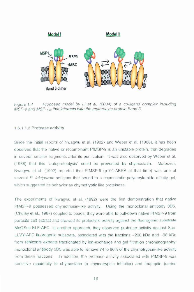
Modell Model II
Band 3 dim6r
Figure 1.4 Proposed model by Li et at. (2004) of a co- ligand complex including MSP-9 and MSP-1..J2 that interacts with the erythrocyte protein Band 3.
1.6 .1.1.2 Protease acti vi ty
Since the initial reports of Nwagwu et al. (1992) and Weber et al. (1988), it has been
observed that the native or recombinant PfMSP-9 is an unstable protein , that deg rades
in several smaller frag ments after its purifi cation. It was also obse rv ed by Weber et al.
(1988) that this "autoproteolysis" could be prevented by chymostatin . Moreover,
Nwagwu et al . (1992) reported that PfMSP-9 (p101 -ABRA at that time) was one of
several P. fa/ciparllm antigens that bound to a chymostatin-polyac rylamide affinity gel,
which suggested its behavior as chymotryptic li ke proteinase.
The expe rim ents of Nwagwu et a l. (1992) were the fi rst demonstration that native
PfMSP-9 possessed chymotrips in -like activity . Using the monoclonal antibody 3D5,
(Chulay et aI. , 1987) coupled to beads, they were able to pu ll -down native PfM SP-9 from
MeOSuc-KLF-AFC. In another approach, they obse rved protease activity against Suc-
LLVY- AFC fl uorogenic substrate, assoc iated with the fractions -200 kDa and - 90 kDa
from schizonts extracts fractionated by ion -exchange and ge l filtration chromatog raphy ;
monoclonal antibody 3D5 was able to remove 74 to 96% of th e chymotrypsin -like activity
from these fractions. In addi ti on, the protease activi ty assoc iated with PfMSP-9 was
sens itive maximall y to chymostatin (a chymotrypsin inhibitor) and leupeptin (se rine
I ~
protease inhibitor but not active against chymotrypsin), but seemed unaffected by
treatment with PMSF or EDT A.
The protease activity of recombinant PfMSP-9 has been reported by Kushwaha et al.
(2000). In this study, they demonstrated that the protease activity of PfMSP-9 resides in
the N-terminal region of the protein. Recombinant fragments representing different
regions of PfMSP-9 were expressed in E. coli with a His tag or in fusion with MBP; the
purified proteins were used for protease assay with fluorogenic substrates. Only the
fragment named ABRA-(P) (aa 24 - 507) exhibited chymotrypsin-like activity against the
substrates Nsuc-LLVY-AMC and Nsuc-AAPF-AMC, but not against Nsuc-AFK-AMC and
NCbz-FR-AMC substrates. As shown for the native PfMSP-9, ABRA-(P) activity was
inhibited by chymostatin, and in contrast to the results of Nwagwu et ai, (Nwagwu et aI.,
1992) the activity of ABRA-(P) was inhibited by PMSF while leupeptin did not have any
effect.
Inspite of the findings described above, there is no significant homology of PfMSP-9 with
any known protease. Nwagwu et al. (1992) proposed that PfMSP-9 could be a serine
protease, based on the homology with the sequence SGG of a chymotrypsin enzyme
from rat. This sequence is predicted to contain the putative catalytic serine, present in
most of the serine proteases and associated with the typical catalytic triad (Ser-195,
Asp-102, His-57). There is only one report by Garber et al. (1993) describing partial
homology PfMSP-9 with a 60 kDa protease from Trichomonas vaginalis; coincidently this
protein also showed degradation during its purification. On the other hand, the serine
present in the SGG sequence is not conserved in rest of the PfMSP-9 homologues
reported until today (Vargas-Serrato et aI., 2002); instead P. vivax, P. cynomolgi, P.
knowlesi and P. coatneyi MSP-9 contain an EGG motif. Vargas-Serrato et al. (2002)
has suggested that two His and Asn residues, 12-24 residues apart, which are
conserved in P. vivax, P. cynomolgi, P. knowlesi and P. fa/ciparum match with the
configuration of the catalytic active site of papain-like cysteine enzymes.
19
1.6.1.1.3 Other predicted functions
Along with the ability to inhibit parasite invasion, the peptide 2149 (aa 141 - 160),
described by Curtidor et al (2001) also showed high homology with a human cytosolic
phospholipase A2. Moreover, this peptide also exhibited haemolytic and anti-microbial
activity in vitro. These findings have raised the hypothesis that PfMSP-9 might be acting
as a phospholipase in the parasite. Interestingly, Roggwiller et al. (1998) has described
the presence of a stage-specific haemolytic activity detected in P. falciparum schizonts,
that resembles a cytosolic group IV phospholipase A2. HELLS (Haloenol Lactone
Suicide Substrate), a specific inhibitor of cytosolic calcium-independent phospholipase
A2 activities, inhibited 50% of the detected haemolytic activity as well as the erythrocyte
reinvasion by 40 to 50%.
The need of phospholipase activity for the merozoites to alter the erythrocyte membrane,
in order to invade is highly probable. It has been reported that P. falciparum infection
drastically reduced the phospholipid unsaturation index in the erythrocyte membrane
(Hsiao et aI., 1991). Recently, the characterization of a surface phospholipase from P.
berghei involved in the migration of sporozoites through cells, has been reported by
Bhanot et al. (2005). This new enzyme contains the motif GXSXG characteristic of
lipases and a catalytic triad of serine, aspartate and histidine that is present in several
phospholipases. By disrupting the open reading frame of this protein, the authors
demonstrated that, the absence of this phospholipase activity reduced the infectivity of
P. berghei sporozoites by 90% and also affected their ability to cross cell membranes in
vitro. It is interesting to notice that the motif GXSXG showed in the alignment of this
protein with other putative lipases of Plasmodium (Bhanot et aI., 2005), overlaps with a
conserved sequence SLGG, which resembles the SGG motif present in PfMSP-9 and
associated with its protease activity.
Another study by Hanada et al (Hanada et al.: 2002) described the characterization of an
enzyme from P. falciparum erythrocytic stages (PfNSM), homologous to a bacterial
sphingomyelin enzyme and with biochemical properties of a phospholipase C. The
addition of the specific inhibitor Scyphostatin inhibited PfNSM recombinant protein
activity as well as the intraerythrocytic proliferation of P. falciparum in a dose-dependent
manner. The morphological changes were observed mainly at the trophozoite stage,
20
which according to the authors indicates that the inhibited activity is important for the
progression from trophozoite to schizont, which coincides with the peak transcription of
PfNSM gene.
1.6.2 Other Merozoite Surface Proteins
1.6.2.1 Merozoite surface protein-1 (MSP-1)
MSP-1 was the first merozoite surface protein to be identified in P. yoe/ii as a 230 kDa
protein, which induced protection against challenge infection when mice were
immunized with the native antigen (Holder and Freeman, 1981). The homologous
protein was subsequently identified in all species of malaria parasites studied to date,
but much of its functions has been studied in relation to PfMSP-1.
This protein is synthesized as a precursor of large molecular mass (180 - 250 kDa
depending on the species) during intra-cellular and hepatic schizogony. It is located over
the entire surface of merozoites both in developing schizonts and in mature forms and is
bound to the surface of the developing merozoite via a GPI anchor (Holder, 1988).
The protein precursor is processed at least twice by protease enzymes into a number of
fragments. At schizont rupture, primary processing occurs, giving rise to 4 major
fragments of approximately 83, 30, 38 and 42 kDa, found as a non-covalently associated
complex, held together on the free merozoite surface by the C-terminal 42 kDa fragment.
This step is strikingly similar in other Plasmodium species, suggesting a fundamentally
conserved role in invasion of erythrocytes (Carruthers and Blackman, 2005; Holder and
Freeman, 1981). A second processing step occurs, which is a pre-requisite for
erythrocyte invasion. At the beginning of this process, when the membrane bound
fragment of the complex is further cleaved at a single juxtamembrane site (Blackman,
2000). The C-terminal product. a tandem epidermal-growth factor like domain ca!!ed
MSP-1 19 remains bound to the invading merozoite surface while the rest of the MSP-1
complex (which also includes fragments of MSP-6 and MSP-7) is shed (Blackman, 2000;
O'Donnell and Blackman, 2005).
21
Comparisons of the deduced primary structure of MSP-1 from different Plasmodium
species have identified two putative epidermal growth factor (EGF)-like domains at the
C-terminus (Blackman et aI., 1991). Many proteins containing these structural motifs are
involved in receptor binding or other cell surface interactions and protein adhesion.
Therefore, it has been suggested that MSP-1, and MSP-1 19 in particular may be involved
in the initial recognition of the red blood cell and have an important role in erythrocyte
invasion (Holder et aI., 1992). The three-dimensional structures of MSP-1 19 from P.
fa/ciparum (Morgan et aI., 1999) and P. cynomolgi (Chitarra et aI., 1999) have now been
elucidated, confirming the double EGF-domain configuration. Transfection experiments
using P. falciparum and P. chabaudi strains in which the MSP-1 19 domain with its two
EGF-like modules was reciprocally exchanged between the two species (O'Donnell et
aI., 2000), have demonstrated the conservation of the function of this domain across
Plasmodium species.
Genes coding for the MSP-1 of P. falciparum and P. vivax both show extensive antigenic
polymorphism with two major allelic forms, which may hamper the development of an
effective vaccine based on this molecule (Gibson et aI., 1992). However, the 19 kDa
fragments of both species are more conserved among strains and show relatively few
amino acid substitutions. The N-terminal 83 kDa of PfMSP-1 is antigenic in human
populations exposed to malaria (Riley et aI., 1993) and it is conceivable that it is
advantageous to the parasite to evoke an antibody response to this variable part of
MSP-1, to counteract an immune response directed against conserved, functionally
essential parts of the molecule. Similarly, shedding of N-terminal processing fragments
of MSP-1, including any immune complexes may be another mechanism of immune
evasion (Holder and Blackman, 1994).
Early experiments established that antibodies that recognize the C-terminal region of
MSP-1 inhibit merozoite invasion in vitro (Chappel and Holder, 1993). Subsequently,
with the development of recombinant proteins, the 42 kDa and 1 9 kDa fragments of
MSP-1 were extensively studies for their protective efficacies in many pre-clinical
immunization trials. At ICGEB, P. falciparum MSP-1 42 and MSP-1 19 these fragments
have been produced in E. coli, along with the corresponding fragments from P. vivax
MSP-1. Studies are going on to fully characterized each of these antigens for an
inclusion in a cocktail malaria vaccine, and a combination of EBA-175/PfMSP-119 will be
22
evaluated in a phase I study (Sachdeva et aI., 2004; Sachdeva et aI., 2006), (Portfolio
Malaria Vaccine Candidates, 2005).
Immunization with E. coli expressed recombinant polypeptides corresponding to the C-
terminus of P. yoelii MSP-1 p19 also resulted in very effective protection against this
parasite (Ling et aI., 1994). Tian et al. (1996) demonstrated that inbred mouse strains
are indeed protected against P. yoe/ii by immunization with P. yoe/ii MSP-1 p19 linked to
GST (GST-MSP-1 p19) with CFA, but are differently protected in an H-2 dependent
manner; H-2b mice are better protected than H_2k1b mice and H_2k mice. GST-fusion
proteins of the 33 kDa N-terminal fragment of MSP-1 p42 (GST-MSP-1 p33) in CFA could
not mediate a protective effect (Ahlborg et aI., 2002).
Since immune response in human populations to MSP-1 19 is relatively low and short-
lived compared with the immunogenicity observed in rodents immunized with this
antigen, recently it has been proposed that the disulfide bonds present in the 19 kDa C-
terminal region, which are critical to stabilized the structure of this antigen, may affect
the antigenic processing and therefore the generation of protective CD4+ T (Hensmann
et aI., 2004).
MSP-1 is one of the most studied malaria vaccine candidate. Immunization in monkeys
with recombinant MSP-142 and MSP-1 19 has been shown to elicit various degrees of
protection against P. falciparum challenge. Research groups are carrying out
preliminary clinical trials in humans with different preparations of MSP-1 antigens. One
of them, Falciparum Merozoite Protein-1 (FMP-1) has been assessed in a phase I
clinical trial in malaria-na·ive individuals, demonstrating excellent immunogenicity and no
safety concerns. However, a phase 2a challenge showed no evidence of protection
from infection. Vaccine candidates based on the 19-kDa C-terminal fragment of MSP-1
are being developed independently as clinical candidates by teams at the University of
Hawaii and the Institute Pasteur. The University of Heidelberg in collaboration with the
USAID is developing a full-length MSP-1 vaccine expressed in E. coli; clinical trials are
being carry out involving alum and AS02A as adjuvants (Ballou et aI., 2004)
23
1.6.2.2 Merozoite surface protein 2 (MSP·2)
It was identified in P. falciparum as a 45-54 kDa protein. Unlike MSP-1, this antigen is
not processed during parasite maturation but it is also anchored to the merozoite
membrane by GPI moiety. The polymorphism in this antigen present in field isolates has
been classified in two major families: FC27 and IC-1/3D7 (Smythe et aI., 1991).
This antigen has been studied also at ICGEB, BALB/c and C57BLl6 mice were
immunized with peptides representing the conserved N-terminal region of MSP-2
protein. Protection was observed only in BALB/c mice that were immunized with the
construct containing the B-cell epitope SNTFINNA and challenged with Plasmodium
yoelii yoe/ii 265BY parasites; however there was no protection observed upon challenge
of immunized mice with lethal Plasmodium yoelii nigeriensis strain. Affinity purified
rabbit anti-SNTFINNA IgG showed more than 60% inhibition of erythrocyte invasion in P.
fa/ciparum culture (Lougovskoi et aI., 1999).
Immunization and challenge studies have been carried out with this antigen in animal
models as well as human volunteers. A study using recombinant vaccinia virus carrying
the complete DNA sequence of MSP-2 gene from the FC27 strain, was used to
immunized Saimiri monkeys, along with constructs encoding RESA, MSP-1 and AMA-1
antigens. The immunized monkeys did not produce significant antibody titers against
MSP-2 after immunization, but they produced significant titers only after challenge. In
addition, none of the monkeys were protected against challenge with P. falciparum
Indochina 1/CDC strain (IC1) (Pye et aI., 1991).
The immunogenicity and protective efficacy of a recombinant protein based on a
conserved region of MSP-2 was studied in human Swiss volunteers that were
challenged with P. falciparum sporozoites. The volunteers were immunized with MSP-2
protein (25 kDa) in combination with a circumsporozoite protein construct with a
molecular mass of 35 kDa. There was no evidence of protection, as all volunteers
develop symptoms of malaria (Sturchler et aI., 1995).
The remarkable polymorphism observed in this antigen has been used for
epidemiological studies of the parasite populations in endemic areas, in relation with
24
clinical manifestations of the disease and to discriminate recrudescence from
reinfections in treated patients (al-Yaman et aI., 1994; Cattamanchi et aI., 2003) Also it
has been observed that MSP-2 polymorphism is a useful marker to analyze the evolution
of parasite populations and the effect of immune response on genetic selection (Ayala
and Rich, 2000; Tonon et aI., 2004)
A phase 1-2b double-blind, randomized, placebo-controlled trial in children of Papua
New Guinea was conducted by Genton et al (Genton et aI., 2002). The vaccine named
Combination B comprises recombinant Plasmodium falciparum ring-infected erythrocyte
surface antigen, MSP-1 and MSP-2 (3D7 allele). There was a reduction of 62% in the
parasite density in the vaccinated children, however the incidence of malaria episodes
was higher in those children and it was associated with FC27-type parasites.
At the moment there are two constructs of MSP-2 that are being pursued as vaccine
candidates, and the studies are at the preclinical development level: MSP-2 long
synthetic peptide (Lausanne) and MSP-2 3D7 (+FC27) E. coli expressed (La Trobe)
(Portfolio Malaria Vaccine Candidates, 2005).
1.6.2.3 Merozoite Surface Protein - 3 (MSP-3)
This surface protein was identified by Oeuvray et al. (Oeuvray et aI., 1994) as the
antigen recognized by antibodies from individuals of endemic areas that had reach a
state of premunition (a non sterilizing type of immunity, progressively acquired by
individuals repeatedly exposed to malaria). These antibodies were also able to mediate
the inhibition of parasite growth in vitro by blood monocytes through a mechanism called
antibody-dependent cellular inhibition (ADCI). The effect of monocytes acting through
this mechanism depends on the cytophilic nature of the antibodies, the authors also
observed that cytophilic clases (lgG1and IgG3) were the most abundant in the sera of
these individuals that reached a state of protection. In all the cases, the antibodies that
were positive by ADCI recognized a doublet protein in parasite extracts of -48 kDa.
Sequence polymorphism is found only in the N-terminal region, which also contains
three blocks of alanine-rich heptad repeats that are conserved and have been predicted
to form an intramolecular coiled-coil (McColl and Anders, 1997). Purified human
25
antibodies against non-overlaping peptides representing the conserved C-terminal
region of MSP-3, have shown to inhibit the parasite growth in vitro through ADCI, which
allowed to define more precisely a region of 70 residues (184 -252aa) which induce the
specific cytophilic antibody response that was correlated with the state of protection from
malaria in these subjects (Singh et aI., 2004).
MSP-3 does not contain a predicted transmembrane domain, but is associated with the
merozoite surface as a peripheral membrane protein (McColl et aI., 1994; Oeuvray et aI.,
1994). In the D10 line of Plasmodium fa/ciparum, MSP-3 is detected as a protein of
62kDa molecular weight at the late thropozoite stage but is subsequently processed to a
form of 44kDa (McColl et aI., 1994). The mature protein contain three blocks of
conserved heptad repeats at the N-terminal and a conserved putative leucine zipper
sequence at the C-terminus. it has been noticed that the presence of Ser-Glu-Thr at
positions P1', P2' and P'3 in the cleavage site is common to other merozoite surface
proteins like MSP-1, MSP-6, and MSP-7, which suggest that maybe the same protease
or closely related proteases are responsible for this processing events (Pachebat et aI.,
2001). One of the candidates for this role is MSP-9, due to its location at the merozoite
surface and its suggested association with the transport of MSP-3 (Mills et aI., 2002;
Pearce et aI., 2004a).
Homologues of MSP-3 have been identified in P. vivax (Bruce et aI., 1999; Galinski et
aI., 2001), and it has been suggested that P. fa/ciparum and P. vivax MSP-3 are
members of a family of structurally related proteins (Galinski et aI., 2001; Trucco et aI.,
2001). Recently, two more genes H101 and H103 have been identified as paralogues of
P. falciparum MSP-3 (Pearce et aI., 2005).
The immunogenicity and vaccine potential of MSP-3 have been studied in animal
models. A chimeric protein combining GLURP (25-500 aa) and MSP-3 (212-382 aa)
produced in Lactococcus lactis have also shown to be immunogenic in BALBc/CF1 mice
inducing antibodies that control parasite growth in vitro through ADCI (Theisen et aI.,
2004).
Aotus nancymai monkeys were immunized with the full-length MSP-3 recombinant
protein expressed in yeast and they were challenged with P. falciparum FVO strain of
26
the immunization. Protection was observed in the MSP-3 immunized monkeys to the
same extent as the protection registered in a control group immunized with MSP-142 .
(Hisaeda et aI., 2002). A vaccination study was carried out using Saimiri sciureus
monkeys that were immunized with MSP-3 (212-380 aa) in AS02 and were challenged
with P. falciparum (Uganda Palo Alto (FUP-SP) strain) infected erythrocytes. Partial or
complete protection was observed in some of the monkeys, and these protection was
related to the prechallenge antibody titers (Carvalho et aI., 2004).
Recently a phase I study in human healthy volunteers has evaluate the safety and
immunogenicity of a long synthetic peptide representing a conserved portion from the C-
terminal region of MSP-3. It was found that MSP-3 long peptide was immunogenic and
induced a strong cytophilic response, although there was adverse reactions reported in
the volunteers immunized with the peptide emulsified in Montanide (Audran et aI., 2005).
1.6.2.4. Merozoite surface protein 4 and 5 (MSP-4) (MSP-5)
The genes msp-4 and msp-5 are clearly related, each of them is composed of 2 exons
and they encode proteins of identical length. The proteins contain hydrophobic signal
sequences, apparent glycosylphosphatidylinositol (GPI) attachment signals and a single
epidermal growth factor-like (EGF-like) domain at their carboxyl termini (Marshall et aI.,
1998)
Merozoite surface protein 4 (MSP-4) of Plasmodium falciparum is a 40-kDa protein (272
residues) that is first synthesized at the late ring stage and transported to the parasite
surface, where it is anchored to the merozoite membrane by a GPI moiety. The most
significant feature of this protein is the presence of a single EGF-like domain at the C
terminus of the protein; it has been observed that all the proteins containing EGF-like
domains are extracellular and they facilitate the interactions between other proteins.
The recent identification of EGF binding sites on human erythrocytes raises the
possibility that MSP-4 and other merozoite surface proteins containing this motif (MSP-
1), are involved in directly binding to erythrocytes during the invasion process. The
observation that the EGF-like domain of MSP-1 is carried into the erythrocyte during
invasion is consistent with this idea (Marshall et aI., 1997).
27

Homologues of P.falciparum MSP-4 and MSP-S have been described in P. vivax, in
which MSP-S protein is localized only at the apical end, in contrast with the localization
of rest of the merozoite surface proteins (Black et aI., 2002). The homologues from the
murine parasites P. berghei, P. yoeJii (Black et aI., 1999; Kedzierski et aI., 2000), and P.
chabaiudi (Black et aI., 1999) has been identified, in these species, there is only a single
gene, designated MSP4/S encoding a single EGF-like domain similar to the EGF-like
domain in both PfMSP4 and PfMSPS. The amino acid sequence of the EGF-like motif is
highly conserved in rodent malaria species and also shows a considerable degree of
similarity with the EGF-like domains found in the P. falciparum proteins (Kedzierski et aI.,
2000).
The antigenic characteristics of MSP-4 protein has been studied by Wang et al. (Wang
et aI., 1999) who has shown that the reactivity of antibodies against recombinant MSP-4
was highly dependent on the protein conformation, as the recognition by antibodies was
affected by reduction and alkylation of the protein, this inhibition was also observed in
case of human antibodies binding to the EGF-like domain, indicating that natural
immune response recognizes conformational epitopes in this protein. Antibody
response to MSP-4 has been detected in 94% of the population in an endemic region of
Vietnam and were predominantly of IgG3 type (Wang etal., 2001).
Studies in mice have shown that immunization with MSP-4/S from different isolates of P.
yoeJii and with MSP-4/S from P. berghei, confers heterologous protection against the
challenge with the lethal strain P. yoeJii yoeJii YM similar to that induced by immunization
with the homologous MSP4/S protein (Gosch nick et aI., 2004; Rainczuk et aI., 2003). In
another study, the authors report variable levels of protection confer by immunization of
mice with MSP-4/S from P. chabaudi adami, depending on the DNA vector and the
vaccination protocol (Rainczuk et aI., 2003).
Kedzierski et al. (Kedzierski et aLi 2002) used the a protein mixture of the EGF-!ike
domains from P. yoel/i MSP1 and MSP4/S, and evaluate the protective efficacy of this
combination. They found that the combination dramatically enhanced the protection
against lethal malaria challenge compared to either protein administered alone. The
authors suggest that the efficacy of multiantigen combinations of different merozoite
surface proteins should be evaluated also. In a similar way, the immunization with the
28
combination MSP-119/MSP-4/5 using the oral route induced similar levels of protection
(Wang et aI., 2004).
1.6.2.5 Merozoite surface protein-6 (MSP-6)
Merozoite surface protein 6 is a membrane protein whose mature form (MSP-636 ) is
found as part of the MSP-1 complex. MSP-6 is a dimorphic antigen, with high degree of
conservation within the sequences of the alleles from each form. The 3D7-type MSP-6
alleles are detected in parasites from all endemic regions of the world, whereas K1-type
MSP-6 alleles have only been detected in parasites from mainland Southeast Asia.
Cleavage of MSP-6, which produces a fragment of 36 kDa in 3D7 -type MSP-6 and
associates with MSP1, also occurs in K1-type MSP-6 but at a different site in the protein
(Pearce et aI., 2004b).
The C-terminus of MSP-6 corresponding to MSP-636 has high degree of similarity with
the C-terminal region of P. fa/ciparum MSP-3. However the heptad-repeat motif present
in MSP-3 is not present in MSP-6. Because of this similarity, the ability of MSP-6
protein to induce protective cytophilic antibodies has been investigated using the same
approach as for MSP-3. Using six overlapping peptides, each representing a different
region of the C-terminal MSP-6, Singh et al. (2005) demonstrated the presence of
specific functional antibodies against all the peptides present in the sera of 30 malaria-
protected African adults. The antibodies were predominantly IgG1 and IgG3 and all of
them exhibit ADCI in vitro.
1.6.2.6 Merozoite surface protein-7 (MSP-7)
Merozoite surface protein 7 from P. fa/ciparum consists of 351 amino acids, mainly
hydrophilic (33% charged residues) with a negative charge cluster from residue 94 to
148. As in case of MSP-6. the cleavage products of MSP-7 (MSP-722 and MSP-719) are
part of the protein complex which also contain MSP-1 fragments. It is a relatively
conserved antigen, with only four sites of sequence variation within the MSP722 region.
The MSP-7 gene is expressed in mature schizonts, at the same time as other merozoite
surface protein genes (Pachebat et aI., 2001).
29
P. yoe/ii MSP-7 homologue (PyMSP-7) has been described (Mello et ai., 2002) along
with a group of new proteins structurally related to PyMSP-7, denominated as merozoite
surface related proteins (MSRPs). The common feature of these proteins is to interact
with the amino-terminal portion of the 83 kDa fragment of MSP-1 from Plasmodium
yoelii. In Plasmodium falciparum there are six of these related protein molecules, three
of these sequences from P. falciparum (MSRP-1 to -3) were localized at the amino-
terminal portion of MSP-1 at the surface of trophozoites. The authors suggest that these
new proteins may playa role as molecular chaperones for the MSP-1 molecule. A
recent study (Mello et ai., 2004) has reported the immunization of mice with P. yoe/ii
MSRP-2 and MSP-7 recombinant proteins and subsequent challenge with P. yoelii 17XL
strain; only P. yoel/i MSRP-2 conferred protection in the immunized mice.
With the idea of studying the role of MSP-7 in erythrocyte invasion and biosynthesis of
MSP-1 complex, Tewari et al. (2005) disrupt the msp-7 gene from P. berghei and they
founded that the synthesis and surface expression of MSP1 was not altered in the
transgenic parasites. These parasites were able to invaded and multiplied within
erythrocytes, however, the overall growth rate was delayed compared with the wild-type
parasites. This effect was restored by the influx of reticulocytes in the mice, suggesting
that the absence of MSP-7 modified to some extent the invasion process.
1.6.2.7 Merozoite surface protein - 8 (MSP-8)
It was identified by a motif search in the Malaria Genome Project databases, as a new
protein of Plasmodium falciparum containing EGF-like domain. It is synthesized as a
mature 80-kDa protein which is rapidly processed to a C-terminal 17-kDa species that
contains the double EGF module, similar to the secondary process of MSP-1. In
contrast with other MSPs, this protein is expressed throughout the parasite asexual life
cycle and is also isolated from free merozoites. There is very limited diversity in the
MSP-8 gene sequences from various P. fa/ciparum laboratory isolates.
Comparison of the protein sequence of P. falciparum MSP-8 with the homologues from
P. yoe/ii (Burns et ai., 2000), P. berghei (Drew et ai., 2004), P. chabaudi, P. vivax and P.
knowlesi reveal common features in this group of proteins like the presence of an N-
terminal asparagines rich domain, which varies in length from 222 aa in PfMSP-8 to 35
30
aa in PcMSP-8 and the presence of double EGF-like domains at the C-terminus followed
by a GPI-anchor moety.
The need of PfMSP-8 for the erythrocytic parasite cycle has been studied by disruption
of the msp-B gene in the P. falciparum strains D10 and 3D7 (Black et aI., 2005; Drewet
aI., 2005). Two independent studies have reported that the absence of MSP-8 protein in
the transgenic parasite lines does not affect the growth in vitro. In addition, the
expression levels of other merozoite surface proteins including MSP1-5, 7 and 10 were
not affected in the mutated parasites. Drew et al. (2005) proposed that MSP-8 is an
intracellular protein that is most probably involved in an early parasitophorous vacuole
function, but is not essential to maintain the erythrocytic cycle of Plasmodium.
In an attempt to study the function of the double EGF-like domains present in the C-
terminal regions of MSP-1 and MSP-8 proteins, Drew et al (Drew et aI., 2004) have
used allelic replacement to construct a chimeric P. fa/ciparum parasite line in which the
double EGF module of PfMSP-1 was replaced by the corresponding region of PbMSP-8.
In spite of only 20% sequence identity, P. berghei EGF motif was able to complement
the function of PfMSP-1 EGF domain, suggesting a common function for these and other
EGF-like domains. According to the authors, this function could be related with the
evasion of the immune response against MSP-1 19.
Recently, Shi et al. (2005) reported that mice immunized with recombinant PyMSP-8
were protected against the challenge with the lethal strain P. yoelii 17XL but no
protection was observed when the mice were challenged with the non-lethal strain P.
yoe/ii 17X. They founded that the expression of msp-B gene was two-fold lower in the
parasites P. yoelii 17XL isolated from protected mice as compared with parasites from
control groups; and there was a considerable increase in the expression of MSP-1,
MSP-4/5, MSP-7 among other genes. The authors concluded that antibodies generated
by PyMSP-8 immunization preferentially suppressed P. yoem 17XL gro\I\.Jth in mature
erythrocytes but not in reticulocytes and that the changes in the expression of these
antigens may influence host cell tropism and allow blood-stage parasites to evade the
protective immune response against MSP-8.
31
The P. vivax MSP-8 homologue has been reported (Perez-Leal et aI., 2004) and is still
not clear if this protein undergoes proteolytic processing as the P. falciparum
homologue. On the other hand, Burns et al. (2000) has reported the isolation of a new
protein in P. yoeJii protein designated PypAg-2, which possess high degree of similarity
with PfMSP-8 protein and also induced protection against P. yoeJii challenge in
immunized mice.
1.6.2.8 Merozoite surface protein-10
As in case of MSP-8, this protein was identified for its EGF-like domains at the C-
terminus. The identity of the EGF-like motifs with those present in other MSP proteins
ranges between 30% and 40%, and antibodies against the EGF-like domains of MSP-10
does not cross-react with the EGF-like domains of MSP-1, MSP-4, MSP-5 or MSP-8.
Another common feature of this protein is the requirement of proteolytic processing,
which generates a C-terminal fragment of 36 kDa from the 80 kDa precursor.
Expression of MSP-10 is detected in trophozoites, schizonts and in isolated merozoites.
Unlike other merozoite surface proteins described until now, MSP-10 seems to have a
dual location in the merozoite surface but also in the apical organelles. Human
antibodies recognize different regions of MSP-10 protein and there is very limited
sequence diversity of this protein in field isolates from Papua New Guinea (Africa) (Black
et aI., 2003). Recently, a specific binding motif for erythrocytes has been identified in P.
falciparum MSP-10 that is common to other MSPs having EGF-like domains (PfMSP1,
PfMSP8, and PfMSP10) (Puentes et aI., 2005).
1.7 Plasmodium berghei infection model
Of the 13 species and subspecies of rodent Plasmodium that exist, mainly P. berghei, P.
yoeli and P. chabaudii have been used for studies on immunogenicity and pathogenesis.
P. berghei is the only mouse species able to induce cerebra! malaria (eM) in mice, rats
and hamsters and for this reason it is widely used for studies on the pathogenesis of this
complication. P. berghei was first isolated from an infected Anopheles dureni in the
Belgian Congo in 1948. It was later shown that Thamnomys surdaster tree rats were the
natural host of this parasite. Four P. berghei species (SP11, ANKA, NK65 and Kyberg
173 (K173)), have been used to study CM.
32
Most strains of mice are susceptible to this complication (CBAlca, CBAlJ, CBAlHN,
C57BLl6J, C57BU6N, SLJ/J, 129/01a, Swiss and NMRI). On the other hand, there are
contradictory reports on the resistance of certain strains, such as C3H, BALB/c, DBAl2
and 129Sv/ev. Nevertheless, P. berghei ANKA in CBAlCa and C57BLl6 mice and P.
berghei K173 in C57BLl6 mice have been the most studied mouse/parasite
combinations. Susceptible mice infected with P. berghei ANKA develop a neurological
syndrome characterized by paralysis, deviation of the head, ataxia, convulsions and
coma. It has been shown that P. berghei ANKA gets sequestrated mainly in the lungs
and in the brain the extent of sequestration varies between parasite species. One major
difference from the human disease is that mice do not develop high fevers but instead
develop a progressive hypothermia in the days prior to death (Engwerda et aI., 2005)
(Landau and Gautret, 1998).
1.8 Some adjuvants used in immunogenicity studies with Plasmodium proteins
Adjuvants have been defined as "agents that act non-specifically to increase the specific
immune response to an antigen. Adjuvants are important in the context of vaccines
because they can define the type of immune response that is generated against the
specific antigen. A wide range of substances have been used as adjuvants, however
their mechanisms of action are poorly understood. They may influence the immune
response at several levels, including the mobilization of appropriate antigen presenting
cells (APCs) to the injected site, enhancing antigen processing and presentation, and
influencing cytokines and co-stimulatory signals necessary for optimal immune
response. Certain adjuvants influence antigen uptake by formation of an antigen depot,
or by formation of microparticles, which facilitate the entry of antigens into APCs. (Cox
and Coulter, 1997; Xiao et aI., 2002).
Two type of adjuvants widely used in experimental studies are Freund's Adjuvant and
Aluminium compounds (Aluminium hydroxide or Aluminium phosphate), referred as
Alum (Brewer et aI., 1999). Both of them, with entirely different nature and therefore,
different effects on the immune system. The Freund's complete adjuvant (CFA)
consists of a water-in oil emulsion including dried, heat-killed Mycobacterium
tuberculosis. Freund's incomplete adjuvant consists of the same emulsion but without
33
without Mycobacteria. Their mode of action is not well defined but it is believed that
they work by creating a deposition of the antigen at the inoculation site and generating
local inflammation that allows accumulation of mononuclear cells that will activate the
specific immune response. Freund's adjuvant typically induce cellular immune
response of the Th-1 type, characterized by high levels of IFNy (Miller et aI., 2005) CFA
produces intense pathologic reactions that can lead to the formation of ganuloma and
abscess, for this reason it use in humans is not recommended.
Alum compounds have been used over the last 70 years and their use is accepted in
humans. It has been observed that these compounds induce basically a Th-2 type
response, with low levels of IFNy and high levels of IL-5, for this reason they are the
choice for vaccines where protection is depending upon the generation of neutralizing
antibodies. It have been demonstrated that Alum can induce potent Th1 responses in
IL-4-deficient mice, suggesting that IL-4 production induced by alum mediates the
inhibition of Th1 responses in intact mice (Brewer et aI., 1999).
34