chapter 17. cilia and flagella chapter 17. actin...
TRANSCRIPT
1
1
Chapter 17. Cilia and Flagella• What are cilia and flagella?
– Eukaryotic flagella and cilia are basically thesame thing.
– Both are very different from bacterial flagella
• Cilia and flagella contain stable MTs movedby dynein.
2
Chapter 17. Cilia and Flagella• Where is the motor?• What powers the motor?• How does the motor result in beating?
– The motor can generate sliding.– Sliding generates bending.
3
Chapter 17. Cilia and Flagella• Evidence that dynein is the motor.
– Dynein is an ATPase.– Dynein is in the right place.– Dynein-less mutants are not motile.– Dynein “concentration” is proportional to beat
frequency.
4
Chapter 17. Actin Filaments
• A reminder: two things I said that we shouldto keep an eye on for each of thecomponents of the cytoskeleton:
– The role of polymerization and depolymerization
– The role of accessory proteins.
5
Chapter 17. Actin filaments
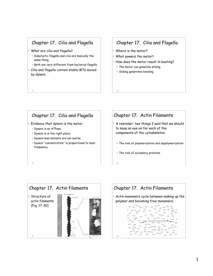
• Structure ofactin filaments(Fig. 17-30)
6
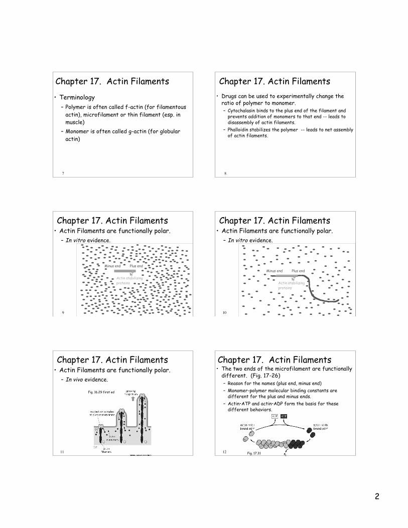
Chapter 17. Actin Filaments
• Actin monomers cycle between making up thepolymer and becoming free monomers.
2
7
Chapter 17. Actin Filaments
• Terminology– Polymer is often called f-actin (for filamentous
actin), microfilament or thin filament (esp. inmuscle)
– Monomer is often called g-actin (for globularactin)
8
Chapter 17. Actin Filaments• Drugs can be used to experimentally change the
ratio of polymer to monomer.– Cytochalasin binds to the plus end of the filament and
prevents addition of monomers to that end -- leads todisassembly of actin filaments.
– Phalloidin stabilizes the polymer -- leads to net assemblyof actin filaments.
9
Chapter 17. Actin Filaments• Actin Filaments are functionally polar.
– In vitro evidence.
Minus end Plus end
Actin stabilizing proteins
10
Chapter 17. Actin Filaments• Actin Filaments are functionally polar.
– In vitro evidence.
AxonemeMinus end Plus end
Actin stabilizing proteins
11
Chapter 17. Actin Filaments• Actin Filaments are functionally polar.
– In vivo evidence.
Fig. 16.29 first ed
12
Chapter 17. Actin Filaments• The two ends of the microfilament are functionally
different. (Fig. 17-26)– Reason for the names (plus end, minus end)– Monomer-polymer molecular binding constants are
different for the plus and minus ends.– Actin•ATP and actin•ADP form the basis for these
different behaviors.
Fig. 17.31
3
13
Chapter 17. Actin Filaments• Similarities and differences between the
dynamic nature of the tubulin / MT systemand the g-actin / f-actin system.– Incredibly similar:
• Short half lives• Plus, minus ends• Role of NTP hydrolysis• Internal and (we will see) external capping proteins
– Different evolutionary history– Similarities due to similar requirements
14
Chapter 17. Actin Filaments• General characteristics of actin filaments
– Actin filaments are thin and flexible.– Actin filaments usually occur in bundles
(exception the red blood cell membrane).– Actin filaments are often associated with the
membrane.– Actin filaments primarily serve as structural
components. Even when involved in motility, theytypically serve as ropes upon which force isgenerated, and do not generate force themselves
15
Chapter 17. Actin Filaments• The importance of actin binding proteins
– There are a very large number of actin bindingproteins in cells.
– Much of our knowledge of actin binding proteinscomes from biochemical studies of “actin richextracts from cells”
– We will consider in vitro interactions first andthen see how they integrate with the suspectedfunctions of actin in cells.
16
Chapter 17. Actin Filaments• The importance of actin binding proteins
17
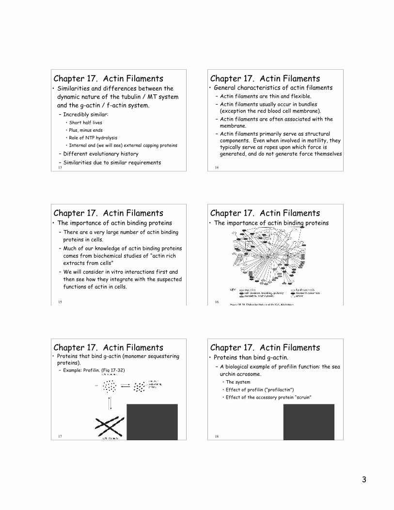
Chapter 17. Actin Filaments• Proteins that bind g-actin (monomer sequestering
proteins).– Example: Profilin. (Fig 17-32)
18
Chapter 17. Actin Filaments• Proteins than bind g-actin.
– A biological example of profilin function: the seaurchin acrosome.• The system• Effect of profilin (“profilactin”)• Effect of the accessory protein “scruin”
4
19
Chapter 17. Actin Filaments• Proteins than bind g-actin.
– Nucleating proteins. (Fig 17-27)
20
Chapter 17. Actin Filaments• Examples of the importance of such
proteins:– Sea urchin acrosomal reaction (discussed
previously)– Initiation of microfilament growth in filapodia,
microvilli and “stereocilia” (to be discussedshortly)
21
Chapter 17. Actin Filaments
22
Chapter 17. Actin Filaments• Examples of the importance of such proteins:
– Sea urchin acrosomal reaction (discussed previously)– Initiation of microfilament growth in filapodia, microvilli and
“stereocilia” (to be discussed shortly)
• Nucleating proteins can be exploited by some diseasecausing bacteria.– Lysteria– Nucleating proteins on its surface.– Rocket propelled.
23
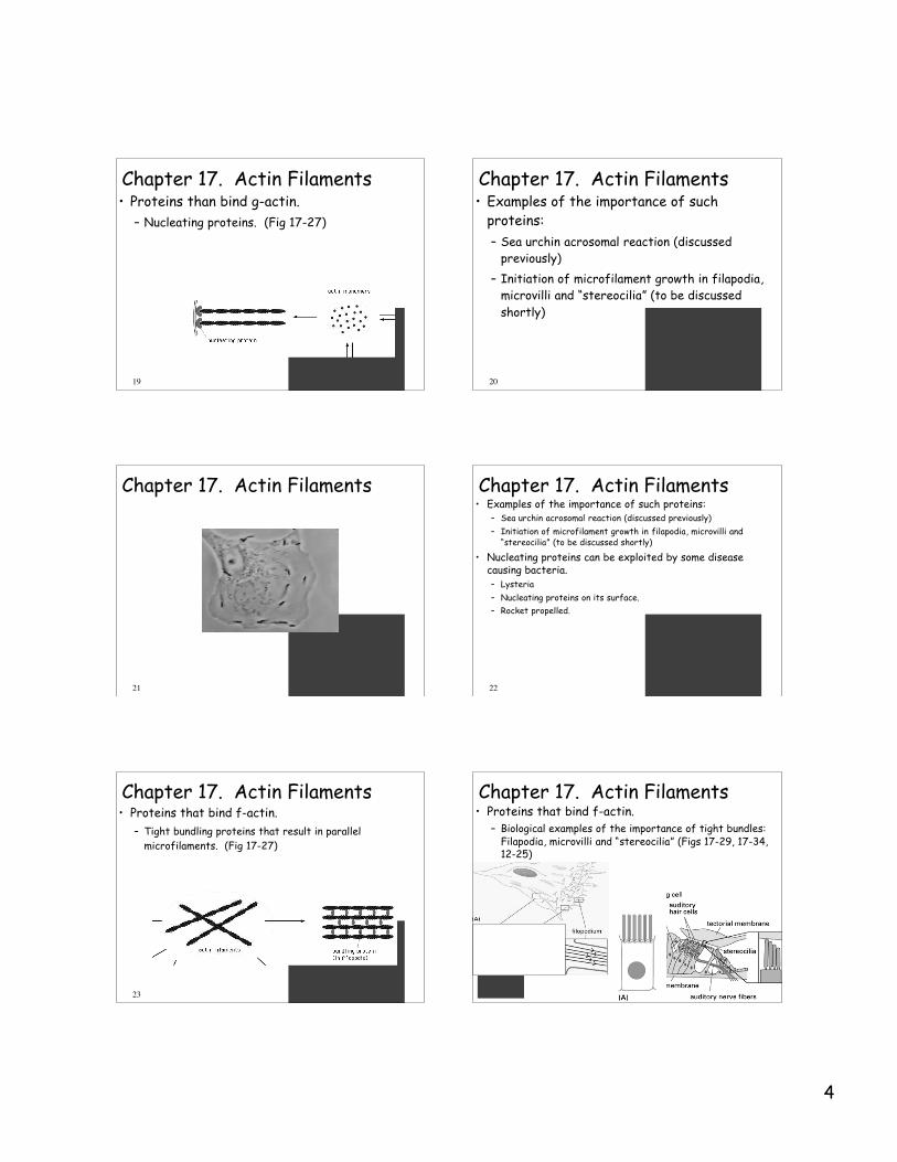
Chapter 17. Actin Filaments• Proteins that bind f-actin.
– Tight bundling proteins that result in parallelmicrofilaments. (Fig 17-27)
24
Chapter 17. Actin Filaments• Proteins that bind f-actin.
– Biological examples of the importance of tight bundles:Filapodia, microvilli and “stereocilia” (Figs 17-29, 17-34,12-25)
5
25
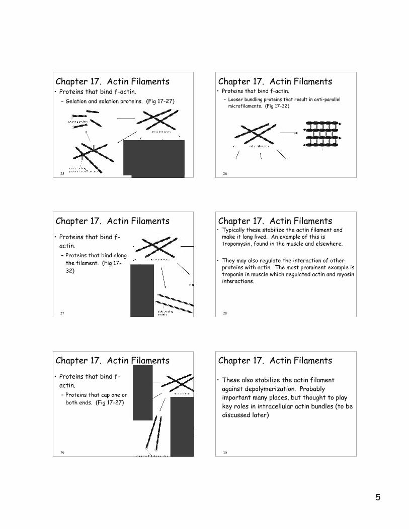
Chapter 17. Actin Filaments• Proteins that bind f-actin.
– Gelation and solation proteins. (Fig 17-27)
26
Chapter 17. Actin Filaments• Proteins that bind f-actin.
– Looser bundling proteins that result in anti-parallelmicrofilaments. (Fig 17-32)
27
Chapter 17. Actin Filaments
• Proteins that bind f-actin.– Proteins that bind along
the filament. (Fig 17-32)
28
Chapter 17. Actin Filaments• Typically these stabilize the actin filament and
make it long lived. An example of this istropomysin, found in the muscle and elsewhere.
• They may also regulate the interaction of otherproteins with actin. The most prominent example istroponin in muscle which regulated actin and myosininteractions.
29
Chapter 17. Actin Filaments
• Proteins that bind f-actin.– Proteins that cap one or
both ends. (Fig 17-27)
30
Chapter 17. Actin Filaments
• These also stabilize the actin filamentagainst depolymerization. Probablyimportant many places, but thought to playkey roles in intracellular actin bundles (to bediscussed later)
6
31
Chapter 17. Actin Filaments• Proteins that bind f-actin.
– Myosin a motor protein (Fig 17-32)
32
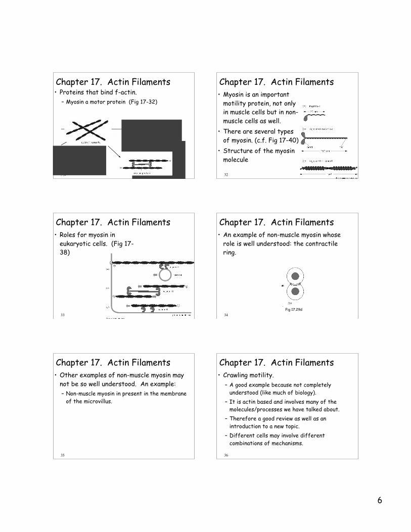
Chapter 17. Actin Filaments• Myosin is an important
motility protein, not onlyin muscle cells but in non-muscle cells as well.
• There are several typesof myosin. (c.f. Fig 17-40)
• Structure of the myosinmolecule
33
Chapter 17. Actin Filaments• Roles for myosin in
eukaryotic cells. (Fig 17-38)
34
Chapter 17. Actin Filaments• An example of non-muscle myosin whose
role is well understood: the contractilering.
Fig 17.29d
35
Chapter 17. Actin Filaments• Other examples of non-muscle myosin may
not be so well understood. An example:– Non-muscle myosin in present in the membrane
of the microvillus.
36
Chapter 17. Actin Filaments• Crawling motility.
– A good example because not completelyunderstood (like much of biology).
– It is actin based and involves many of themolecules/processes we have talked about.
– Therefore a good review as well as anintroduction to a new topic.
– Different cells may involve differentcombinations of mechanisms.
7
37
Chapter 17. Actin Filaments• The importance and mechanism of new
polymerization.– The leading edge (= lamellipodium)
• Plus ends towards membrane.• New polymerization involves actin-related proteins
(ARP).• New growth extends lamellipodia.• The back of the actin array depolymerizes.• The probable mechanism (next slide)
38
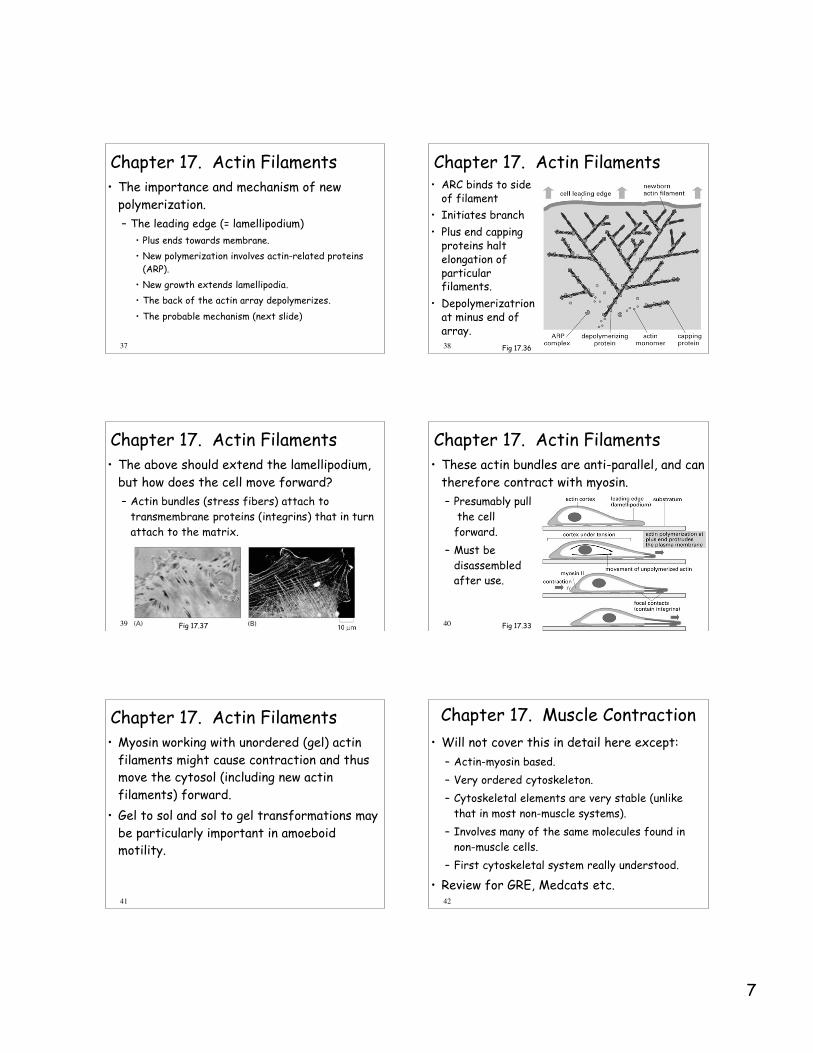
Chapter 17. Actin Filaments• ARC binds to side
of filament• Initiates branch• Plus end capping
proteins haltelongation ofparticularfilaments.
• Depolymerizatrionat minus end ofarray.
Fig 17.36
39
Chapter 17. Actin Filaments• The above should extend the lamellipodium,
but how does the cell move forward?– Actin bundles (stress fibers) attach to
transmembrane proteins (integrins) that in turnattach to the matrix.
Fig 17.37 40
Chapter 17. Actin Filaments• These actin bundles are anti-parallel, and can
therefore contract with myosin.– Presumably pull
the cellforward.
– Must bedisassembledafter use.
Fig 17.33
41
Chapter 17. Actin Filaments• Myosin working with unordered (gel) actin
filaments might cause contraction and thusmove the cytosol (including new actinfilaments) forward.
• Gel to sol and sol to gel transformations maybe particularly important in amoeboidmotility.
42
Chapter 17. Muscle Contraction• Will not cover this in detail here except:
– Actin-myosin based.– Very ordered cytoskeleton.– Cytoskeletal elements are very stable (unlike
that in most non-muscle systems).– Involves many of the same molecules found in
non-muscle cells.– First cytoskeletal system really understood.
• Review for GRE, Medcats etc.