chapter 15 and 16: spectroscopy. determining molecular...
TRANSCRIPT
Chapter 15 and 16: Spectroscopy. Determining Molecular Structure
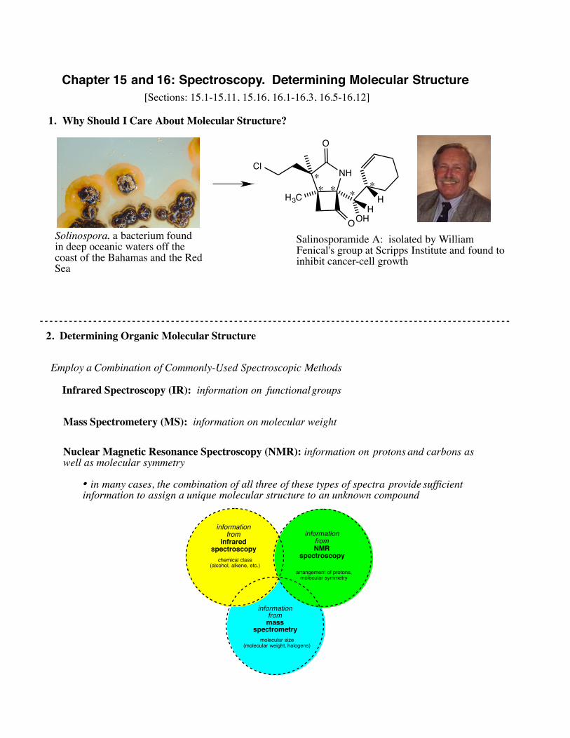
1. Why Should I Care About Molecular Structure?
Solinospora, a bacterium foundin deep oceanic waters off the coast of the Bahamas and the RedSea
NH
O
H3C
OHH
H
O
Cl
Salinosporamide A: isolated by William Fenical's group at Scripps Institute and found toinhibit cancer-cell growth
2. Determining Organic Molecular Structure
[Sections: 15.1-15.11, 15.16, 16.1-16.3, 16.5-16.12]
informationfrom
infraredspectroscopy
chemical class(alcohol, alkene, etc.)
informationfrom NMR
spectroscopy
arrangement of protons,molecular symmetry
informationfrom mass
spectrometrymolecular size
(molecular weight, halogens)
Employ a Combination of Commonly-Used Spectroscopic Methods
Infrared Spectroscopy (IR): information on functional groups
Mass Spectrometery (MS): information on molecular weight
Nuclear Magnetic Resonance Spectroscopy (NMR): information on protons and carbons as well as molecular symmetry
• in many cases, the combination of all three of these types of spectra provide sufficient information to assign a unique molecular structure to an unknown compound
**
* **

! !!
!
ROR'
O
HOR'
O
R
O
H
C CR
RR
R
C C RR
R O R'
R
O
R'
R OH
alkene
alkyne
alcohol
ether
ketone
aldehyde
ester
carboxylicacid
alkyl benzene(aromatic)
OH
O
O
H
O
OCH3
O
OH
O
R'
general structure class example


3. 1H Nuclear Magnetic Resonance Spectroscopy (NMR Spectroscopy)• Moving charges give rise to magnetic fields. • Therefore, movement of negatively charged electrons result in the generation of magnetic fields.• The natural "spin" of an electron also gives rise to a tiny magnetic field even though the electron is "trapped" within a bond or lone pair.• Positively charged protons (like electrons) have spin. The spin gives rise to a tiny magnetic field. So protons act as tiny magnets.• Normally, the tiny magnetic fields generated by protons are randomly oriented.
B0
• In the presence of a strong (external) magnetic field (B0), the magnetic fields of the individual protons align themselves in the same direction of the field, although they may be in the same direction (parallel) or opposite direction (anti-parallel). The parallel spin is of slightly lower energy than the anti-parallel, although both are populated.
B0irradiate
withradio
frequencies
Ε
• when the protons are irradiated with the proper amount of energy, the proton can flip from one direction (parallel, for example) to the opposite direction (anti-parallel). This is the point of nuclear magnetic resonance! • the energy at which this occurs can be measured and plotted• if all protons "flipped" (or, are at resonance) at exactly the same energy, different types of protons could not be differentiated• however, the energy at which the resonance occurs is dependent upon the "chemical environment" of those particular protons

4. Chemical Environments in 1H NMR Spectroscopy
C
H
HH
H C
Cl
HH
H C
H
ClH
H C
H
HClH C
H
HH
Cl
• methane has four hydrogens but all are considered to be in the same chemical environment (chemically equivalent) since replacing any one of the four hydrogens leads to the same compound• therefore, methane has ONE chemical environment and will give rise to ONE signal in the 1H NMR spectrum
H
C C
HH
H
HH
H
C C
HH
Cl
HH
Cl
C C
HH
H
HH
H
C C
HH
H
HCl
H
C C
HH
H
ClH
H
C C
ClH
H
HH
H
C C
HCl
H
HH
• ethane has six hydrogens but all are considered to be in the same chemical environment (chemically equivalent) since replacing any one of the four hydrogens leads to the same compound• therefore, ethane has ONE chemical environment and will give rise to ONE signal in the 1H NMR spectrum
NOTE: the three hydrogen atoms of a methyl group will always be chemically equivalent
H3C CH3
Analyze
H3C CH2 CH3
Number of chemical environments?Number of signals in the 1H NMR spectrum expected?
Analyze
NOTE: the two hydrogen atoms of a methylene (CH2) group will USUALLY be chemically equivalent
Predict the number of chemical environments and expected signals in the 1H NMR spectrum:

O O
Br
H
O CH3Cl
CH3
OH
H
H
H
H
H
HB HA
low electron density area( downfield or deshielded)
high electron density area(upfield or shielded)
Si
CH3
CH3H3C
H3C
• NMR scale = ppm = parts per million• always set relative to a standard; usually tetramethylsilane (TMS) at 0 ppm• protons in environments of "high" electron density are found closer to 0 ppm (said to be "upfield" or "shielded"• protons in environments of "low" electron density are found closer to 10 ppm (said to be "downfield" or "deshielded"• in the example above, HA must be in a chemical environment in which it experiences higher electron density than does HB• the power of 1H NMR is that certain types of protons consistently appear in the same area of the NMR spectrum, according to their chemical "environment"
5. Basic Analysis of a 1H NMR spectrum
TMS

6. Predicting Chemical Shifts: Where do the Different Chemical Environments Appear on the Spectrum?
• use the "Typical Shifts" chart and webpage to predict the approximate range for the type of proton of interest
O
O
O
O
Three Rules of 1H NMR Spectroscopychemically equivalent protons absorb at the same chemical shift
chemically inequivalent protons absorb at different chemical shiftschemically similar protons absorb at similar chemical shift

OCH
R
HHR
RR
CO
RHR
RCNR
2
HRRC
XHR
R
12
34
56
78
910
11
vinyl Hbenzenering H
aldehyde
OCO
R
ppm
carboxylic acid H
TYPICAL CHEMICAL SHIFTS IN
1H NMR SPECTRO
SCOPY
H
saturated CHsthat are onlyattached toother saturatedcarbons
RC
R
HR
methine
RC
R
HH
methylene
RC
HH
H
methyl
RC
OH
Ralpha to a
carbonyl
RC H
R
allylic
RC H
R
benzylic
H's on a Cattached to anelectronegative
atom

O
OO
O11
1
3
7. Integration• from integration the area under each curve, a value is assigned to each signal that provides information on the relative number of protons in each chemical environment. NOTE: these may NOT be the same as the actual number of protons in that environment!
• 1H NMR spectroscopy provides information on the chemical environment of the protons in the molecule AND the relative numbers of protons in those enviroments
Predict the number of signals and expected chemical shifts in the 1H NMR spectrum of 2-butanone:
O

O
• protons, like electrons, generate small magnetic fields• these magnetic fields impact the Heff of neighboring protons• the result is that simple "singlet" signals are split into more complicated "multiplets"• the size of the multiplet can be predicted by counting the number of neighboring chemically inequivalent protons• the multiplet will be equivalent to n+1 where n = number of neighboring chemically inequivalent protons
8. Multiplets: when does splitting occur? what will the multiplet look like?
C C
HH
vicinal protons that arechemical inequivalent
C
H H
geminal protons thatare chemical inequivalent
HC
CC
H
NO: too far awayfrom each other
C C
H
Cl
Cl
H
Cl
ClNO: chemically
equivalent
# neighbors 0 1 2 3 4 5 6
expected integration?
n+1?

O
O
O
9. Predict the number of signals and expected chemical shifts, multiplicities, and integrations in the 1H NMR spectrum for each of the following. Properly label the NMR spectra.

OH
• OH protons have variable chemical shift, dependent upon the concentration of the sample that is made up. Generally, the appear as broad singlets between 2-6 ppm
common "pairs" of multiplets
CH2CH3CH3CH
CH3
CH3
methyl ethyl
isopropyl
2 31 6
~ 0-1.5
~ 0-1.5
CH3
CH3
CH3
tert-butyl
3
9
10. Miscellaneous (but important!) features of 1H NMR Spectroscopy

P: 16.7 (use our table and see if their predicted answer falls in the range), 16.8, 16.9, 16.11-16.17, 16.21, 16.34, 16.37, 16.48, 16.55, 16.57
Cl Cl Cl I
a reminder about splitting patterns!
more on chemical shifts!
OH
predict:
actual:
A B C D • even though not directly attached, a substituent can affect the chemical shifts of other chemical environments• the effect drops off very rapidly as the distance to the substituent increases (greatest impact is with neighboring protons)
H3CO
CH3 H3CO O
CH3
• additional substituents will exert nearly additive effects on chemical shifts

The IR and 1H NMR spectra for a compound of molecular formula C5H10O are provided below. Provide a compound structure consistent with the spectra.
i. examine IR spectrum and try to determine class of compoundii. examine 1H NMR and look for characteristic absorptionsiii. make some guesses and see which fit to 1H NMR spectrum
Plan of Action:
P: 15.9, 15.10, 15.12, 15.41
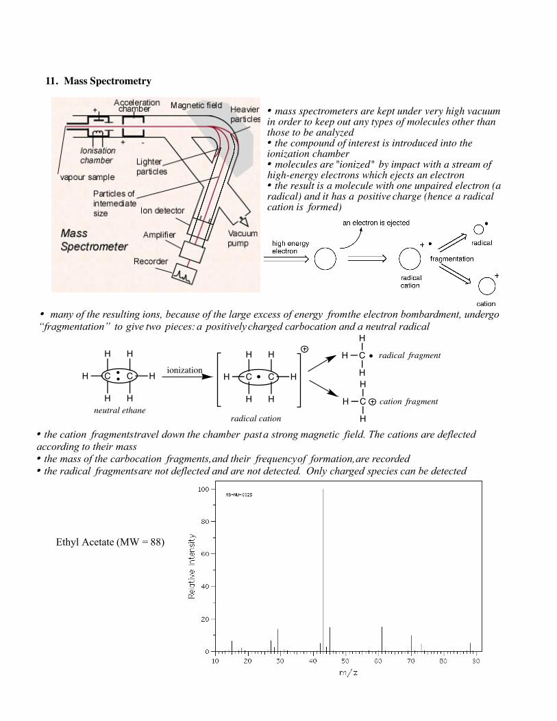
11. Mass Spectrometry
• mass spectrometers are kept under very high vacuum in order to keep out any types of molecules other than those to be analyzed• the compound of interest is introduced into the ionization chamber• molecules are "ionized" by impact with a stream of high-energy electrons which ejects an electron• the result is a molecule with one unpaired electron (a radical) and it has a positive charge (hence a radical cation is formed)
• many of the resulting ions, because of the large excess of energy from the electron bombardment, undergo “fragmentation” to give two pieces: a positively charged carbocation and a neutral radical
C C
H
H
H
H
H
H
ionizationC C
H
H
H
H
H
H
H C
H
H
H C
H
Hneutral ethane
radical cation
radical fragment
cation fragment
• the cation fragments travel down the chamber past a strong magnetic field. The cations are deflected according to their mass • the mass of the carbocation fragments, and their frequency of formation, are recorded• the radical fragments are not deflected and are not detected. Only charged species can be detected
Ethyl Acetate (MW = 88)
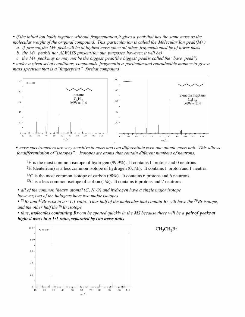
• if the initial ion holds together without fragmentation, it gives a peak that has the same mass as the molecular weight of the original compound. This particular ion is called the Molecular Ion peak (M+) a. if present, the M+ peak will be at highest mass since all other fragments must be of lower mass b. the M+ peak is not ALWAYS present (for our purposes, however, it will be) c. the M+ peak may or may not be the biggest peak (the biggest peak is called the “base peak”)• under a given set of conditions, compounds fragment in a particular and reproducible manner to give a mass spectrum that is a “fingerprint” for that compound
octaneC6H18
MW = 114
2-methylheptaneC6H18
MW = 114
• mass spectrometers are very sensitive to mass and can differentiate even one atomic mass unit. This allows for differentiation of “isotopes”. Isotopes are atoms that contain different numbers of neutrons.
• all of the common "heavy atoms" (C, N, O) and hydrogen have a single major isotopehowever, two of the halogens have two major isotopes• 79Br and 81Br exist in a ~ 1:1 ratio. Thus half of the molecules that contain Br will have the 79Br isotope, and the other half the 81Br isotope• thus, molecules containing Br can be spotted quickly in the MS because there will be a pair of peaks at highest mass in a 1:1 ratio, separated by two mass units
1H is the most common isotope of hydrogen (99.9%). It contains 1 protons and 0 neutrons 2H (deuterium) is a less common isotope of hydrogen (0.1%). It contains 1 proton and 1 neutron
CH3CH2Br
12C is the most common isotope of carbon (98%). It contains 6 protons and 6 neutrons 13C is a less common isotope of carbon (1%). It contains 6 protons and 7 neutrons
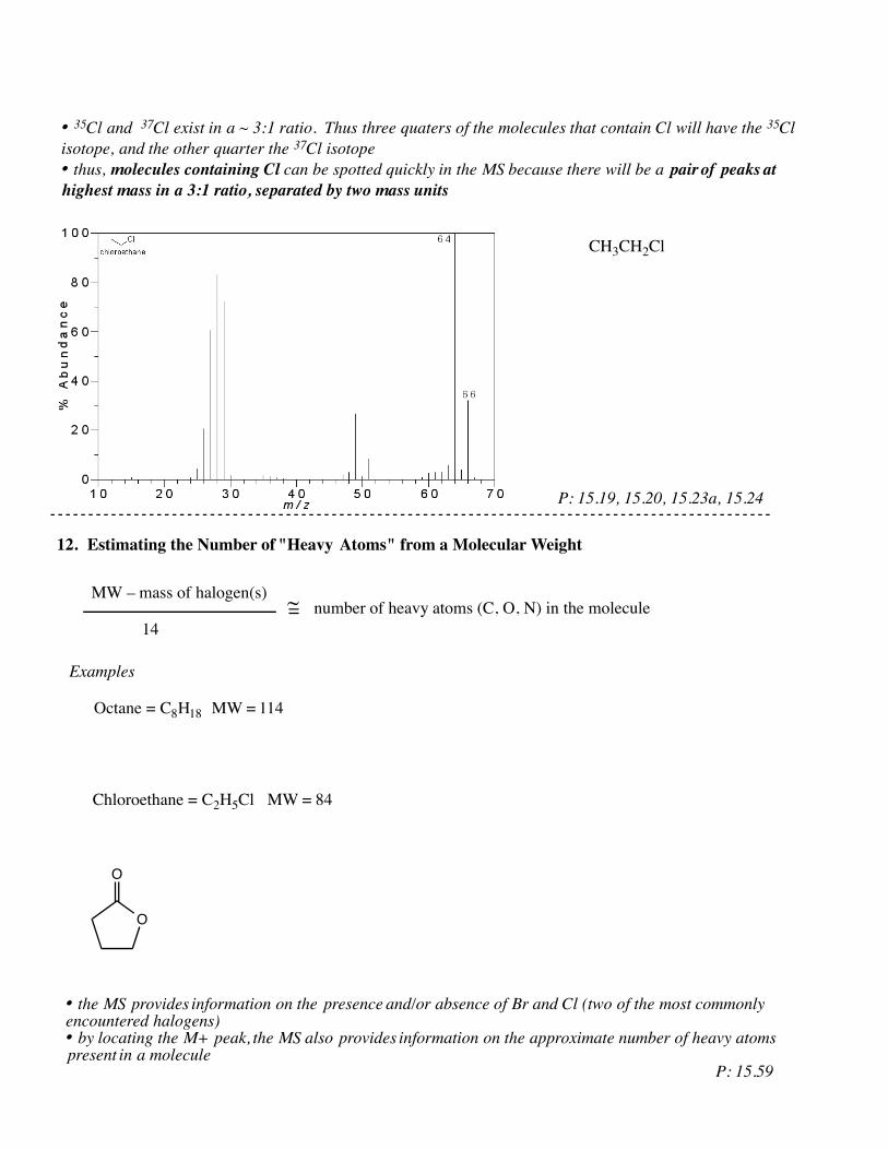
• 35Cl and 37Cl exist in a ~ 3:1 ratio. Thus three quaters of the molecules that contain Cl will have the 35Cl isotope, and the other quarter the 37Cl isotope• thus, molecules containing Cl can be spotted quickly in the MS because there will be a pair of peaks at highest mass in a 3:1 ratio, separated by two mass units
CH3CH2Cl
12. Estimating the Number of "Heavy Atoms" from a Molecular Weight
MW – mass of halogen(s)
14~= number of heavy atoms (C, O, N) in the molecule
Octane = C8H18 MW = 114
Examples
Chloroethane = C2H5Cl MW = 84
O
O
• the MS provides information on the presence and/or absence of Br and Cl (two of the most commonly encountered halogens)• by locating the M+ peak, the MS also provides information on the approximate number of heavy atoms present in a molecule
P: 15.19, 15.20, 15.23a, 15.24
P: 15.59