chapter 11 the auditory and vestibular systems. introduction sensory systems –sense of hearing,...
Post on 19-Dec-2015
228 views
TRANSCRIPT

Chapter 11 The Auditory and
Vestibular Systems

Introduction
Sensory Systems– Sense of hearing, audition
• Detect sound• Perceive and interpret nuances
– Sense of balance, vestibular system• Head and body location• Head and body movements
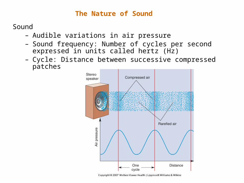
The Nature of Sound
Sound– Audible variations in air pressure– Sound frequency: Number of cycles per second expressed
in units called hertz (Hz)– Cycle: Distance between successive compressed patches

The Nature of Sound
Sound– Range: 20 Hz to 20,000 Hz– Pitch: High pitch = high frequency; low frequency = low
pitch– Intensity: High intensity louder than low intensity

The Structure of the Auditory System

The Structure of the Auditory System
Auditory pathway stages – Sound waves– Tympanic membrane– Ossicles– Oval window– Cochlear fluid– Sensory neuron response

Components of the Middle Ear
The Middle Ear

5 – Stapedius muscle
9 – Tensor Tympani muscle

• Sound Force Amplification by the Ossicles– Pressure: Force by surface area– Greater pressure at oval window than tympanic
membrane, moves fluids
• The Attenuation Reflex– Response where onset of loud sound causes tensor
tympani and stapedius muscle contraction– Function: Adapt ear to loud sounds, understand speech
better
The Middle Ear

The Inner Ear
• Anatomy of the Cochlea• Perilymph: Fluid in scala vestibuli and scala tympani• Endolymph: Fluid in scala media• Endocochlear potential: Endolymph electric potential 80
mV more positive than perilymph

• Physiology of the Cochlea– Pressure at oval window, pushes perilymph into
scala vestibuli, round window membrane bulges out
• The Response of Basilar Membrane to Sound– Structural properties: Wider at apex, stiffness
decreases from base to apex• Research: Georg von Békésy
– Endolymph movement bends basilar membrane near base, wave moves towards apex
The Inner Ear

Georg von Békésy - Hungarian biophysicist born in Budapest.In 1961, he was awarded the Nobel Prize in Physiology or Medicine for his research
on the function of the cochlea in the mammalian hearing .

Travelling wave in the Basilar Membrane
The Inner Ear

The Organ of Corti and Associated Structures
The Inner Ear

Transduction by Hair Cells– Research: A.J.
Hudspeth.– Sound: Basilar
membrane upward, reticular lamina up and stereocilia bends outward
The Inner Ear

Fluids in cochlear canals
Upper and middle
Internal earExternal ear
PinnaExternalacousticmeatus
Air
Tympanicmembrane
Malleus, incus,stapes
(ossicles)
Ovalwindow Lower
Middle ear
Onevibration
TimeSpiral organ
(of Corti)stimulated
Amplificationin middle ear
Amplitude
Pre
ssu
re

Central Auditory Processes
Auditory Pathway

• Techniques for Sound Localization– Horizontal: Left-right, Vertical: Up-down
• Localization of Sound in Horizontal Plane– Interaural time delay: Time taken for sound to reach
from ear to ear– Interaural intensity difference: Sound at high
frequency from one side of ear– Duplex theory of sound localization:
• Interaural time delay: 20-2000 Hz• Interaural intensity difference: 2000-20000 Hz
Mechanisms of Sound Localization

Interaural time delay and interaural intensity difference
Mechanisms of Sound LocalizationMechanisms of Sound Localization

The Sensitivity of Binaural Neurons to Sound Location
Mechanisms of Sound LocalizationMechanisms of Sound Localization

Mechanisms of Sound Localization
Delay Lines and Neuronal Sensitivity to Interaural Delay
– Sound from left side, activity in left cochlear nucleus, sent to superior olive
– Sound reaches right ear, activity in right cochlear nucleus, first impulse far
– Impulses reach olivary neuron at the same time summation action potential

Mechanisms of Sound Localization
Localization of Sound in Vertical Plane– Vertical sound localization based on reflections from the
pinna

Primary Auditory Cortex– Axons leaving MGN project to auditory cortex via
internal capsule in an array– Structure of A1 and secondary auditory areas:
Similar to corresponding visual cortex areas
Auditory Cortex

The Vestibular System
• Importance of Vestibular System– Balance,
equilibrium, posture, head, body, eye movement
• Vestibular Labyrinth– Otolith organs -
gravity and tilt– Semicircular canals
- head rotation– Use hair cells, like
auditory system, to detect changes

Human Anatomy and Physiology, 7eby Elaine Marieb & Katja Hoehn
Copyright © 2007 Pearson Education, Inc.,publishing as Benjamin Cummings.
Figure 15.35: Structure of a macula, p. 594.
Macula ofutricle
Macula ofsaccule
Otoliths
Hair bundle
KinociliumStereocilia Otolithic
membrane
Vestibularnerve fibers
Hair cells
Supportingcells

Human Anatomy and Physiology, 7eby Elaine Marieb & Katja Hoehn
Copyright © 2007 Pearson Education, Inc.,publishing as Benjamin Cummings.
Figure 15.36: The effect of gravitational pull on a macula receptor cell in the utricle, p. 595.
Otolithicmembrane
Kinocilium
Ster eocilia
Receptorpotential
Nerveimpulsesgenerated investibular fiber
Depolarization
(Hairs bent towarkinocilium)
dHyperpolarization
(Hairs bent awayfrom kinocilium)
Increasedimpulse frequency
Excitation
Decreasedimpulse frequency
Inhibition

Human Anatomy and Physiology, 7eby Elaine Marieb & Katja Hoehn
Copyright © 2007 Pearson Education, Inc.,publishing as Benjamin Cummings.
Figure 15.37: Location and sturcture of a crista ampullaris, p. 596.
(a)
(c) (d)
(b)
CupulaCupula at rest
Position of cupuladuring turn
Turning motion
Fluid motion inducts
Afferent fibers of vestibular nerve
Increased firing Decreased firing
Position of cupuladuring turn
Ampulla of left ear
Ampulla ofright ear
Horizontal ducts
Flow ofendolymph
Cupula
Cristaampullaris
Fibers ofvestibular nerve

The Vestibular System
The Semicircular CanalStructure

Push-Pull Activation of Semicircular Canals– Three semicircular
canals on one side• Helps sense all
possible head-rotation angles
– Each paired with another on opposite side of head
– Push-pull arrangement of vestibular axons:
The Vestibular System

The Vestibulo-Ocular Reflex (VOR)
The Vestibular System