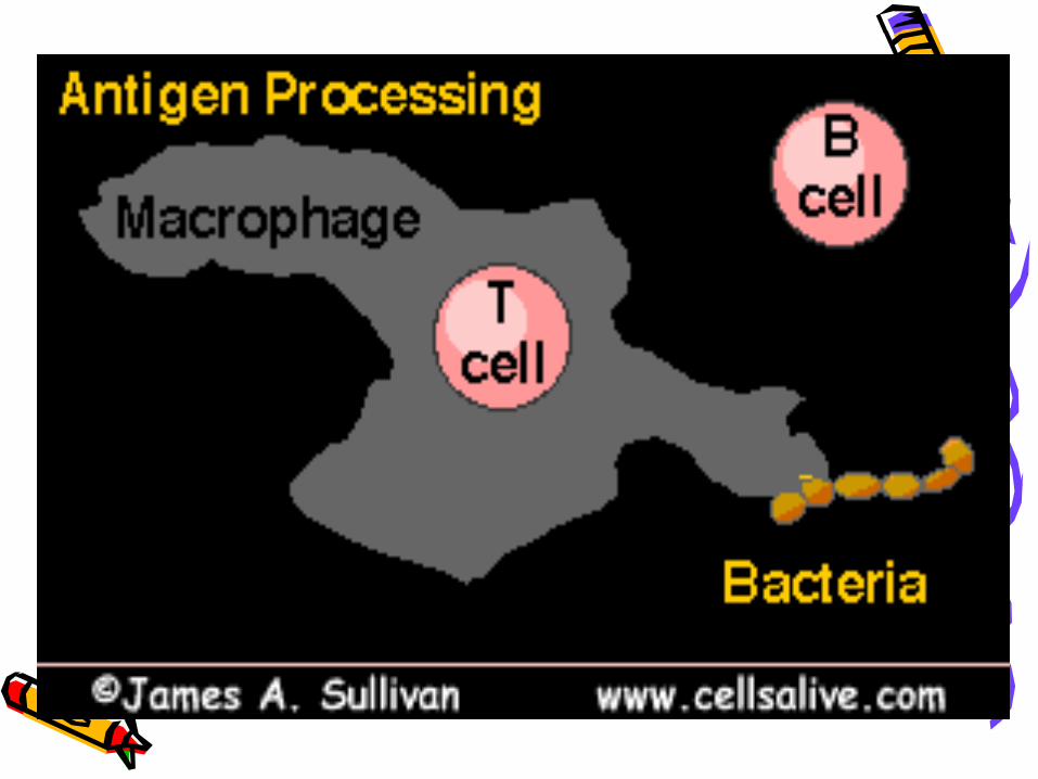
chapter 11 antigen processing and presentation. t cells do not recognise native antigens y y y y y y...
TRANSCRIPT
Chapter 11 Chapter 11
Antigen Processing and PresentAntigen Processing and Present
ationation
Chapter 11 Chapter 11
Antigen Processing and PresentAntigen Processing and Present
ationation

T cells do not recognise native antigens
YY Y Y YYY
BY
T
Y
T
Proliferation and antibody production
No proliferationNo cytokine release
Cross-linking of surface membrane Ig
Y
B
Y
B Y
B
Y
B
Y
B Y
B
Y
BYY
B

Cell surfacepeptides
of Ag
Antigens must be processed in orderto be recognised by T cells
YT
T cellresponse
No T cellresponse
No T cellresponse
No T cellresponse
No T cellresponse
Solublenative Ag
Cell surfacenative Ag
Soluble peptides
of Ag
Cell surface peptides of Ag presented by cells that express MHC molecules
ANTIGENANTIGENPROCESSINGPROCESSING
APCAPC

Contents Part Introduction--conceptsⅠ Part Characteristics of APCs Ⅱ Part Ag Processing and presentation Ⅲ
Chapter 11 Antigen Processing and Presentation

PartⅠ Introduction--conceptsEndogenous Ags: antigens synthesized within cells, in
cluding self and unself protein----class Ⅰ MHC molecules.Exogenous Ags: antigens comes outside the cells, incl
uding self and unself protein----class Ⅱ MHC molecules.Antigen processing: the conversion of native protein
s to peptides which can combine with MHC molecules.Antigen presentation: the course of formation and
display of peptide-MHC complexes on the surface of APCs and the course of peptide-MHC complexes recognition by T cells.
Ag capturing----Endocytosis (internalization) Phagocytosis, Pinocytosis, Receptor-mediated endocytosis

Production of endogenous Ags and exogenous Ags

YThe site of pathogen replication or mechanism of antigen uptake determines the antigen processing pathway used
Y
Cytosolic compartmentEndogenous processing(Viral, tumor antigens )
Vesicular CompartmentContiguous with extracellular fluid
Exogenous processing(Streptococcal, tumor antigens)
INTRACELLULAR REPLICATION
EXTRACELLULAR ORENDOSOMAL REPLICATION

Cell surfacepeptides
of Ag
Antigens must be processed in orderto be recognised by T cells
YT
T cellresponse
No T cellresponse
No T cellresponse
No T cellresponse
No T cellresponse
Solublenative Ag
Cell surfacenative Ag
Soluble peptides
of Ag
Cell surface peptides of Ag presented by cells that
express MHC antigens
ANTIGEN PROCESSING
APCAPC
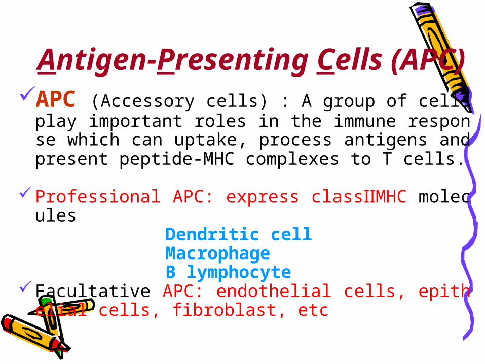
Antigen-Presenting Cells (APC)
APC (Accessory cells) : A group of cells play important roles in the immune response which can uptake, process antigens and present peptide-MHC complexes to T cells.
Professional APC: express classⅡMHC molecules Dendritic cell Macrophage B lymphocyte Facultative APC: endothelial cells, epithelial cells,
fibroblast, etc

APC
• Express classⅠ, Ⅱ MHC molecules and co-stimulatory molecules
• Uptake, process endogenous/exogenous antigens and present peptide-MHC to T cells
• Including dendritic cells, macrophages and B cells

PartⅡ Characteristics of APCs
Dendritic cell (DC) Macrophage B lymphocyte

1. Dendritic cell (DC)• History: DCs were first found by Steinman in 1973 ,
named for their special spinelike projections. DCs were cultured successfully in vitro in 1993 by Inaba.
• Characteristic: The most efficient APC, can present antigens to naive T cells to elicit primary immune response.

Fig2-4 Mature DC suspended in media by colony ( ×400 )

Fig2-5 scattered mature DC ( ×400 )

Scanning electron micrograph
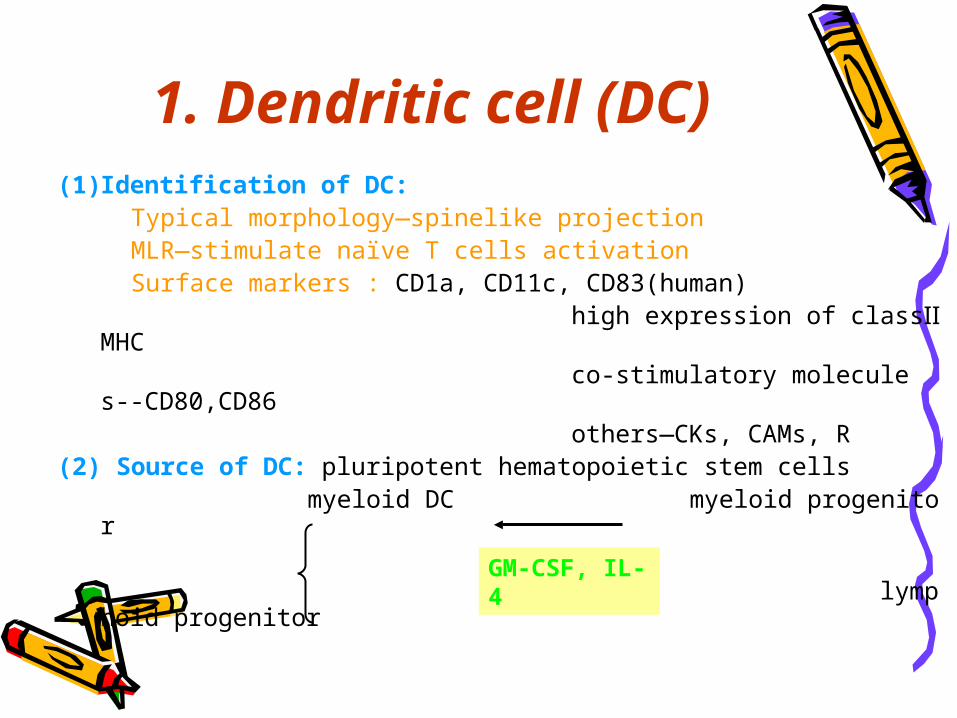
(1) Identification of DC: Typical morphology—spinelike projection MLR—stimulate naïve T cells activation Surface markers : CD1a, CD11c, CD83(human) high expression of classⅡMHC co-stimulatory molecules--CD80,CD86 others—CKs, CAMs, R(2) Source of DC: pluripotent hematopoietic stem cells myeloid DC myeloid progenitor lymphoid DC lymphoid progenitor
1. Dendritic cell (DC)
GM-CSF, IL-4

(3) Classification of DC : DC in lymphoid tissue: Interdigitating DC (IDC) , Folicular DC (FDC) DC in non lymphoid tissue: Langerhans cell (LC) DC in body fluid: Veiled cell, Blood DC
1. Dendritic cell (DC)

Interdigitating DC( IDC )
Express high level of classⅠ, Ⅱ MHC molecules and B7 , lack of FcR and CR, can stimulate T cells.

FDC
B cell
Folicular DC ( FDC )
Lie in follicle of LN, no expression of class Ⅱ MHC, high level of FcR and C3bR.
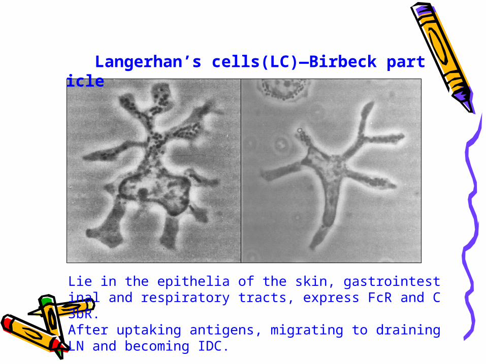
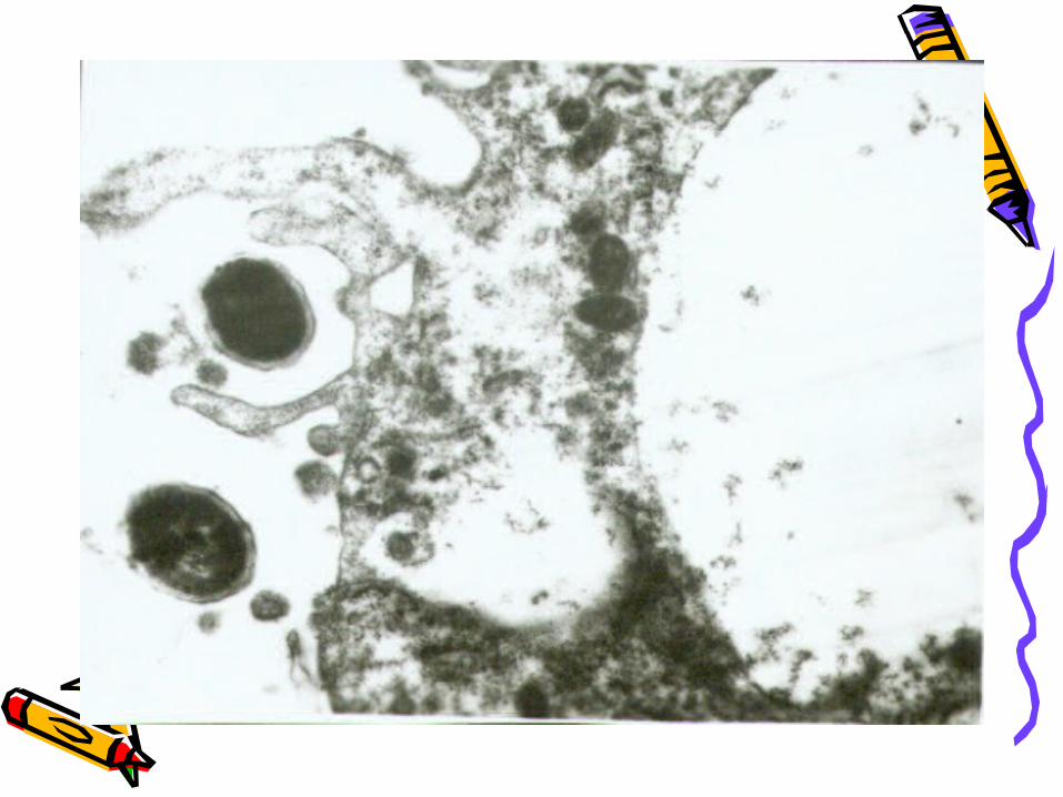
Langerhan’s cells(LC)—Birbeck particle
Lie in the epithelia of the skin, gastrointestinal and respiratory tracts, express FcR and C3bR. After uptaking antigens, migrating to draining LN and becoming IDC.

(4) Development and Maturation of DC
Pre-DC phase Immature DC ( iDC ) phase Migration phase Mature DC ( mDC ) phase
1. Dendritic cell (DC)
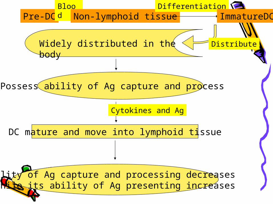
Pre-DCBlood
Non-lymphoid tissueDifferentiation
ImmatureDC
DistributeWidely distributed in the body
Possess ability of Ag capture and process
Cytokines and Ag
DC mature and move into lymphoid tissue
Ability of Ag capture and processing decreases while its ability of Ag presenting increases

Difference between iDC and mDC
• Ability of uptaking and processing antigens decreases.
• Ability of antigen presentation increases.• Express high level of MHC, co-stimulator
y molecules(CD80,CD86), CAMs(ICAM-1).• Ability to stimulate naïve T cell activatio
n increases.

(5) Antigens capturing:
• Phagocytosis—cell, bacteria
• Pinocytosis—soluble antigen
• Receptor-mediated endocytosis
FcγRⅡ, C3bR, mannose receptor
1. Dendritic cell (DC)

(6) Function of DC :
• Capture, process, present antigens—APC• Stimulate T or B lymphocytes—mature
DC• Induce immune tolerance—immature DC
1. Dendritic cell (DC)

2. Macrophage ( MФ )
• Stem from monocytes in blood• Have strong phagocytosis (big phagocyte)• Can not stimulate naïve T cells • Capture antigens by phagocytosis, pinocy
tosis, receptor-mediated endocytosis


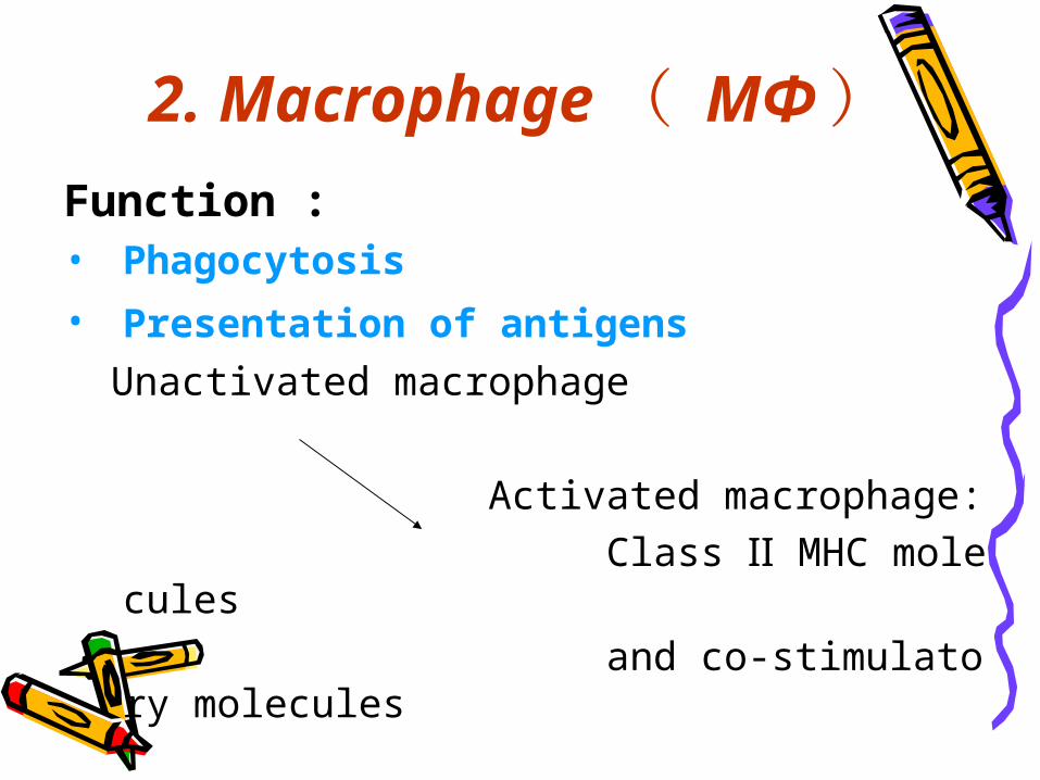
Function : • Phagocytosis • Presentation of antigens Unactivated macrophage Activated macrophage: Class Ⅱ MHC molecules and co-stimulatory molecules
2. Macrophage ( MФ )

3. B cellsFunctions• Mediate humoral immune response • Immunological regulation• Present antigens to T cell Soluble Ag--pinocytosis Specific receptor-mediated endocytosis
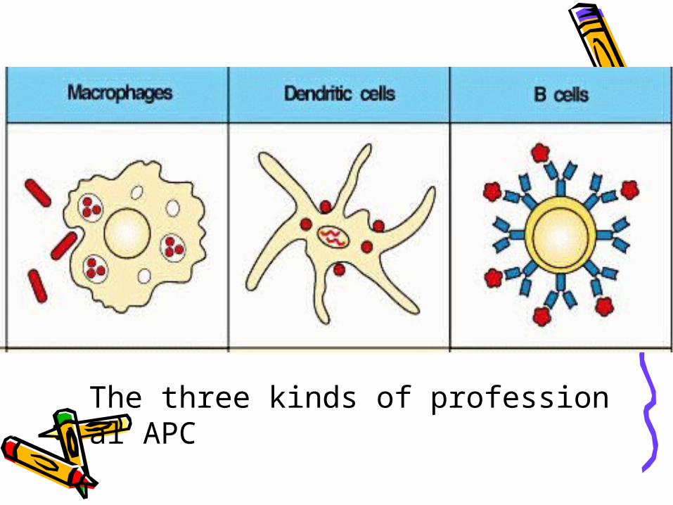
The three kinds of professional APC

Cell surfacepeptides
of Ag
Antigens must be processed in orderto be recognised by T cells
YT
T cellresponse
No T cellresponse
No T cellresponse
No T cellresponse
No T cellresponse
Solublenative Ag
Cell surfacenative Ag
Soluble peptides
of Ag
Cell surface peptides of Ag presented by cells that
express MHC antigens
ANTIGEN PROCESSING
APCAPC

PartⅢ Ag Processing and Presentation
Class Ⅱ MHC pathway ------exogenous antigens
Class Ⅰ MHC pathway ------endogenous antigens
Cross – presentation of antigen

SectionⅠ Class Ⅱ MHC pathway
1. Capture of exogenous Ag
2. Processing of Ag
3. Synthesis and transportation of class Ⅱ MHC molecules
4. Peptide loading of classⅡ MHC molecules
5. Presenting to CD4+T cells

1. Capture of exogenous Ag
• Endocytosis: Phagocytosis: particles or granules Pinocytosis: soluble antigens Receptor-mediated endocytosis: • Form endosome

Y Y
Pinocytosis
Phagocytosis
Membrane Igreceptor mediateduptake
Y
Uptake of exogenous antigens
Complement receptormediated phagocytosis Y
Fc receptor mediated phagocytosis

2. Processing of Ag
endosome + lysosome
Ag antigen peptides(10-30aa) Cathepsin
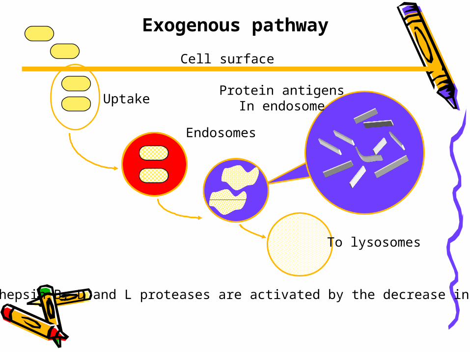
Endosomes
Exogenous pathway
Cell surface
To lysosomes
UptakeProtein antigens
In endosome
Cathepsin B, D and L proteases are activated by the decrease in pH

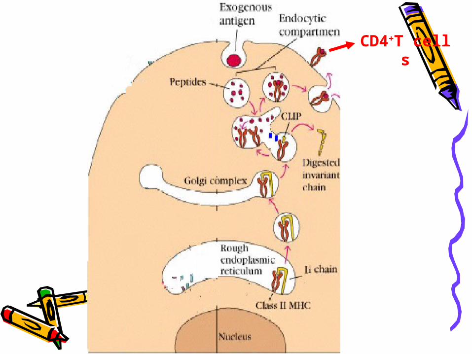
3. Synthesis and transportation of class Ⅱ MHC molecules
Synthesis of class Ⅱ MHC molecules in ER
Ii chain --- class Ⅱ MHC molecule (Ii3α3β3 ) ①Promote formation of class Ⅱ MHC dimer ②Preventing endogenous peptide from combining with c
lassⅡMHC molecules within ER ③Leading classⅡMHC molecules into endosome from ER
Endosome (MIIC)*Ii chain: Ia-associated invariant chain

A peptide of the invariant chain blocks the MHC molecule binding site.This peptide is called the CLass Ⅱ associated Invariant chain Peptide
(CLIP)
Invariant chain CLIP peptide
and chains of MHC class II molecules
CLIP
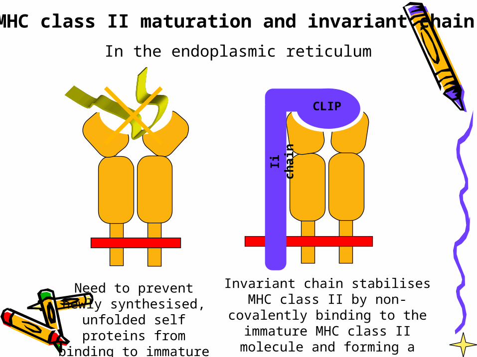
Need to prevent newly synthesised, unfolded
self proteins from binding to immature MHC
Invariant chain stabilises MHC class II by non- covalently binding to the
immature MHC class II molecule and forming a nonomeric complex
In the endoplasmic reticulum
MHC class II maturation and invariant chain
Ii c
hai
n
CLIP

4. Peptide loading of class Ⅱ MHC molecules
Ii - class Ⅱ MHC molecules
protease Ii chain cleaving
CLIP - class Ⅱ MHC molecules
HLA-DM CLIP releasing
Antigen peptide - class Ⅱ MHC complexes

Endosomes
Cell surface
Uptake
Class II associated invariant chain peptide (CLIP)
(inv)3 complexesdirected towardsendosomes byinvariant chain
Cathepsin L degrades Invariant chain
CLIP blocks groove in MHC molecule
MHC Class IIcontaining vesiclesfuse with antigen
containing vesicles
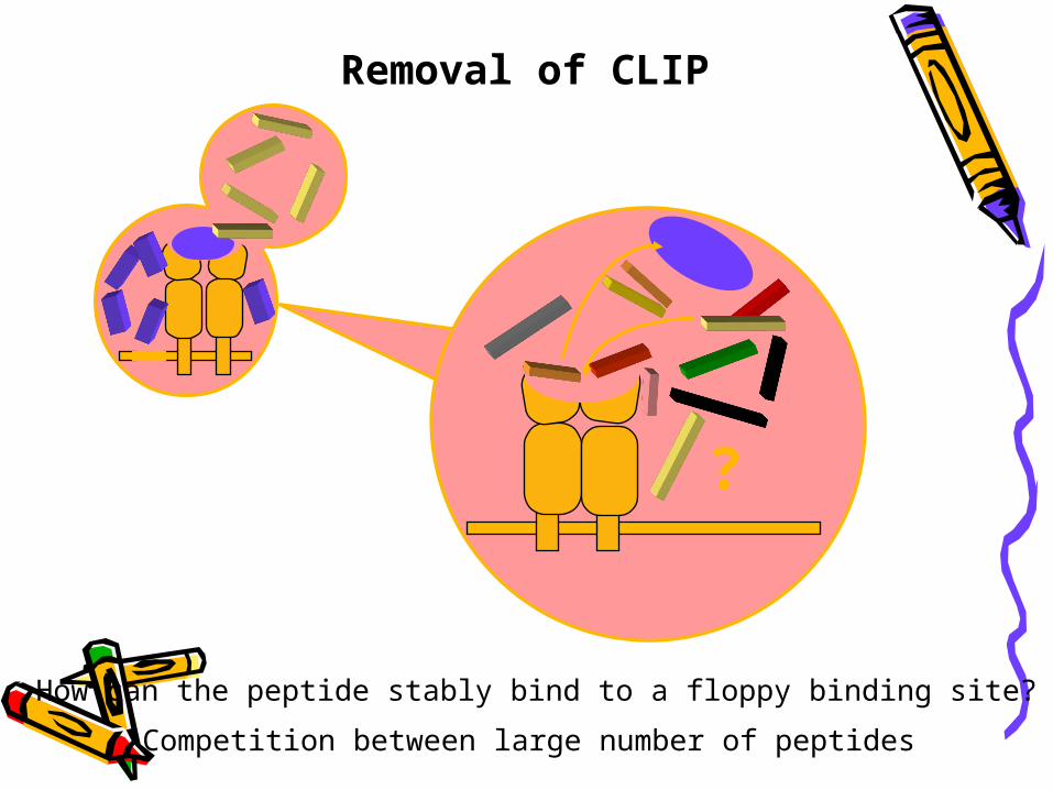
Removal of CLIP
?
How can the peptide stably bind to a floppy binding site?
Competition between large number of peptides

HLA-DM catalyses the removal of CLIP
MIIC compartment
HLA-DM
Replaces CLIP with a peptide antigen using a
catalytic mechanism (i.e. efficient at sub-
stoichiometric levels)
Discovered using mutant cell lines that failed to
present antigen
HLA-DO may also play a role in peptide exchange
Sequence in cytoplasmic tail retains HLA-DM in endosomes
HLA-DMHLA-DR

5.Presenting to CD4+T cells
Antigen peptide-class Ⅱ MHC molecuels presented on cell membrane by exocytos
is
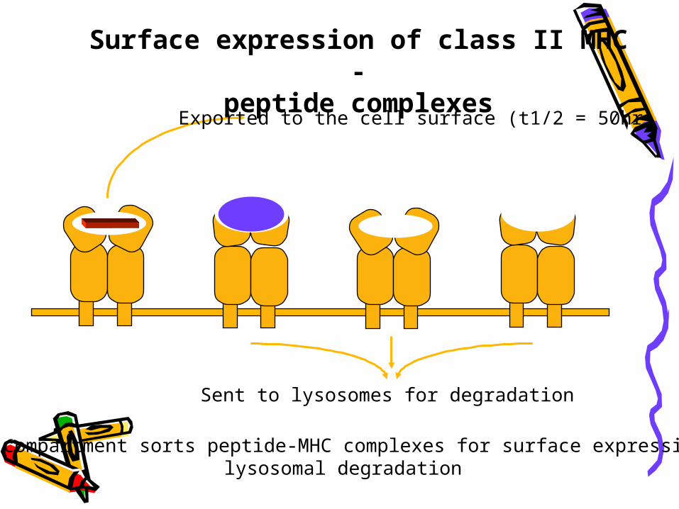
MIIC compartment sorts peptide-MHC complexes for surface expression orlysosomal degradation
Surface expression of class II MHC -peptide complexes
Exported to the cell surface (t1/2 = 50hr)
Sent to lysosomes for degradation


CD4+T cells
sectionⅡ class Ⅰ MHC pathway
1. Processing of endogenous Ag
2. Transporting of antigen peptide into ER
3. Peptide loading of class Ⅰ MHC molecules
4. Presenting to CD8+T cells

1. Processing of endogenous Ag • Proteosome : 20S, 26S• Low molecular weight polypeptide (LMP) : LMP2, LMP7 , LMP10• Ag antigen peptides (6-30aa)

Degradation in the proteasome
The components of the proteasome include LMP2, LMP7, MECL-1 ( LMP10)
*MECL-1 : Multicatalytic endopeptidase complex subunit
Cytoplasmic cellular proteins, including non-self proteinsare degraded continuously by a multicatalytic protease
Binding ubiquitin

2. Transporting of antigen peptide into ER
TAP(transporter associated with antigen processing):
Consisting of TAP1 and TAP2 ATP dependent transporter Selective transporting (8-
15aa)

ENDOPLASMIC RETICULUM
CYTOSOL
Peptide antigens produced in the cytoplasm are physically separated from newly formed class I MHC
Newly synthesisedclass I MHC molecules
Peptides needaccess to the ER in
order to be loaded onto class I MHC molecules

ER membrane
Lumen of ER
Cytosol
Transporters associated withantigen processing (TAP1 & 2)
Transporter has preference for >8 amino acid peptideswith hydrophobic C termini.
TAP-1 TAP-2
Peptide
TAP-1 TAP-2
PeptideTAP-1 TAP-2
Peptide
TAP-1 TAP-2
PeptideTAP-1 TAP-2
Peptide
TAP-1 TAP-2
PeptideTAP-1 TAP-2
Peptide
TAP-1 TAP-2
PeptideTAP-1 TAP-2
Peptide
TAP-1 TAP-2
Peptide
ER membrane
Lumen of ER
Cytosol
TAP-1 TAP-2
Peptide
ATP-binding cassette(ABC) domain
Hydrophobictransmembranedomain
Peptide antigensfrom proteasome
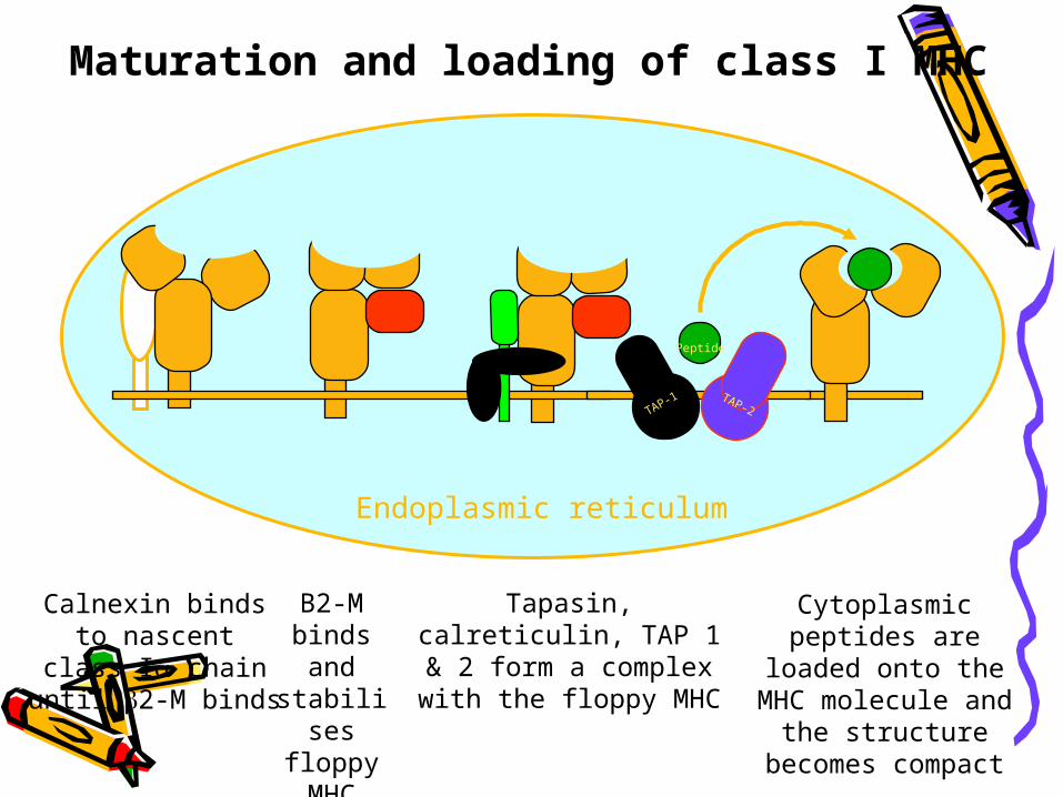
3. Peptide loading of class Ⅰ MHC molecules ER: antigen peptide—class Ⅰ MHC complexes
Endoplasmic reticulum
Calnexin bindsto nascent
class I chainuntil 2-M binds
TAP-1 TAP-2
Peptide
TAP-1 TAP-2
PeptideTAP-1 TAP-2
Peptide
TAP-1 TAP-2
PeptideTAP-1 TAP-2
Peptide
TAP-1 TAP-2
PeptideTAP-1 TAP-2
Peptide
TAP-1 TAP-2
PeptideTAP-1 TAP-2
Peptide
TAP-1 TAP-2
PeptideTAP-1 TAP-2
Peptide
B2-M binds and stabilises
floppy MHC
Tapasin, calreticulin, TAP 1 & 2 form a complex with
the floppy MHC
Cytoplasmic peptides are loaded onto the
MHC molecule and the structure becomes
compact
Maturation and loading of class I MHC
4. Presenting to CD8+T cells Antigen peptide-class Ⅰ MHC molecuels
presented on cell membrane by exocytosis

Fate of class I MHC
Sent to lysosomes for degradation
Exported to the cell surface

Ag(cytosolic protein)
Proteasome proteolytic degradation
Ag peptide
TAP complex transporting into ER
antigen peptide-class Ⅰ MHC complexes
Golgi complex exocyotsis
Presenting to CD8+T cells

CD8+T cells

CD8+T cells
CD4+T cells


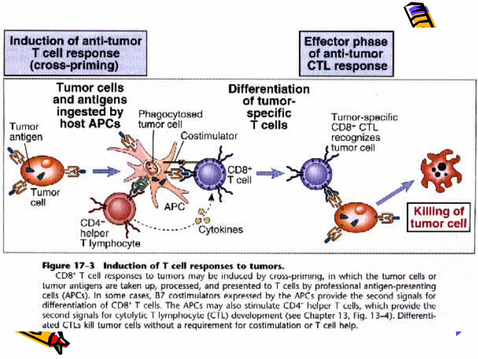
SectionⅢ Cross-presentation of antigens
Cross-priming:• Class Ⅰ MHC molecules also present
exogenous antigens to CD8+T cells• Class Ⅱ MHC molecules also present
endogenous antigens to CD4+T cells
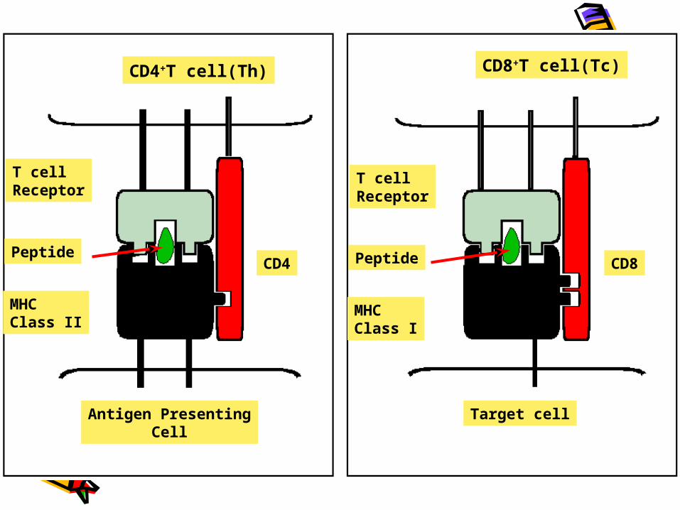
CD4+T cell(Th) CD8+T cell(Tc)
T cellReceptor
Peptide
MHCClass II
T cellReceptor
Peptide
MHCClass I
Antigen PresentingCell
Target cell
CD4 CD8