chapter 1 synthesis, characterization and applications of...
TRANSCRIPT
1
Chapter 1
Synthesis, Characterization and Applications of Gold
Nanoparticles
1.1. Introduction
The field of nanoscience and nanotechnology deals with development and understanding of
materials with at least one of its dimensions in nanoscale in the range 1–100 nm (Figure 1.1).
Properties of these nanomaterials have been found to be significantly different from that of
the compositional atoms as well as corresponding bulk materials [1,2]. Most importantly,
properties of materials change as their size approaches the nanoscale and the percentage of
atoms at the surface of a material becomes more significant. Nanostructures, whether
synthetic or natural, exhibit fascinating properties e.g. quantum confinement in
semiconductor particles, surface plasmon resonance in noble metal particles,
superparamagnetism in magnetic materials, metallic or semiconducting properties of single
wall carbon nanotubes depending upon their diameter, extremely high electron mobility of
graphene, significant decrease in electrical resistance in presence of a magnetic field for giant
magnetoresistance etc [3-5]. Nanoparticles, with all the three dimensions in nanoscale,
represent the most widespread current form of nanomaterials and their striking features have
been widely exploited for various multidisciplinary applications in sensing, photonics,
catalysis, biomedical, electronics etc [6-9]. These advances have been made possible with the
development of controlled synthesis methodologies and advanced characterization
techniques.

Chapter 1: Synthesis, Characterization and Applications of Gold Nanoparticles
2
Figure 1.1. The length scale of interest in nanoscience (1–100 nm) and its comparison with
smaller (atomic) and larger (macroscopic) structures.
Many nano forms of matter exist around us and their historical milestones spans over
centuries. One of the earliest nano-sized objects known to us was made of gold. Faraday
prepared colloidal gold in 1856 and called the particles he made the „divided state of gold‟
which can be suspended in water [10]. In 1890, the German bacteriologist Robert Koch found
that compounds made with gold inhibited the growth of bacteria and for this he was awarded
Nobel Prize for medicine in 1905. The use of gold in medicinal preparations is not new. In
the Indian medical system called Ayurveda, gold is used in several preparations. One popular
preparation is called „Saraswatharishtam‟, prescribed for memory enhancement. All these
preparations use finely ground gold. The metal was also used for medical purposes in ancient
Egypt where the Egyptians used gold in dentistry [11]. Colloidal gold had been incorporated
in glasses and vases to give them colour [12]. The oldest of these is the 4th
Century AD

Chapter 1: Synthesis, Characterization and Applications of Gold Nanoparticles
3
Lycurgus cup made by the Romans. The cup appears red in transmitted light (if a light source
is kept within the cup) and appears green in reflected light (if the light source is outside).
Modern chemical analysis showed that the glass is not much different from that used today
but contains very small amounts of gold (about 40 parts per million) and silver (about 300
parts per million) in the form of nanoparticles to give the cup a dichroic property [13,14].
The science of nanometer scale objects however was not discussed until much later.
The Nobel Prize winning physicist, Richard P. Feynman in 1959 gave a talk at the annual
meeting of the American Physical Society entitled “There‟s plenty of room at the bottom‟,
stating “The principles of physics, as far as I can see, do not speak against the possibility of
maneuvering things atom by atom” [15,16]. He, in a way, suggested the bottom-up approach,
“... it is interesting that it would be, in principle, possible (I think) for a physicist to
synthesize any chemical substance that the chemist writes down. Give the orders and the
physicist synthesizes it. How? Put the atoms down where the chemist says, and so you make
the substance. The problems of chemistry and biology can be greatly helped if our ability to
see what we are doing, and to do things on an atomic level, is ultimately developed–a
development which I think cannot be avoided” [15,16]. However, the world had to wait a
long time to put down atoms at the required place. Many would credit this talk as the genesis
of the modern field of nanotechnology, the science of manipulating molecular- and atomic-
level structures to engineer microscopic devices. Gold nanoparticles have recently become a
fundamental building block in nanotechnology due to their unique optical, electronic,
catalytic and chemical properties. The high surface-to-volume ratio, size and shape dependent
optical features, their size-dependent electrochemistry, high chemical stability and facile
surface chemistry have made them the model system of choice for exploring a wide range of
phenomena including self-assembly, bio-labeling, catalysis etc. Additional functionality can

Chapter 1: Synthesis, Characterization and Applications of Gold Nanoparticles
4
be imparted to these particles when they are modified with ligands such as small molecules,
polymers or biomolecules [6].
One attractive feature of gold nanoparticles is that their surfaces can be derivatized
with thiols, phosphines, alkynes and amines in both aqueous and organic solvents, allowing a
range of chemistry to be utilized in particle modification [17]. Gold nanoparticles are often
modified by soaking the colloid in a solution of the ligand of interest, making modification
straightforward. Another advantage is that gold nanoparticle size can be easily modified to
suit the needs of the experiment. For example, larger gold nanoparticles (> 80 nm) scatter
light very effectively, making them useful labels in optical microscopy. In contrast, smaller
nanoparticles (~ 5 nm) can be used as a size-control template for biomimetic high density
lipoprotein structures. A fascinating and useful trait of gold nanoparticles is that their
electronic interactions cause a distance dependent color change. This effect is observed in
solutions when the particles come within less than one particle diameter of each other.
Importantly, almost any surface modification that can be made to the gold nanoparticle that
can cause particle cross linking in the presence of a specific analyte, in principle, can result in
a colorimetric sensor. Again, the facile and flexible surface chemistry of gold nanoparticles
allows for a very wide range of creative surface modifications to achieve this effect. The gold
nanoparticle surface enables one to create tailorable, multivalent interfaces, directing the
particle to interact with its environment in a highly programmable manner in three
dimensions [18]. Gold nanoparticles, thus unmodified or modified, have been of great recent
interest in the context of its diverse applications due to their unique properties. They can be
synthesized by different ways depending on their application requirements. This thesis
provides insight into the synthesis and characterization of gold nanoparticles for a recently
developed novel method using block copolymers.

Chapter 1: Synthesis, Characterization and Applications of Gold Nanoparticles
5
1.2. Characteristics of Gold Nanoparticles
Gold nanoparticles are one of the most commonly used nanoparticles for various applications
because of their unique optical, electronic, surface and thermal properties [19].
(i) Optical Properties
Noble metals including gold nanoparticles exhibit different colours depending on the particle
size due to surface plasmon resonance (SPR) which is both metal and size dependent. SPR
excitation is based on the interaction with the electromagnetic field of the incoming light
resulting in a collective oscillation of the electrons on the nanoparticle surface [20,21]. The
SPR for gold nanoparticles occur throughout the visible and near-infrared region of the
electromagnetic spectrum depending on the size of the nanoparticles (Figure 1.2). Besides
size, the peak position is influenced by the nanostructure shape and the surrounding media,
including the nature of the ligand shell and the interparticle distances in dispersions [22]. In
the case when anisotropy is added to the nanoparticle, such as growth of nanorods, the optical
properties of the nanoparticles change dramatically.
Figure 1.2. (a) Gold nanoparticles change colours depending on the particle size (blue to red
colour is obtained with decreasing nanoparticle size). (b) Gold nanoparticles absorption for
various sizes and shapes.

Chapter 1: Synthesis, Characterization and Applications of Gold Nanoparticles
6
Many applications became possible due to the large enhancement of the surface
electric field on the gold nanoparticles surface. The plasmon resonance absorption has an
absorption coefficient orders of magnitude larger than strongly absorbing dyes. Anisotropic
shapes have plasmon resonance absorptions that are even stronger, leading to increased
detection sensitivity. Gold nanoparticles generate enhanced electromagnetic fields that affect
the local environment. The field is determined by the geometry of the nanoparticle and can
enhance fluorescence of the metal itself, the Raman signal of a molecule on the surface, and
the scattering of light. The optical properties of noble gold nanoparticles lead to many uses as
sensing and imaging techniques. The use of DNA has been pioneered in assembling and
studying their interaction and their application in colorimetric detection of biological targets
based on the binding events of target DNA [23,24]. Also the use of gold nanoparticles in the
field of photonics is immense.
(ii) Electronic Properties
Gold nanoparticles, in particular, exhibit good chemical stability. In principle, they can be
surface functionalized with almost every type of electron-donating molecule including
biomolecules. Beyond that, in the meantime, several protocols have been developed that
allow their assembly into one, two and three dimensions. Altogether, these facts triggered the
development of concepts for the design of novel materials with very specific properties based
on the unique size-dependent properties of single nanoparticles and their collective properties
in assemblies, owing to dipolar, magnetic or electronic coupling. Single nanoparticles with
sizes in the range of a few nanometers exhibit an electronic structure that corresponds to an
intermediate electronic structure between the band structure of the bulk metal and the discrete
energy levels of molecules with a characteristic highest occupied molecular orbital (HOMO)–
lowest unoccupied molecular orbital (LUMO) gap [25].

Chapter 1: Synthesis, Characterization and Applications of Gold Nanoparticles
7
In the size range of approximately 2 nm and below, single particles can be considered
as quantum dots. With modern microelectronics, transistors and other microelectronic
devices get smaller and smaller. Along with miniaturization, distances between transistors
and related switching elements on a chip get shorter and quantum effects become relevant.
Today‟s nanolithographic fabrication techniques allow scaling down to 50nm or below. This
has already made a great impact on the performance of traditional semiconductor circuits, and
it opens up new opportunities utilizing quantum effects. Following the utilization of charging
effects, the so-called Coulomb effects, in metallic circuits comprising tunnel junctions with
submicron sizes, allow us to handle individual charge carriers. This field has been named
single electronics (SE). It relies on the discreteness of the electric charge, and the tunneling of
electrons [single electron tunneling (SET)] in a system of such junctions can be affected by
Coulomb interaction of electrons, which can be varied by an externally applied voltage or by
injected charges [8,26]. As the continuous miniaturization in microelectronics reaches its
physical limits, new concepts are used to achieve component sizes of tens of nanometers or
less, or, ideally, the molecular level. Thus, the idea of utilizing the principle of SE for the
development of logic and memory cells, which in principle could lead to the construction of a
computer working on single electrons, realizing a „single-electron logic‟, has triggered
intense research activities related to SET phenomena.
(iii) Surface Properties
The surface properties of nanoparticles including surface reactivity are distinctly different
from larger particles and have an effect on surface composition, termination, charge and
functionalization for nanoparticles [27,28]. Gold nanoparticles are surrounded by a shell of
stabilizing molecules. With one of their ends these molecules are either adsorbed or
chemically linked to the gold surface, while the other end points towards the solution and

Chapter 1: Synthesis, Characterization and Applications of Gold Nanoparticles
8
provides colloidal stability. After synthesis of the particles the stabilizer molecules can be
replaced by other stabilizer molecules in a ligand exchange reaction. As thiol moieties bind
with high affinity to gold surfaces, most frequently thiol-modified ligands are used which
bind to the surface of the gold nanoparticles by formation of Au–sulfur bonds. Ligand
exchange is motivated by several aspects [29]. Ligand exchange allows, for example, the
transfer of gold particles from an aqueous to an organic phase (and vice versa) by exchanging
hydrophilic surfactants with hydrophobic surfactants (and vice versa). In this way, by
choosing the surfactant molecules, it is possible to adjust the surface properties of the
particles.
Figure 1.3. Schematic of a ligand-conjugated gold nanoparticle. The gold core (yellow) is
surrounded by stabilizer molecules (red) which provide colloidal stability. Ligands (blue) can
be either linked to the shell of stabilizer molecules (as shown here) or directly attached to the
gold surface by replacing part of the stabilizer molecules.
Biological molecules can be attached to the particles in several ways. If the biological
molecules have a functional group which can bind to the gold surface (like thiols or specific
peptide sequences), the biological molecules can replace some of the original stabilizer
molecules when they are added directly to the particle solution [7]. Figure 1.3 shows the
schematic of ligand-conjugated gold nanoparticle. In this way, molecules like
Chapter 1: Synthesis, Characterization and Applications of Gold Nanoparticles
9
oligonucleotides, peptides or PEG can be readily linked to gold nanoparticles and subsequent
sorting techniques even allow particles with an exactly defined number of attached molecules
per particle to be obtained. Alternatively, biological molecules can also be attached to the
shell of stabilizer molecules around the gold nanoparticles by bioconjugate chemistry. The
most common protocol is the linkage of amino-groups on the biological molecules with
carboxy groups at the free ends of stabilizer molecules by using EDC (1-ethyl-3-(3-
dimethylaminopropyl)-carbodiimide-HCl). With related strategies almost all kinds of
biological molecules can be attached to the particle surface. Though such protocols are
relatively well established, bioconjugation of gold nanoparticles still is not trivial and
characterization of synthesized conjugates is necessary, in particular to rule out aggregation
effects or unspecific binding during the conjugation reaction.
(iv) Thermal Properties
The remarkable optical properties of gold nanoparticles associated with the surface plasmon
resonance phenomenon have usually been thought mostly responsible for its applications
such as in the nano-photonics. However, one cannot but notice that the main recent
breakthroughs have rather been achieved in the domain of thermal applications of these
optical properties. Indeed, as optical and thermal responses are in fact closely bound, gold
nanoparticles can be considered together as nanometric heat sources and probes for local
temperature variations via their optical behaviour. The energetic conversion realized by gold
nanoparticles which are able to transform at the nanoscale an electromagnetic radiation into
heat emitted toward their environment may be relevant in numerous fields. For example, in
plasmonic devices local heating may alter the guiding of the electromagnetic wave by gold
nanostructures and therefore requires to be well controlled. Gold nanoparticles are also
expected to be used in microscopy for labelling biologic cells: nanoparticle heating by light
Chapter 1: Synthesis, Characterization and Applications of Gold Nanoparticles
10
absorption enables to modify the optical response of their local environment [9]. In the
medical area, photo-thermal cancer therapy based on gold nanoparticles is a very promising
technique, where gold nanoparticles absorb light energy transmitted through biologic tissues
and transform it into heat which diffuses toward local environment. By using an appropriate
targeting method for carrying particles close to affected cells, the latter will be destroyed by
overheating. One may also take advantage of this local heating around particles for inducing
local phase or morphology transformation in the surrounding medium. On the one hand, this
can enable the measurement of nanoscale heat transfer through the investigation of such
phase transformations. On the other hand, this could be used to modify the global medium
optical properties. This effect has been supposed to be at the origin of the optical limitation
phenomenon in colloidal solutions (induced light scattering by formation of gas bubbles
around gold colloids) [30]. Metal nanoparticles are also considered as model defects for
studying the damage of optical devices induced by powerful lasers. The dynamics of the
light-heat conversion in a gold nanoparticle and of the thermal release toward its environment
appears then to be a relevant issue in all these domains.
1.3. Synthesis of Gold Nanoparticles
Methods to synthesize gold nanoparticles have been known for centuries, but only in the last
half century have reliable methods been developed to synthesize them in high yield and in a
variety of sizes and shapes [31-35]. Since most of the applications, particularly biological, are
dependent on size and shape of gold nanoparticles, therefore use of appropriate method for
their controlled synthesis is one the important issues of consideration. Gold nanoparticles can
be synthesized in organic or aqueous media. There are two approaches for synthesis of
nanomaterials, top-down and bottom-up, as shown in Figure 1.4. Both approaches play very
important role in modern industry involving nanotechnology [36].

Chapter 1: Synthesis, Characterization and Applications of Gold Nanoparticles
11
Figure 1.4. Schematic representation of the building up of nanoparticles.
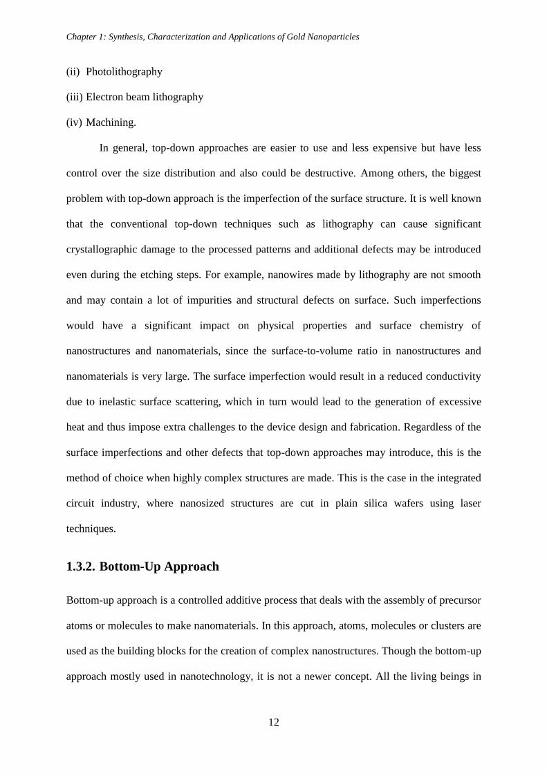
1.3.1. Top-Down Approach
The top-down approach is a subtractive process starting from bulk materials to make
nanomaterials. This approach involves division of bulk material or miniaturization of bulk
fabrication process to produce the desired structure with the appropriate properties. This
includes some of the following commonly used methods:
(i) Attrition or Ball milling
Chapter 1: Synthesis, Characterization and Applications of Gold Nanoparticles
12
(ii) Photolithography
(iii) Electron beam lithography
(iv) Machining.
In general, top-down approaches are easier to use and less expensive but have less
control over the size distribution and also could be destructive. Among others, the biggest
problem with top-down approach is the imperfection of the surface structure. It is well known
that the conventional top-down techniques such as lithography can cause significant
crystallographic damage to the processed patterns and additional defects may be introduced
even during the etching steps. For example, nanowires made by lithography are not smooth
and may contain a lot of impurities and structural defects on surface. Such imperfections
would have a significant impact on physical properties and surface chemistry of
nanostructures and nanomaterials, since the surface-to-volume ratio in nanostructures and
nanomaterials is very large. The surface imperfection would result in a reduced conductivity
due to inelastic surface scattering, which in turn would lead to the generation of excessive
heat and thus impose extra challenges to the device design and fabrication. Regardless of the
surface imperfections and other defects that top-down approaches may introduce, this is the
method of choice when highly complex structures are made. This is the case in the integrated
circuit industry, where nanosized structures are cut in plain silica wafers using laser
techniques.
1.3.2. Bottom-Up Approach
Bottom-up approach is a controlled additive process that deals with the assembly of precursor
atoms or molecules to make nanomaterials. In this approach, atoms, molecules or clusters are
used as the building blocks for the creation of complex nanostructures. Though the bottom-up
approach mostly used in nanotechnology, it is not a newer concept. All the living beings in
Chapter 1: Synthesis, Characterization and Applications of Gold Nanoparticles
13
nature observe growth by this approach only. Bottom-up methods are chemically controllable
and non-destructive. The synthesis of nanoparticles from molecular solutions is a good
example of a bottom-up approach. The size of the nanostructures, which can be obtained with
a bottom-op approach, spans the full nano scale. An advantage of the bottom-up approach is
the better possibilities to obtain nanostructures with less defects and more homogeneous
chemical compositions. This is due the mechanisms utilized in the synthesis of nanostructures
reducing the Gibbs free energy, so that the produced nanostructures are in a state closer to a
thermodynamic equilibrium [1]. Some of the important methods involved are:
(i) Sol-gel method
(ii) Vapour phase deposition method
(iii) Chemical reduction method.
The bottom-up approach usually employs solution-phase colloid chemistry for the
synthesis. In a typical colloidal synthesis, atoms of the desired component are produced in the
solution at very high supersaturation to induce the assimilation of these atoms into particles to
reduce the system Gibbs free energy. Due to the flexibility in selecting different reducing
agents, particle capping agents, solvent systems as well as synthesis conditions, colloidal
synthesis offers a great variety of options for composition, shape, size and surface chemistry
control. The bottom-up approach is also suitable for controlling monodispersity of the
nanoparticles. With all these advantages, the bottom-up approach has become the main route
to nanomaterial production.
Among all bottom-up methods, the chemical reduction of the metal salt in an aqueous,
an organic phase or two phases, is one of the most popular routes as nanoparticles of a wide
range of sizes and shapes can be prepared by controlling the reaction conditions. The
reduction of gold salts in existing of a stabilizing agent is a facile and easy technique to
produce desired sizes of nanoparticles [37]. A stabilizing agent, also called as capping

Chapter 1: Synthesis, Characterization and Applications of Gold Nanoparticles
14
material, prevents aggregation and precipitation of metal nanoparticles as well as plays a role
in determining size and shape of gold nanoparticles. Table 1.1 summarizes some of the
popular and widely used synthesizing methods for various size gold nanoparticles.
Table 1.1. Some of the widely used gold nanoparticle synthesis methods.
Nanoparticle
Size Methods
Capping
Agents
1 – 2 nm AuCl(PPh3) reduction by diborane or sodium
borohydride [38] Phosphine
2 – 5 nm Biphasic reduction of HAuCl4 by sodium
borohydride with thiol as a capping agent [39,40] Alkanethiol
10 – 100 nm HAuCl4 reduction with sodium citrate in water
[31,32,41] Citrate
1.4. Characterization Techniques
Several techniques are available under the broad umbrella of characterization of materials,
which may be used to study nanoparticles in one way or the other. The resulting information
can be processed to yield images or spectra which reveal the topographic, geometric,
structural, chemical or physical details of the nanomaterials. Different techniques based on
the use of photon (light and X-ray), electron and neutron probes, which are complementary
with respect to their sensitivity on different length scales, have been used. These techniques
can be broadly classified into three categories: (i) spectroscopic, (ii) microscopic and (iii)
scattering techniques.
1.4.1. Spectroscopic Techniques
Optical spectroscopic techniques are widely used in the study of optical properties of
different materials including nanoparticles. The different techniques are usually based on

Chapter 1: Synthesis, Characterization and Applications of Gold Nanoparticles
15
measuring absorption, scattering or emission of light that contains information about
properties of the materials. Commonly used techniques include UV-visible electronic
absorption spectroscopy, photoluminescence, infrared absorption and Raman scattering.
These different techniques can provide different information about the nanoparticle properties
of interest [42].
(i) UV-Visible Spectroscopy
The basic operating principle of electronic absorption spectroscopy is based on the
measurement of light absorption due to electronic transitions in a sample. Since the
wavelength of light required for electronic transitions is typically in the UV and visible
region of the electromagnetic radiation spectrum, electronic absorption spectroscopy is
usually called UV-visible or UV-vis spectroscopy [43]. It is named electronic absorption
spectroscopy because the absorption in the UV-visible regions involves mostly electronic
transitions. The spectrum is characteristic of a given sample and reflects the fundamental
electronic properties of the sample. For nanoparticles, UV-visible spectroscopy provides vital
information of nanoparticles through surface plasmon resonance (SPR) studies. This
absorption strongly depends on the particle size, dielectric medium and chemical
surroundings [44].
(ii) Photoluminescence Spectroscopy
At the fundamental level, the principle underlying photoluminescence (PL) spectroscopy is
very similar to that of electronic absorption spectroscopy. They both involve electronic
transition of initial and final states coupled by the electrical dipole operator. The main
difference is that the transition involved in PL is from a higher energy level or state to a lower
energy level [45]. There is also an important practical difference between the two techniques

Chapter 1: Synthesis, Characterization and Applications of Gold Nanoparticles
16
in that PL is a zero background experiment, i.e. no signal detected when there is no PL,
which is in contrast to absorption spectroscopy that is a non-zero background experiment.
A typical PL spectrum is just a plot of the PL intensity as a function of wavelength for
a fixed excitation wavelength. A photoluminescence excitation spectrum, however, is a
measure of PL at a fixed emission wavelength as a function of excitation wavelength. Gold
nanoparticles show PL, which has been correlated with their well-defined plasmon
resonances [46]. It is found that there is strong relationship between PL and surface plasmon
peak. For example, PL is very intense if SPR is broad and PL intensity is reduced when the
plasmon absorption sharpens.
(iii) Infrared Spectroscopy
The mechanical molecular and crystal vibrations are at very high frequencies ranging from
1012
to 1014
Hz (3–300 m wavelength), which falls in the infrared (IR) region of the
electromagnetic spectrum. In infrared Spectroscopy, the oscillations induced by certain
vibrational frequencies provide a means for matter to couple with an impinging beam of
infrared electromagnetic radiation and to exchange energy with it when the frequencies are in
resonance [47]. These absorption frequencies represent excitations of vibrations of the
chemical bonds and thus, are specific to the type of bond and the group of atoms involved in
the vibration. In Fourier transform infrared spectroscopy, the intensity-time output of the
interferometer is subjected to a Fourier transform to convert it to the familiar infrared
spectrum (intensity-frequency) and atomic arrangement, surrounding environments and
concentrations of the chemical bonds that are present in the sample can be determined. The
studies relating the quantification of the coverage and binding strength of ligands, surfactants
etc. on the gold nanoparticle surface are usually investigated using FTIR spectroscopy [48].

Chapter 1: Synthesis, Characterization and Applications of Gold Nanoparticles
17
(iv) Raman Scattering
Raman scattering is another vibrational technique and differs from the infrared spectroscopy
by an indirect coupling of high-frequency radiation with vibrations of chemical bonds. When
the incident photon interacts with the chemical bond, the chemical bond is excited to a higher
energy state. The scattering process is inelastic and thus the scattered light can have a lower
(Stokes, by depositing energy into the molecule) or higher energy (anti-Stokes, by gaining
energy from the molecule) than the incident light (Rayleigh scattering). The energy shift is
characteristic for the chemical structure where the scattering occurred and complex molecules
have therefore a characteristic Raman spectrum that allows for detection and identification. A
Raman spectrum serves as a “molecular fingerprint” of a sample, yielding information on
molecular bonds, conformations, and intermolecular interactions. In spite of its advantages,
its practical uses have been significantly limited because the Raman scattering signal is
intrinsically weaker than most other fluorescence signals. Methods of enhancement have been
developed to extend the detection limit. Among various methods, enhancement with noble
metal nanostructures, a technique termed surface-enhanced Raman scattering (SERS), has
been found to enhance the efficiency dramatically [49,50]. Using this method, it is possible to
probe single molecules adsorbed onto a single gold nanoparticle [51].
1.4.2. Microscopic Techniques
Microscopic techniques for the characterization of nanoparticles involve interaction
of electron beams with the specimen, and the subsequent collection of transmitted or
scattered electrons in order to create an image. This process may be carried out by scanning
of a fine beam over the sample (e.g. scanning electron microscopy) or by wide-field
irradiation of the sample (e.g. transmission electron microscopy). Scanning probe microscopy
involves the interaction of a scanning probe with the surface of the object of interest. The
Chapter 1: Synthesis, Characterization and Applications of Gold Nanoparticles
18
advantage of microscopic techniques is that it allows the direct visualization of the
nanoparticles [1,42].
(i) Scanning Electron Microscopy
Scanning electron microscopy (SEM) is a powerful and popular technique for imaging the
surfaces of almost any material with a resolution down to about 1 nm. The image resolution
offered by SEM depends not only on the property of the electron probe, but also on the
interaction of the electron probe with the specimen. The interaction of an incident electron
beam with the specimen produces secondary electrons, the emission efficiency of which
sensitively depends on surface geometry, surface chemical characteristics and bulk chemical
composition. SEM can thus provide information about the surface topology, morphology and
chemical composition [52]. The high resolution capability afforded by SEM makes it
convenient for probing nanoparticles of which the structural features on the nanoscale are
critical to their properties and functionalities.
Interaction between the electron beam and the sample generates back scattered
electrons (BSE), X-ray, secondary electrons (SE) and Auger electrons in a thick or bulk
sample. These various electrons are detected in SEM and the signal detected contains
information about the specimen under investigation. BSE is more sensitive to heavier
elements than SE. The X-ray radiation can be detected in a technique called energy dispersive
X-ray (EDX) spectroscopy that can be used to identify specific elements [53].
(ii) Transmission Electron Microscopy
Transmission electron microscopy (TEM) is a high spatial resolution structural and chemical
characterization tool. A modern TEM has the capability to directly image atoms in crystalline
specimens at resolutions close to 0.1 nm, smaller than interatomic distance. An electron beam
can also be focused allowing quantitative chemical analysis from a single nanoparticle. This

Chapter 1: Synthesis, Characterization and Applications of Gold Nanoparticles
19
type of analysis is extremely important for characterizing materials at a length scale from
atoms to hundreds of nanometers. TEM can be used to characterize nanomaterials to gain
information about particle size, shape, crystallinity, and interparticle interaction [42,54].
One major difference between SEM and TEM is that TEM detects transmitted
electrons whereas SEM detects backscattered and/or secondary electrons. While both
techniques can provide topological, morphological and compositional information about the
sample, TEM can provide crystallographic information as well. In addition, TEM allows for
diffraction patterns to be detected that also contain useful crystallographic information about
the sample [42].
(iii) Scanning Probe Microscopy
Scanning probe microscopy (SPM) represents a group of techniques, including scanning
tunneling microscopy (STM) and atomic force microscopy (AFM), that have been
extensively applied to characterize nanostructures with atomic or subatomic spatial
resolution. A common characteristic of these techniques is that an atom sharp tip scans across
the specimen surface and images are formed by either measuring the current flowing through
the tip or the force acting on the tip. SPM can be operated in a number of environmental
conditions, in a variety of different liquids or gases, allowing direct imaging of nanoparticle
surfaces. It allows viewing and manipulation of objects on the nanoscale and its invention is a
major milestone in nanotechnology [55].
The STM is based on the concept of quantum tunneling. When a conducting tip is
brought very near to the surface to be examined, a bias (voltage difference) applied between
the two can allow electrons to tunnel through the vacuum between them. The
resulting tunneling current is a function of tip position, applied voltage and the local density
of states of the sample. Information is acquired by monitoring the current as the tip's position
Chapter 1: Synthesis, Characterization and Applications of Gold Nanoparticles
20
scans across the surface, and is displayed in image form. STM is applicable mainly for
conductive samples [1,55].
For nonconductive nanomaterials, AFM is a better choice. AFM is based on
measuring the force between the tip and the solid surface. The interaction between two atoms
is repulsive at short-range and attractive at long-range. The force acting on the tip reflects the
distance from the tip atom(s) to the surface atom, thus images can be formed by detecting the
force while the tip is scanned across the specimen [1,55].
1.4.3. Scattering Techniques
Scattering techniques constitute powerful probes for characterizing nanoparticles. Different
techniques based on different radiations (light, X-ray and neutron) have been extensively
used. The important techniques used are X-ray diffraction, light scattering (static light
scattering and dynamic light scattering) and small-angle scattering (small-angle X-ray
scattering and small-angle neutron scattering). In each of these techniques the radiation (light,
X-ray or neutron) is scattered by a sample and the resulting scattering pattern is analyzed to
provide information about the structure (shape and size), interaction, ordering in the sample.
These techniques can be utilized over a wide range of length scales 1 to 1000 nm [56]. Since
most of the measurements are performed in solution, these techniques provide unique
structural information under different conditions. Moreover, it is also possible to investigate
structural evolutions. These techniques are often used as a complementary tool with each
other, providing detailed information about the system.
(i) X-Ray Diffraction
X-ray diffraction (XRD) is an important experimental technique that has long been used to
address the issues related to the crystal structure of solids, including lattice constants and
geometry, identification of unknown materials, orientation of single crystals, preferred

Chapter 1: Synthesis, Characterization and Applications of Gold Nanoparticles
21
orientation of polycrystals, defects, stresses, etc. In XRD, a collimated beam of X-rays, with a
wavelength typically ranging from 0.7 to 2 Å, is incident on a specimen and is diffracted by
the crystalline phases in the specimen according to Bragg's law: 2d sin = n, where d is the
spacing between atomic planes in the crystalline phase and is the X-ray wavelength. The
intensity of the diffracted X-rays is measured as a function of the diffraction angle 2 and the
specimen's orientation. This diffraction pattern is used to identify the specimen's crystalline
phases and measure its structural properties. XRD is non-destructive and does not require
elaborate sample preparation, which explains the wide usage of XRD method in materials
characterization. One of the disadvantages of XRD is the low intensity of diffracted X-rays,
particularly for low-Z materials. XRD is more sensitive to high-Z materials and for low-Z
materials, neutron diffraction is more suitable [57].
The XRD from the powder nanoparticles is also used for determining the size of the
particle (D) using the Scherrer equation as [58]
(1.1) cos B
KD
B
where is the X-ray wavelength, B is the full width of height maximum (FWHM) of a
diffraction peak, B is the diffraction angle and K is the shape factor. The dimensionless
shape factor has a typical value of about 0.9, but varies with the actual shape of the
crystallite. The Scherrer equation is limited to nanoscale particles and not applicable to sizes
larger than about 0.1 μm. It is important to realize that the Scherrer formula provides a lower
bound on the particle size. The reason for this is that a variety of factors can contribute to the
width of a diffraction peak besides crystallite size, the most important of these are usually
inhomogeneous strain and instrumental effects. If all of these other contributions to the peak
width were zero, then the peak width would be determined solely by the crystallite size and
the Scherrer formula would apply. If the other contributions to the width are non-zero, then
Chapter 1: Synthesis, Characterization and Applications of Gold Nanoparticles
22
the crystallite size can be larger than that predicted by the Scherrer formula, with the
additional peak width coming from the other factors [59].
(ii) Light Scattering
Light scattering experiments can be performed as a function of two variables: the scattering
angle (θ) and the observation time (t). There are two classes of light scattering techniques:
(1) the static (elastic) light scattering (SLS) and (2) the dynamic (quasielastic) light scattering
(DLS). In SLS one measure the time averaged scattered intensity as a function of the
scattering angle. The scattered intensity bears information on the static properties of the
scattering medium such as size and shape of the scatterers. SLS is sensitive to the length
scales that are of the order of wavelength of light (~ 100 nm). It cannot therefore measure the
nanoparticles as such but is useful to see the aggregation of nanoparticles [60].
DLS is based on the scattering of light by diffusing particles and can measure length
scale in the range 1 to 1000 nm. At any instant the suspended particles will have a particular
set of positions within the scattering volume. The particles scatter the light to the detector, but
the relative phase of scattered wavelets differs, due to differing incident phases which they
experienced and due to different particle-detector distances. The intensity at the detector is
the superposition of all the scattered wavelets and will have a value I(t) at time t. At the time
(t+), which is very small time later than t, the diffusing particles will have new positions and
the intensity at the detector will have a value I(t+). As time progress, the intensity at the
detector will fluctuate as the Brownian processes in the sample volume continue [61]. Small
rapidly diffusing particles will yield fast fluctuations, whereas larger particles and aggregates
generate relatively slow fluctuations [62]. The rate of fluctuations can be determined through
the technique of autocorrelation analysis and the particle hydrodynamic radius RH is
calculated by the Stokes-Einstein relation. If the system is monodisperse, there should only
be one population, whereas a polydisperse system would show multiple particle populations.

Chapter 1: Synthesis, Characterization and Applications of Gold Nanoparticles
23
Stability studies can be done conveniently using DLS. Periodical DLS measurements of a
sample can show whether the particles aggregate over time by seeing whether the
hydrodynamic radius of the particle increases. If particles aggregate, there will be a larger
population of particles with a larger radius [60,63].
(iii) Small-angle Scattering
Small-angle scattering (SAS) is a technique to study the materials at nanoscale. It is a
diffraction experiment, covering a small wave vector transfer Q, typically in the range of 10-3
to 1 Å-1
. Since the smallest Q values occur at small scattering angles (~ 1o), the technique is
called as small-angle neutron scattering. In SAS experiment one measures the scattered
intensity as given by I(Q) ~ (p–m)2
P(Q) S(Q), where P(Q) is the intra-particle structure
factor and S(Q) is the inter-particle structure factor. P(Q) is the square of the particle form
factor and is decided by the shape and size of the particle. S(Q) depends on the spatial
arrangement of particles and is thereby sensitive to inter-particle interactions. SAS
thus gives the information about the structure (shape and size) and the interactions of the
particles dispersed in a medium [64].
The term (p–s)2 is referred as a contrast factor. The scattering expressions are same
for both the SAXS and the SANS experiments. The contrast factor, however, depends on the
radiation used. The values of p and s depend on the chemical composition of the particle
and the solvent and are different for neutrons and X-rays. The differences in values for
neutrons and X-rays arise from the fact that while neutrons are scattered by the nucleus of an
atom, the X-rays are scattered by the electron clods around the nucleus. It is seen that as one
goes across the periodic table, the neutron scattering lengths vary in a random way and the X-
ray scattering lengths increase with the atomic number of the atom [60,65]. For example,
unlike X-rays where s (H2O) = s (D2O), the values of s changes significantly for neutrons
when solvent is changed from H2O to D2O. X-rays are scattered more strongly from heavy
Chapter 1: Synthesis, Characterization and Applications of Gold Nanoparticles
24
elements as compared to light elements such as C, H etc. This means whereas SAXS will be
mostly sensitive to the scattering from gold nanoparticles, SANS can be used to measure both
the gold nanoparticles and the functional group attached to it [65,66].
1.5. Applications of Gold Nanoparticles
Nanoparticles are at the forefront of the nanotechnology wave. The ability to fabricate and
control the structure of nanoparticles allows influencing the resulting properties and
ultimately to design materials to get desired properties. The current and potential applications
for nanoparticles are growing and cover an extremely broad range of markets industries
including biomedical and cancer treatment, renewable energy, environmental protection,
pharmaceuticals, electronics, personal care, surface coatings, plastics, textiles, food, building
materials, automotives etc [1,19]. Some of these applications are discussed below.
1.5.1.Biology
Gold nanoparticles have led to new and exciting developments with enormous potential in
biology. The uses of gold nanoparticles are classified into four concepts of applications as
labeling, delivering, heating and sensing [9].
(i) Gold Nanoparticles for Labeling
Traditionally, gold nanoparticles have been primarily used for labeling applications. In this
regard, the particles are directed and enriched at the region of interest and they provide
contrast for the observation and visualization of this region. Gold nanoparticles are a very
attractive contrast agent as they can be visualized with a large variety of different techniques.
The most prominent detection techniques are based on the interaction between gold
nanoparticles and light. Gold particles strongly absorb and scatter visible light. In particular,
close to the surface plasmon resonance frequency the absorption cross-section is very high.

Chapter 1: Synthesis, Characterization and Applications of Gold Nanoparticles
25
Absorbed light ultimately leads to heating of the particles and upon heat transport
subsequently to heating of the particle environment [67]. This can be observed in two ways.
Photothermal imaging records density fluctuations (i.e. local variations of the refractive
index) of the liquid environment around the particles by differential interference contrast
microscopy. Photoacoustic imaging, on the other hand, makes use of the fact that the liquids
expand due to heat. A local heat-pulse due to light absorption leads to expansion of the liquid
surrounding the gold particles and thus to the creation of a sound wave which can be detected
by a microphone. Both photothermal and photoacoustic imaging make use of the large light
absorption cross-section of gold nanoparticles. Small gold particles have recently also been
reported to emit fluorescence upon photo-excitation and thus can be visualized with
fluorescence microscopy. All of the above mentioned methods involving photoexcitation
(phase contrast/interference contrast microscopy, dark field microscopy, photothermal
imaging, photoacoustic imaging and fluorescence microscopy) provide sufficient sensitivity
to allow for detection at the single particle level [68].
Besides the interaction with visible light, the interaction with both electron waves and
X-rays can also be used for visualization of gold nanoparticles. Due to their high atomic
weight gold nanoparticles provide high contrast in transmission electron microscopy. Gold
particles also scatter X-rays efficiently and thus provide contrast in X-ray imaging [69]. Gold
nanoparticles can also be radioactively labeled by neutron activation and can be detected in
this way by gamma radiation.
(ii) Gold Nanoparticles as a Vehicle for Delivery
Gold nanoparticles have been used for a long time for delivery of molecules into cells. For
such delivery applications gold nanoparticles is exploited as they are small, optically active,
colloidally stable, inert and relatively easy to conjugate with ligands [70]. For this purpose
the molecules are adsorbed on the surface of the nanoparticles and the whole conjugate is
Chapter 1: Synthesis, Characterization and Applications of Gold Nanoparticles
26
introduced into the cells. Introduction into cells can either be forced as in the case of gene
guns or achieved naturally by particle ingestion. Inside cells the molecules will eventually
detach themselves from the gold nanoparticles. In gene guns, the nanoparticles are shot as a
ballistic projectile into the cells. The ballistic acceleration of the drug-loaded micro- or
nanoparticles is realized by different means like macroscopic bullets, gas pressure or electric
discharges and some types of guns are commercially available [71].
Cells naturally ingest colloidal nanoparticles whereby particle incorporation can be
specific (via receptor–ligand interaction) or nonspecific. The goal is again to transfer
molecules which are adsorbed on the surface of the gold nanoparticles into the cells. For
specific uptake ligands specific to receptors on the cell membrane, such as transferring which
binds to membrane-bound transferrin receptors, are conjugated to the surface of the gold
particles. As specific uptake is more effective than nonspecific uptake, in this way ligand-
modified nanoparticles are predominantly incorporated by cells which possess receptors for
these ligands, but not by other cells. In this way, it is for example possible to direct particles
specifically to cancer cells by conjugating them with ligands specific to receptors which are
over expressed on the surface of cancer cells but that are less present on healthy cells [72].
After incorporation nanoparticles are stored in endosomal/lysosomal vesicular structures
inside cells. In order to release the particles from the vesicular structures to the cytosol their
surface can be coated with membrane-disruptive peptides or the particles can be modified
with peptides which allow for direct transfer across the cell membrane [73]. In this way it is
possible to deliver molecules which are adsorbed on the surface of the gold nanoparticles
upon particle incorporation inside the cells.
(iii) Gold Nanoparticles as a Heat Source
When gold particles absorb light the free electrons in the gold particles are excited. Excitation
at the plasmon resonance frequency causes a collective oscillation of the free electrons. Upon
Chapter 1: Synthesis, Characterization and Applications of Gold Nanoparticles
27
interaction between the electrons and the crystal lattice of the gold particles, the electrons
relax and the thermal energy is transferred to the lattice. Subsequently the heat from the gold
particles is dissipated into the surrounding environment [74]. Besides its combination with
imaging techniques (see above in section 3), controlled heating of gold particles can be used
in several ways for manipulating the surrounding tissues [75,76].
Cells are very sensitive to small increases in temperature. Even temperature rises of a
few degrees can lead to cell death. For human beings temperatures above 37 oC lead to fever
and temperatures above 42 oC are lethal. This fact can be harnessed for anti-cancer therapy in
a concept called hyperthermia. The idea is to direct colloidal nanoparticles to the cancerous
tissue. This can be done by conjugating the particle surface with ligands that are specific to
receptors over expressed on cancer cells. The particles are then locally enriched in the
cancerous tissue (either adherent to the cell membranes or inside the cells after
internalization). If the particles can be heated by external stimuli then the temperature of cells
close to the particles is raised and in this way cells in the vicinity of the particles can be
selectively killed [77]. As mentioned above, Au particles can be heated by absorption of
light, whereby the absorbed light energy is converted into thermal energy. Thus the idea is to
enrich cancerous tissues with gold nanoparticles and to illuminate the tissue. Due to the heat
mediated by the gold particles to the surrounding tissue, cancerous tissues can be destroyed
locally without exposing the entire organism to elevated temperatures.
(iv) Gold Nanoparticles as Sensors
Besides using gold nanoparticles as (passive) labels they can also be used for (active) sensor
applications. Their aim in a sensor is to specifically register the presence of analyte molecules
and provide a read-out that indicates the concentration of the analyte. When an optical read-
out is used, the presence of analyte can, for example, be indicated by changes in the optical
Chapter 1: Synthesis, Characterization and Applications of Gold Nanoparticles
28
properties of gold nanoparticles. Due to their small size, gold particle-based sensors could
have an important impact in diagnostics [78].
The plasmon resonance frequency is a very reliable intrinsic feature present in gold
nanoparticles (with wavelengths around 510–530 nm for Au nanoparticles of around 4–40 nm
diameter) that can be used for sensing [79]. The binding of molecules to the particle surface
can change the Plasmon resonance frequency directly, which will lead to a change in the
color of the solution. Besides the detection of analytes, such colour changes can also be used
to measure lengths. The concept of such „rulers on the nanometer scale‟ is again based on
colour changes of gold particles if the gold particles are in close proximity. Different sites of
a macromolecule can be linked to gold particles. By observing the colour of the gold particles
the distance between these sites can be measured and in this way for example conformation
changes in molecules can be observed [80,81].
1.5.2. Nanoelectronics
According to the present paradigm of electronic information storage and exchange,
continuous miniaturization of microelectronic devices turns out to be the only evident
concept for improving the performance of integrated circuits. One of the most promising
concepts is the development of single-electron (SE) devices which retain their scalability
down to the molecular level [8,82]. At present, due to exploitation of charging effects or so-
called Coulomb effects in metallic single-electron devices comprising tunnel junctions with
sub-micrometer size, individual charge carriers can be handled. The discreteness of the
electric charge becomes essential and the tunneling of electrons in a system (SET) of such
junctions can be affected by the Coulomb interaction of electrons which can be varied by an
externally applied voltage or by injected charges. The simplest arrangement for a two-
terminal device is a metal island between two metallic electrodes separated from each other

Chapter 1: Synthesis, Characterization and Applications of Gold Nanoparticles
29
by a dielectric environment. By transferring a single electron from the electrodes to the island
by applying a certain voltage, the island is charged negatively and the electrodes keep the
positive image charge, whereas the overall charge is kept to zero. In this situation, the
electrostatic energy, i.e. the single electron charging energy Ec = e2/2C where e is the
elementary charge and C is the self-capacitance of the metallic island, is stored in the
arrangement. If Ec >> kBT, thermal fluctuation of the charge is suppressed and the threshold
voltage has to overcome the Coulomb blockade to add an electron via the source electrode or
to let it leave via the drain electrode. If, for instance, the diameter of the metal island is about
micron size, Ec exceeds kBT only for very low temperature around 10 K. Consequently, by
decreasing the island size down to nanoscale (1–2 nm), single electron movements can be
controlled even in the range of room temperature [8].
Gold nanoparticles have been synthesized with diameters as small as 2 nm. However,
integrating individual nanoparticle into devices and gating them effectively can be extremely
challenging. This is due to the challenges in (i) fabricating nanometer-spaced electrodes and
(ii) precise placement of the nanoparticles between the electrodes. Several techniques have
been developed to realize the fabrication of metallic SETs using gold nanoparticles [83,84].
In some cases, individual SETs have been fabricated by creating sub-10 nm electrodes via
electromigration of ultra-thin gold nanowires defined by high resolution electron beam
lithography and then depositing gold nanoparticles via physical vapor deposition of gold or
via self-assembly from solution. In other case, lithographically defined electrodes have been
used of a gap size larger than 50 nm to assemble gold nanoparticles [85,86]. However,
because of the larger gap size multiple particles were assembled, leading to multiple SETs
connected in series.

Chapter 1: Synthesis, Characterization and Applications of Gold Nanoparticles
30
1.5.3. Catalysis
Catalysis drives many reactions, with the ability to lower the activation energy of the
reaction, and thus increases the rate of reaction and the yield of the desired products. The use
of nanoparticles as catalysts has increased exponentially as nanoparticle properties and
reactions are better understood. The possibility of using less material and having different
properties for different shapes of nanoparticles is very attractive. Nanoparticle catalysis has
been investigated for both homogeneous (catalyst and reactants are both in solution) and
heterogeneous (catalyst supported on a substrate) systems. In homogeneous catalysis, it has
been that shapes with more corners and edge atoms have a higher reactivity than similar
nanoparticles with fewer corner and edge atoms. Thus shape and crystal structure difference
can lead to different catalytic rates. There are a lot of studies being carried out to observe the
connection between structure and function for nanoscale catalysts [87].
Although gold nanoparticles have been used for many different purposes, their
catalytic properties were for decades considered to be weak or absent [87]. It was an exciting
discovery when Haruta and Hutchings simultaneously and independently showed that gold
could be very active, in particular, for the heterogeneous low-temperature oxidation of CO
[88,89]. It was found that bare gold nanoparticles were not active but when on a metal oxide
support, such as Co3O4, Fe2O3 or TiO2, became excellent catalysts for the oxidation of CO. It
was first considered that the high activity resulted from a new type of composite oxide
catalyst, but after a detailed electron microscopy study, it was found that the most active
catalysts were small gold nanoparticles approximately 2–5 nm in diameter. The catalytically
active nanoparticles form a reconstructed structure with the substrate and CO adsorption
would proceed on the adjacent metal oxide. The reaction is thought to involve carbonate-like
intermediates decomposing to CO2 upon desorption from the surface. This catalytic discovery
has spurred a substantial body of other studies on heterogeneous gold catalysis, including the

Chapter 1: Synthesis, Characterization and Applications of Gold Nanoparticles
31
hydrogenation of alkenes or alkynes, hydrosilylation, oxidation of alcohols and
photocatalysis [90].
Many possible explanations have been proposed for the difference in reactivity
between nanoparticles and bulk gold. They include the electronic and chemical properties of
nanoparticles or the shape, size and oxidation state of the nanoparticles. The surface support
is also suggested to be responsible for the catalytic activity. The crystal structure of gold has
also been proposed to be important in the catalytic properties. This demonstrates new
properties for nanoparticles, which are unexpected based on bulk behavior since bulk gold
has no catalytic activity and nano particles are efficient catalysts, generating further interest
in nanomaterials as new functionality is present on the nanoscale [90,91].
1.6. Objective of the Thesis
This thesis aims to look into the synthesis and characterization of one of the novel ways of
forming gold nanoparticles. The development of simple and versatile methods for the
preparation of nanoparticles in a size- or shape-selected and controlled manner has been a
challenging. The nanoparticle morphology often emerges as a result of a competitive growth
of different crystallographic surfaces. This is typically achieved by altering the relative
growth rates of different facets by the selective localization of surface-modifying or capping
agents, but also by the modulation of nucleation and reaction parameters such as time,
temperature, reagent concentration and pH. The preparation of metal nanoparticles in solution
is most commonly based on the chemical reduction of metal ions and invariably involves
organic solvents and ligands [92,93]. The gold nanoparticles thus synthesized are covered
with strongly bound ligands that render them difficult to disperse in water and may hinder
further surface modification and functionalization of particles for particular applications.

Chapter 1: Synthesis, Characterization and Applications of Gold Nanoparticles
32
A methodology based on the use of water as the solvent would provide an
environmentally benign route to the production of gold nanoparticles and result in a product
that can be easily integrated in applications involving aqueous media. In aqueous solutions,
gold nanoparticles have been typically produced from the chemical reduction of AuCl4- ions
by reducing agents such as citric acid and ascorbic acid. Such reduction takes place in the
presence of one or more water-soluble polymers, surfactants or capping agents, and with the
aid of externally supplied energy such as photo-irradiation, ultrasound irradiation or heating
[31,32,38,39]. These methods allow for adequate control of the size and concentration of the
dispersed particles. Moreover, the surface-modifying or capping agents confer colloidal
stabilization and prevent nanoparticle aggregation. While the most common strategy to
achieve colloidal stability proceeds via the chemical binding of ligands at the surface of the
nanoparticles, a covalent linkage between the ligand and the nanoparticle may alter the
properties of the nanoparticles through a modification of their electronic density and the
dielectric constant of the surrounding medium. A strategy based on the physical adsorption of
ligands on the surface of the nanoparticles may be preferable, in order to maintain the
intended properties of the nanomaterial. Despite the progress achieved, concerns and
problems with the preparation of metal nanoparticles remain, such as the byproducts from the
reducing agent, the multiple steps often required, and the high concentration of protective
agents [94].
The utilization of nontoxic chemicals, environmentally benign solvents, and
renewable materials are emerging issues that merit important consideration in the
development of synthetic strategies. Recently, it has been discovered that poly(ethylene
oxide)-poly(propylene oxide)-poly(ethylene oxide) (PEO-PPO-PEO) block copolymers can
act as a reductant and stabilizer in the single-step synthesis and stabilization of gold

Chapter 1: Synthesis, Characterization and Applications of Gold Nanoparticles
33
nanoparticles from hydrogen tetrachloroaureate(III) hydrate (HAuCl4.3H2O) in aqueous
solutions, at ambient temperature, in the absence of any additional reductants or energy input
[95]. This synthesis proceeds quite fast and is environmentally benign and economical since
it involves only water and nontoxic polymers. The gold nanoparticle dispersions formed are
highly stable [96-98]. The thesis provide insight into the role of various components and
solution conditions used in this novel block copolymer-mediated synthesis of gold
nanoparticles on the optimization of various parameters of synthesis such as formation rate,
yield, stability and structure of nanoparticles.
1.7. Summary
Gold nanoparticles have been of recent great interest in the context of its diverse applications
due to their unique optical, electronic, catalytic and chemical properties. The unusual optical
properties of this noble metal, their size-dependent electrochemistry and high chemical
stability have made them the model system of choice for exploring a wide range of
phenomena including self-assembly, bio-labeling, catalysis etc. They can be synthesized by
different ways depending on their applications. All the methods of synthesis of gold
nanoparticles are broadly classified in two categories as top-down and bottom-up methods.
The top-down approach is a subtractive process starting from bulk materials to make
nanomaterials while bottom-up is a controlled additive process that deals with the assembly
of precursor atoms or molecules to make nanomaterials. On the other hand, bottom-up
methods are chemically controllable and non-destructive. Among all bottom-up methods, the
chemical reduction of the metal salt in an aqueous, an organic phase, or two phases, is one of
the most popular routes as nanoparticles of a wide range of sizes and shapes can be prepared
by controlling the reaction conditions. There is variety of techniques for the characterization
of nanoparticles. Spectroscopic techniques (e.g. UV-visible, photoluminescence, IR and

Chapter 1: Synthesis, Characterization and Applications of Gold Nanoparticles
34
Raman scattering) are employed for the confirmation of the presence of molecular species
and electronic transitions, monitoring phase transitions and band gap calculations, studying
luminescence, fluorescence and chemical species etc. Microscopic techniques (e.g. SEM,
TEM, STM and AFM) give the direct visualization of the morphology, particle size, phases,
defects etc. Scattering techniques (e.g. XRD, DLS, SAXS and SANS) are extremely reliable
for finding the particle size, shape, number density, interactions and crystal structure. Gold
nanoparticles are extensively used in the fields of biology, catalysis, electronics, sensing etc.
In biology and medicine, gold nanoparticles are used for drug delivery, labeling, sensing and
heating. Gold is very popular for being chemically inert and one of the most stable metals,
thus resistant to oxidation. Catalysis with gold nanoparticles, in particular the very active
oxide-supported ones, is now an expanding area, and a large number of new catalytic systems
for various reactions are now being explored. Further, electronic conduction correlated with
single-electron tunnelling involving gold nanoparticles are being studied as basis for future
nanoelectronics. Excellent sensory and environmental devices are becoming available for
various applications by tuning the spectroscopy, fluorescence, luminescence, and
electrochemical characteristics of gold nanoparticles with different substrates including DNA,
sugars and other biological molecules. For all these applications, synthesis and
characterization of gold nanoparticle play an important role for achieving the better results.
A novel method of gold nanoparticles synthesis using block copolymers has been
investigated in this thesis.