chapter 1 overview of ms - hse.ie€¦ · 1.5.2 where is the immune system ... the economic impact...
TRANSCRIPT

CHAPTER 1 Overview of MS 1
Chapter 1 Overview of MS
CONTENTS
1.1 Introduction ----------------------------------------------------------------------------------------3
1.2 Overview ---------------------------------------------------------------------------------------------4
1.3 MSprofile --------------------------------------------------------------------------------------------4 1.3.1 Introduction and learning objectives ------------------------------------------------------------------------------------ 4
1.3.2 Classification of MS ----------------------------------------------------------------------------------------------------------------------- 5
1.3.3 Prevalence --------------------------------------------------------------------------------------------------------------------------------------- 9
1.3.4 MS distribution -----------------------------------------------------------------------------------------------------------------------------11
1.3.5 References -------------------------------------------------------------------------------------------------------------------------------------14
1.3.6 Suggested reading ---------------------------------------------------------------------------------------------------------------------16
1.4 The nervous system -------------------------------------------------------------------------17 1.4.1 Introduction and learning objectives ---------------------------------------------------------------------------------- 17
1.4.2 Central nervous system ------------------------------------------------------------------------------------------------------------17
1.4.3 Demyelination and axon damage -----------------------------------------------------------------------------------------22
1.5 The immune system -----------------------------------------------------------------------23 1.5.1 Introduction and learning objectives ----------------------------------------------------------------------------------23
1.5.2 Where is the immune system? ------------------------------------------------------------------------------------------------23
1.5.3 An immunopathogenic model of MS -----------------------------------------------------------------------------------25
1.5.4 References -------------------------------------------------------------------------------------------------------------------------------------26

2 A GUIDE TO BEST PRACTICE MULTIPLE SCLEROSIS SPECIALIST NURSING IN IRELAND
1.6 Inflammation,demyelinationandremyelination ---------------------27 1.6.1 Introduction and learning objectives ----------------------------------------------------------------------------------27
1.6.2 The MS plaque -----------------------------------------------------------------------------------------------------------------------------27
1.6.3 Inflammation and disease activity ---------------------------------------------------------------------------------------28
1.6.4 What happens during a relapse? -------------------------------------------------------------------------------------------28
1.6.5 Predisposing factors for relapse -------------------------------------------------------------------------------------------30
1.6.6 Pseudoexacerbation in MS ------------------------------------------------------------------------------------------------------30
1.6.7 Epidemiology of the demyelination process --------------------------------------------------------------------30
1.6.8 Consequences of demyelination -------------------------------------------------------------------------------------------31
1.6.9 Recovery of function ------------------------------------------------------------------------------------------------------------------31
1.6.10 Role of axonal pathology in MS ---------------------------------------------------------------------------------------------32
1.6.11 Implications for treatment with disease-modifying agents ---------------------------------------32
1.6.12 Summary ----------------------------------------------------------------------------------------------------------------------------------------33
1.6.13 References -------------------------------------------------------------------------------------------------------------------------------------33
1.6.14 Suggested reading ---------------------------------------------------------------------------------------------------------------------34
1.7 Clinicalmanifestations -------------------------------------------------------------------35 1.7.1 Introduction and learning objectives ----------------------------------------------------------------------------------35
1.7.2 Symptoms of MS -------------------------------------------------------------------------------------------------------------------------35
1.7.3 Common sites of demyelination --------------------------------------------------------------------------------------------36
1.7.4 Rare signs and symptoms, but can be attributed to MS -----------------------------------------------38
1.7.5 References -------------------------------------------------------------------------------------------------------------------------------------39
1.8 Progress check ---------------------------------------------------------------------------------40
1.9 Progresscheckanswers-----------------------------------------------------------------45
1.10 Glossary ---------------------------------------------------------------------------------------------49

CHAPTER 1 Overview of MS 3
1.1 INTRODUCTIONMultiple sclerosis (MS) is an autoimmune-mediated inflammatory disease of the central nervous system (CNS) that usually presents with a history of an acute onset of neurological symptoms (relapse), or with progressive neurological impairment. According to Burgess (2010), MS is the main cause of neurological disability in young adults with a varying severity of symptoms. The cause of this disease is unknown although, in general, it is believed that a non-specific viral infection may trigger an autoimmune reaction in a genetically susceptible individual (Hawkins and Wolinsky, 2000).
The disease affects people almost worldwide with an incidence of 0.1% (Richman and Schub, 2011), although there is an established epidemiological variation indicating that a higher prevalence is associated with an increasing distance from the equator. It is now accepted that MS is an autoimmune disease and, as is characteristic of autoimmune disorders, it has a predilection for women at a ratio of approximately 2:1, and is diagnosed in Caucasians more often than in any other racial/ethic group (Richman and Schub, 2011). There is a hereditary element in the predisposition to MS although there is no single “MS gene”. Approximately one in 25 PWMS have another affected family member (whereas in the commhhsk is 1/600). This genetic predisposition is due to the inheritance of a number of genes that increase the risk of developing MS: many individuals also inherit these genes but do not develop MS. It is probable that a combination of a number of inherited genes plus a number of as yet unknown environmental factors lead to the development of MS in any one individual. Although MS can occur at any age, it is most likely to be diagnosed in young adults, aged 20–40 (Paty et al, 1999; Richman and Schub, 2011).
The symptoms associated with MS vary depending on the location of the lesions (Richman and Schub, 2011). Common symptoms include clumsiness of the hands, leg weakness with difficulty in walking, numbness and pins and needles, double vision, unsteadiness in walking, problems with bladder/bowel control and sexual function, temporary vision loss or blurred vision, extreme fatigue, painful sensations, depression and cognitive impairment (Richman and Schub, 2011). Typically, when MS starts, there are distinct attacks – perhaps one or two per year – from which the patient may make a full recovery. As the disease progresses, recovery from attacks may not be complete and there is a gradual accumulation of disability (Richman and Schub, 2011).
Overview of MSCHAPTER 1

4 A GUIDE TO BEST PRACTICE MULTIPLE SCLEROSIS SPECIALIST NURSING IN IRELAND
1.2 OVERVIEWThis chapter contains the following five sections:
1. MS profile
2. The nervous system
3. The immune system
4. Inflammation, demyelination and remyelination
5. Clinical manifestations.
At the end of this chapter, please find a section entitled ‘Progress check’ – this section tests your knowledge of the information presented in this chapter.
1.3 MS PROFILE
1.3.1IntroductionandlearningobjectivesIn this section, the classification of MS is reviewed. Prevalence, distribution, and genetic links are discussed. In addition, the economic impact of the disease is examined.
In this chapter you will find several references to the Kurtzke Expanded Disability Status Scale (EDSS). This scale is given in Appendix 1.
After completing this section, the reader will be able to:
· Describe the types of MS as well as their prevalence rates
· Describe Marburg’s variant of MS
· Discuss the difficulties associated with disease classification
· Understand prevalence and distribution of MS
· Discuss the importance of 'twin studies' in MS
· Discuss the risks involved if one family member develops MS.
CHAPTER 1 Overview of MS 5
1.3.2ClassificationofMSOnce an individual is diagnosed with MS, they often ask what type of MS they have and what clinical course this will take. This has, of late, become a particularly pertinent issue with the prospect of assigning patients to research trials or prescribing them the most appropriate disease-modifying treatments. Fernstein (1999), however, questions the significance of assigning someone to a disease classification. He argues that, as MS is such an unpredictable disease, some individuals with MS may experience very mild neurological symptoms for many years and enjoy a full and active lifestyle while others, for no apparent reason, may suddenly enter a more progressive phase and rapidly accumulate disability.
In clinical practice, it is not always a straightforward process to identify the type of MS an individual has – it often requires regular and ongoing assessment of his or her clinical history and clinical course. MS shows individual variability and patients may not fit neatly into clinical categories. As a consequence, it is essential that appropriate care is taken when discussing disease types and ensuring that the individual realises that their relapsing-remitting MS (RRMS), for example, will not take on the same course as it will in another individual. Not all patients will reach the same level of disability and there can be wide variations in prognosis and functional difficulties between individuals (Burgess, 2002).
Some experts suggest that, based on recent pathological findings and results from clinical trials, a more pragmatic approach to classifying MS is to distinguish between active (relapsing) and non-active disease. Such classification is difficult but is required as it may influence prescription choice.
Lublin and Reingold (1996) carried out an international survey of leading MS specialists with the aim of establishing an agreement pertaining to the various descriptive terms used to describe the disease course and type. This survey revealed the following terms: benign MS, relapsing-remitting MS, secondary-progressive MS and primary-progressive MS.
Talbot (2010) suggests that the following terms more appropriately describe the disease types in MS:
· Relapsing-remitting MS (RRMS)
· Progressive-relapsing MS (PRMS)
· Secondary-progressive MS (SPMS)
· Primary-progressive MS (PPMS).
1.3.2.1 Benign MS
There is debate among MS specialists regarding the use of the term ‘benign’ when describing an MS disease course. Talbot (2010) suggests that this term should be used with caution and with the benefit of hindsight. Hurwitz (2009) states that benign MS should be diagnosed in retrospect as autopsy evidence has indicated undiagnosed MS in some patients without clinical features of MS, therefore indicating a benign course of disease.
In benign MS, there is complete recovery from isolated attacks, with little or no accumulation of disability. The attacks may be separated by 10 years or more. Typically, patients with benign MS have a Kurtzke EDSS score of less than 3.0 after 10 – 15 years of illness (a score of 3.0 indicates moderate disability in one functional system or mild disability in three or four functional systems though the patient is fully ambulatory) (Hawkins and Wolinsky, 2000). This type of MS often goes undiagnosed for several years and in many instances, benign MS is diagnosed post-mortem. Figure 1.1 depicts the course of benign MS.

6 A GUIDE TO BEST PRACTICE MULTIPLE SCLEROSIS SPECIALIST NURSING IN IRELAND
Figure 1.1. Clinical course of the types of MS (Lublin and Reingold, 1996).
Time
Increasingdisability
A
Time
Increasingdisability
B
Time
Increasingdisability
A
Time
Increasingdisability
B
Time
Increasingdisability
A
Time
Increasingdisability
B
Time
Increasingdisability
A
Time
Increasingdisability
B
i) Relapsing-remitting MS is characterised by clearly defined acute attacks with full recovery (A) or with sequelae and residual deficit upon recovery (B). Periods between disease relapses are characterised by lack of disease progression.
Time
Increasingdisability
A
Time
Increasingdisability
B
Time
Increasingdisability
A
Time
Increasingdisability
B
Time
Increasingdisability
A
Time
Increasingdisability
B
Time
Increasingdisability
A
Time
Increasingdisability
B
ii) Primary-progressive MS is characterised by disease showing progression of disability from onset (A) without plateaus or remissions or (B) with occasional plateaus and temporary minor improvements.
Time
Increasingdisability
A
Time
Increasingdisability
B
Time
Increasingdisability
A
Time
Increasingdisability
B
Time
Increasingdisability
A
Time
Increasingdisability
B
Time
Increasingdisability
A
Time
Increasingdisability
B
iii) Secondary progressive MS begins with an initial relapsing-remitting course, followed by (A) progression of variable rate that may also include (B) occasional relapses and minor remissions.
Time
Increasingdisability
A
Time
Increasingdisability
B
Time
Increasingdisability
A
Time
Increasingdisability
B
Time
Increasingdisability
A
Time
Increasingdisability
B
Time
Increasingdisability
A
Time
Increasingdisability
B
iv) Progressive-relapsing MS shows progression from onset but with clear acute relapses (A) with or (B) without full recovery.

CHAPTER 1 Overview of MS 7
1.3.2.2 Relapsing-remitting MS (RRMS)
The majority of patients (85%) who develop MS will present with RRMS (Thompson et al, 2000 and Newland et al, 2010). A relapse is an acute episode of neurological symptoms that worsen for some days and then improve or completely subside over time (Hurwitz, 2009; Poser et al, 1983). For the purposes of clinical trials, a relapse lasts at least 24 hours in the context of a normal body temperature. A normal body temperature is essential as there is a syndrome called Uhthoff’s phenomenon, which can occur in MS and is associated with fever (resulting from infection) resulting in a reduction of visual acuity). A fever may worsen disability in patients with established MS; even mild degrees of temperature elevation (such as after a bath, a hot meal, or exercise) can cause a re-appearance or worsening of symptoms and increased disability. Once the body temperature returns to normal, these worsened symptoms disappear. This phenomenon is due to reversible temperature related conduction block in demyelinated neurones and IS NOT A RELAPSE.
By definition, a period of 30 days should separate the onset of two events for them to be distinguished as separate attacks (McDonald et al, 2001). The person may not experience a full recovery from their relapse, but relapses are clearly distinguishable from periods of remission, when there is characteristically a lack of disease progression (see Figure 1.1).
RRMS varies greatly in severity from individual to individual. The annual relapse rate in the first few years of RRMS averages about 2–2.5 (Barnes, 2000) and thereafter drops to approximately one per year or one every two years (Benz and Reynolds, 2005). It is considered a poor prognostic sign if the person experiences frequent relapses, especially at the onset of the disease (Weinshenker, 1995).
According to Blumhardt (ed) (2004), by 10 years after disease onset, 50% of patients will have entered the SPMS phase; by 20 years after disease onset, this figure will have risen to 80%. The EDSS and MRI changes (Blumhardt [ed], 2004) can provide the neurologist with an indicator of whether the patient is entering the SPMS phase. Weinshenker et al (1991) state that patients with an EDSS of 4–5.5 are at the highest risk of developing SPMS.
1.3.2.3 Progressive-relapsing MS (PRMS)
PRMS can be identified by the pattern of slowly progressive increasing disability with relapses superimposed in the first few years of illness. Although recognised as a separate entity, epidemiological studies indicate that PRMS and PPMS have a similar course in relation to disability progression and outcome. For that reason in clinical trials of PPMS patients with PRMS are also included. Lublin and Reingold (1996) define PRMS as when patients have two or more relapses in one year after the first year of diagnosis in the setting of slowly accumulating disability (usually a progressive paraparesis).
1.3.2.4 Secondary-progressive MS (SPMS)
An initial relapsing-remitting nature of disease tends to develop into a steadily progressive phase of the disease. SPMS is defined as progression of clinical disability (with or without relapses and minor fluctuations) after a relapsing-remitting onset (Barnes, 2000; Blumhardt (ed), 2004). The patient does not recover from relapses/attacks and disability progresses, even in between the relapses (see Figure 1.1). When assessing patients, it can be difficult, at times, to establish when they are converting from RRMS to SPMS. This may only become apparent over a significant length of time.

8 A GUIDE TO BEST PRACTICE MULTIPLE SCLEROSIS SPECIALIST NURSING IN IRELAND
1.3.2.5 Primary-progressive MS (PPMS)
PPMS occurs in approximately 10–15% (Talbot, 2010) of the MS population and, unlike other forms of MS, the gender ratio is equal (Cottrell et al, 1999). The course is progressive from onset without any discernible relapses or remissions (see Figure 1.1). The unique clinical characteristics of this form of MS make its diagnosis difficult (Thompson et al, 2000). Typically, the age of onset is later, with patients experiencing their first symptoms in their 40s or after, but it can occur at an earlier age. It typically presents with an increasing spastic gait with significant disability developing within a few years of illness. The prognosis is poorer – the time taken to reach an EDSS of 6 is approximately 6 years (McDonald and Thompson, 1997). Confavreux et al (2003), cited in Talbot (2010), states that half of patients with PPMS will require a walking aid within 7 years of disease onset. Diagnostic criteria for definite PPMS include clinical progression for at least a year (Thompson et al, 2000). The MRI of the brain can look normal as the plaques characteristically form in the spinal cord. Consequently, it is essential for a patient to have a spinal MRI in order to diagnose progressive MS.
1.3.2.6 Clinically isolated syndrome (CIS)
According to the MS Trust (2011), 85% of MS patients will experience an initial symptom(s) lasting for at least 24 hours, known as clinically isolated syndrome. Those who experience CIS will not automatically develop MS although abnormal MRI brain findings at the time of the CIS presentation, increase the likelihood of MS developing. CIS can be defined as “an acute or subacute neurological episode indicative of demyelination in the CNS and often associated with silent lesions on MRI” (Moser et al, 2009 cited in Halper and Holland, 2011). Typical CIS symptoms include those of unilateral visual loss (optic neuritis), diplopia (brain-stem lesion) and numb clumsy hand (posterior columns in cervical cord).
1.3.2.7 Radiologically isolated syndrome (RIS)
Halper and Holland (2011) state that a radiologically isolated syndrome involves changes to MRIs without any clinical indications. This issue can pose difficulties for neurologists in terms of deciding whether or not treatment should be initiated. Both CIS and RIS will be discussed further in Chapter 2.
1.3.2.8 Malignant MS (Marburg’s variant)
In addition to the types of MS described earlier, there is a variant known as malignant MS. This is a rare and severe form of MS characterised by multiple large lesions scattered throughout the CNS. This form of MS has a rapid progressive course which leads to severe disability or death soon after disease onset (Blumhardt [ed], 2004). The demyelination and loss of axons are much more extensive and result in a rapid accumulation of significant disability. It is such an atypical form that diagnosis is often very difficult (Burgess, 2002). However, it will generally progress rapidly without any lasting remission.
CHAPTER 1 Overview of MS 9
1.3.3PrevalenceMS affects 1.1–2.5 million persons worldwide. The distribution of MS varies throughout the world and appears to be related
to geographical location and genetic background. Areas with high rates of MS include North America, Northern Europe, and
Australasia (see Table 1.1 and Figure 1.2). The prevalence rate is about 1 in 1,000 adults in North America and Northern Europe.
It has long since been recognised that both environmental and genetic factors contribute to the aetiology of MS and that genetic
factors contribute to MS susceptibility. MS is particularly prevalent in people from Northern Europe and their descendants,
including those living in Australia, New Zealand and North America. It has been suggested that MS is more frequent in areas
settled by Vikings and Goths and that migrants from these areas spread this susceptibility throughout Europe, the New World,
South Africa, Australia and New Zealand (Kurtzke, 1997).

10 A GUIDE TO BEST PRACTICE MULTIPLE SCLEROSIS SPECIALIST NURSING IN IRELAND
Table 1.1. Reported prevalence rates of MS in different geographical regions.
GEOGRAPHICAL REGION REPORTED PREVALENCE RATESIreland 120–180 per 100,000 (McGuigan et al, 2004)
United Kingdom (Northern Ireland, Scotland, England and Wales) 74–193 per 100 000 (Rosati, 2001)
Scandinavia (Norway, Sweden, Denmark, Finland and Iceland)
21–132 per 100 000 (Rosati, 2001)
Northern United States (above 37oN) 46–160 per 100 000 (Rosati, 2001)
Australia and New Zealand 11–69 per 100 000 (Rosati, 2001)
Asia (China, Japan, Taiwan) 1–4 per 100 000 (Rosati, 2001)
Africa 3–13 per 100 000 (Rosati, 2001)
South America 2–30 per 100 000 (Rosati, 2001)
Canada 90–248 per 100,000 (Rosati, 2001)
Other than the Irish prevalence rate, figures were obtained from Rosati (2001) (systematic study of prevalence of MS worldwide).
Figure 1.2. Prevalence of MS per 100,000 of the general population.
Prevalence of MS per 100,000
Over 100
80 to 100
60 to 80
30 to 60
Less than 30
No data
Data from National MS Society Estimates
CHAPTER 1 Overview of MS 11
1.3.4MSdistribution
1.3.4.1 Geography
It is recognised that MS is unevenly but non-randomly distributed throughout the world (Ebers and Sadovnick, 1994) and that environmental factors play a significant role in the onset of MS. Many epidemiology studies have been performed to investigate these phenomena. Several of these studies support the existence of a gradient of MS prevalence, which increases with distance from the equator in both northern and southern hemispheres. Within Caucasian populations, MS occurs more frequently in countries or regions of higher northern latitudes in the Northern Hemisphere and in those of higher southern latitudes in the Southern Hemisphere. A study by Rothwell and Charlton (1998) suggests that the Orkney and Shetland Islands and South East Scotland have the highest prevalence rates in the world. Similarly, a systematic review of prevalence studies of MS throughout the world found that the highest frequency of MS is in Scotland (Rosati, 2001).
1.3.4.2 Race
MS affects Caucasians more than other races. Older studies indicate that MS was virtually unknown among black Africans (Dean, 1967) but a low incidence of MS has been reported in this population. Interestingly, Weinstoch–Guttman et al (2010) found that, although African–American people develop MS less often than Caucasian Americans, they develop a more aggressive disease.
Migration studies are particularly interesting when studying the cause of MS. Data have suggested that the potential for developing MS may be established in early life and probably by the age of 15 (Ascherio and Munger, 2007; Paty and Ebers, 1998). Thus, if a person is born in a high-risk area (Northern Europe, Northern USA, Southern Canada, Southern Australia and New Zealand) but moves to a low-risk area (Asia, Latin America, Middle East) before the age of 15, he will assume the low-risk potential.
1.3.4.3 Age and MS
Although MS can occur at any age, the average age at diagnosis of MS is approximately 30 years. The age-specific incidence of MS is similar worldwide (see Figure 1.3). Childhood MS is rare (less than 4% of cases [Paty and Ebers, 1998]) although some specialist centres and some specialist MS nurses will have children in their caseload. Childhood MS is more frequent in females; who normally present with a relapse of sensory symptoms and follow a relapsing-remitting course (Duquette et al, 1987; Patel et al, 2009).

12 A GUIDE TO BEST PRACTICE MULTIPLE SCLEROSIS SPECIALIST NURSING IN IRELAND
Figure 1.3. Age-specific incidence of MS (women).
Washington, USA 1965–1969
California, USA 1965–1969
Hobart, Tasmania 1971–1981
Perth, WA 1971–1981
12
10
8
6
3
2
0
Age
0 70605040302010
Annu
al in
cide
nce
per 1
00,0
00 p
opul
atio
n
1.3.4.4 Gender and MS
Like the majority of other autoimmune diseases, MS predominately affects women. The ratio of women to men was 2:1, however an increasing incidence of MS now suggests the ratio to be 3:1 (Confavrex, 2012; Orton et al, 2006; Wallin et al, 2012). The only exception to this is PPMS, in which the female preponderance is absent. The disease tends to be more severe in men and the male gender is typically associated with a poorer prognosis (Barnes, 2000; Hurwitz, 2009).
1.3.4.5 Genetics
Despite many studies investigating the genetic link, there is still no one specific genetic marker that has been identified as being responsible for predisposing an individual to the development of MS. The inherited pattern that occurs does not resemble patterns seen in other genetic diseases that characteristically display dominant, recessive or X-linked modes of inheritance, but it is established that MS will occur in families in a rather irregular pattern.
The rate of MS among family members of an individual affected by the disease is higher than would be expected by chance, with 5% of PWMS having a family history of at least one additional case of MS. However, this cannot be entirely attributed to genetics, as it has to be assumed that most family members share a similar environment and similar lifestyle.
Although complicated, it is important for nurses to understand this genetic link in order to answer one of the most common questions a newly diagnosed patient will ask, “Is MS inherited?” When counselling MS patients and their relatives it should be explained that the risk for first-degree relatives of PWMS is greater than the risk to second-degree relatives. Overall, siblings have the highest age-adjusted risk followed by parents then children then uncles, aunts and cousins (Sandovnick and Ebers, 1994).

CHAPTER 1 Overview of MS 13
One UK study (Robertson et al, 1996) examined the risks of developing MS in both first and second-degree relatives of a PWMS and reported the following figures:
· Sister: 4.4%
· Brother: 3.2%
· Parent: 2.1%
· Child: 1.8%
Where both parents have MS, the risk to their children is higher, approaching 20%.
A European study (Hensiek et al, 2007) found that age of disease onset is similar in families, particularly sibling pairs.
Twin studies
There have been a number of twin studies performed looking at concordance rates. In theory, if genes were solely responsible for determining the risk of the development of MS, then it would follow that if one monozygotic twin were diagnosed with MS, then there would be a 100% chance of the other twin also developing the disease. In fact this is not the case. In a Canadian study of twin pairs, Sadovnick et al (1993) followed up their study group for 7.5 years. They discovered the concordance rate is approximately 30% in monozygotic twins, which contrasts with the rate in dizygotic twins of approximately 4.7%. This is roughly the same risk as in non-twin siblings. However, a later Canadian suggests that dizygotic twins share the same number of genes as do siblings but show a much stronger age of onset correlation (Sadovnick et al, 2009).
1.3.4.6 Climate
Climate is a well-known risk factor. Temperate climates in the Northern hemisphere show an increasing frequency of MS (Burgess, 2002; MS Trust, 2008). An Irish study carried out in 2010 stated the role of the environment is supported by the distinctive geographical distribution of MS with the greatest incidences recorded at high latitudes north and south of the equator (Lonergan et al, 2011). There is also some indication that lack of vitamin D may be implicated (Barnes et al, 2007; Lonergan et al, 2011). This is supported by the fact that areas closer to the equator with sunnier climates and more exposure to vitamin D have a lower incidence of MS (Burgess, 2002).
14 A GUIDE TO BEST PRACTICE MULTIPLE SCLEROSIS SPECIALIST NURSING IN IRELAND
1.3.5 References
Ascherio A, Munger K. Environmental risk factors for multiple sclerosis. Part I: the role of infection. Anal Neurology 2007; 61(4): 288–299.
Barnes D. Multiple sclerosis. Questions and answers. Merit Publishing International; Surrey, 2000.
Benz C, Reynolds R. Coping with multiple sclerosis: a practical guide to understanding and living with MS. Vermilion; London, 2005.
Blumhardt L (ed.) Dictionary of multiple sclerosis. Martin Dunitz; London, 2004.
Burgess M. Diagnosing multiple sclerosis: recognising symptoms and diagnostic testing. J Neurosci Nurs 2010; 6(3): 112–115.
Burgess M. Multiple sclerosis. theory and practice for nurses. Whurr Publishers; London, 2002.
Confavreux C. An unchanging man faced with changing times. Brain 2012; 135: 1663–1667.
Cottrell DA, Kremenchutzky M, Rice GP et al. The natural history of multiple sclerosis: a geographically based study. The clinical features and natural history of primary-progressive multiple sclerosis. Brain 1999; 122: 625–639.
Dean G. Annual incidence, prevalence and mortality rates of MS in white South African born and in white immigrants to South Africa. BMJ 1967; 2: 724–730.
Duqette P, Murray TJ, Pleines J et al. Multiple sclerosis in childhood: clinical profile in 125 patients. J Pediatr 1987; 111: 359–363.
Ebers GC, Sandovnick AD. The role of genetic factors in multiple sclerosis susceptibility. J Neuroimmunol 1994; 54(1–2): 1–17.
Fernstein A. The clinical neuropsychiatry of multiple sclerosis. University of Cambridge; Cambridge, 1999.
Halper J, Holland N. Comprehensive nursing care in multiple sclerosis. 3rd ed. Springer Publishing Company; New York, 2011.
Hawkins CP, Wolinsky J. Principles of treatments in multiple sclerosis. Butterworth–Heinermannto; Oxford, 2000.
Hensiek AE, Seaman SR, Barcellos LF et al. Familial effects on the clinical course of multiple sclerosis. Neurology 2007; 68(5): 376–383.
Hurwitz B. The diagnosis of multiple sclerosis and the clinical subtypes. Ann Indian Acad Neurol 2009; 12(4): 226–230.
Kurtzke J. The epidemiology of multiple sclerosis. In: Multiple sclerosis. Clinical and pathogenetic basis. (Eds Raine CS, McFarland HF, Tourlette WW). Chapman and Hall Medical; London, 1997.
Lonergan R, Kinsella K, Fitzpatrick P et al. Multiple sclerosis prevalence in Ireland: relationship to vitamin D status and HLA genotype. J Neurol Neurosurg Psychiatry 2011; 82(3): 317–322.
Lublin FD, Reingold SC. Defining the clinical course of multiple sclerosis. Results of an international survey. Neurology 1996; 46: 907–911.
McDonald WI, Compston A, Edan G et al. Recommended diagnostic criteria for multiple sclerosis: guidelines from the international panel on the diagnosis of multiple sclerosis. Ann Neurol 2001; 50: 121–127.

CHAPTER 1 Overview of MS 15
McDonald WI, Thompson AJ. How many types of multiple sclerosis are there? In: Multiple sclerosis. Clinical challenges and controversies. (Eds Thompson AJ, Polman C, Hohfeld R). Martin Dunitz; London, 1997.
McGuigan C, McCarthy A, Quigley C et al. Latitudinal variation in the prevalence of multiple sclerosis in Ireland, an effect of genetic diversity. J Neurol Neurosurg Psychiatry 2004; 75: 572–576.
MS Trust. MS explained: a straight forward guide to multiple sclerosis. 2nd ed. MS Trust; Hertfordshire, 2008.
Newland PK, Riley MA, Fearing AD et al. Pain in women with relapsing-remitting multiple sclerosis and healthy women: relationship to demographic variables MedSurg Nursing 2010; 19(3): 177–182.
Orton SM et al. Sex ratio of multiple sclerosis in Canada: a longitudinal study. Lancet Neurology 2006; 5: 932-936.
Patel Y, Bhise V, Krupp L. Paediatric multiple sclerosis. Ann Indian Acad Neurol 2009; 12(4): 238–245.
Paty DW, Ebers GC (eds). Multiple sclerosis. FA Davis Company; Philadelphia, 1998.
Paty DW, Hartung H-P, Ebers GC et al. Management of relapsing-remitting multiple sclerosis: diagnosis and treatment guidelines. Eur J Neurol 1999; 6(suppl 1): S1–S35.
Poser CM, Paty DW, Sheinberg L et al. New diagnostic criteria for multiple sclerosis: guidelines for research protocols. Ann Neurol 1983; 13: 227–231.
Richman S, Schub E. Multiple sclerosis. Cinahl information systems; Ipswich, 2011.
Robertson NP, Fraser M, Deans J et al. Age-adjusted recurrence risks for relatives of patients with multiple sclerosis. Brain 1996; 119: 449–455.
Rosati G. The prevalence of multiple sclerosis in the world: an update. Neurol Sci 2001; 22: 117–139.
Rothwell PM, Charlton D. High incidence and prevalence of multiple sclerosis in Southeast Scotland: evidence of a genetic predisposition. J Neurol Neurosurg Psychiatry 1998; 64: 730–735.
Sadovnick AD, Armstrong H, Rice GP et al. A population-based study of multiple sclerosis in twins: update. Ann Neurol 1993; 33: 281–285.
Sadovnick AD, Ebers GC. Genetic factors in the pathogenesis of MS. International MS Journal 1994; 1(1): 17–24.
Talbot P. Primary-progressive multiple sclerosis: a separate disease entity or part of a clinical continuum? J Neurosci Nurs 2010; 6(5): 236–240.
Talbot P. Understanding the types of multiple sclerosis and prognostic indicators J Neurosci Nurs 2010; 6(4): 161–166.
Thompson AJ, Montalban X, Barkhof F et al. Diagnostic criteria for primary-progressive multiple sclerosis: a position paper. Ann Neurol 2000; 47(6): 831–835.
Wallin MT et al. The Gulf War era multiple sclerosis cohort; age and incidence by race, sex and service. Brain 2012; 135: 1778-1785
Weinshenker BG, Bass B, Ricce GPA et al. The natural history of multiple sclerosis: a geographically based study. 1. Clinical course and disability. Brain 1989; 112: 133–146.
Weinshenker BG, Rice GP, Noseworthy JH et al. The natural history of multiple sclerosis: a geographically based study. 3. Multivariate analysis of predictive factors and models of outcome. Brain 1991; 114: 1045–1056.
Weinshenker BG. The natural history of multiple sclerosis. Neurol Clin 1995; 13: 119–146.

16 A GUIDE TO BEST PRACTICE MULTIPLE SCLEROSIS SPECIALIST NURSING IN IRELAND
1.3.6Suggestedreading
Costelloe L, Thompson A, Walsh C et al. Long-term clinical relevance of criteria for designating multiple sclerosis as benign after 10 years of disease. J Neurol Neurosurg Psychiatry 2008; 79: 1245–1248.
Cree B, Waubant E. Does race matter for multiple sclerosis? Neurology 2010; 74: 532–533.
Holland N. Bladder management in multiple sclerosis. MS Management 1994; 1:7–8
Lassmann H. Neuropathology in multiple sclerosis: new concepts. Multiple Sclerosis 1998; 4(3): 93–98.
Matthews WB (ed). McAlpines multiple sclerosis. Churchill Livingstone; London, 1996.
Matthews WB. Multiple sclerosis: the facts. Oxford: Oxford University Press; 1994.
Morrison W. Multiple sclerosis: an overview for nurses. Axon 1999; 30(3): 55–61.
Murray T J. The psychosocial aspect of multiple sclerosis. Neurologic Clinics 1995; 13: 197–218.
Robertson N, Compston A. Surveying multiple sclerosis in the United Kingdom. J Neurol Neurosurg Psychiatry 1995; 58(1): 2–6.
Rovaris M, Barkhof F, Calabrese M et al. MRI features of benign multiple sclerosis. Neurology 2009; 72: 1693–1701.
Scheinberg LC, Holland NJ (eds). Multiple sclerosis. Raven Press; New York, 1987.
The MS Society of Ireland. Diagnosed with multiple sclerosis. 2nd ed. 2000. Dublin, Ireland.
Whetton–Goldstein K, Sloan F, Conover C et al. The economic burden of multiple sclerosis. MS Management 1996; 3(1): 33–37.
Williams R, Rigby AS, Airey M et al. Multiple sclerosis: its epidemiological, genetic, and healthcare impact. J Epidemiol Community Health 1995; 49: 563–569.
CHAPTER 1 Overview of MS 17
1.4 THE NERVOUS SYSTEM
1.4.1IntroductionandlearningobjectivesThe CNS pathology of MS is characterised by the breakdown of the blood–brain barrier followed by inflammation and then neuronal damage. The myelin sheath and the axons are the primary target of the inflammation. Myelin is a fatty coating of the nerve axon and has an insulating effect, enabling electrical impulses to move faster from the brain to the rest of the body and back again. If the myelin is damaged, there will be a disturbance of information as it travels along the axons. There is increasing evidence that, without the myelin to protect them, the axons themselves will become damaged and can either break or disintegrate. Axonal loss leads to permanent disability.
After completing this section, the reader will be able to:
· List the components of the CNS
· Describe the function of sensory, motor, and association neurons
· Describe the structure of neurons
· Explain the function of myelin
· List and describe three important types of glial cells.
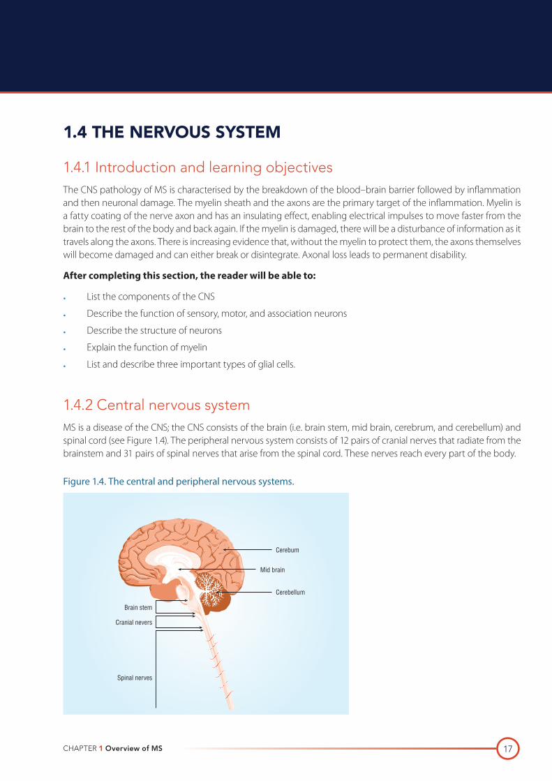
1.4.2CentralnervoussystemMS is a disease of the CNS; the CNS consists of the brain (i.e. brain stem, mid brain, cerebrum, and cerebellum) and spinal cord (see Figure 1.4). The peripheral nervous system consists of 12 pairs of cranial nerves that radiate from the brainstem and 31 pairs of spinal nerves that arise from the spinal cord. These nerves reach every part of the body.
Figure 1.4. The central and peripheral nervous systems.
Cerebum
Cerebellum
Brain stem
Cranial nevers
Spinal nerves
Mid brain
18 A GUIDE TO BEST PRACTICE MULTIPLE SCLEROSIS SPECIALIST NURSING IN IRELAND
The central and peripheral nervous systems consist of networks of neurons (nerve cells). The peripheral nervous system consists of sensory and motor neurons. Sensory neurons respond to stimuli (e.g. heat, pain, colour, etc.) and send impulses to the CNS. Motor neurons transmit messages in the other direction, from the CNS to the peripheral organs and tissues. Therefore, in reality, peripheral nerves are actually bundles of both sensory and motor neurons.
1.4.2.1 Association neurons
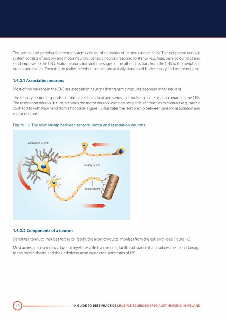
Most of the neurons in the CNS are association neurons that transmit impulses between other neurons.
The sensory neuron responds to a stimulus such as heat and sends an impulse to an association neuron in the CNS. The association neuron, in turn, activates the motor neuron which causes particular muscles to contract (e.g. muscle contracts to withdraw hand from a hot plate). Figure 1.5 illustrates the relationship between sensory, association and motor neurons.
Figure 1.5. The relationship between sensory, motor and association neurons.
Motor neuron
Sensory neuron
Association neuron
1.4.2.2 Components of a neuron
Dendrites conduct impulses to the cell body; the axon conducts impulses from the cell body (see Figure 1.6).
Most axons are covered by a layer of myelin. Myelin is a complex, fat-like substance that insulates the axon. Damage to the myelin sheath and the underlying axon causes the symptoms of MS.

CHAPTER 1 Overview of MS 19
Figure 1.6. Structure of a typical neuron.
Nucleus
Passage of nerve impulse
Cell body
Dendrite
Myelin
Axon
Information
The CNS is not made up entirely of neurons: 40% of the total volume consists of neuroglia or glial cells (glia is the Greek word for glue).
1.4.2.3 Neuroglia
Neuroglia consist of three important types of cells: oligodendrocytes, astrocytes,and microglia (see Figure 1.7).
Figure 1.7. Neuroglia.
Blood vessel
Axon
Astrocyte
Microglial cell
Axon
Neuron cell body
Oligodendrocyte

20 A GUIDE TO BEST PRACTICE MULTIPLE SCLEROSIS SPECIALIST NURSING IN IRELAND
Oligodendrocytes
Oligodendrocytes synthesise myelin. Each oligodendrocyte has many processes that wrap themselves around short sections of nearby axons. While doing so, these processes also produce layers of myelin. Therefore, the myelin sheath of an axon is not continuous, but rather is made up from the processes of many oligodendrocytes, with short lengths of non-myelinated axon between them (see Figure 1.7). These unmyelinated regions are referred to as the nodes of Ranvier.
In the peripheral nervous system, Schwann cells produce myelin (equivalent to oligodendrocytes in the CNS).
Astrocytes
Astrocytes are star-shaped cells that provide the CNS with its physical structure. Some astrocytes have processes that are in contact with the endothelial cells of cerebral blood vessels, helping to maintain tight junctions between them and thereby excluding large circulating cells. This is the main factor that constitutes the blood–brain barrier and normally prevents circulating cells from coming into contact with CNS neurons and neuroglia. In MS, this barrier is breached.
Microglia (microglial cells)
Microglia have the same precursors as circulating macrophages and fulfil similar functions – digestion of cellular debris and foreign matter. In MS, the function of microglia is corrupted, causing them to attack and destroy myelin (see ‘The Immune System‘ in this Chapter for a more detailed discussion of this process).
1.4.2.4 Myelin
Nerve impulses are created by small amounts of electricity flowing along neurons. Myelin acts to insulate the neuron from this flow of electricity. The non-myelinated areas of axons are known as nodes of Ranvier. When impulses flow along myelinated axons, the impulses jump over the myelinated areas from one node to the next (saltatory conduction). This makes the flow of impulses faster and more energy efficient than in non-myelinated axons.
As well as helping the flow of nerve impulses, myelin and oligodendrocytes provide axons with physical support (literally holding them in place and helping preserve their structure) and functional support (by producing chemicals that keep the neurons functioning properly).
1.4.2.5 Grey and white matter
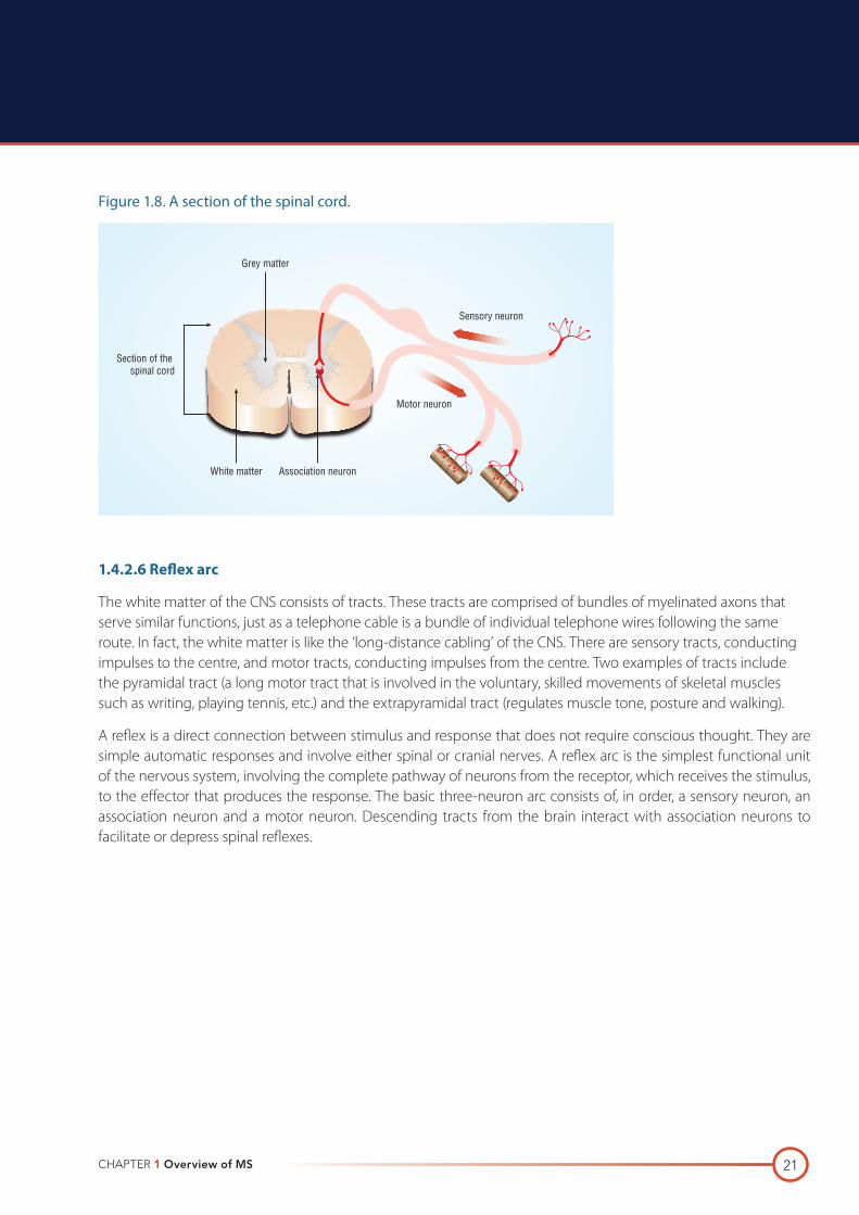
To the naked eye, some of the CNS looks grey, and some of it looks white – hence the terms ‘grey matter’ and ‘white matter.’
Grey matter is a mass of neuronal cell bodies and dendrites. White matter includes bundles of myelinated axons. The same neuron can have its cell body in grey matter and its axon in white matter.
The cerebrum consists of a thick, outer layer of grey matter – the cortex – which covers an inner core of white matter. An H-shaped core of grey matter runs through the middle of the spinal cord and is surrounded by white matter (see Figure 1.8).
CHAPTER 1 Overview of MS 21
Figure 1.8. A section of the spinal cord.
Sensory neuron
Motor neuron
White matter
Grey matter
Association neuron
Section of the spinal cord
1.4.2.6 Reflex arc
The white matter of the CNS consists of tracts. These tracts are comprised of bundles of myelinated axons that serve similar functions, just as a telephone cable is a bundle of individual telephone wires following the same route. In fact, the white matter is like the ‘long-distance cabling’ of the CNS. There are sensory tracts, conducting impulses to the centre, and motor tracts, conducting impulses from the centre. Two examples of tracts include the pyramidal tract (a long motor tract that is involved in the voluntary, skilled movements of skeletal muscles such as writing, playing tennis, etc.) and the extrapyramidal tract (regulates muscle tone, posture and walking).
A reflex is a direct connection between stimulus and response that does not require conscious thought. They are simple automatic responses and involve either spinal or cranial nerves. A reflex arc is the simplest functional unit of the nervous system, involving the complete pathway of neurons from the receptor, which receives the stimulus, to the effector that produces the response. The basic three-neuron arc consists of, in order, a sensory neuron, an association neuron and a motor neuron. Descending tracts from the brain interact with association neurons to facilitate or depress spinal reflexes.

22 A GUIDE TO BEST PRACTICE MULTIPLE SCLEROSIS SPECIALIST NURSING IN IRELAND
1.4.3DemyelinationandaxondamageIn MS, axons in the CNS lose their myelin sheath and, as a result, may break and disintegrate. Many of the typical symptoms of MS affect walking, balance, coordination, bladder function, etc. All of these functions depend on the cabling connecting the brain to the limbs or bladder via the spinal cord. Pain and other sensory disturbances can be explained by demyelination of sensory axons discharging spontaneously or short-circuiting. Loss of muscular control can also result from motor axon damage (i.e. breakage, transection and degeneration).
Higher intellectual functions such as language, memory, and reasoning depend on the grey matter of the cortex. Therefore, these functions are often unaffected by MS. Nevertheless, the different parts of the cortex are connected by white matter, so some subtle intellectual impairment is consistent with demyelination and axon damage.
1.4.3.1 Why does MS not affect the peripheral nervous system?
Peripheral neurons are also myelinated, but the myelin is produced in a different way, by Schwann cells, rather than oligodendrocytes. Schwann cells are not affected in MS.

CHAPTER 1 Overview of MS 23
1.5 THE IMMUNE SYSTEM
1.5.1IntroductionandlearningobjectivesThe immune system is the body’s defence mechanism against infection by bacteria, viruses, fungi, parasites, cancerous agents, and other foreign material. Although the immune system usually combats disease, it can also cause diseases or illnesses. For example, asthma, hay fever, and other allergic disorders are dysfunctional or exaggerated responses of the immune system to foreign agents such as dust and pollen.
In autoimmune diseases such as MS, the immune system is disrupted in such a way that it attacks the body’s own cells. Rheumatoid arthritis and systemic lupus erythematosus are other common autoimmune diseases.
This section reviews the immune system and presents an immunopathogenic model of MS.
After completing this section, the reader will be able to:
· Describe the functions of T-cells, B-cells, cytokines and antibodies
· Understand the role the immune system plays in MS
· Describe an immunopathogenic model of MS.
1.5.2 Where is the immune system?Although the immune system is as vital for sustaining life as the heart or the liver, it is not situated in a single organ (see Figure 1.9). All immune cells, like every other circulating cell, originate from precursors in bone marrow.

24 A GUIDE TO BEST PRACTICE MULTIPLE SCLEROSIS SPECIALIST NURSING IN IRELAND
Figure 1.9. The primary and secondary lymphoid organs.
Primary Lymphoid Organs
Thymus1
1
2
3
4
5
7
Bone marrow2
Secondary Lymphoid Organs & tissues
Lymph nodes, tonsils & adenoids3
Lymph nodes4
Spleen5
Mucosa-associated lymphoid tissues (MALT)6
Lymph nodes7
6
1.5.2.1 Lymphocyte maturation
B-cells mature and differentiate in bone marrow, then migrate into the spleen, lymph nodes and mucosal-associated lymphoid tissue (MALT) – the so-called secondary lymphoid organs. B-cells are involved in specific humoral immunity. Antibodies are made by plasma cells derived from B-cells. These antibodies bind to specific recognition sites on an antigen and destroy or inactivate the antigen in a variety of ways.
T-cells mature and differentiate (i.e. acquire their specificity) in the thymus then also concentrate in the secondary lymphoid organs. T-cells are involved in specific cell-mediated immunity. Each T-cell has an individual receptor that recognises an antigen. These T-cells will, however, only recognise an antigen when it is presented on the surface of a body cell together with a cell marker called major histocompatibility complex (MHC). For instance, T-helper cells recognise antigens that are presented with class II MHC on the surface of macrophages. These T-cells release cytokines to activate the macrophage and enable it to kill intracellular parasites. Cytotoxic T-cells recognise specific antigens when presented with Class I MHC on the surface of infected cells. These T-cells release toxic chemicals, which kill the virus and also release other cytokines.

CHAPTER 1 Overview of MS 25
1.5.3AnimmunopathogenicmodelofMSAlthough the cause of MS is unknown, it is generally believed that environmental factors (possible viral infections) trigger an immunological-mediated process in individuals of certain genetic backgrounds (Polman, 2001; Voumvourakis et al, 2010). When a genetically susceptible person encounters a specific, unknown environmental trigger, a dormant pool of autoreactive T-lymphocytes become active.
Two recent studies, performed in Greece (Voumvourakis et al, 2010) and the USA (Ascherio and Munger, 2007), discuss two possible viral causative agents for the trigger of MS – human herpes virus 6 and Epstein–Barr virus. However, Voumvourakis et al (2010) point out several limitations to their systematic review study, including subjective bias. Ascherio and Munger (2007) conclude their systematic review study by suggesting that not all features of MS epidemiology are explained by Epstein–Barr virus, and this implies involvement of other infectious or non-infectious agents.
1.5.3.1 Proposed sequence of events
The proposed sequence of events is as follows:
· The activated T-lymphocytes cross the blood–brain barrier and search for their target auto-antigen.
· The steps associated with inflammation are as follows:
– An antigen + an antigen presenting cell + a concentration of T-lymphocytes (specific for that antigen) are stimulated to a high threshold
– On reaching threshold the activated T-lymphocytes release gamma interferon
– This process attracts other T-lymphocytes and activates the macrophages.
· Once activated the macrophages:
– Attack and phagocytose the myelin sheath
– Release cytokines
– Present more antigen to T-lymphocytes.
· The T–lymphocytes multiply by cloning and release even more toxic cytokines.
· Overall, this inflammatory reaction:
– Damages myelin
– Damages oligodendrocytes
– Damages the blood–brain barrier allowing other immune cells such as B-lymphocytes, antibodies and complement to leak in and cause further damage.
· The immune cells that leak through cause the following damage:
– B-lymphocytes may produce autoantibodies that help the T-lymphocytes damage the myelin and oliogodendrocytes
– Autoantibody encourage macrophage attack
– Complement destroys oligodendrocytes.
· This cascade of events continues until, for some as yet unidentified reason, the inflammatory response subsides and the blood–brain barrier is restored.

26 A GUIDE TO BEST PRACTICE MULTIPLE SCLEROSIS SPECIALIST NURSING IN IRELAND
1.5.4 References
Ascherio A, Munger K. Environmental risk factors for multiple sclerosis. Part I: The role of infection. Ann Neurol 2007; 61: 288–299.
Polman CH, Thompson AJ, Murray TJ et al. Multiple sclerosis: the guide to treatment and management. Demos Medical Publishing; New York, 2001.
Voumvourakis KI, Kitsos DK, Tsiodras S et al. Human Herpes virus 6 infection as a trigger of multiple sclerosis. Mayo clinic Proc 2010; 11: 1023–1030.

CHAPTER 1 Overview of MS 27
1.6 INFLAMMATION, DEMYELINATION AND REMYELINATION
1.6.1IntroductionandlearningobjectivesMS is a chronic, inflammatory, demyelinating disease of the CNS with secondary axonal involvement. The inflammatory process is driven by a T-cell-mediated immune reaction that leads an attack against both the myelin sheath and the cells that produce myelin, the oligodendrocytes.
As a result of this inflammatory process, myelin can be stripped from the axon and the process of demyelination can take place. Demyelination will slow down the conduction of impulses as they travel along the nerves and will lead to interruption or loss of function. Although remyelination can occur in early or acute MS (Bruck et al, 1994), this process of recovery is limited in the more progressive forms of the disease when repair of the damaged myelin sheath will become impossible (Bramow et al, 2010).
As a result of the demyelination, a process of gliosis (or sclerosis) takes place, scar tissue will form at the lesion site and axonal loss will occur. If the axonal loss occurs in a strategic area or pathway within the CNS, this will lead to irreversible and chronic clinical symptoms.
After completing this section, the reader will be able to:
· Describe the process of demyelination
· Understand the role that inflammation plays in disease activity
· Explain the consequences of demyelination
· Understand how remyelination may occur.
1.6.2TheMSplaqueThe demyelinated plaque is the hallmark of the pathology in MS (Lassmann, 1998). This can occur wherever there is CNS myelin and particularly in the more vascular regions. The pattern of plaque formation will be different in each individual and, although it characteristically occurs in the white matter, grey matter can also become involved. When examining the images from an MRI scan, plaque formation is particularly evident in the periventricular area, although it can also be seen along the optic nerve, the midbrain (particularly around the Aqueduct of Sylvius), the pons, the medulla oblongata and the cerebellum. It also commonly occurs in the spinal cord, particularly the cervical area. These spinal cord lesions show no regularity in their distribution, and so both lesions and associated atrophy can be scattered through its length (Raine, 1993).
It is important to acknowledge that plaques can occur without any clinical signs. It is commonly reported that MRI images demonstrate large asymptomatic plaques. This is particularly the case in PPMS where there is often little correlation between number of lesions and level of disability.

28 A GUIDE TO BEST PRACTICE MULTIPLE SCLEROSIS SPECIALIST NURSING IN IRELAND
1.6.3Inflammationanddiseaseactivity
‘The pathology of progressive MS is fully consistent with that of a classical inflammatory disease’ Frischer et al, 2008
Inflammation within the brain can occur independently of demyelination and can, again, affect both white and grey matter. During the inflammatory process, the meninges can be involved and macrophages, lymphocytes and plasma cells can also be identified in the patient’s subarachnoid space. It was considered likely that in the development of the plaque, inflammation is the primary event, with demyelination a secondary sequel, but according to Lassman (1998), there is an ongoing debate whether this is indeed the case or whether the inflammation is secondary to the demyelination. Inflammation is a consistent feature of both RRMS and SPMS, but is much less of a component in PPMS (Revesz, 1994). An Austrian study of MS autopsy reports (Fischer et al, 2009) found that inflammation declines in older patients at a late stage of disease.
1.6.4Whathappensduringarelapse?An acute exacerbation, or relapse, of MS is defined as “the occurrence of a symptom or symptoms of neurological dysfunction with or without objective confirmation lasting more than 24 hours” (McGriff, 2008; Poser et al, 1983; Sellebjerg et al, 2005 cited in Burgess, 2011).
Typically a relapse will progress over a few days to weeks, reaching a maximum level after several weeks before slowly resolving. Complete recovery of symptoms is common during the initial stages of the disease, which is probably because the oligodendrocytes are more likely to survive during the inflammatory process and remyelination can then occur (Lassmann, 1998). An understanding of the pathological disease process that occurs during a relapse is essential for the effective care and management of people with RRMS. This ensures that the individual receives the necessary information to understand what is happening to them and they are then able to understand the rationale behind any treatments that may be offered to them.
The initial process involved in a relapse is immune-mediated damage to the blood–brain barrier. There will be an activation of the T-cells which target the vascular wall and, by secreting cytokines, will recruit other cells including macrophages. Adhesion molecules expressed on the lymphocyte and endothelial cell will allow the adhesion molecules to attach to the vessel wall and then T-cells pierce through the cell and into the CNS. According to Burgess (2011), a relapse is caused by an increase in autoimmune activity, which weakens the blood–brain barrier, allowing inflammatory cells to enter the CNS causing lesions and demyelination.
CHAPTER 1 Overview of MS 29
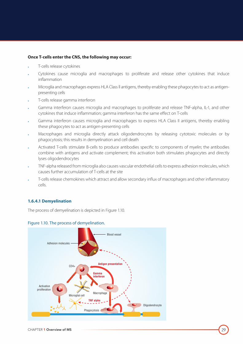
Once T-cells enter the CNS, the following may occur:
· T-cells release cytokines
· Cytokines cause microglia and macrophages to proliferate and release other cytokines that induce inflammation
· Microglia and macrophages express HLA Class ll antigens, thereby enabling these phagocytes to act as antigen-presenting cells
· T-cells release gamma interferon
· Gamma interferon causes microglia and macrophages to proliferate and release TNF-alpha, IL-1, and other cytokines that induce inflammation; gamma interferon has the same effect on T-cells
· Gamma interferon causes microglia and macrophages to express HLA Class II antigens, thereby enabling these phagocytes to act as antigen-presenting cells
· Macrophages and microglia directly attack oligodendrocytes by releasing cytotoxic molecules or by phagocytosis; this results in demyelination and cell death
· Activated T-cells stimulate B-cells to produce antibodies specific to components of myelin; the antibodies combine with antigens and activate complement; this activation both stimulates phagocytes and directly lyses oligodendrocytes
· TNF-alpha released from microglia also causes vascular endothelial cells to express adhesion molecules, which causes further accumulation of T-cells at the site
· T-cells release chemokines which attract and allow secondary influx of macrophages and other inflammatory cells.
1.6.4.1 Demyelination
The process of demyelination is depicted in Figure 1.10.
Figure 1.10. The process of demyelination.
Blood vessel
Oligodendrocyte
Macrophage
Antigen presentation
Gamma interferon
TNF alpha
Activation proliferation
CD4+
Microglial cell
Phagocytosis
Adhesion molecules
B
T

30 A GUIDE TO BEST PRACTICE MULTIPLE SCLEROSIS SPECIALIST NURSING IN IRELAND
It is presumed that the endpoint of this process may be transient inflammation of myelin or, if continued, irreversible destruction of oligodendrocytes and ultimately axonal loss.
Although it may appear that this process could lead to rapid and complete demyelination within hours, many individuals with MS recover from attacks, even without treatment. This is because the process is not uncontrolled. T-suppressor cells and subsets of T-helper cells may play a role here by releasing cytokines (such as IL-4, IL-10) and transforming growth factor (TGF) beta which downregulate the immune response. Beta interferon may enhance the action of T-suppressor cells and the relevant T-helper cells.
1.6.5PredisposingfactorsforrelapseWhen a patient presents with a relapse, it is important to consider predisposing factors. For example, women are thought to more likely relapse in the first 3–6 months post-partum (Vukusic et al, 2004). Other factors that could increase the relapse rate include stress, infection or extreme temperature exposure. According to Vukusic et al (2004), addressing these lifestyle issues, by, for example, restarting treatment post-partum, managing stress, preventing infection or reducing heat exposure, could help to avoid triggering a relapse.
1.6.6PseudoexacerbationinMSA pseudoexacerbation or pseudo-relapse involves a worsening of previous symptoms, which is thought to be due to fever or flu-like symptoms secondary to disease-modifying drugs (Villoslada et al, 2009). According to Sadiq, cited in Rowland (ed) (2005), this transient event disappears soon after the provoking activity is stopped, i.e. when the infection is cleared.
1.6.7EpidemiologyofthedemyelinationprocessA combination of genetic and environmental factors may play a role in the demyelination process of MS.
1.6.7.1 Viruses
A number of known facts suggest a viral cause for MS:
· Viruses can cause other demyelinating diseases in humans and other species
· Tropical spastic paraparesis is caused by the human T-cell leukaemia virus type 1 (HTLV 1)
· Viral infection is consistent with the distribution of MS in cold climates
· Studies of migrants suggest that susceptibility to MS depends on where a person lived up to the age of 15 years, not where he/she currently lives; therefore, childhood factors appear to play a role in the development of MS
· Viral infections stimulate the production of interferons – gamma interferon, in particular, appears to play a key role in MS.

CHAPTER 1 Overview of MS 31
One hypothesis suggests that, in genetically susceptible individuals, MS results from delayed exposure to a common infectious agent. Although many agents have been examined, such as measles, canine distemper, rubella, and herpes zoster and simplex, no single pathogen has been identified as causing the disease.
The possible role of viral infections in MS was the original rationale for the study of gamma interferon as a treatment for MS (since the interferons were known to have antiviral properties). The fact that gamma interferon actually worsened MS helped to elucidate some of the mechanisms of the disease.
1.6.7.2 Genetics
Evidence from a variety of studies indicates that certain people are genetically predisposed to MS (Halper and Holland, 2011). For example, 5% of siblings of a person with MS develop the disease. In fact, the identical twin of a person with MS has a much higher likelihood of developing MS than the non-identical twin of a person with MS. However, siblings also tend to share the same environment and, therefore, this increased risk among siblings does not prove that MS is inherited.
1.6.8ConsequencesofdemyelinationDemyelination makes axons susceptible to inflammation, causing them to break and subsequently degenerate.
Severely demyelinated axons lack normal support from oligodendrocytes; many of these axons break or simply degenerate and lead to irreversible neuronal death.
1.6.9 Recovery of functionIt used to be thought that remyelination could not occur in MS. However, it is now known that remyelination can take place, leaving increased numbers of glial cells in otherwise normal white matter (Lassmann, 1983). On MRI scans, areas of remyelination show up as pale ‘shadow plaques’.
The pathological features of remyelination are uniformly thin myelin and short distances between the nodes of Ranvier (Prineas, 1993). The type and amount of remyelination varies between different disease patterns of MS. The factors that determine whether a plaque will or will not remyelinate are not yet known.
In common with many aspects of MS, the process appears to be complicated, as there seems to be a feedback mechanism between demyelination and remyelination. It is hypothesised that during demyelination, myelin breakdown products stimulate oligodendrocytes to produce more myelin (Raine, 1993). In early MS, remyelination is a prominent feature of the disease, which may be because many oligodendrocytes survive at this stage of the disease. As the disease progresses, less remyelination occurs (Burgess, 2010), which may be because oligodendrocytes are targeted at this stage.
Overall, the pathology of remyelination demonstrates the dynamic nature of MS, which accounts for the variance seen in the symptoms presented by individuals.
32 A GUIDE TO BEST PRACTICE MULTIPLE SCLEROSIS SPECIALIST NURSING IN IRELAND
1.6.10RoleofaxonalpathologyinMSAxonal pathology may play a role in MS. For example, the results of post-mortem studies of patients with MS have noted a large number of broken (transected) axons in active MS lesions. Active lesions are defined as having extensive inflammation. Inflammation is an early event in the pathological process and is more pronounced in the earlier stages of MS (Dutta and Trapp, 2007). These transected axons were also found, in smaller numbers, on the borders of chronic lesions; very few were found in chronic lesions or normal brain tissues.
Axonal breakage has also been noted in patients with both short (2 weeks) and long (26 years) durations of MS, suggesting that axonal transection starts early in the disease process. Axon transection is irreversible; axons degenerate after breaking, leading to permanent disability. In a UK study involving a review of magnetic resonance spectroscopy (MRS) and MRIs of MS patients with severe cerebellar involvement, Davie et al (1995) found that axonal loss is important in the development of persistent clinical disability in MS.
The fact that transected axons were found mainly in inflamed and demyelinated areas suggests that demyelinated axons may be susceptible to inflammatory changes that cause axon damage. This is probably the process that leads to axon transection in the early stages of the MS. It should be noted, however, that patients do not show permanent disability in the early phases of MS because the excess capacity of the brain can compensate for the damage through the rerouting of the affected functions. This rerouting is dangerous because it masks the real damage and leads to the misleading perception that the patient is doing well while irreversible damage is accumulating.
In SPMS, where inflammation is no longer a key feature, axonal degeneration (Dutta and Trapp, 2007) and accumulation of permanent disability continues. The damage appears to result from the chronic demyelination of axons, which means that the axons lack the support normally provided by oligodendrocytes (this helps to keep the axons healthy).
1.6.11Implicationsfortreatmentwithdisease-modifyingagentsThe finding that axon damage, although present, does not produce symptoms in the early stages of MS (due to rerouting of affected functions) has profound implications for the treatment of MS. The goal of treatment should be to slow irreversible damage to oligodendrocytes and axons and, therefore, delay progression of the disease to irreversible SPMS. The axon and myelin damage is linked to inflammation, which is very pronounced during the first years of MS. In the early stages of MS, for example, new lesions occur 10 times more frequently than clinical signs and are detected by MRI, even when patients are in remission. Therefore, treatment initiated at the start of the disease may prevent inflammatory changes that lead to demyelination and axon damage. Interferon dosing is also an important consideration, since higher doses of some types of beta-interferon have been shown to be more effective at suppressing inflammatory activity (see Chapter 3: ‘Treatments’ for a more detailed discussion of interferons).
CHAPTER 1 Overview of MS 33
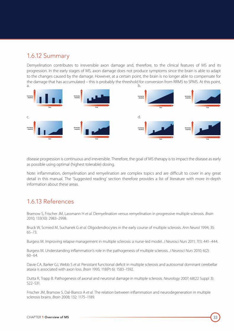
1.6.12SummaryDemyelination contributes to irreversible axon damage and, therefore, to the clinical features of MS and its progression. In the early stages of MS, axon damage does not produce symptoms since the brain is able to adapt to the changes caused by the damage. However, at a certain point, the brain is no longer able to compensate for the damage that has accumulated – this is probably the threshold for conversion from RRMS to SPMS. At this point,
disease progression is continuous and irreversible. Therefore, the goal of MS therapy is to impact the disease as early as possible using optimal (highest tolerable) dosing.
Note: inflammation, demyelination and remyelination are complex topics and are difficult to cover in any great detail in this manual. The ‘Suggested reading’ section therefore provides a list of literature with more in-depth information about these areas.
1.6.13 References
Bramow S, Frischer JM, Lassmann H et al. Demyelination versus remyelination in progressive multiple sclerosis. Brain 2010; 133(10): 2983–2998.
Bruck W, Scmied M, Suchanek G et al. Oligodendrocytes in the early course of multiple sclerosis. Ann Neurol 1994; 35: 65–73.
Burgess M. Improving relapse management in multiple sclerosis: a nurse-led model. J Neurosci Nurs 2011; 7(1): 441–444.
Burgess M. Understanding inflammation’s role in the pathogenesis of multiple sclerosis. J Neurosci Nurs 2010; 6(2): 60–64.
Davie CA, Barker GJ, Webb S et al. Persistant functional deficit in multiple sclerosis and autosomal dominant cerebellar ataxia is associated with axon loss. Brain 1995; 118(Pt 6): 1583–1592.
Dutta R, Trapp B. Pathogenesis of axonal and neuronal damage in multiple sclerosis. Neurology 2007; 68(22 Suppl 3): S22–S31.
Frischer JM, Bramow S, Dal-Bianco A et al. The relation between inflammation and neurodegeneration in multiple sclerosis brains. Brain 2008; 132: 1175–1189.
Time
Increasingdisability
A
Time
Increasingdisability
B
Time
Increasingdisability
A
Time
Increasingdisability
B
Time
Increasingdisability
A
Time
Increasingdisability
B
Time
Increasingdisability
A
Time
Increasingdisability
B
Time
Increasingdisability
A
Time
Increasingdisability
B
Time
Increasingdisability
A
Time
Increasingdisability
B
Time
Increasingdisability
A
Time
Increasingdisability
B
Time
Increasingdisability
A
Time
Increasingdisability
B
Time
Increasingdisability
A
Time
Increasingdisability
B
Time
Increasingdisability
A
Time
Increasingdisability
B
Time
Increasingdisability
A
Time
Increasingdisability
B
Time
Increasingdisability
A
Time
Increasingdisability
B
Time
Increasingdisability
A
Time
Increasingdisability
B
Time
Increasingdisability
A
Time
Increasingdisability
B
Time
Increasingdisability
A
Time
Increasingdisability
B
Time
Increasingdisability
A
Time
Increasingdisability
B
a.
c.
b.
d.

34 A GUIDE TO BEST PRACTICE MULTIPLE SCLEROSIS SPECIALIST NURSING IN IRELAND
Halper J, Holland N. Comprehensive nursing care in multiple sclerosis. 3rd ed. Springer Publishing Company; New York, 2011.
Lassmann H. Comparative neuropathology of chronic experimental allergic encephalomyelitis and multiple sclerosis. Springer–Verlag, Berlin 1983.
Lassmann H. Pathology of multiple sclerosis. In: Compston A, Ebers G, Lassmann H et al (eds). McAlpines Multiple Sclerosis. 3rd ed. Churchill Livingstone; London, 1998.
McGriff. The dangerous fight against plaque: the use of natalizumab in the treatment of multiple sclerosis. Drug Formulary Review March 2008.
Poser CM, Paty DW, Sheinberg L et al. New diagnostic criteria for multiple sclerosis: guidelines for research protocols. Ann Neurol 1983; 13: 227–231.
Prineas JW, Barnard RO, Kwon EE et al. Multiple sclerosis: remyelination of nascent lesions. Ann Neurol 1993; 52(3): 199–204.
Raine CS, Wu E. Multiple sclerosis: remyelination in acute lesions. J Neuropathol Exp Neurol 1993; 52(3): 199–204.
Revesz T, Kidd D, Thompson AJ et al. A comparison of the pathology of primary and secondary-progressive multiple sclerosis. Brain 1994; 117(Pt 4): 759–765.
Villoslada P, Arrondo G, Sepulcre J et al. Memantine induces reversible neurologic impairment in patient with MS. Neurology 2009; 72(19): 1630–1633.
Vukusic S, Hutchinson M, Hours M et al. Pregnancy and multiple sclerosis (the PRIMS study): clinical predictors of post-partum relapse. Brain 2004 (Pt 6); 127: 1353–1360.
1.6.14Suggestedreading
Allen IV, Glover G, Anderson R. Abnormalities in the macroscopically normal white matter in cases of mild or spinal multiple sclerosis. Acta Neuropathol 1981; 7: 176–178.
Allen IV, McKeown SR. A histological, histochemical and biochemical study of the macroscopically normal white matter in multiple sclerosis. J Neurol Sci 1979; 41(1): 81–91.
De Stefano N, Narayanan S, Francis GS et al. Evidence of axonal damage in the early stages of multiple sclerosis and its relevance to disability. Arch Neurol 2001; 58(1): 65–70.
Prineas JW. The pathology of multiple sclerosis. In: Handbook of multiple sclerosis (Ed Cook S). Marcel Decker Inc.; New York, 2001.
Smith KJ, McDonald WI. The pathophysiology of MS: the mechanisms underlying the symptoms and the natural history of the disease. Philos Trans R Soc Lon B Biol Sci 1999; 354(1390): 1649–1673.
Thomson AJ, McDonald WI. Multiple sclerosis and its pathophysiology. In: Ashbury AK, McKhann GM, McDonald WI (eds). Diseases of the nervous system; 1209–1228. Philadelphia: WB Saunders Company; 1992.
Trapp BD, Ransohoff RM, Fisher E. Neurodegeneration in multiple sclerosis: relationship to neurological disability. The Neuroscientist 1999; 5(1): 48–57.
Wingerchuk DM, Lucchinetti CF, Noseworthy JH. Multiple sclerosis: current pathophysiological concepts. Lab Invest 2001; 81(3): 263–281.

CHAPTER 1 Overview of MS 35
1.7 CLINICAL MANIFESTATIONS
1.7.1IntroductionandlearningobjectivesAs a result of inflammation, demyelination or axonal loss, PWMS can experience a diverse range of neurological symptoms. There is no doubt that there are sites of predilection for demyelination (the optic nerves, cervical cord and brainstem) but in clinical practice, the symptomatic experience of an individual can never really be predicted. Some people will develop a variety of irritating symptoms that are troublesome but do not necessarily affect quality of life, whereas others develop a whole range of disabling symptoms producing a dramatic impact on their psychosocial functioning. Whatever the symptoms are, it is the treatment of them that is fundamental in the management of MS (Thompson, 1998).
It cannot be overemphasised that not all symptoms are due to MS and it is crucial that other causes for symptoms are excluded.
After completing this section, the reader will be able to:
· Identify the common sites within the CNS that are more prone to demyelination
· List and discuss the symptoms that a person may experience if they have spinal lesions
· List and discuss the symptoms that a person may experience if they have brainstem lesions
· List the paroxysmal symptoms that PWMS may experience
· Identify the rarer symptoms that can be attributed to MS.
1.7.2 Symptoms of MSThe type of neurological symptoms experienced will generally reflect the parts of the CNS that have been the most heavily demyelinated (Mathews, 1998). As these symptoms are extensive and can interact in complex ways, the management of MS can be difficult (Thompson, 1998). For example, if a person has mobility difficulties, they may also have spasticity and ataxia. Symptoms can fluctuate in presence and severity on a day-to-day, even hour-to-hour span in time, and can manifest without any real stimulus to trigger them. Fatigue and/or heat can also worsen symptoms, resulting in more severe disability, albeit on a transient basis.
PWMS may experience co-occurring disorders such as thyroid disease or diabetes that are totally unrelated to their underlying MS. It is essential that, as nurses, we are familiar with the typical clinical symptoms that can be produced as a result of demyelination, thus enabling us to educate patients on what is typical of MS and what may be uncharacteristic and require further exploration. Most books that are written on MS will detail the clinical features that people will exhibit.

36 A GUIDE TO BEST PRACTICE MULTIPLE SCLEROSIS SPECIALIST NURSING IN IRELAND
1.7.3Commonsitesofdemyelination
1.7.3.1 Optic nerves
Abnormalities of vision are commonly due to plaque formation along the optic nerve or chiasm. They can also be due to damage to the optic tract or optic radiation. Optic neuritis is a common presenting symptom of MS occurring in 14–23% of cases. Bonhomme et al (2009) note that after acute unilateral optic neuritis, adults have a 38% chance of developing MS within 10 years. It is generally unilateral, and the patient will complain of dimming vision, photophobia and pain on eye movement. They can also experience loss of colour vision, a central scotoma or loss of visual acuity, but visual loss is seldom total.
1.7.3.2 Spinal cord
Lesions within the spinal cord can produce the following symptoms:
· Bladder dysfunction: The patient will complain of detrusor instability (frequency, urgency, nocturia or incontinence) or detrusor sphincter dyssynergia (hesitancy, retention or overflow incontinence). Some will experience a combination of both. According to DasGupta and Fowler (2003), bladder dysfunction affects 75% of patients with MS and it is incontinence that causes social inconvenience to patients.
· Bowel dysfunction: The most common experience is constipation, but urgency and incontinence are also possible.
· Limb weakness: Although this in itself is very incapacitating, it can be compounded by ataxia and tremor. It is often associated with brisk reflexes.
· Lhermittes’s sign: This is a tingling or electric shock-like sensation felt in the limbs on neck flexion.
· Sensory changes: These can be acute (intense, burning, shooting, stabbing) or chronic (dysaesthetic pain – i.e. burning, itching, crawling, prickling, tingling). Most commonly sensory symptoms consist of numbness and pins and needles type of paraesthesiae.
· Sexual dysfunction: In males, erectile dysfunction is estimated at 70% (DasGupta and Fowler, 2003); ejaculatory dysfunction, lack of libido and diminished orgasmic capacity can also occur. Females report lack of libido, orgasmic dysfunction and vaginal dryness.
· Spasticity: This is commonly seen in legs, although it can also occur in the arms. This will contribute to fatigue. The person may experience flexor or extensor spasms, clonus and contractures.

CHAPTER 1 Overview of MS 37
1.7.3.3 Brainstem
Lesions within the brainstem can produce the following symptoms:
· Dysarthria (slurring or indistinctness of speech): This is more common in advanced MS. A particular type of
speech disturbance known as ‘scanning speech’ is typical of MS: this consists of a particular rhythm in which
each word and syllable is given emphasis, there are pauses after each syllable and the syllable is pronounced
slowly.
· Dysphagia: This is usually experienced in association with dysarthria and is more common in advanced MS.
The patient may have a pseudobulbar palsy.
· Facial palsy: This probably results from a plaque along the facial nerve.
· Impairment of ocular motility: This can include nystagmus (usually horizontal, but can also be rotary, upbeating
and downbeating), blurred vision, oscillopsia (images jumping) and double vision.
· Internucleur ophthalmoplegia: This can be unilateral or bilateral. It consists of failure of the eye ipsilateral to the
lesion to adduct, whereas the contralateral eye abducts fully, but with horizontal nystagmus.
· Trigeminal neuralgia: Involves one, two or three divisions of the trigeminal nerve. Can be excruciating.
Generally remits.
“Symptomatic trigeminal neuralgia is frequently related to MS, with MS patients having a
20 fold increased risk of developing trigeminal neuralgia” - Cruccu et al, 2009
· Vertigo: This occurs as an initial symptom in 5% of patients, and can eventually occur in as many as 50% of
people. It can be transient in nature.
1.7.3.4 Cerebellar
Cerebellar lesions can produce the following symptoms:
· Ataxia: This is one of the most disabling symptoms, and includes head titubation, gait ataxia and truncal ataxia.
· Dysarthria: This consists of ataxic or scanning speech.
· Tremor: This can be an intention and/or postural tremor.

38 A GUIDE TO BEST PRACTICE MULTIPLE SCLEROSIS SPECIALIST NURSING IN IRELAND
1.7.3.5 Cerebral hemispheres
Lesions within the cerebral hemispheres can produce the following symptoms:
· Cognitive impairment: This includes memory, conceptual reasoning (especially with frontal lobe lesions), attention, word finding, decreased speed of information processing and emotional lability. Mattioli et al (2011) note that cognitive impairment affects 43–70% of MS patients.
· Depression: This will be experienced by one in every two people (Sadovnick et al, 1996). Mattioloi et al (2011) state that depression affects 22–50% of MS patients. It is unknown whether it is a primary or secondary symptom.
· Fatigue: This causes a major impact on quality of life. It is unrelated to activity – fatigue can be present on waking – and can relapse and remit. It is noted to occur in 90% of MS patients (Mattioli et al, 2011).
1.7.3.6 Sensory impairment – pain
Kratz et al (2011) note that pain in MS is likely to originate from a variety of sources, directly, indirectly or coincidental to MS.
There are 3 types of pain that PWMS may complain of can be directly or indirectly related to their MS:
· Dysaesthetic (or neurogenic) pain: This tends to be experienced in the hands and feet, and is described as burning, pricking, tingling, dull or nagging. The pathology is unknown, but it is probably due to transmission of abnormal electrical discharges across a demyelinated plaque (Paty and Ebers, 1998).
· Musculoskeletal: This can occur in the spinal area or anywhere where there is a joint. It is usually due to mechanical problems, such as poor gait and hyperextension of the knee joint.
· Paroxysmal: This pain tends to be abrupt in onset, brief, commonly lasting 30–60 seconds, but is severe in nature, and recovers spontaneously. It is not associated with or predictive of disability. Examples include trigeminal neuralgia (pain along the trigeminal nerve), Lhermitte’s phenomenon (electric shock sensation when the patient bends their head into their chest), tonic spasms (often triggered by movement and results in a tonic spasm involving either flexion or extension of a limb. This spasm can last for several seconds and can occur frequently. It tends to settle spontaneously rather than in response to treatment), pruritus (can last 20 minutes or more), optic neuritis (will experience shooting pain behind the eye – this can be spontaneous or on eye movement).
1.7.4Raresignsandsymptoms,butcanbeattributedtoMS
· Anosmia
· Aphasia
· Auditory disturbance
· Deafness
· Hiccoughs
· Sleep disturbance
· Sweating
· Yawning

CHAPTER 1 Overview of MS 39
1.7.5 References
Bashir K, Whitaker JN. Handbook of multiple sclerosis. Lipincott Williams and Wilkins; Philadelphia, 2002.
Bonhomme GR, Waldman AT, Balcer LJ et al. Pediatrin optic neuritis. Neurology 2009; 72(10): 881–885.
Cruccu G, Biasiotta A, Di Rezze S et al. Trigeminal neuralgia and pain related multiple sclerosis. Pain 2009; 143(3): 186–191.
Das Gupta R, Fowler C. Bladder, bowel and sexual dysfunction in multiple sclerosis. Drugs 2003; 63(2): 153–166.
Kratz AL, Molton IR, Jensen MP A et al. Further evaluation of the motivational model of pain self-management: coping with chronic pain in multiple sclerosis. Ann Behav Med 2011; 41(3): 391–400
Mathews B. Symptoms and signs of multiple sclerosis. In: McAlpines multiple sclerosis (Compston A, Ebers G, Lassmann H et al (Eds). 3rd ed. Churchill Livingstone; London, 1998.
Mattioli F, Bellomi F, Stampatori C et al. Depression, disability and cognitive impairment in multiple sclerosis: a cross sectional Italian study. Neurol Sci 2011; 32(5): 825–832.
Paty DW, Ebers GC. Clinical features. In: Multiple sclerosis (Eds Paty DW, Ebers GC ). FA Davis Company; Philadelphia, 1998.
Sadovnick AD, Remick RA, Allen J et al. Depression and multiple sclerosis. Neurology 1996; 46(3): 628–632.
Thompson AJ. Symptomatic treatment in multiple sclerosis. Curr Opin Neurol 1998; 11(4): 305–309.
Van den Noort S, Holland NJ. Multiple sclerosis in clinical practice. Desmos Medical Publishing; New York, 1999.

40 A GUIDE TO BEST PRACTICE MULTIPLE SCLEROSIS SPECIALIST NURSING IN IRELAND
1.8 PROGRESS CHECK 1. The prevalence of PPMS is approximately _________ %. The prevalence of RRMS is approximately _________ %.
2. Name each type of MS depicted below.
a.
b.
c.
d.

CHAPTER 1 Overview of MS 41
3. The peak age of MS onset is _________ years.
4. The prevalence rate of MS in Ireland is about _________ in _______________ adults.
5. The risk of developing MS for a person with a sister with MS is _________ %.
6. Answer true or false to each of the following statements.
a. There is a genetic marker that is significant in MS
True False
b. MS affects Caucasians more than any other race.
True False
c. Within Caucasian populations, MS occurs more frequently in countries or regions of southern latitudes in the Northern Hemisphere and northern latitudes in the Southern Hemisphere.
True False
d. MS is more common in men than in women.
True False
e. The concordance rate of monozygotic twins being diagnosed with MS is 30%.
True False

42 A GUIDE TO BEST PRACTICE MULTIPLE SCLEROSIS SPECIALIST NURSING IN IRELAND
7. Label the figure.
8. Most of the neurons in the CNS are _______________ _______________ neurons.
9. Explain the main function of the following:
a. Axons ___________________________________________________________________________
b. Myelin ___________________________________________________________________________
c. Oligodendrocytes __________________________________________________________________
d. Schwann cells _____________________________________________________________________
e. Microglia _________________________________________________________________________
10. What do the following consist of?
a. Grey matter _______________________________________________________________________
b. White matter ______________________________________________________________________

CHAPTER 1 Overview of MS 43
11. Label the figure.
12. What is the consequence of demyelination?
__________________________________________________________________________________
__________________________________________________________________________________
13. Although axon damage is present during the early stages of MS, the damage does not produce symptoms. Why is this dangerous?
__________________________________________________________________________________
__________________________________________________________________________________
__________________________________________________________________________________
__________________________________________________________________________________
14. List five symptoms and/or signs a person will present with if they have demyelination along their optic nerve.
a. ________________________________________________________________________________
b. ________________________________________________________________________________
c. ________________________________________________________________________________
d. ________________________________________________________________________________
e. ________________________________________________________________________________
B
T

44 A GUIDE TO BEST PRACTICE MULTIPLE SCLEROSIS SPECIALIST NURSING IN IRELAND
15. List eight symptoms and/or signs a person will present with if they have demyelination in their brainstem.
a. ________________________________________________________________________________
b. ________________________________________________________________________________
c. ________________________________________________________________________________
d. ________________________________________________________________________________
e. ________________________________________________________________________________
f. ________________________________________________________________________________
g. ________________________________________________________________________________
h ________________________________________________________________________________

CHAPTER 1 Overview of MS 45
1.9 PROGRESS CHECK ANSWERS1. The prevalence of PPMS is approximately 10%. The prevalence of RRMS is approximately 40%.
2. Name each type of MS depicted below.
3. The peak age of MS onset is 30 years.
4. The prevalence rate of MS in Ireland is about 1 in 1,000 adults.
5. The risk of developing MS for a person with a sister with MS is 4.4%.
6. Answer true or false to each of the following statements.
a. There is a genetic marker that is significant in MS
False. There is still no one specific genetic marker that has been identified as being responsible for the predisposition to the development of MS
b. MS affects Caucasians more than any other race.
True
c. Within Caucasian populations, MS occurs more frequently in countries or regions of southern latitudes in the Northern Hemisphere and northern latitudes in the Southern Hemisphere.
False. Within Caucasian populations, MS occurs more frequently in countries or regions of higher northern latitudes in the Northern Hemisphere and higher southern latitudes in the Southern Hemisphere
d. MS is more common in men than in women.
False. MS has a predilection for women at a ratio of approximately 3:1
e. The concordance rate of monozygotic twins being diagnosed with MS is 30%.
True
Time
Increasingdisability
A
Time
Increasingdisability
B
Time
Increasingdisability
A
Time
Increasingdisability
B
Time
Increasingdisability
A
Time
Increasingdisability
B
Time
Increasingdisability
A
Time
Increasingdisability
B
Time
Increasingdisability
A
Time
Increasingdisability
B
Time
Increasingdisability
A
Time
Increasingdisability
B
Time
Increasingdisability
A
Time
Increasingdisability
B
Time
Increasingdisability
A
Time
Increasingdisability
B
Time
Increasingdisability
A
Time
Increasingdisability
B
Time
Increasingdisability
A
Time
Increasingdisability
B
Time
Increasingdisability
A
Time
Increasingdisability
B
Time
Increasingdisability
A
Time
Increasingdisability
B
Time
Increasingdisability
A
Time
Increasingdisability
B
Time
Increasingdisability
A
Time
Increasingdisability
B
Time
Increasingdisability
A
Time
Increasingdisability
B
Time
Increasingdisability
A
Time
Increasingdisability
B
a. Relapsing-remitting MS (RRMS)
c. Secondary-progressive MS (SPMS)
b. Primary-progressive MS (PPMS)
d. Progressive-relapsing MS (PRMS)

46 A GUIDE TO BEST PRACTICE MULTIPLE SCLEROSIS SPECIALIST NURSING IN IRELAND
7. Label the figure.
Cerebum
Cerebellum
Brain stem
Cranial nevers
Spinal nerves
Mid brain
8. Most of the neurons in the CNS are association neurons.
9. Explain the main function of the following:
a. Axons
The primary role of axons is to conduct impulses from the cell body.
b. Myelin
Myelin protects and insulates neurons and also enhances the conduction of nerve impulses along axons.
c. Oligodendrocytes
Oligodendrocytes are primarily responsible for synthesising myelin in the CNS
d. Schwann cells
Schwann cells are primarily responsible for synthesising myelin in the peripheral nervous system.
e. Microglia
Microglia are responsible for the digestion of cellular debris and foreign matter.

CHAPTER 1 Overview of MS 47
10. What do the following consist of?
a. Grey matter
Grey matter consists of neuronal cell bodies and dendrites.
b. White matter
White matter consists of bundles of myelinated axons.
11. Label the figure.
Blood vessel
Oligodendrocyte
Macrophage
Antigen presentation
Gamma interferon
TNF alpha
Activation proliferation
CD4+
Microglial cell
Phagocytosis
Adhesion molecules
B
T
12. What is the consequence of demyelination?
Demyelination makes axons susceptible to inflammation, causing them to break and subsequently degenerate.
13. Although axon damage is present during the early stages of MS, the damage does not produce symptoms. Why is this dangerous?
Patients do not yet show permanent disability in the early stages of MS because the excess capacity of the brain can compensate for the axonal damage by re-routing of the affected functions. This re-routing is dangerous because it masks the real damage and leads to the misleading perception that the patient is doing well while irreversible damage is accumulating.
48 A GUIDE TO BEST PRACTICE MULTIPLE SCLEROSIS SPECIALIST NURSING IN IRELAND
14. List five symptoms and/or signs a person will present with if they have demyelination along their optic nerve.
a. Dimming vision
b. Photophobia
c. Pain on movement of the eye
d. Loss of colour vision
e. Loss of visual acuity
15. List eight symptoms and/or signs a person will present with if they have demyelination in their brainstem.
a. Auditory disturbance (rare)
b. Dysarthria
c. Dysphagia
d. Facial palsy
e. Impairment of ocular motility
f. Internucleur ophthalmoplegia
g. Trigeminal neuralgia
h. Vertigo
CHAPTER 1 Overview of MS 49
1.10 GLOSSARYCIS Clinically isolated syndrome
CNS Central nervous system
EDSS Expanded disability status scale
MHC Major histocompatibility complex
MRI Magnetic resonance imaging
MRS Magnetic resonance spectroscopy
MS Multiple sclerosis
PRMS Progressive relapsing multiple sclerosis
RIS Radiological isolated syndrome
SPMS Secondary progressive multiple sclerosis
PPMS Primary progressive multiple sclerosis
PWMS People with multiple sclerosis
RRMS Relapsing remitting multiple sclerosis

50 A GUIDE TO BEST PRACTICE MULTIPLE SCLEROSIS SPECIALIST NURSING IN IRELAND