chapter 1 lipids: definitions, naming, methods and a guide
TRANSCRIPT
CHAPTER 1
Lipids: definitions, naming, methods and aguide to the contents of this book
1.1 Introduction
Lipids occur throughout the living world in microorgan-
isms, fungi, higher plants and animals. They occur in all
cell types and contribute to cellular structure, provide
energy stores and participate in many biological pro-
cesses, ranging from transcription of genes to regulation
of vital metabolic pathways and physiological responses.
In this book, they will be described mainly in terms of
their functions, although on occasion it will be conve-
nient, even necessary, to deal with lipid classes based on
their chemical structures and properties. In the conclud-
ing section of this chapter, we provide a ‘roadmap’ to
help students find their way around the book, so as to
make best use of it.
1.2 Definitions
Lipids are defined on the basis of their solubility propert-
ies, not primarily their chemical structure.
The word ‘lipid’ is used by chemists to denote a
chemically heterogeneous group of substances having
in common the property of insolubility in water, but
solubility in nonaqueous solvents such as chloroform,
hydrocarbons or alcohols. The class of natural substances
called ‘lipids’ thus contrasts with proteins, carbohydrates
and nucleic acids, which are chemically well defined.
The terms ‘fat’ and ‘lipid’ are often used interchange-
ably. The term fat is more familiar to the layman for
substances that are clearly fatty in nature, greasy in
texture and immiscible with water. Familiar examples
are butter and the fatty parts of meats. Fats are generally
solid in texture, as distinct from oils which are liquid at
ambient temperatures. Natural fats and oils are
composed predominantly of esters of the three-carbon
alcohol glycerol with fatty acids, often referred to as ‘acyl
lipids’ (or more generally, ‘complex lipids’). These are
called triacylglycerols (TAG, see Section 2.2: often called
‘triglycerides’ in older literature) and are chemically
quite distinct from the oils used in the petroleum indus-
try, which are generally hydrocarbons. Alternatively, in
many glycerol-based lipids, one of the glycerol hydroxyl
groups is esterified with phosphorus and other groups
(phospholipids, see Sections 2.3.2.1 & 2.3.2.2) or sugars
(glycolipids, see Section 2.3.2.3). Yet other lipids are
based on sphingosine (an 18-carbon amino-alcohol
with an unsaturated carbon chain, or its derivatives)
rather than glycerol, many of which also contain sugars
(see Section 2.3.3), while others (isoprenoids, steroids
and hopanoids, see Section 2.3.4) are based on the five-
carbon hydrocarbon isoprene.
Chapter 2 deals mainly with lipid structures, Chapters
3 and 4 with biochemistry and Chapter 5 with lipids in
cellular membranes. Aspects of the biology and health
implications of these lipids are discussed in parts of
Chapters 6–10 and their biotechnology in Chapter 11.
The term ‘lipid’ to the chemist thus embraces a huge and
chemically diverse range of fatty substances, which are
described in this book.
1.3 Structural chemistry andnomenclature
1.3.1 Nomenclature, generalNaming systems are complex and have to be learned. The
naming of lipids often poses problems. When the subject
was in its infancy, research workers gave names to
substances that they had newly discovered. Often, these
1
Lipids: Biochemistry, Biotechnology and Health, Sixth Edition. Michael I. Gurr, John L. Harwood, Keith N. Frayn,Denis J. Murphy and Robert H. Michell.© 2016 John Wiley & Sons, Ltd. Published 2016 by John Wiley & Sons, Ltd.
COPYRIG
HTED M
ATERIAL

substanceswould turn out to be impuremixtures but as the
chemical structures of individual lipids became established,
rather more systematic naming systems came into being
and are still evolving. Later, these were further formalized
under naming conventions laid down by the International
Union of Pure and Applied Chemistry (IUPAC) and the
International Union of Biochemistry (IUB). Thus, the term
‘triacylglycerols’ (TAGs – see Index – the main constituents
ofmost fats and oils) is nowpreferred to ‘triglyceride’but, as
the latter is still frequently used especially by nutritionists
and clinicians, you will need to learn both. Likewise, out-
datednames for phospholipids (major componentsofmany
biomembranes), for example ‘lecithin’, for phosphatidyl-
choline (PtdCho) and ‘cephalin’, for an ill-defined mixture
of phosphatidylethanolamine (PtdEtn) and phosphatidyl-
serine (PtdSer) will be mostly avoided in this book, but you
should be aware of their existence in older literature.
Further reference to lipid naming and structures will be
given in appropriate chapters. A routine system for abbre-
viation of these cumbersome phospholipid names is given
below.
1.3.2 Nomenclature, fatty acidsThe very complex naming of the fatty acids (FAs) is
discussed in more detail in Chapter 2, where their
structures are described. Giving the full names and
numbering of FAs (and complex lipids) at each mention
can be extremely cumbersome. Therefore a ‘shorthand’
system has been devised and used extensively in this
book and will be described fully in Section 2.1, Box 2.1.
This describes the official system for naming and num-
bering FAs according to the IUPAC/IUB, which we shall
use routinely. An old system used Greek letters to
identify carbon atoms in relation to the carboxyl carbon
as C1. Thus, C2 was the α-carbon, C3 the β-carbon and
so on, ending with the ω-carbon as the last in the chain,
furthest from the carboxyl carbon. Remnants of this
system still survive and will be noted as they arise.
Thus, we shall use ‘3-hydroxybutyrate’, not ‘β-hydroxy-butyrate’ etc.
While on the subject of chain length, it is common to
classify FAs into groups according to their range of
chain lengths. There is no standard definition of these
groups but we shall use the following definitions in this
book: short-chain fatty acids, 2C–10C; medium-chain,
12C–14C; long-chain, 16C–18C; very long-chain
>18C. Alternative definitions may be used by other
authors.
1.3.3 Isomerism in unsaturatedfatty acids
An important aspect of unsaturated fatty acids (UFA) is
the opportunity for isomerism, which may be either
positional or geometric. Positional isomers occur when
double bonds are located at different positions in
the carbon chain. Thus, for example, a 16C mono-
unsaturated (sometimes called monoenoic, see below)
fatty acid (MUFA) may have positional isomeric forms
with double bonds at C7-8 or C9-10, sometimes written
Δ7 or Δ9 (see Box 2.1). (The position of unsaturation is
numbered with reference to the first of the pair of carbon
atoms between which the double bond occurs, counting
from the carboxyl carbon.) Two positional isomers of an
18C diunsaturated acid are illustrated in Fig. 1.1(c,d).
HH
CC
CH3(CH2)yCH2 CH2(CH2)xCOOH
H
H
CC
CH3(CH2)yCH2
CH2(CH2)xCOOH
(a) cis (Z) (c) cis, cis –9, 12–octadecadienoic acid
(b) trans (E) (d) cis, cis –6, 9–octadecadienoic acid
CH3(CH2)4CH CH.CH2.CH CH(CH2)7COOH
912
CH3(CH2)7CH CH.CH2.CH CH(CH2)4COOH
69
Fig. 1.1 Isomerism in fatty acids. (a) cis-double bond; (b) a trans-double bond; (c) c,c-9,12-18:2; (d) c,c-6,9-18:2.
2 Chapter 1

Geometric isomerism refers to the possibility that the
configuration at the double bond can be cis or trans.
(Although the convention Z/E is now preferred by chem-
ists instead of cis/trans, we shall use the more traditional
and more common cis/trans nomenclature throughout
this book.) In the cis form, the two hydrogen substituents
are on the same side of the molecule, while in the trans
form they are on opposite sides (Fig. 1.1a,b). Cis and trans
will be routinely abbreviated to c,t (see Box 2.1).
1.3.4 Alternative namesStudents also need to be aware that the term ‘ene’
indicates the presence of a double bond in a FA. Conse-
quently, mono-, di-, tri-, poly- (etc.) unsaturated FAs
may also be referred to as mono-, di-, tri- or poly- (etc.)
enoic FAs (or sometimes mono-, di-, tri- or poly-enes).
Although we have normally used ‘unsaturated’ in this
book, we may not have been entirely consistent and
‘-enoic’ may sometimes be encountered! Furthermore it
is important to note that some terms are used in the
popular literature that might be regarded as too
unspecific in the research literature. Thus shorthand
terms such as ‘saturates’, ‘monounsaturates’, ‘polyunsa-
turates’ etc. will be avoided in much of this text but,
because some chapters deal with matters of more interest
to the general public, such as health (Chapter 10) and
food science or biotechnology (Chapter 11), we have
introduced them where appropriate, for example when
discussing such issues as food labelling.
1.3.5 StereochemistryAnother important feature of biological molecules is their
stereochemistry. In lipids based on glycerol, for example,
there is an inherent asymmetry at the central carbon atom
of glycerol. Thus, chemical synthesis of phosphoglycerides
yields an equal mixture of two stereoisomeric forms,
whereas almost all naturally occurring phosphoglycerides
have a single stereochemical configuration, much in the
same way as most natural amino acids are of the L (or S)
series. Students interested in the details of the stereo-
chemistry of glycerol derivatives should consult previous
editionsof this book(seeGurr et al. (1971,1975,1980,1991,
2002) and other references in Further reading). The
IUPAC/IUB convention has now abolished the DL (or
even the more recent RS) terminology and has provided
rules for the unambiguous numbering of the glycerol car-
bon atoms. Under this system, the phosphoglyceride,
phosphatidylcholine, becomes 1,2-diacyl-sn-glycero-3-
phosphorylcholine or,more shortly, 3-sn-phosphatidylcho-
line (PtdCho; Fig. 1.2). The letters sn denote ‘stereochemical
numbering’ and indicate that this system is being used. The
stereochemical numbering system is too cumbersome to
use routinely in a book of this type and, therefore, we shall
normally use the terms ‘phosphatidylcholine’ etc. or their
relevant abbreviations, but introduce the more precise
name when necessary.
1.3.6 Abbreviation of complex lipidnames and other biochemical terms
Students will appreciate that the official names of complex
lipids (andmany other biochemicals) are cumbersome and
researchworkers have evolved different systems for abbre-
viating them. In this latest edition we have incorporated all
abbreviations into the index. At the first mention of each
term in the text, we shall give the full authorized name
followed by the abbreviation in parentheses. This will be
repeated at the first mention in each subsequent chapter. Stu-
dents should be aware that, unlike the IUB/IUPAC naming
system,which is nowgenerally accepted andexpected to be
used, the abbreviation system is still very much a matter of
personal choice. Therefore students may expect to find
alternative phospholipid abbreviations in some publica-
tions, for example PC, PE, PS and PI for
CHO
HHO
CH2OH
C HHO
CH2OH
C
CH2OH1
2
3
R2.C. HO
CH2.O.P.O.CH2
.CH2.N+(CH3)3
C
CH2.O.C.R11
2
3
L–Glyceraldehyde Glycerol
O
O
O
O–
1, 2–Diacyl–sn–glycero–3–phosphorylcholine
Fig. 1.2 The stereochemical numbering of lipids derived fromglycerol.
Lipids: definitions, naming, methods and a guide to the contents of this book 3
phosphatidylcholine, -ethanolamine, -serine and –inositol,
instead of the PtdCho, PtdEtn, PtdSer and PtdIns used here.
With very few exceptions we have not defined abbrevia-
tions forwell-known substances in the general biochemical
literature, such as ATP, ADP, NAD(H), NADP(H), FMN,
FAD etc.
Another field in which nomenclature has grown up
haphazardly is that of the enzymes of lipid metabolism.
This has now been formalized to some extent under the
Enzyme Commission (EC) nomenclature. The system is
incomplete and not all lipid enzymes have EC names and
numbers. Moreover, the system is very cumbersome for
routine use and we have decided not to use it here. You
will find a reference to this nomenclature in Further
reading should you wish to learn about it.
Since the last edition was published in 2002, there
have been huge advances in molecular biology and, in
particular, in identifying the genes for an ever-increasing
number of proteins. Where appropriate, we have
referred to a protein involved in human lipid metabo-
lism, of which the gene has been identified and have
placed the gene name in parentheses after it (protein
name in Roman, gene name in Italic script).
1.4 Lipidomics
1.4.1 IntroductionSince the last edition of this book in 2002, there have been
very considerable advances in analysing and identifying
natural lipids. Much modern research in this field is con-
cerned with the profiling of lipid molecular species in cells,
tissues and biofluids. This has come to be known as ‘lip-
idomics’, similar to the terms ‘genomics’ for profiling the
gene complement of a cell or ‘proteomics’ for its proteins.
Some older methods of lipid analysis, presented in
previous editions, will be described only briefly here
and the student is referred to Further reading for
books, reviews and original papers for more detail.
Before describing the modern approach to lipidomics,
we describe briefly the steps needed to prepare lipids for
analysis and the various analytical methods, many of
which are still widely used.
1.4.2 Extraction of lipids fromnatural samples
This is normally accomplished by disrupting the tissue
sample in the presence of organic solvents. Binary
mixtures are frequently used, for example chloroform
and methanol. One component should have some water
miscibility and hydrogen-bonding ability in order to split
lipid-protein complexes in the sample, such as those
encountered in membranes (Chapter 5). Precautions
are needed to avoid oxidation of, for example, UFAs.
Control of temperature is important, as well as steps to
inhibit breakdown of lipids by lipases (see Sections 4.2 &
4.6). The extract is finally ‘cleaned up’ by removingwater
and associated water-soluble substances (see Further
reading).
1.4.3 Chromatographic methods forseparating lipids
Once a sample has been prepared for analysis, chroma-
tography can be used to separate its many lipid constitu-
ents. A chromatograph comprises two immiscible phases:
one is kept stationary by being held on a microporous
support; the other (moving phase) percolates continu-
ously through the stationary phase. The stationary phase
may be located in a long narrow bore column of metal,
glass or plastic (column chromatography), coated onto a
glass plate or plastic strip (thin layer chromatography,
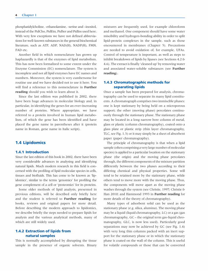
TLC, see Fig. 1.3) or it may simply be a sheet of absorbent
paper (paper chromatography).
The principle of chromatography is that when a lipid
sample (often comprisingavery largenumberofmolecular
species) is applied to a particular location on the stationary
phase (the origin) and the moving phase percolates
through, the different components of themixture partition
differently between the two phases according to their
differing chemical and physical properties. Some will
tend to be retained more by the stationary phase, while
others tend to move more with the moving phase. Thus,
the components will move apart as the moving phase
washes through the system (see Christie, 1997; Christie &
Han 2010; and Hammond 1993 in Further reading for
more details of the theory of chromatography).
Many types of adsorbent solid can be used as the
stationary phase (e.g. silica, alumina). The moving phase
may be a liquid (liquid chromatography, LC) or a gas (gas
chromatography, GC – the original term gas-liquid chro-
matography, GLC, is now less used). Particularly good
separations may now be achieved by GC (see Fig. 1.4)
with very long thin columns packed with an inert sup-
port for the stationary phase or in which the stationary
phase is coated on the wall of the column. This is useful
for volatile compounds or those that can be converted
4 Chapter 1
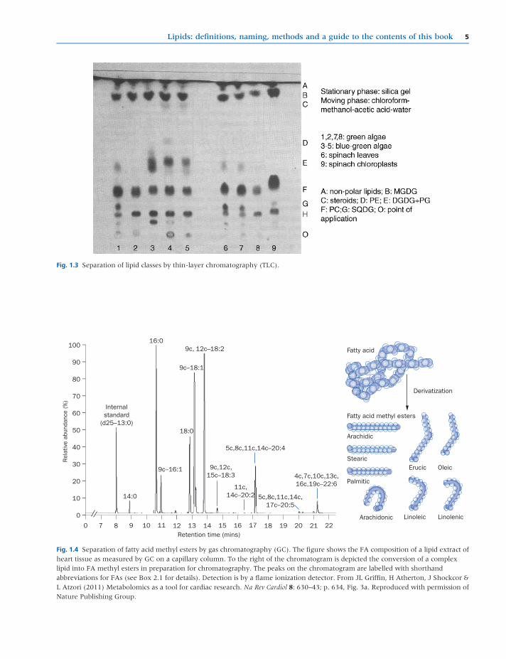
Fig. 1.3 Separation of lipid classes by thin-layer chromatography (TLC).
100 –
90 –
80 –
70 –
60 –
50 –
40 –
30 –
20 –
10 –
0 –
0
Internalstandard
(d25–13:0)
14:0
16:09c, 12c–18:2 Fatty acid
Derivatization
Arachidic
Stearic
Arachidonic Linoleic Linolenic
Erucic Oleic
9c–18:1
18:0
9c–16:1
5c,8c,11c,14c–20:4
4c,7c,10c,13c,16c,19c–22:6
9c,12c,15c–18:3
11c,14c–20:2 5c,8c,11c,14c,
17c–20:5
7 8Retention time (mins)
Rel
ativ
e ab
unda
nce
(%)
9 10 11 12 13 14 15 16 17 18 19 21 2220
Fatty acid methyl esters
Palmitic
Fig. 1.4 Separation of fatty acid methyl esters by gas chromatography (GC). The figure shows the FA composition of a lipid extract ofheart tissue as measured by GC on a capillary column. To the right of the chromatogram is depicted the conversion of a complexlipid into FA methyl esters in preparation for chromatography. The peaks on the chromatogram are labelled with shorthandabbreviations for FAs (see Box 2.1 for details). Detection is by a flame ionization detector. From JL Griffin, H Atherton, J Shockcor &L Atzori (2011) Metabolomics as a tool for cardiac research. Na Rev Cardiol 8: 630–43; p. 634, Fig. 3a. Reproduced with permission ofNature Publishing Group.
Lipids: definitions, naming, methods and a guide to the contents of this book 5
into more volatile ones, such as the methyl esters of FAs
(see Sections 2.1.8.1 & 11.2.4.2 for further details of the
preparation of FA methyl esters). For less volatile com-
plex lipids, LC in thin columns through which the mov-
ing phase is passed under pressure can produce superior
separations: this is called high performance liquid chro-
matography (HPLC).
Once the components have been separated, they can
be collected as they emerge from the column for further
identification and analysis (see Section 1.4.4). Com-
pounds separated on plates or strips can be eluted
from the stationary phase by solvents or analysed
in situ by various means. (Further information on meth-
ods of detection can be found in Christie & Han (2010)
and Kates (2010) in Further reading.)
The power of modern lipidomics has been made pos-
sible by the combination of GC or LC with improved
methods of mass spectrometry (MS) to provide detailed
and sophisticated analyses of complex natural lipid mix-
tures and this is the subject of the next section.
1.4.4 Modern lipidomics employs acombination of liquidchromatography or gaschromatography with massspectrometry to yield detailedprofiles of natural lipids – the‘lipidome’
While individual FAs can be readily measured by gas
chromatography-mass spectrometry (GC-MS), the com-
monestmethod to perform this analysis relies on cleaving
FAs from the head groups that they are associated with
and converting them into methyl esters by transester-
ification. This process is used to make the FAs volatile at
the temperature used by GC-MS, but during this process
information is lost, particularly about which lipid species
are enriched in a given FA.
An alternative is to use LC-MS. In this approach, lipid
extracts from biofluids and tissues can be analysed
directly. The lipids are dissolved in an organic solvent
and injected directly onto the HPLC column. Columns
can contain a variety of chemicals immobilized to form a
surface (stationary phase) that the analytes interact with.
For the analysis of lipids, columns containing long chains
of alkyl groups are most commonly used, in particular 8C
and 18C columns, which have side-chain lengths of 8
and 18 carbons, respectively. The most commonly used
HPLC method is referred to as ‘reverse phase’, whereby
lipids are initially loaded onto a HPLC column and then
the HPLC solvent is varied from something that is pre-
dominantly aqueous to a solvent that is predominantly
organic, across what is termed a gradient. The solvents
are referred to as the mobile phases. During this process,
lipids are initially adsorbed on to the stationary phase,
until their solubility increases to the point that they begin
to dissolve in themobile phase. In this manner, polar and
nonpolar lipids can readily be separated and typically, in
a lipid extract, lipid molecular species would elute in the
order of nonesterified fatty acids (NEFAs), phospholipids,
cholesteryl esters and TAGs. The chromatography serves
two important purposes. Firstly, it reduces the complex-
ity of the subsequent mass spectra generated by the mass
spectrometer, making metabolite identification more
convenient. Secondly, some metabolites can ionize
more readily than others and this can produce an effect
called ‘ion suppression’ where one metabolite ionizes
more easily and reduces the energy available for the
ionization of other species. As a result, the mass spec-
trometer may detect only the metabolite that ionizes
readily and miss the other metabolites that do not readily
form ions.
LC-MS is most commonly used with ‘electrospray ion-
ization’ where the analytes are introduced to the mass
spectrometer in the form of a spray of solvent. They are
accelerated over an electric field across the capillary that
introduces them into the mass spectrometer and the
nebulization of the spray is often assisted by the flow of
an inert gas. The inert gas causes the solvent to evaporate
(desolvate), producing a fine spray of droplets. As the
solvent evaporates, charges build up in the droplets until
they explode into smaller droplets, finally producing an
ion that is introduced into the mass spectrometer. While
this may sound relatively destructive, this form of ioniza-
tion is relatively ‘soft’, ensuring that the molecule itself or
an adduct (a combination of the molecule and another
charged species such as H+, Na+, K+ or other ions present
in the solvent) is formed. The ions are then detected by the
mass spectrometer (Fig. 1.5).
While there are numerous designs of mass spectrome-
ter, two common methods are often used in lipidomics.
In high resolutionMS, the mass accuracy achievable is so
great that chemical formulae can be determined with
reasonable precision. This is because only carbon-12 has
a mass of exactly 12 atomic mass units, while other
nuclides all have masses that slightly differ from a whole
number. These mass deficits can be used to predict what
6 Chapter 1

nuclides are present and estimate a small number of
chemical formulas that may be responsible for the ion.
The accuracy of modern high resolution mass spectrom-
eters is so high, often less than 3–5 parts per million, that
it is possible in lipidomics to determine what species are
being detected by their exact mass and references to
databases such as LIPID MAPS (http://www.lipidmaps.
org/). However, even in cases where only one formula is
identified this could still belong to a range of potential
lipid species. For example, if we take the PtdCho (36:2 –
i.e. total FA chains of 36 carbon atoms with a total of 2
double bonds), this could be due to a PtdCho containing
two C18:1 FAs, one C18:0 and one C18:2 or a variety of
other isomers. To further define the chemical structure,
fragmentation can be performed. In this process the ion is
accelerated through a low pressure of inert gas, produc-
ing collisions and fragmentation of the parent ion. The
daughter fragments can then be used to work out the
parent structure, with head groups and FAs commonly
being lost in the process (Fig. 1.6).
In the other form of commonly used lipidomics, a triple
quadrupole mass spectrometer is used. In this instru-
ment, the mass spectrometer consists of three electro-
magnet gates called quadrupoles. The first is used to
select for one ion, which is usually the parent ion of
the lipid species being detected. The second quadrupole
acts as a fragmentation cell where the ion is fragmented.
The third quadrupole then selects a particular fragment
ion. While many lipid species may have the same parent
mass, it is very unlikely that they will fragment in the
same manner and thus this method is highly selective. In
addition, these instruments can be made to be quantita-
tive and are particularly appropriate for targeted analyses
where a limited number of species is to be assayed.
Furthermore, in an approach termed ‘shotgun lipido-
mics’, the assay can be set up to scan for particular lipid
Phospholipid
Lysophospholipid Triglyceride
Fatty acid
100 –
90 –
80 –
70 –
60 –
50 –
40 –
30 –
20 –
10 –
0 –
0 21
0.03
0.86
1.40
1.62
2.15
3.98
4.26
4.40
4.67 5.27
5.90
5.35
5.44
7.21
8.57
8.79
9.02
9.25 12.807.76
8.346.45
6.94
5.13
3Retention time (mins)
Rel
ativ
e ab
unda
nce
(%)
4 5 6 7 8 9 10 11 12 13 14 15
Gly
cero
l
Gly
cero
l
Gly
cero
l
Phospholipid head group
Fig. 1.5 Separation and identification of heart lipidome by liquid chromatography-mass spectrometry (GC-MS). Intact lipids from anextract of heart tissue have been separated, detected and identified by GC-MS. Chromatography separates the intact lipids accordingto their polarity and high resolution MS identifies individual lipid molecular species. From JL Griffin, H Atherton, J Shockcor & LAtzori (2011) Metabolomics as a tool for cardiac research. Nat Rev Cardiol 8: 630–43; p. 634, Fig. 3b. Reproduced with permission ofNature Publishing Group.
Lipids: definitions, naming, methods and a guide to the contents of this book 7

species either by virtue of the head group present (e.g.
scanning for PtdCho species) or particular FAs (e.g.
identifying lipid species that contain a particular FA
such as arachidonic acid, all-c5,8,11,14-20:4, n-6).
More detailed accounts of these methods can be found
in Further reading.
1.5 A guide to the contents of this book
The purpose of this section is to provide a ‘roadmap’ to
enable students to find their way around and make best
use of the information provided in this book.
Continuing the scheme adopted in this chapter, each
chapter is divided into numbered sections; the first number
of thesectionwill indicate thechapternumber.Therewill be
extensive cross-referencing between sections within chap-
ters and between chapters. Although there are severalways
wecouldhavearranged the successionof chapters,wehope
that the one we have chosen will be a logical one.
At the end of each chapter there is a ‘Key points’
section that provides a concise summary of the
principal information in the chapter. This is followed
by a section on ‘Further reading’, which provides a
selection of useful reviews and also some original
research publications to give students a flavour of
important and exciting recent advances. Although
items in Further reading will be referenced through-
out each chapter, there are limited references to specific
pieces of literature in the main text. The number of
references in Further reading could not be unlimited.
We have attempted to cite those most useful that were
available at the time of writing but additional references
and/or diagrams are available on the companion web-
site. Information in the text will be supplemented with
figures and tables, and ‘boxes’ will be used to provide
more detail on specific topics where inclusion in the
text might interfere with the flow.
Chapter 2 introduces the chemical structures of the
different types of lipids in three sections. These deal with
(1) FAs, (2) lipids mainly involved in energy storage and
(3) those predominantly associated with cellular mem-
branes and also involved in physiological processes such
100 –
90 –
80 –
70 –
60 –
50 –
40 –
30 –
20 –
10 –
0 –
0
502.3
503.3
506.3520.3
[–H20]
18:0/18:2 [M–C18:0]
[M–C18:2]
521.3 526.3
525.3
O
OH HO
O
OP
O
N+
CH3
CH3H3C
524.3
507.3
504.3
505500 510
Rel
ativ
e ab
unda
nce
(%)
515 520 525 525 530
O
O
HO
HO
O
OP
O
N+
CH3
CH3H3C
H3C
H3C
O
Mass-to-charge ratio (m/z)
Fig. 1.6 Fragmentation of two phosphocholines derived from phosphatidylcholines (PtdChos). This figure demonstrates the furthercharacterization of the lipidome by the technique of ‘tandem MS’. One of the main challenges of LC-MS is lipid identificationbecause of the large numbers of isomers present. In this technique, chromatography is dispensed with altogether and the sample isdirectly infused into a high resolution MS instrument. Figure 1.6 illustrates the identification of two phosphocholine isomersproduced by fragmentation of PtdChos that would have been esterified with 18:0/18:2 and 18:1/18:1 respectively. From JL Griffin,H Atherton, J Shockcor & L Atzori (2011) Metabolomics as a tool for cardiac research. Nat Rev Cardiol 8: 630–43; p. 636, Fig. 4a.Reproduced with permission of Nature Publishing Group.
8 Chapter 1

as cell signalling. Of course there will be overlap between
these functions: it is impossible (and undesirable) fully to
compartmentalize lipid forms and functions. These sec-
tions discuss how the chemical structures of lipids relate
to their physical and physiological properties and point
the way to aspects of their metabolism, function and
utilization in subsequent chapters. The FA section con-
tains Box 2.1, which provides useful information on the
complex topic of FA nomenclature.
Chapter 3 covers the metabolism of the FAs. This starts
with their biosynthesis and discusses up-to-date knowl-
edge of biosynthetic pathways, the enzymes involved in
their biosynthesis and the genes coding for them. The
degradation of FAs by oxidative pathways is then dis-
cussed in detail with particular reference to the genera-
tion of metabolic energy. A key section in this and other
chapters concerns the all-important matter of how these
metabolic pathways are controlled and integrated.
In the discussions of the biosynthesis of the poly-
unsaturated fatty acids (PUFAs) and their subsequent oxi-
dation to form physiologically active products such as the
‘eicosanoids’, reference will be made to later chapters that
describe the role of suchmolecules in cell signalling (Chap-
ter 8) and as mediators in such physiological processes as
immunity and the implications for health and disease
(Chapter 10). Some PUFAs described in Chapter 3 are
essential components of the diet (‘essential fatty acids’,
EFAs) and their roles will be discussed further in Chapter 6.
Chapter 4. Just as Chapter 3 discusses the metabolism
of the FAs, Chapter 4 deals with the metabolism of the
complex lipids. Many of these (TAGs, phosphoglycerides,
glycosylglycerides etc.) are FA esters of glycerol but the
chapter also covers the sphingolipids (derivatives of the
base sphingosine rather than glycerol, many of which
incorporate sugars in the molecule) and the isoprenoids
(also called terpenoids), in which the sterols, such as
cholesterol and the plant steroids, are included.
The formation of TAGs is related to their role in energy
storage in adipose tissue. This has ramifications for influ-
ence of dietary fats on the fat stores (Chapter 6) and on
the relationships between the energy stores and health
problems such as obesity, insulin resistance, diabetes,
immune function, cancer and cardiovascular diseases,
all of which are discussed in more detail in Chapter 10.
Numerous seed oils of commercial importance store
TAGs as an energy source. This too, has implications
for the type and amounts of lipids in the diet (Chapter 6),
their implications for health (Chapter 10) and their
biotechnological modification to provide useful products
(Chapter 11).
An important section in this chapter discusses the
many lipases (see Sections 4.2 & 4.6) that degrade lipids.
Some are involved in the digestion of dietary lipids
(Chapter 7), many others are involved in modifying
the FA composition of lipids to suit the needs of particular
cell types and cell structures (Chapters 4, 5, 7, 9 & 10),
others are utilized in biotechnological processes (Chap-
ter 11) and yet others are involved in the release of
components of lipid molecules that are destined to
become cell-signalling molecules (Chapters 8 & 10).
Failure to degrade certain glycolipids, mainly owing to
gene defects, can result in several lethal diseases of the
nervous system that are addressed in Chapter 10.
Failure in the regulation of the metabolism of choles-
terol in human beings, as a result of gene defects (Chap-
ters 4 & 7) or dietary imbalance (Chapters 7 & 10) has
implications for cardiovascular diseases that are also
explored in Chapter 10.
Chapter 4 also mentions the biosynthesis of lipids that
have specific functions and points the way to more
detailed discussion in later chapters – for example the
platelet activating factor and the lung surfactant lipid in
Chapter 10.
Chapter 5 discusses the various ways in which dif-
ferent lipids can associate with each other and with
proteins as a result of their chemical and physical
properties. Such lipid assemblies are crucial to the
structure and function of cells and cell organelles
and in this chapter, we explore what is currently
known about how lipids have shaped the evolution
of living cells. Light is cast on the way in which, for
example, the evolution of the bacteria and the archaea
depended on the development of lipids with quite
different chemical structures. Of particular importance
is the development of different types of membranes
whose lipid composition is crucial to their functions.
Membranes are important for the topics discussed in
each of the chapters of this book because of their role
in cell structure, function and integrity, as a location
for many metabolic pathways, their involvement in
inter- and intracellular signalling processes, in the
trafficking of biochemical substances within and
between cells and because the development of many
disease processes results from defects in the integrity of
many membranes. As well as their presence in mem-
branes, lipids accumulate as droplets (LDs) in cells (see
Lipids: definitions, naming, methods and a guide to the contents of this book 9

Section 5.5) where they act as energy stores or sources
of molecules involved in the mediation of metabolic
processes. Some lipid assemblies are involved in
processes outside cells, for example in the formation
of surface layers with barrier properties (see Sec-
tion 2.2.4) or, as lipoproteins, in the transport of lipids
in the bloodstream (see Sections 7.2 & 10.5).
Chapter 6 discusses the types of lipids in food and the
diet and their biological roles. (Chapter 7, which follows,
explains how these dietary lipids are digested, absorbed
and the digestion products transported in the blood to the
tissues of the body.) These two chapters are devoted
mainly to human diets but there is also discussion of
other simple-stomached animals, such as rats, mice and
pigs, which are often used as so-called ‘animal models’.
This is because, in animal studies, procedures can be
more easily controlled and the experimental design
can be more rigorous. The disadvantage is that the
biochemistry and physiology may sometimes differ
between species, leaving open some doubt as to their
relevance to Man.
Much of the food we now eat is processed in some
way – industrially and domestically. There is some
discussion here of how such processes may affect die-
tary lipids but reference is made to Chapter 11, which
provides more detail on food processing and bio-
technological developments. Dietary fats provide meta-
bolic energy and although the subject is introduced
here, readers will find more detailed information in
Chapter 9. Dietary fats also supply many essential
nutrients. This chapter picks up on the EFAs – PUFAs
that are essential for health but cannot be made in the
body – that were first introduced in Chapters 2 and 3.
Also essential for good health are the fat-soluble vita-
mins, which are required in only milligram or micro-
gram quantities as distinct from the gram or almost
gram quantities of the EFAs. While knowledge of them
developed in the late 19th and early 20th centuries, it is
only in the last few decades that the full extent of their
physiological roles as, for example, hormones and signal-
ling molecules and regulators of metabolism has been
realized. The molecular biology revolution has indicated
the key involvement of some of them in the regulation of
gene expression. The chapter ends with a thorough dis-
cussion of the role of lipids in foetal and postnatal
development.
Chapter 7 describes in detail the processes by which
lipid components of the diet are digested and the
digestion products absorbed from different parts of the
intestinal tract. Once within the intestinal absorbing cells
(enterocytes) they are ‘remodelled’ and combined with
proteins (‘lipoproteins’) for transport around the body in
the bloodstream. The proteins not only help to solubilize
the lipids but also direct them to sites of further metabo-
lism. The different types of lipoproteins are described and
also the elaborate system for the control of their metab-
olism and their movement to appropriate tissues. Such a
complex system is vulnerable to defects either from gene
mutations or from ‘dietary overload’ and the reader is
pointed to Chapter 10, which describes the involvement
of various lipids in health and disease.
Chapter 8 is concerned entirely with molecules that
send signals to different cells of the body. The emphasis in
this chapter is mainly on two types: the phosphoinosi-
tides and the sphingolipids. Before the mid-1960s, lipids
were thought of as having three main biological func-
tions: as structural components of membranes, as energy
stores and as a barrier against the environment or pro-
viders of insulation. Phosphatidylinositol (PtdIns) was
already known as a widespread membrane component
but everything changed when it was discovered that
inositol phospholipids with additional phosphate groups
esterified in different positions on the inositol ring could,
when cells were stimulated by agonist molecules such as
hormones, be catabolized to yield compounds that sent
signals across the membrane that then resulted in a
variety of metabolic changes. Even a molecule such as
diacylglycerol (DAG), it was then discovered, could act as
a ‘messenger’. Similar roles were discovered also for a
variety of sphingolipids. Several other lipid molecules
with signalling functions are described in other chapters,
for example: platelet activating factor (PAF, an ‘ether’
phospholipid) in Chapters 4 and 10; endocannabinoids in
Chapters 4 and 8.
Chapter 9 is devoted entirely to the role of lipids as
energy stores in animals and plants. The first part goes
into detail in the animal storage organs – white and
brown adipose tissue. The white form is the main storage
tissue for TAGs; it is widely dispersed around the body
rather than being a discrete organ like the liver or brain. It
contains smaller amounts of other lipid molecules and as
well as a storage organ it is now known to have endo-
crine properties, producing hormones. The uptake of
TAGs into the fat cells and their mobilization for energy
supply is discussed in relation to the biochemistry already
described in Chapters 3 and 4.
10 Chapter 1

The cells of the brown form of adipose tissue contain
many small LDs (in contrast to white adipose tissue’s
unilocular droplet) and these are surrounded by
mitochondria that accept FAs released from the fat drop-
lets and oxidize them by the process of β-oxidation,which is described in detail in Chapter 3.
Lipid storage by some plants is important for supply-
ing the metabolic energy for seed development and
germination. The different storage locations – fruits,
seeds and pollen grains – and the types of lipids
involved, are described. Plant storage fats are impor-
tant in diets (Chapter 6) and require industrial proc-
essing (Chapter 11). New methods of introducing
genes for the biosynthesis of specific lipids that may
not be native to a particular plant are now becoming
available (Chapter 11).
Chapter 10 addresses the subject of lipids in health and
disease. It opens with a discussion of various inborn
errors of metabolism, describing the genetic background
and the implications for dietary lipids. There are relevant
pointers to other chapters in which the biochemical
basics are discussed (Chapters 3, 4, 7 & 9). A section
on cancer examines the influence of dietary lipids (both
in development and treatment), the roles of specific lipids
in physiological functions associated with cancer devel-
opment and the involvement of the immune system. A
whole section is devoted to the ways in which lipids may
be involved in aspects of immune function, including
theirmodification of gene expression. Once again there is
comprehensive referencing to the biochemistry of lipids
in Chapters 3, 4, 5, 7 and 9. The conditions of obesity and
diabetes (see also Chapter 9) and disorders of lipoprotein
metabolism (in their association with cardiovascular
diseases, Chapter 7) are similarly related to preceding
biochemical background (Chapters 3 & 4).
Chapter 11 discusses the industrial processing of
lipids and lipid-containing foods as well as how bio-
technology is being applied in the development of new
products with very specific properties. Nonfood aspects
include the properties and production of soaps, deter-
gents, biofuels and oleochemicals. Many of these topics
are introduced for the first time but reference is made
back to Chapter 7 when discussing the detergent prop-
erties of the bile salts. The functional properties of lipid-
based foods such as spreads are discussed in terms of
their enhancement of palatability and their role as
carriers for fat-soluble vitamins, with reference back
to Chapters 6 and 10. Foods that supply different types
of FAs and their relevance to health (e.g the n-3 PUFAs
and plant sterols) and disease (e.g. the trans-FAs) are
discussed with reference back to Chapters 6 and 10.
Finally, recent advances in the use of genetic modifica-
tion to produce crops and livestock with novel lipid
profiles are described.
KEY POINTS
• In contrast to carbohydrates, proteins and nucleic acids, lipids are defined on the basis of their physical properties (insolubility inwater) rather than on the basis of consistent chemical features. For this reason, the student will need to learn and remember awide range of different chemical types and their rather complex nomenclature.
• Lipids can usually be extracted easily from tissues by making use of their hydrophobic characteristics. However, such extractionsyield a complex mixture of different lipid classes which have to be purified further for quantitative analysis. Moreover, the crudelipid extract may be contaminated by other hydrophobic molecules, e.g. by intrinsic membrane proteins, and need to be‘cleaned up’.
• Of the various types of separation, thin layer and column chromatography are most useful for intact lipids. A powerful tool forquantitation of volatile lipids or derivatives is GC but HPLC has become increasingly used.
• Current research is increasingly concerned with identifying complete profiles of the extremely complex lipid constituents ofliving tissues – the so-called ‘lipidome’. Modern ‘lipidomics’ utilizes a combination of either GC-MS or LC-MS to define thelipidome.
• With this background to what lipids are and how they are studied, the ‘roadmap’ then guides the student through theremaining ten chapters.
Lipids: definitions, naming, methods and a guide to the contents of this book 11

Further reading
Ceve G, ed., (1993) Phospholipids Handbook, Marcel Dekker, Basel.
Christie WW (1989) Gas Chromatography and Lipids. The Oily
Press, Ayr, UK.
Christie WW, ed., (1997) Advances in Lipid Methodology, 4 vols.
The Oily Press, Ayr, UK.
Christie WW & Han X (2010) Lipid Analysis, 4th edn. The Oily
Press, Bridgwater, UK.
Fahy E, Subramaniam S, Murphy RC, et al. (2005) A compre-
hensive classification system for lipids. J Lipid Res, 46:839–61.
Griffin JL, Atherton H, Shockcor J & Atzori L (2011) Metab-
olomics as a tool for cardiac research. Na Rev Cardiol
8:630–43.
Gross RW&Han X (2009) Shotgun lipidomics of neutral lipids as
an enabling technology for elucidation of lipid-related dis-
eases, Am J Physiol Endoc-M 297 E297–303.
Gunstone FD, Harwood JL& Dijkstra AJ, eds. (2007) The Lipid
Handbook, 3rd edn. CRC Press, Boca Raton, USA.
Gurr MI, James AT, Harwood JL, et al. (1971, 1975, 1980, 1991,
2002)LipidBiochemistry:AnIntroduction, Editions1–4,Chapman&
Hall, London; Edition 5, Blackwell Science, Oxford, UK.
Hamilton RJ& Hamilton S, eds. (1992) Lipid Analysis: A Practical
Approach, IRL Press, Oxford, UK.
Hammond EW (1993) Chromatography for the Separation of Lipids.
CRC Press, Boca Raton, USA.
International Union of Biochemistry and Molecular Biology
(1992) Biochemical Nomenclature and Related Documents, 2nd
edn. Portland Press, London, UK.
IUPAC–IUB Commission on Biochemical Nomenclature (1989)
Eur J Biochem 186:429–58.
Kates M (1986) Techniques of Lipidology, 2nd edn. Elsevier Sci-
ence, Amsterdam, The Netherlands. (This classic book lacks
details about recent advances (e.g. HPLC) but still contains a
wealth of basic information.)
Kates M (2010) Techniques of Lipidology, 3rd edn. Newport Som-
erville Innovation Ltd, Ottawa, Canada.
Leray C (2013) Introduction to Lipidomics: From Bacteria to Man.
CRC Press, Boca Raton, USA.
Lipid Library (http://lipidlibrary.aocs.org)
Nomenclature Committee of the International Union of Bio-
chemistry (1984) Enzyme Nomenclature, Academic Press, Lon-
don, UK. (The most up-to-date information on enzyme
nomenclature can be found by accessing: http://www.chem.
qmw.ac.uk/iubmb/enzyme/ (last accessed 4 December 2015).)
Nygren H, Seppänen-Laakso T, Castillo S, Hyötyläinen T&Oresic M (2011) Liquid chromatography-mass spectrometry (LC-
MS)-based lipidomics for studies of body fluids and tissues.
Method Mol Biol 708:247–57.
Roberts LD, Koulman A & Griffin JL (2014) Methods for per-
forming lipidomics in white adipose tissue, Method Enzymol
538:211–31.
12 Chapter 1