changes in bacterial number at different sites of oral ... · j appl oral sci. abstract submitted:...
TRANSCRIPT

J Appl Oral Sci.
Abstract
Submitted: October 26, 2017Modification: January 15, 2018
Accepted: January 29, 2018
Changes in bacterial number at different sites of oral cavity during perioperative oral care management in gastrointestinal cancer patients: preliminary study
Objective: The objective of this study was to clarify differences in bacterial accumulation between gastrointestinal cancer patients who underwent severely invasive surgery and those who underwent minimally invasive surgery. Material and Methods: We performed a preliminary investigation of gastrointestinal cancer patients who were treated at the Department of Surgery, Takarazuka Municipal Hospital, from 2015 to 2017 (n=71; 42 laparoscopic surgery, 29 open surgery) to determine changes in bacterial numbers at different sites of the oral cavity (tongue dorsum, gingiva of upper anterior teeth, palatoglossal arch), as well as mouth dryness and tongue coating indices. Specifically, patients received professional tooth cleaning (PTC), scaling, tongue cleaning, and self-care instruction regarding tooth brushing from a dental hygienist a day before the operation. Professional oral health care was also performed by a dental hygienist two and seven days after surgery. Oral bacteria numbers were determined using a bacterial counter with a dielectrophoretic impedance measurement method. Results: The number of bacteria at all three examined sites were significantly higher in the open surgery group when compared to the laparoscopic surgery group on the second postoperative day. Relevantly, bacterial count in samples from the gingiva of the upper anterior teeth remained greater seven days after the operation in patients who underwent open surgery. Furthermore, the dry mouth index level was higher in the open surgery group when compared to the laparoscopic surgery group on postoperative days 2 and 7. Conclusions: Even with regular oral health care, bacterial numbers remained high in the upper incisor tooth gingiva in gastrointestinal cancer patients who received open surgery. Additional procedures are likely needed to effectively reduce the number of bacteria in the gingival area associated with the upper anterior teeth.
Keywords: Oral health. Perioperative care. Bacterial count. Gastrointestinal cancer.
Tomoko KAWANO1,2
Hideo SHIGEISHI1
Eri FUKADA1
Takamichi YANAGISAWA2
Nobukazu KURODA3
Toshinobu TAKEMOTO4
Masaru SUGIYAMA1
Original Articlehttp://dx.doi.org/10.1590/1678-7757-2017-0516
1Hiroshima University, Graduate School of Biomedical & Health Sciences, Program of Oral Health Sciences, Department of Public Oral Health, Hiroshima, Japan.2Takarazuka Municipal Hospital, Department of Dentistry & Oral Surgery, Takarazuka, Japan.3Takarazuka Municipal Hospital, Department of Surgery, Takarazuka, Japan.4Hiroshima University, Graduate School of Biomedical & Health Sciences, Program of Oral Health Sciences, Department of Oral Health Management, Hiroshima, Japan.
Corresponding address:Hideo Shigeishi
Department of Public Oral Health -Program of Oral Health Sciences -
Graduate School of Biomedical & Health Sciences - Hiroshima University - 1-2-3 - Kasumi, Minami-ku -
Hiroshima - 734-8553 - Japan.Phone: +81-82-257-5945 - Fax: +81-82-257-5945
e-mail: [email protected]
2018;26:e201705161/12
J Appl Oral Sci. 2018;26:e201705162/12
Introduction
Performing dental treatments, such as periodontal
and dental caries treatments, extraction of non-
preservable teeth and replacement of missing teeth,
are required before starting cancer treatment to reduce
postoperative complications. In addition, perioperative
professional oral health care is recognized to be
important for cancer patients18,26.
Colorectal cancer is a predominant type of cancer
and the third leading cause of cancer-associated death
worldwide1, while gastric cancer is also one of the most
common types of malignancies throughout the world2.
Surgical resection is the primary procedure to treat
patients with gastrointestinal cancer, with laparoscopic
surgery being widely adopted for gastrointestinal
cancers, such as gastric and colorectal cancer10,20.
There are reports that perioperative oral care may
improve respiratory function in patients who undergo
gastrointestinal surgery by reducing the number of
bacteria species25, indicating that perioperative oral
health care may play a significant role in reducing
post-operative complications in those patients.
We consider that gastrointestinal cancer patients
undergoing severely invasive surgery (i.e., open
surgery) may have worse postoperative oral hygiene
when compared to those who undergo minimally
invasive surgery (i.e., laparoscopic surgery), thus
oral health care may help to reduce infectious
diseases in those patients. However, few reports
have shown differences on the oral hygiene status
between patients who underwent severely invasive
gastrointestinal surgery and those who underwent
minimally invasive surgery. In this study we performed
a preliminary investigation of changes in bacterial
numbers at different sites of the oral cavity, including
the tongue dorsum, gingiva of the upper anterior
teeth, and palatoglossal arch, as well as dry mouth
and tongue coating indices in gastrointestinal cancer
patients undergoing surgical treatment who received
perioperative oral care. In addition, inflammatory
response and nutritional status were investigated in
these cases.
Material and methods
SubjectsWe registered 71 patients (55 men, 16 women;
mean age of 72.0 years, ranging from 39 to 97 years)
with gastrointestinal cancer who underwent surgery
at the Department of Surgery, Takarazuka Municipal
Hospital, from November 2015 to February 2017 and
received perioperative oral health care. The study
design was approved by the Ethics Committee of the
Takarazuka Municipal Hospital and all patients signed
an informed consent form. Those who suffered from
tumor recurrence and received chemotherapy were
excluded. Clinical data obtained included patient
age, sex, tumor grade, medical history, blood loss
volume, operation duration time and hospitalization
duration. Tumors were classified using the TNM
staging system according to the seventh edition of
the American Joint Committee on Cancer Staging
Manual6. We divided patients based on their surgical
treatment into 2 groups, as follows. The minimally
invasive surgery group was comprised of those who
underwent laparoscopic surgery (n=42; 12 gastric,
22 colon, 8 rectal cancer), while the severely invasive
surgery group included patients who underwent open
surgical resection (n=29; 20 gastric, 6 colon, 3 rectal
cancer). Cephem antibiotics (cefazolin at 1.0 g/day
for gastric cancer patients or cefmetazole at 1.0 g/
day for rectal cancer patients) were administered to
all patients 1-2 days after the operation to prevent
surgical site infection. The minimum effective dosage
and duration of the antibiotics administrations
were determined based on previous reports by the
randomized control trial (RCT)8,12. None of the selected
patients had problems associated with denture wearing
(i.e., instability, unfitness, fracture). Furthermore,
none wore their appliance during the fasting period,
during this period it was kept in clean water. Following
the fasting period, each participant washed their
denture and cleaned it with a soft-bristle brush after
taking a meal, resting it in clean water overnight.
Clinicopathological factors in the gastrointestinal
cancer patients are summarized in Table 1.
Oral health careThe protocol for oral care given to the patients
was standardized among all dental hygienists who
participated in this study. Each patient received self-
care instruction on tooth brushing and tongue cleaning,
Changes in bacterial number at different sites of oral cavity during perioperative oral care management in gastrointestinal cancer patients: preliminary study

J Appl Oral Sci. 2018;26:e201705163/12
Clinical factors Laparoscopic surgery (42) Open surgery (29) p-value
Sex
Men (55) 34 (61.8%) 21 (38.2%) 0.57
Women (16) 8 (50.0%) 8 (50.0%)
Age in years
<65 (14) 12 (85.7%) 2 (14.3%) 0.051
≥65 (57) 30 (52.6%) 27 (47.4%)
Tumor stage
Stage I/II (39) 27 (69.2%) 12 (30.8%) 0.096
Stage III/IV (32) 15 (46.9%) 17 (53.1%)
BMI (kg/m2) 22.3±2.9 20.6±2.9 0.28
Number of remaining teeth
≥20 (31) 18 (58.1%) 13 (41.9%) 0.65
10–19 (16) 11 (68.8%) 5 (31.3%)
0–9 (24) 13 (54.2%) 11 (45.8%)
Average number of remaining teeth 15.4±8.7 14.4±9.6 0.71
Denture
Non-user (28) 19 (67.9%) 9 (32.1%) 0.25
Partial denture (37) 21 (56.8%) 16 (43.2%)
Full denture (6) 2 (33.3%) 4 (66.7%)
Smoking
Non-smoker (37) 17 (45.9%) 20 (54.1%) 0.055
Former smoker (25) 19 (76.0%) 6 (24.0%)
Current smoker (9) 6 (66.7%) 3 (33.3%)
Diabetes
(-) (55) 35 (77.8%) 20 (22.2%) 0.26
(+) (16) 7 (43.8%) 9 (56.3%)
Hypertension
(-) (40) 26 (65.0%) 14 (35.0%) 0.38
(+) (31) 16 (51.6%) 15 (48.4%)
Cardiovascular disease
(-) (64) 38 (59.4%) 26 (40.6%) 1.0
(+) (7) 4 (57.1%) 3 (42.9%)
Respiratory disease
(-) (65) 39 (60.0%) 26 (40.0%) 0.68
(+) (6) 3 (50.0%) 3 (50.0%)
Cerebrovascular disease
(-) (65) 38 (58.5%) 27 (41.5%) 1.0
(+) (6) 4 (66.7%) 2 (33.3%)
Renal disease
(-) (64) 37 (57.8%) 27 (42.2%) 0.69
(+) (7) 5 (71.4%) 2 (28.6%)
Operation time (min) 278.2±123.0 309.0±121.3 0.46
Blood loss volume (ml) 116.7±150.8 590.5±556.8 <0.001
Fasting duration (day) 2.9±1.2 4.9±2.1 <0.001
Hospitalization duration (day) 18.6±6.7 31.9±16.9 <0.001
Fisher’s exact test or Mann-Whitney’s U test was used for statistical analysis, with p<0.05 were considered statistically significant
Table 1- Clinical factors of gastrointestinal cancer patients
KAWANO T, SHIGEISHI H, FUKADA E, YANAGISAWA T, KURODA N, TAKEMOTO T, SUGIYAMA M
J Appl Oral Sci. 2018;26:e201705164/12
as well as scaling and professional tooth cleaning (PTC)
by a dental hygienist at the Oral Surgery Department a
day before the operation. Two days after the operation
the patients received oral health care and self-care
instruction in the inpatient ward by a dental hygienist,
then seven days after the operation oral health care
such as PTC was given again. In addition, a 0.025%
benzalkonium chloride solution was used to disinfect
the dorsum of the tongue and upper incisor gingiva.
Oral bacterial countThe number of oral bacteria in patient samples
were determined using a Bacterial Counter (Panasonic
Healthcare Co., Ltd., Tokyo, Japan), with the
dielectrophoretic impedance change converted to
bacterial concentration per ml for each sample
[colony-forming units (CFU)/ml]. This device ensures
that only viable bacteria are counted. The samples
were obtained from the tongue dorsum, gingiva of
the upper anterior teeth, and palatoglossal arch, also
known as the fauces, using a cotton swab, according
to the instructions of the manufacturer. The number
of oral bacteria were determined before and after oral
health care on preoperative day 1, and again prior to
oral care on postoperative days 2 and 7. The samples
were collected from 2:00 to 3:00 PM on preoperative
day 1, and at 8:00 AM on postoperative days 2 and
7. Furthermore, the same dental hygienist with
experience to use the apparatus obtained all samples
to maintain reproducibility.
Evaluation of dry mouthChanges in dry mouth, also known as xerostomia,
and subjective symptoms (i.e., severely dry,
moderately dry, slightly dry, moderately wet, fully
wet) were evaluated based on interviews performed
on preoperative day 1, and on postoperative days 2
and 7. In addition, dry mouth was also classified as
normal, light (increased saliva viscosity), moderate
(small bubbles observed in saliva), and severe (dry
tongue surface without saliva), using a previously
reported method14.
Tongue coating indexTongue coating index (TCI) values were determined
on preoperative day 1, and postoperative days 2 and 7.
To determine both the extension and thickness of the
tongue coating, we used a detailed index previously
proposed29. With this method, the tongue dorsum is
divided into 9 sections, including 1 middle and 2 lateral
areas for each of the posterior, middle, and anterior
thirds of the tongue. The presence of tongue coating
was classified as none (Score 0), light-thin (Score 1,
pink color underneath coating visible), and heavy-
thick coating (Score 2, no pink color observed) for
each section. We obtained a personal tongue coating
index by adding the scores for all 9 sections (total
score 0-18).
Clinical examinationsWe examined clinical markers such as body
temperature (BT), white blood cell (WBC) count, and
C-reactive protein (CRP) level to evaluate inflammatory
response 1 day before, and 1, 3, and 7 days after the
surgery. Increased WBC count is commonly related to
inflammatory response, while CRP is a major plasma
protein which presents dramatically elevated levels
during acute-phase inflammation. In addition, albumin
(ALB) level is considered a reliable marker of nutritional
status in patients following surgery24. We also noted
the occurrence of complications after surgery, such
as surgical site infection (SSI), anastomotic leakage,
and aspiration pneumonia.
Statistical analysisWe used the Wilcoxon signed-rank test to compare
paired data as a non-parametric analysis alternative to
a paired t-test, statistically significant p-values were
considered p<0.05. We also used Mann-Whitney’s U
test and Fisher’s exact test for statistical analysis.
Results
Clinical factors of the gastrointestinal cancer patients
The clinical factors of the gastrointestinal cancer
patients are summarized in Table 1. There were no
statistical differences for sex, age, tumor stage, body
mass index (BMI), number of remaining teeth, or
operation duration between the laparoscopic surgery
and open surgery groups, however, blood loss volume
was significantly higher in the open surgery group
(p<0.001 Mann-Whitney’s U test). Two of 29 patients
in the laparoscopic surgery group and 11 of 42 in the
open surgery group received a blood transfusion. In
addition, the open surgery group had a significantly
longer average length of fasting and hospital stay than
the laparoscopic surgery group (4.9±2.1 vs. 2.9±1.2
Changes in bacterial number at different sites of oral cavity during perioperative oral care management in gastrointestinal cancer patients: preliminary study

J Appl Oral Sci. 2018;26:e201705165/12
days and 31.9±16.9 vs. 18.6±6.7 days, respectively)
(p<0.001 and p<0.001, respectively, Mann-Whitney’s
U test).
Inflammatory response and ALB level in laparoscopic surgery and open surgery groups
The results of WBC count, CRP level, BT, and ALB
are summarized in Table 2. There was a significant
difference in mean WBC count between the groups
at 1 day after surgery (p=0.026, Mann-Whitney’s
U test) (Figure 1A). Mean CRP in the laparoscopic
surgery group was lower than that in the open surgery
group at all moments after surgery. In addition, on
postoperative days 1 and 3, the level of CRP was
significantly lower in the laparoscopic surgery group
(p<0.001 and p=0.020, respectively, Mann-Whitney’s
Perioperative day Day 1 Day 3 Day 7
WBC (/mm3)
Laparoscopic surgery group 5548.6±1833.0 8210.0±3427.4 7451.8±2371.4 6507.1±3363.8
Open surgery group 5185.8±2299.3 9776.3±3229.8 8787.0±3815.4 9089.9±5910.5
CRP (mg/dl)
Laparoscopic surgery group 0.37±0.74 5.87±3.52 7.98±7.72 3.17±4.04
Open surgery group 0.34±0.48 8.67±3.15 11.7±6.08 4.31±4.16
BT (oC)
Laparoscopic surgery group 36.5±0.32 37.1±0.68 36.7±0.60 36.6±0.40
Open surgery group 36.4±0.57 37.3±0.68 37.0±0.48 36.9±0.62
ALB (g/dl)
Laparoscopic surgery group 4.08±0.48 3.06±0.46 3.19±0.50 3.20±0.46
Open surgery group 3.43±0.73 2.40±0.46 2.28±0.66 2.58±0.50
Table 2- Changes in WBC count, CRP level, BT, and ALB
Figure 1- Changes in (A) WBC count, (B) CRP level, (C) BT, and (D) ALB in laparoscopic surgery and open surgery groups. Error bars represent the mean±SD. There were statistically significant differences between the groups. *p<0.05, ***p<0.001
KAWANO T, SHIGEISHI H, FUKADA E, YANAGISAWA T, KURODA N, TAKEMOTO T, SUGIYAMA M
J Appl Oral Sci. 2018;26:e201705166/12
U test) (Figure 1B). Regarding mean BT, there was a
statistically significant difference between the groups
on postoperative days 3 and 7 (p=0.028 and p=0.017,
respectively, Mann-Whitney’s U test) (Figure 1C). The
mean ALB level was significantly lower in the open
surgery group at each moment (p<0.001 for each,
Mann-Whitney’s U test) (Figure 1D). No patients from
either group suffered from any postoperative infectious
complications (e.g., SSI, anastomotic leak, aspiration
pneumonia).
Comparison of number of oral bacteria between laparoscopic surgery and open surgery groups
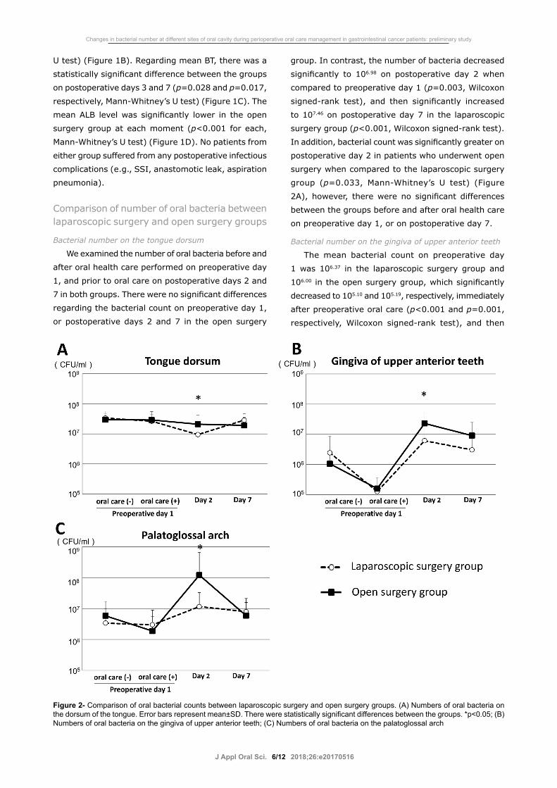
Bacterial number on the tongue dorsum
We examined the number of oral bacteria before and
after oral health care performed on preoperative day
1, and prior to oral care on postoperative days 2 and
7 in both groups. There were no significant differences
regarding the bacterial count on preoperative day 1,
or postoperative days 2 and 7 in the open surgery
group. In contrast, the number of bacteria decreased
significantly to 106.98 on postoperative day 2 when
compared to preoperative day 1 (p=0.003, Wilcoxon
signed-rank test), and then significantly increased
to 107.46 on postoperative day 7 in the laparoscopic
surgery group (p<0.001, Wilcoxon signed-rank test).
In addition, bacterial count was significantly greater on
postoperative day 2 in patients who underwent open
surgery when compared to the laparoscopic surgery
group (p=0.033, Mann-Whitney’s U test) (Figure
2A), however, there were no significant differences
between the groups before and after oral health care
on preoperative day 1, or on postoperative day 7.
Bacterial number on the gingiva of upper anterior teeth
The mean bacterial count on preoperative day
1 was 106.37 in the laparoscopic surgery group and
106.00 in the open surgery group, which significantly
decreased to 105.10 and 105.19, respectively, immediately
after preoperative oral care (p<0.001 and p=0.001,
respectively, Wilcoxon signed-rank test), and then
Figure 2- Comparison of oral bacterial counts between laparoscopic surgery and open surgery groups. (A) Numbers of oral bacteria on the dorsum of the tongue. Error bars represent mean±SD. There were statistically significant differences between the groups. *p<0.05; (B) Numbers of oral bacteria on the gingiva of upper anterior teeth; (C) Numbers of oral bacteria on the palatoglossal arch
Changes in bacterial number at different sites of oral cavity during perioperative oral care management in gastrointestinal cancer patients: preliminary study

J Appl Oral Sci. 2018;26:e201705167/12
significantly increased to 106.79 and 107.36, respectively,
on postoperative day 2 (p<0.001, Wilcoxon signed-rank
test). In the open surgery group, the mean bacterial
count significantly decreased on postoperative day 7
when compared to day 2 (p=0.027, Wilcoxon signed-
rank test). On postoperative day 2, the mean bacterial
count was significantly higher in the open surgery
group (P<0.001, Mann-Whitney’s U test) (Figure 2B)
and showed a trend to be higher on postoperative day
7, when compared to the laparoscopic surgery group.
Furthermore, in the open surgery group, the levels of
oral bacteria remained high in the gingiva of the upper
anterior teeth even after undergoing postoperative
oral health care by a dental hygienist and performing
self-care procedures.
Bacterial count on the palatoglossal arch
The mean bacterial count on the palatoglossal arch
of the open surgery group increased significantly to
108.08 on postoperative day 2 when compared to after
oral health care on preoperative day 1 (p<0.001,
Wilcoxon signed-rank test), and then decreased
significantly to 106.77 on postoperative day 7 (p=0.001,
Wilcoxon signed-rank test). The bacterial count in
the laparoscopic surgery group increased to 107.06
on postoperative day 2 when compared to after oral
care on preoperative day 1. The mean bacterial count
was significantly higher in the open surgery group
when compared to the laparoscopic surgery group
on postoperative day 2 (p<0.001, Mann-Whitney’s U
test) (Figure 2C). Both groups presented very similar
values on postoperative day 7.
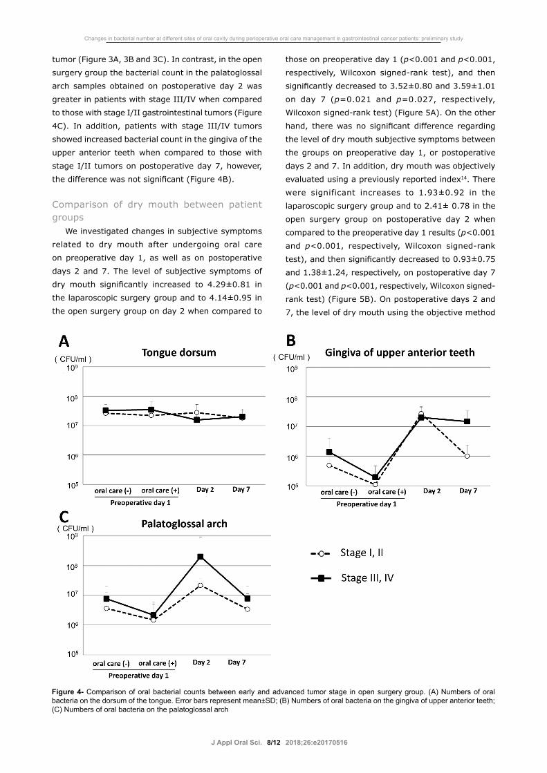
Comparison of number of oral bacteria between early and advanced tumor stage
We compared the number of oral bacteria between
patients with early and advanced tumor stage in both
groups. For the laparoscopic surgery group there was
no significant difference regarding bacterial count
between those with a stage I/II and a stage III/IV
Figure 3- Comparison of oral bacterial counts between early and advanced tumor stage in laparoscopic surgery group. (A) Numbers of oral bacteria on the dorsum of the tongue. Error bars represent mean±SD; (B) Numbers of oral bacteria on the gingiva of upper anterior teeth; (C) Numbers of oral bacteria on the palatoglossal arch
KAWANO T, SHIGEISHI H, FUKADA E, YANAGISAWA T, KURODA N, TAKEMOTO T, SUGIYAMA M
J Appl Oral Sci. 2018;26:e201705168/12
tumor (Figure 3A, 3B and 3C). In contrast, in the open
surgery group the bacterial count in the palatoglossal
arch samples obtained on postoperative day 2 was
greater in patients with stage III/IV when compared
to those with stage I/II gastrointestinal tumors (Figure
4C). In addition, patients with stage III/IV tumors
showed increased bacterial count in the gingiva of the
upper anterior teeth when compared to those with
stage I/II tumors on postoperative day 7, however,
the difference was not significant (Figure 4B).
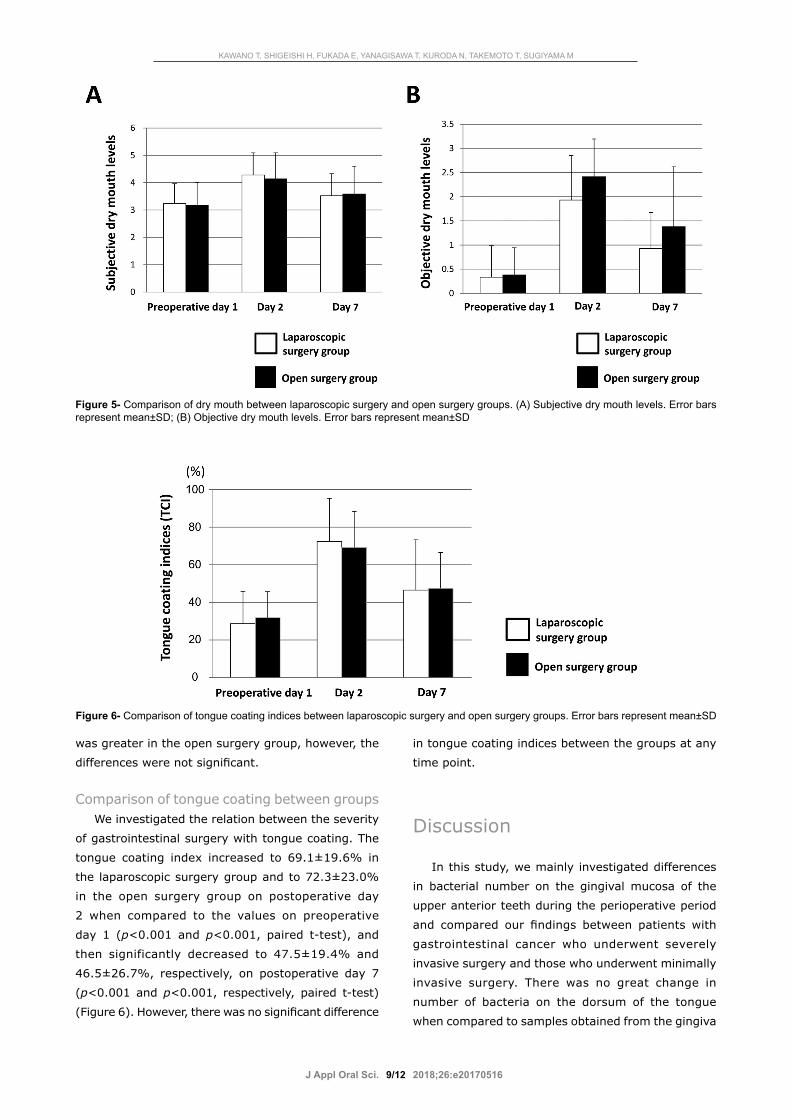
Comparison of dry mouth between patient groups
We investigated changes in subjective symptoms
related to dry mouth after undergoing oral care
on preoperative day 1, as well as on postoperative
days 2 and 7. The level of subjective symptoms of
dry mouth significantly increased to 4.29±0.81 in
the laparoscopic surgery group and to 4.14±0.95 in
the open surgery group on day 2 when compared to
those on preoperative day 1 (p<0.001 and p<0.001,
respectively, Wilcoxon signed-rank test), and then
significantly decreased to 3.52±0.80 and 3.59±1.01
on day 7 (p=0.021 and p=0.027, respectively,
Wilcoxon signed-rank test) (Figure 5A). On the other
hand, there was no significant difference regarding
the level of dry mouth subjective symptoms between
the groups on preoperative day 1, or postoperative
days 2 and 7. In addition, dry mouth was objectively
evaluated using a previously reported index14. There
were significant increases to 1.93±0.92 in the
laparoscopic surgery group and to 2.41± 0.78 in the
open surgery group on postoperative day 2 when
compared to the preoperative day 1 results (p<0.001
and p<0.001, respectively, Wilcoxon signed-rank
test), and then significantly decreased to 0.93±0.75
and 1.38±1.24, respectively, on postoperative day 7
(p<0.001 and p<0.001, respectively, Wilcoxon signed-
rank test) (Figure 5B). On postoperative days 2 and
7, the level of dry mouth using the objective method
Figure 4- Comparison of oral bacterial counts between early and advanced tumor stage in open surgery group. (A) Numbers of oral bacteria on the dorsum of the tongue. Error bars represent mean±SD; (B) Numbers of oral bacteria on the gingiva of upper anterior teeth; (C) Numbers of oral bacteria on the palatoglossal arch
Changes in bacterial number at different sites of oral cavity during perioperative oral care management in gastrointestinal cancer patients: preliminary study
J Appl Oral Sci. 2018;26:e201705169/12
was greater in the open surgery group, however, the
differences were not significant.
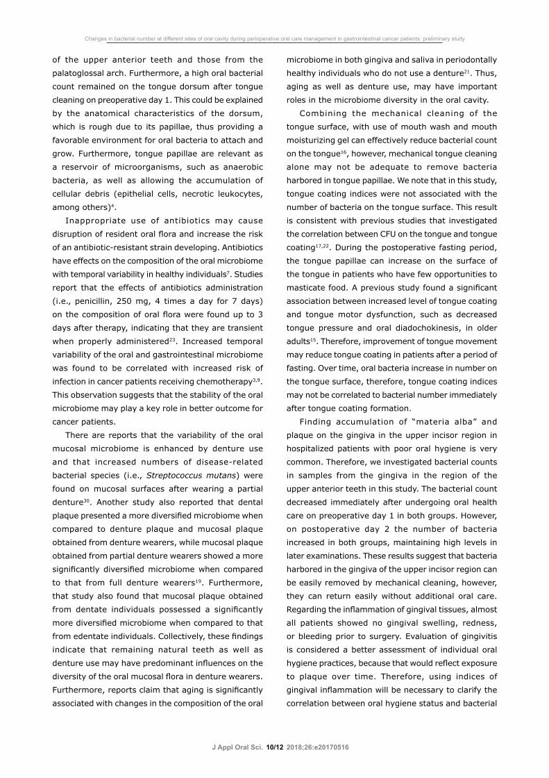
Comparison of tongue coating between groupsWe investigated the relation between the severity
of gastrointestinal surgery with tongue coating. The
tongue coating index increased to 69.1±19.6% in
the laparoscopic surgery group and to 72.3±23.0%
in the open surgery group on postoperative day
2 when compared to the values on preoperative
day 1 (p<0.001 and p<0.001, paired t-test), and
then significantly decreased to 47.5±19.4% and
46.5±26.7%, respectively, on postoperative day 7
(p<0.001 and p<0.001, respectively, paired t-test)
(Figure 6). However, there was no significant difference
in tongue coating indices between the groups at any
time point.
Discussion
In this study, we mainly investigated differences
in bacterial number on the gingival mucosa of the
upper anterior teeth during the perioperative period
and compared our findings between patients with
gastrointestinal cancer who underwent severely
invasive surgery and those who underwent minimally
invasive surgery. There was no great change in
number of bacteria on the dorsum of the tongue
when compared to samples obtained from the gingiva
Figure 5- Comparison of dry mouth between laparoscopic surgery and open surgery groups. (A) Subjective dry mouth levels. Error bars represent mean±SD; (B) Objective dry mouth levels. Error bars represent mean±SD
Figure 6- Comparison of tongue coating indices between laparoscopic surgery and open surgery groups. Error bars represent mean±SD
KAWANO T, SHIGEISHI H, FUKADA E, YANAGISAWA T, KURODA N, TAKEMOTO T, SUGIYAMA M
J Appl Oral Sci. 2018;26:e2017051610/12
of the upper anterior teeth and those from the
palatoglossal arch. Furthermore, a high oral bacterial
count remained on the tongue dorsum after tongue
cleaning on preoperative day 1. This could be explained
by the anatomical characteristics of the dorsum,
which is rough due to its papillae, thus providing a
favorable environment for oral bacteria to attach and
grow. Furthermore, tongue papillae are relevant as
a reservoir of microorganisms, such as anaerobic
bacteria, as well as allowing the accumulation of
cellular debris (epithelial cells, necrotic leukocytes,
among others)4.
Inappropriate use of antibiotics may cause
disruption of resident oral flora and increase the risk
of an antibiotic-resistant strain developing. Antibiotics
have effects on the composition of the oral microbiome
with temporal variability in healthy individuals7. Studies
report that the effects of antibiotics administration
(i.e., penicillin, 250 mg, 4 times a day for 7 days)
on the composition of oral flora were found up to 3
days after therapy, indicating that they are transient
when properly administered23. Increased temporal
variability of the oral and gastrointestinal microbiome
was found to be correlated with increased risk of
infection in cancer patients receiving chemotherapy3,9.
This observation suggests that the stability of the oral
microbiome may play a key role in better outcome for
cancer patients.
There are reports that the variability of the oral
mucosal microbiome is enhanced by denture use
and that increased numbers of disease-related
bacterial species (i.e., Streptococcus mutans) were
found on mucosal surfaces after wearing a partial
denture30. Another study also reported that dental
plaque presented a more diversified microbiome when
compared to denture plaque and mucosal plaque
obtained from denture wearers, while mucosal plaque
obtained from partial denture wearers showed a more
significantly diversified microbiome when compared
to that from full denture wearers19. Furthermore,
that study also found that mucosal plaque obtained
from dentate individuals possessed a significantly
more diversified microbiome when compared to that
from edentate individuals. Collectively, these findings
indicate that remaining natural teeth as well as
denture use may have predominant influences on the
diversity of the oral mucosal flora in denture wearers.
Furthermore, reports claim that aging is significantly
associated with changes in the composition of the oral
microbiome in both gingiva and saliva in periodontally
healthy individuals who do not use a denture21. Thus,
aging as well as denture use, may have important
roles in the microbiome diversity in the oral cavity.
Combining the mechanical cleaning of the
tongue surface, with use of mouth wash and mouth
moisturizing gel can effectively reduce bacterial count
on the tongue16, however, mechanical tongue cleaning
alone may not be adequate to remove bacteria
harbored in tongue papillae. We note that in this study,
tongue coating indices were not associated with the
number of bacteria on the tongue surface. This result
is consistent with previous studies that investigated
the correlation between CFU on the tongue and tongue
coating17,22. During the postoperative fasting period,
the tongue papillae can increase on the surface of
the tongue in patients who have few opportunities to
masticate food. A previous study found a significant
association between increased level of tongue coating
and tongue motor dysfunction, such as decreased
tongue pressure and oral diadochokinesis, in older
adults15. Therefore, improvement of tongue movement
may reduce tongue coating in patients after a period of
fasting. Over time, oral bacteria increase in number on
the tongue surface, therefore, tongue coating indices
may not be correlated to bacterial number immediately
after tongue coating formation.
Finding accumulation of “materia alba” and
plaque on the gingiva in the upper incisor region in
hospitalized patients with poor oral hygiene is very
common. Therefore, we investigated bacterial counts
in samples from the gingiva in the region of the
upper anterior teeth in this study. The bacterial count
decreased immediately after undergoing oral health
care on preoperative day 1 in both groups. However,
on postoperative day 2 the number of bacteria
increased in both groups, maintaining high levels in
later examinations. These results suggest that bacteria
harbored in the gingiva of the upper incisor region can
be easily removed by mechanical cleaning, however,
they can return easily without additional oral care.
Regarding the inflammation of gingival tissues, almost
all patients showed no gingival swelling, redness,
or bleeding prior to surgery. Evaluation of gingivitis
is considered a better assessment of individual oral
hygiene practices, because that would reflect exposure
to plaque over time. Therefore, using indices of
gingival inflammation will be necessary to clarify the
correlation between oral hygiene status and bacterial
Changes in bacterial number at different sites of oral cavity during perioperative oral care management in gastrointestinal cancer patients: preliminary study

J Appl Oral Sci. 2018;26:e2017051611/12
count in a future study.
Saliva has several roles including oral cleaning (i.e.,
washing away food debris and plaque) and antibacterial
functions, which are associated with the regulation of
the number of bacteria harbored in the oral cavity13.
Additionally, the bacterial buffering capacity of saliva
varies at different sites in the oral cavity27,28. Reduced
salivary flow may have had effects on the number of
oral bacteria during the postoperative fasting period
in the patients. Generally, dry mouth is related to an
increase in the number of oral bacteria11. In our study,
the peak of xerostomia was found at 2 days after the
operation in both groups, likely because most had no
opportunity to gargle during the postsurgical period.
Xerostomia and elevated numbers of bacteria in the
gingiva of the upper incisor region may be correlated.
Furthermore, the decrease in activities of daily living
associated with recovery after surgery may have
been associated with poor cleaning of the gingiva in
the upper incisor region. Some of the open surgery
patients remained in bed for 3 or more days after
the operation, thus having a longer period of fasting,
which may have caused the higher bacterial count in
the open surgery group when compared to the count
on the laparoscopic surgery group on postoperative
days 2 and 7.
Usually, mechanical cleaning should not be applied
to the fauces, because it can initiate vomiting reflex. In
this study, even though oral health care of the fauces
was not performed, there was no distinct difference
in bacterial number in the palatoglossal arch samples
before and after the operation in the laparoscopic
surgery group. On the other hand, the bacterial
number in those samples increased significantly
in the open surgery group on postoperative day
2, returning to nearly the same number as the
preoperative number on postoperative day 7. This
increased bacterial count in the open surgery group
may have been caused by the worsened dry mouth
condition induced by staying in bed for a long duration
without oral self-care. Inadequate salivary flow was
shown to contribute to severe dry mouth, which
led to development of mucositis and colonization of
oropharyngeal bacteria in intubated intensive care
unit patients5. These observations suggest that the
improvement of dry mouth may reduce the number
of bacteria in the fauces and oropharyngeal regions.
Thus, perioperative oral care performed by a dental
hygienist is considered as vital to improve the oral
health status (i.e., improvement of dry mouth) in
patients who have difficulty to perform self-care.
The CRP level was significantly lower in the
laparoscopic surgery group on postoperative days
1 and 3 when compared to the open surgery group,
indicating that severe damage caused by the open
surgery procedures led to distinct differences in CRP
between the groups. After undergoing severely invasive
surgery, patients with advanced-stage gastrointestinal
tumors seem to have a greater increase in bacterial
number when compared to those with early-stage
tumors. The patients with advanced-stage tumors of
the open surgery group showed longer durations of bed
resting (2.87±0.77 vs. 1.62±1.63 days) and fasting
(5.44±2.13 vs. 4.15±1.63 days) after the operation
than those with early-stage tumors. As a result, those
with an advanced-stage tumor likely did not perform
self-care and had a decrease in the cleaning function
of their saliva, resulting in an increased number of
oral bacteria. We note that the decreased ability to
perform physical activity soon after the procedure
may have been associated with a reduced amount
of oral self-care performed by patients treated for
an advanced stage tumor. In conclusion, there
was a significant difference for oral bacterial count
between patients who underwent laparoscopic surgery
and those who received abdominal open surgery
during the perioperative period, which included oral
management treatments. Even when regular oral care
was performed, the level of oral bacteria remained
high in the gingiva of the upper anterior teeth in the
gastrointestinal cancer patients who underwent open
surgery. We speculate that the gingiva of the upper
incisor region is less susceptible to anti-bacterial
functions of saliva when compared to other regions
in the oral cavity. Additional procedures are likely
needed to effectively reduce the bacterial number in
gingiva in that region. Additional studies are required
to develop evidence-based perioperative oral health
care procedures.
References1- Arnold M, Sierra MS, Laversanne M, Soerjomataram I, Jemal A, Bray F. Global patterns and trends in colorectal cancer incidence and mortality. Gut. 2017;66(4):683-91.2- Balakrishnan M, George R, Sharma A, Graham DY. Changing trends in stomach cancer throughout the world. Curr Gastroenterol Rep. 2017;19(8):36.
KAWANO T, SHIGEISHI H, FUKADA E, YANAGISAWA T, KURODA N, TAKEMOTO T, SUGIYAMA M

J Appl Oral Sci. 2018;26:e2017051612/12
3- Chen X, Winckler B, Lu M, Cheng H, Yuan Z, Yang Y, et al. Oral microbiota and risk for esophageal squamous cell carcinoma in a high-risk area of China. PLoS One. 2015;10:e0143603.4- Danser MM, Gómez SM, Van der Weijden GA. Tongue coating and tongue brushing: a literature review. Int J Dent Hyg. 2003;1(3):151-8.5- Dennesen P, van der Ven A, Vlasveld M, Lokker L, Ramsay G, Kessels A, et al. Inadequate salivary flow and poor oral mucosal status in intubated intensive care unit patients. Crit Care Med. 2003;31(3):781-6.6- Edge SB, Byrd DR, Compton CC, Fritz AG, Greene FL, Trotti A, editors. AJCC cancer staging manual. 7th ed. New York: Springer; 2010.7- Flores GE, Caporaso JG, Henley JB, Rideout JR, Domogala D, Chase J, et al. Temporal variability is a personalized feature of the human microbiome. Genome Biol. 2014;15(12):531.8- Fujita S, Saito N, Yamada T, Takii Y, Kondo K, Ohue M, et al. Randomized, multicenter trial of antibiotic prophylaxis in elective colorectal surgery: single dose vs. 3 doses of a second-generation cephalosporin without metronidazole and oral antibiotics. Arch Surg. 2007;142(7):657-61.9- Galloway-Peña JR, Smith DP, Sahasrabhojane P, Wadsworth WD, Fellman BM, Ajami NJ, et al. Characterization of oral and gut microbiome temporal variability in hospitalized cancer patients. Genome Med. 2017;9(1):21.10- Gholami S, Cassidy MR, Strong VE. Minimally invasive surgical approaches to gastric resection. Surg Clin North Am. 2017;97(2):249-64.11- Guobis Ž, Kareivienė V, Basevičienė N, Paipalienė P, Niedzelskienė I, Sabalys G, et al. Microflora of the oral cavity in patients with xerostomia. Medicina (Kaunas). 2011;47(12):646-51.12- Haga N, Ishida H, Ishiguro T, Kumamoto K, Ishibashi K, Tsuji Y, et al. A prospective randomized study to assess the optimal duration of intravenous antimicrobial prophylaxis in elective gastric cancer surgery. Int Surg. 2012;97(2):169-76.13- Kagami H, Wang S, Hai B. Restoring the function of salivary glands. Oral Dis. 2008;14(1):15-24.14- Kakinoki Y, Nishihara T, Arita M, Shibuya K, Ishikawa M. Usefulness of new wetness tester for diagnosis of dry mouth in disabled patients. Gerodontology. 2004;21(4):229-31.15- Kikutani T, Tamura F, Nishiwaki K, Suda M, Kayanaka H, Machida R, et al. The degree of tongue-coating reflects lingual motor function in the elderly. Gerodontology. 2009;26(4):291-6.16- Kobayashi K, Ryu M, Izumi S, Ueda T, Sakurai K. Effect of oral cleaning using mouthwash and a mouth moisturizing gel on bacterial number and moisture level of the tongue surface of older adults requiring nursing care. Geriatr Gerontol Int. 2017;17(1):116-21.17- Mantilla Gómez S, Danser MM, Sipos PM, Rowshani B, van der Velden U, van der Weijden GA. Tongue coating and salivary bacterial counts in healthy/gingivitis subjects and periodontitis patients. J Clin Periodontol. 2001;28(10):970-8.
18- National Institutes of Health Consensus Development Conference on Oral Complications of Cancer Therapies: Diagnosis, Prevention, and Treatment. Bethesda, Maryland, April 17-19, 1989. NCI Monogr. 1990;(9):1-184.19- O'Donnell LE, Robertson D, Nile CJ, Cross LJ, Riggio M, Sherriff A, et al. The oral microbiome of denture wearers is influenced by levels of natural dentition. PLoS One. 2015;10(9):e0137717.20- Parker JM, Feldmann TF, Cologne KG. Advances in laparoscopic colorectal surgery. Surg Clin North Am. 2017;97(3):547-60.21- Percival RS, Challacombe SJ, Marsh PD. Age-related microbiological changes in the salivary and plaque microflora of healthy adults. J Med Microbiol 1991;35(1):5-11.22- Quirynen M, Mongardini C, van Steenberghe D. The effect of a 1-stage full-mouth disinfection on oral malodor and microbial colonization of the tongue in periodontitis. A pilot study. J Periodontol. 1998;69(3):374-82.23- Sanders CC, Sanders WE Jr, Harrowe DJ. Bacterial interference: effects of oral antibiotics on the normal throat flora and its ability to interfere with group A streptococci. Infect Immun. 1976;13(3):808-12.24- Santos NS, Draibe SA, Kamimura MA, Canziani ME, Cendoroglo M, Júnior AG, et al. Is serum albumin a marker of nutritional status in hemodialysis patients without evidence of inflammation? Artif Organs. 2003;27(8):681-6.25- Sato M, Yoshihara A, Miyazaki H. Preliminary study on the effect of oral care on recovery from surgery in elderly patients. J Oral Rehabil. 2006;33(11):820-6.26- Shigeishi H, Ohta K, Fujimoto S, Nakagawa T, Mizuta K, Ono S, et al. Preoperative oral health care reduces postoperative inflammation and complications in oral cancer patients. Exp Ther Med. 2016;12(3):1922-8.27- Suse K, Tanase K, Eda T, Kataumi T, Miki A, Nakamura K, et al. Salivary clearance of acid and the pH in the different regions of the mouth. Dent Oral Craniofac Res. 2017;3(3):1-4.28- Suzuki A, Watanabe S, Ono Y, Ohashi H, Pai C, Xing X, et al. Influence of the location of the parotid duct orifice on oral clearance. Arch Oral Biol. 2009;54(3):274-8.29- Winkel EG, Roldán S, Van Winkelhoff AJ, Herrera D, Sanz M. Clinical effects of a new mouthrinse containing chlorhexidine, cetylpyridinium chloride and zinc-lactate on oral halitosis. A dual-center, double-blind placebo-controlled study. J Clin Periodontol. 2003;30(4):300-6.30- Zhu X, Wang S, Gu Y, Li X, Yan H, Yan H, et al. Possible variation of the human oral bacterial community after wearing removable partial dentures by DGGE. World J Microbiol Biotechnol. 2012;28(5):2229-36.
Changes in bacterial number at different sites of oral cavity during perioperative oral care management in gastrointestinal cancer patients: preliminary study