changed expression of e-cadherin and galectin-9 in oral...
TRANSCRIPT

Asian Pacific Journal of Cancer Prevention, Vol 15, 2014 2145
DOI:http://dx.doi.org/10.7314/APJCP.2014.15.5.2145Changed Expression of E-cadherin and Galectin-9 in Oral SCCs but no Potential as Prognostic Markers
Asian Pac J Cancer Prev, 15 (5), 2145-2152
Introduction
Oral squamous cell carcinoma (OSCC) ranks among the top ten most common cancers worldwide (Stewart and Kleihues, 2008; Rao et al., 2013). The survival rate for OSCC has remained generally unchanged in the past three decades due to late detection despite the curious fact that the oral cavity can be readily examined (Neville and Day, 2002; Rao et al., 2013). One promising strategy for the treatment of OSCC and other cancers, which has developed as a result of breakthrough in the fields of molecular biology, cancer genetics, and cancer biology, is molecular targeted therapy (Sahu and Grandis, 2011). These molecular markers are also being explored for use in developing clinical diagnostic aids, detecting malignant cells in biopsies, predicting recurrence at tumour surgical margins, diagnosing unsuspected nodal metastasis and to serve as adjuncts to routine histopathological examination to aid prognostication and effective management (Scully and Bagan, 2009). Some biomarkers are already being used as clinical tools but it is still in a juvenile stage, hence the need for more biomarkers to be developed (Balan et al., 2010). Cadherins are a class of type-1 trans-membrane 1Department of Oro-Maxillofacial Surgical and Medical Sciences, 2Oral Cancer Research and Coordinating Center, Faculty of Dentistry, University of Malaya, Kuala Lumpur, 3Department of Oral & Maxillofacial Surgery, Hospital Tengku Ampuan Rahimah, Selangor, Malaysia *For correspondence: [email protected]
Abstract
Background: The survival rate for oral squamous cell carcinoma (OSCC) has remained generally unchanged in the past three decades, underlining the need for more biomarkers to be developed to aid prognostication and effective management. The prognostic potential of E-cadherin expression in OSCCs has been variable in previous studies while galectin-9 expression has been correlated with improved prognosis in other cancers. The aim of the present study was to investigate the expression of galectin-9 and E-cadherin in OSCC and their potential as prognostic biomarkers. Materials and Methods: E-cadherin and Galectin-9 expression was examined by immunohistochemistry in 32 cases of OSCC of the buccal mucosa (13 with and 19 without lymph node metastasis), as well as 6 samples of reactive lesions and 5 of normal buccal mucosa. Results: The expression of E-cadherin in OSCC was significantly lower than the control tissues but galectin-9 expression was conversely higher. Median E-cadherin HSCOREs between OSCCs positive and negative for nodal metastasis were not significantly different. Mean HSCOREs for galectin-9 in OSCC without lymph node metastasis (127.7±81.8) was higher than OSCC with lymph node metastasis (97.9±62.9) but this difference was not statistically significant. Conclusions: E-cadherin expression is reduced whilst galectin-9 expression is increased in OSCC. However, the present results suggest that E-cadherin and galectin-9 expression may not be useful as prognostic markers for OSCC. Keywords: E-cadherin - galectin-9 - oral squamous cell carcinoma - prognostic markers
RESEARCH ARTICLE
Changed Expression of E-cadherin and Galectin-9 in Oral Squamous Cell Carcinomas but Lack of Potential as Prognostic MarkersSiew Wui Chan1*, Thomas George Kallarakkal1,2, Mannil Thomas Abraham3
proteins known for its calcium-dependent cell-cell adhesion property (Marsh and Brackenbury, 1996). E-cadherins are members of this protein family found in epithelial cells which serve important roles in cell adhesion by ensuring that cells within tissues are bound together (Alberts et al., 2002). The loss of E-cadherin function or expression has been implicated in cancer progression and metastasis (Christofori and Semb, 1999). Galectins are a family of beta-galactoside-binding proteins (Leffler et al., 2004) which are generally present in the cytoplasm and in the nucleus as well as extracellularly on the cell surface and especially within the extracellular matrix (Cooper and Barondes, 1999). Galectin-9 was identified as a potent T cell derived eosinophil chemoattractant (Matsumoto et al., 1998). Since eosinophil accumulation has been linked to a better prognosis for OSCC (Dorta et al., 2002), it is hypothesized that expression of Galectin-9 is related to a better prognosis (Hirashima, 2003). In addition, Galectin-9 induces adhesion and aggregation of certain cell types and can be a prognostic factor in patients with melanoma and breast cancer (Hirashima, 2003). Although the prognostic potential of E-cadherin expression has been widely studied, conflicting results

Siew Wui Chan et al
Asian Pacific Journal of Cancer Prevention, Vol 15, 20142146
cast doubts over its reliability and hamper its recognition as a biomarker. Galectins are a group of promising and relatively untapped source in the exhaustive quest for potential biomarkers in cancers. Galectin-9 expression has already been correlated with improved prognosis in malignant melanoma and breast cancer. The aim of this study was to investigate the expression of Galectin-9 and E-cadherin in OSCC and their potential as prognostic markers.
Materials and Methods
Study design The present study was a cross-sectional study investigating the expression of E-cadherin and Galectin-9 in OSCC in comparison to epithelium of normal mucosa and reactive lesions. Expression of E-cadherin and Galectin-9 was also compared in OSCC with and without lymph node involvement. Ethical clearance was duly obtained from the relevant authorities: DF OP1008/0043(P).
Tissue samples Formalin-fixed paraffin-embedded (FFPE) tumour tissues of histologically diagnosed cases of OSCC were obtained from the archives of the Department of Oro-Maxillofacial Surgical and Medical Sciences and Oral Cancer Research and Coordinating Center (OCRCC). Clinicopathological details of all suitable cases were recovered from the departmental registry and reviewed. Available samples from the year 2000 till June 2012 which satisfied all inclusion criteria were selected.
Inclusion criteria Our study group consisted of cases of primary OSCC which had not undergone radiotherapy or chemotherapy, as evidenced by previous biopsy reports or clinical notes. This was to avoid therapy-induced changes which would have interfered with targeted tumour cells in subsequent immunohistochemical staining (Suzuki et al., 2004; Schneider et al., 2005). Any previous treatment would also have altered normal lymphatic drainage, resulting in unusual distribution of regional spread of disease (Edge et al., 2010). Only OSCCs of the buccal mucosa were chosen as an attempt to standardize samples because prognosis as indicated by the involvement of lymph nodes may change according to different tumour sites (Woolgar, 2006). Buccal mucosa refers to all membranous lining of inner surface of the cheeks and lips from the line of contact between opposing lips to the line of attachment of mucosa of the alveolar ridge (upper and lower) and pterygomandibular raphe (Edge et al., 2010). Degree of differentiation was not emphasized because histological grade alone is weakly correlated to outcome (Woolgar, 2006). Excisional biopsies or surgically removed specimens of OSCC with report on lymph node involvement (when relevant) were selected because the prognostic importance of presence or absence of lymph node metastasis, which is the focus of our study, has been recognized for decades
(Woolgar, 2006).
Exclusion criteria Cases with incomplete or ambiguous documented evidence for the above inclusion criteria were excluded. Sample size PS (Power and Sample Size Calculation) Software Version 3.0.43 was used to calculate the sample size for this study. Based on the expected mean difference for immunostaining score between patients with and without lymph node metastasis of 53 (Li et al., 2012), at least 23 cases in each sample group would be needed to be able to reject the null hypothesis that the staining score means of both sample groups are equal with the probability (power) of 0.8 (80%). The Type I error probability associated with this test of the null hypothesis is 0.05 (p=0.05). In fact, based on previous studies in which sample size ranged from 8 (Yamada et al., 1997) to 83 (Liu et al., 2010) for E-cadherin and 9 (Pioche-Durieu et al., 2005) to 65 (Alves et al., 2011) for Galectin-9, significant difference was elicited in samples as small as 30 (Mahomed et al., 2007). Due to various constraints, only 34 OSCC cases which fulfilled the above criteria were available when this research was initiated. Of these, 14 cases exhibited positive lymph node metastasis and 20 without metastasis. However, two cases had to be excluded after the immunohistochemical staining because the amount of tissue remaining was inadequate to allow assessment of our markers. Therefore at final count, our sample groups were as follows: i) 13 OSCC cases with lymph node metastasis; ii)19 OSCC cases without lymph node metastasis; iii) 6 samples of oral mucosa from reactive lesions (fibroepithelial polyp); iv) 5 samples of normal buccal mucosa (taken with consent from patients undergoing surgical removal of impacted third molar)
Antibodies Dako monoclonal mouse anti-human antibody against E-cadherin, clone NCH-38 (DAKO Mo a hu E-Cadherin, clone NCH-38) and Abcam rabbit polyclonal antibody to Galectin-9 were purchased from Bita Lifescience Sdn Bhd with PPP grant P0035/2010B.
Immunohistochemical staining Immunohistochemical (IHC) staining was performed with antibodies to Galectin-9 and E-cadherin by the Immunoperoxidase Envision technique on sections of FFPE tissues from the above mentioned sample groups. Optimization was achieved for Galectin-9 at 1:1000 dilution and E-cadherin at 1:100 dilution with Dako REALTM Antibody Diluent. Sections were heated at 60oC for 1 hour in the microwave oven after which they were deparaffinized in xylene and rehydrated in graded ethanol according to the standard procedure. Antigen retrieval was performed by incubating sections immersed in Tris-EDTA at pH 9.0 in a decloaking chamber at 121oC for 30 seconds after which the sections were left to cool at room temperature for 20 minutes before they were washed in running tap water for

Asian Pacific Journal of Cancer Prevention, Vol 15, 2014 2147
DOI:http://dx.doi.org/10.7314/APJCP.2014.15.5.2145Changed Expression of E-cadherin and Galectin-9 in Oral SCCs but no Potential as Prognostic Markers
5 minutes. Sections were subjected to endogenase peroxidase blocking with Dako REALTM Peroxidase-Blocking Solution for 10 minutes in a humidified chamber. Sections were later washed in 2 baths of Phosphate Buffered Saline pH 7.4 (PBS). Next, sections were incubated with primary antibodies (antibodies to E-cadherin or Galectin-9) for 30 minutes at room temperature. After the requisite PBS rinse, sections were incubated in a second antibody and subsequently staining was visualized using the Dako REALTM EnVisionTM Detection System, Peroxidase/DAB+, Rabbit/Mouse kit. The photosensitive 3,3’-diaminobenzidine (DAB) forms a brown end product upon oxidation at the site of target antigen, which is interpreted as positive staining. Later, sections were washed gently in running water for 5 minutes. Sections were then counterstained with hematoxylin, dehydrated in increasing concentrations of ethanol, cleared in xylene before they were eventually mounted in DPX (Di-N-Butyle Phthalate in Xylene), a synthetic resin mounting media. Normal oral mucosal tissues were used as positive control for E-cadherin and Non-Hodgkin lymphoma (T cell lineage) for Galectin-9. For the negative controls, primary antibodies were omitted and replaced by TBS buffer.
IHC staining evaluation The intensity and percentage of stained tumour cells in each section were determined independently by two observers without prior knowledge of the clinical or histopathological data. Before evaluation, a calibration exercise was conducted between the two observers to reduce inter- and intra- observer discrepancies. The immunostaining intensity (Figure 1) was graded 0 for no detectable staining, 1 for weak staining, 2 for clearly positive staining and 3 for strongly positive staining (Irie et al., 2005). A semiquantitative index known as the HSCORE (histochemical score) (Kageshita et al., 2002) which incorporates both intensity and percentage of stained cells was used for staining evaluation. H-SCORE is a summation of the proportion of cell staining at each intensity multiplied by the intensity of the staining (Berchuck et al., 1989) represented by the following formula (Kitawaki et al., 1999; Liang et al., 2008): HSCORE=∑Pi ( i )
where i=intensity of staining (0-3) and Pi=percentage of stained cells for each given i (0-100%). A total of 500 tumour cells (100 cells each in 5 high power fields) were assessed in each section to calculate the HSCORE. This score theoretically ranges from a minimum of 0 in cases with no staining to a maximum of 300 in cases where all the tumour cells stain with maximal intensity. The final HSCORE is a mean of two values calculated by the two independent observers. Differences of greater than 10% were resolved by consensus (Berchuck et al., 1989).
Statistical analysis Statistical analysis was performed with a series of parametric and non-parametric tests using the SPSS package (version 15) to investigate the expression of the markers (Galectin-9 and E-cadherin) in OSCCs compared to control group, as well as association between the staining pattern of the markers and the metastatic potential of OSCC (lymph node status). A P-value of less than 0.05 was considered to be statistically significant.
Results
Clinicopathological details Our OSCC cases had a mean age at presentation of 60 years with a range of 36 to 89 years. Of the 32 patients, an overwhelming majority of 87.5% was female and likewise Indians constituted a great percentage at 81.3%. The histopathological grading in our cases was vastly imbalanced with moderately differentiated OSCC amounting to 22 cases, 9 cases being well-differentiated and only a singular case of poorly differentiated OSCC. 13 of our cases exhibited positive metastasis to the lymph nodes as opposed to 19 without lymph node involvement. In addition, there were 11 cases of normal and reactive tissues, consisting of 5 normal buccal mucosa and 6 fibroepithelial hyperplasia (FEH), considered collectively as the control group.
Expression of E-cadherin and Galectin-9 in OSCC of buccal mucosa in comparison to normal oral mucosa and reactive lesions Close examination of the epithelium from the control
Figure 1. Immunostaining Intensity for Galectin-9 (A-D) and E-cadherin (E-H) is Graded 0 for No Detectable Staining (A, E), 1 for Weak Staining (B, F), 2 for Clearly Positive Staining (C, G) and 3 for Strongly Positive Staining (D, H). [X400 magnification]
Figure 2. Distribution of E-cadherin Expression in Epithelium from the Control Group was Concentrated at the Basal 2/3 of the Epithelium
Siew Wui Chan et al
Asian Pacific Journal of Cancer Prevention, Vol 15, 20142148
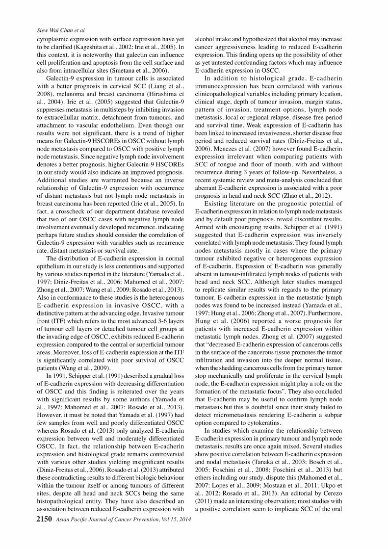
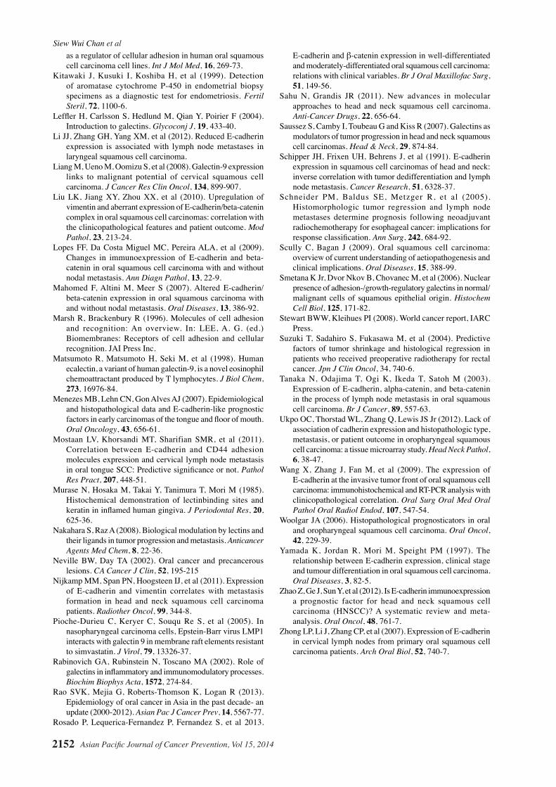
group revealed strong, positive membranous staining for E-cadherin at the basal and parabasal layer (Figure 2). The staining gradually lost its intensity as it approached the superficial spinous layer and the keratotic layer. In general, E-cadherin staining comprised approximately the basal 2/3 of the epithelium, excluding the basal surface. E-cadherin showed similar membranous staining of the tumour islands, especially with better differentiated tumour cells. Central parts of tumour islands stained more prominently than the peripheral part (Figure 4 A and B). Mann-Whitney test was conducted to compare HSCOREs for E-cadherin expression between the control group and OSCC because HSCOREs for E-cadherin were not normally distributed. E-cadherin expression in OSCC was greatly reduced compared to normal epithelium (Table 1). Galectin-9 staining was less predictable but a peculiar pattern of staining was noted when examining the surface epithelium of fibroepithelial hyperplasias (Figure 3 A and B). The spinous layer of the epithelium showed weak
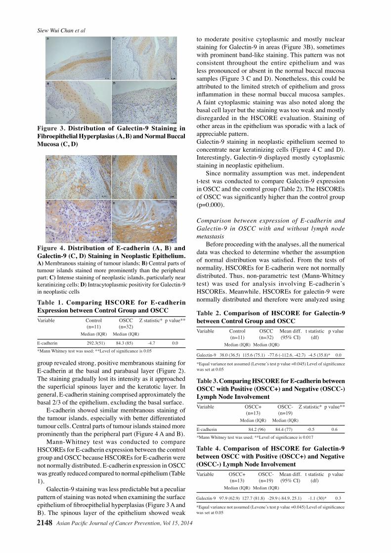
to moderate positive cytoplasmic and mostly nuclear staining for Galectin-9 in areas (Figure 3B), sometimes with prominent band-like staining. This pattern was not consistent throughout the entire epithelium and was less pronounced or absent in the normal buccal mucosa samples (Figure 3 C and D). Nonetheless, this could be attributed to the limited stretch of epithelium and gross inflammation in these normal buccal mucosa samples. A faint cytoplasmic staining was also noted along the basal cell layer but the staining was too weak and mostly disregarded in the HSCORE evaluation. Staining of other areas in the epithelium was sporadic with a lack of appreciable pattern. Galectin-9 staining in neoplastic epithelium seemed to concentrate near keratinizing cells (Figure 4 C and D). Interestingly, Galectin-9 displayed mostly cytoplasmic staining in neoplastic epithelium. Since normality assumption was met, independent t-test was conducted to compare Galectin-9 expression in OSCC and the control group (Table 2). The HSCOREs of OSCC was significantly higher than the control group (p=0.000).
Comparison between expression of E-cadherin and Galectin-9 in OSCC with and without lymph node metastasis Before proceeding with the analyses, all the numerical data was checked to determine whether the assumption of normal distribution was satisfied. From the tests of normality, HSCOREs for E-cadherin were not normally distributed. Thus, non-parametric test (Mann-Whitney test) was used for analysis involving E-cadherin’s HSCOREs. Meanwhile, HSCOREs for galectin-9 were normally distributed and therefore were analyzed using
Figure 3. Distribution of Galectin-9 Staining in Fibroepithelial Hyperplasias (A, B) and Normal Buccal Mucosa (C, D)
Figure 4. Distribution of E-cadherin (A, B) and Galectin-9 (C, D) Staining in Neoplastic Epithelium. A) Membranous staining of tumour islands; B) Central parts of tumour islands stained more prominently than the peripheral part; C) Intense staining of neoplastic islands, particularly near keratinizing cells; D) Intracytoplasmic positivity for Galectin-9 in neoplastic cells
Table 1. Comparing HSCORE for E-cadherin Expression between Control Group and OSCCVariable Control OSCC Z statistic* p value** (n=11) (n=32) Median (IQR) Median (IQR)
E-cadherin 292.3(51) 84.3 (85) -4.7 0.0
*Mann Whitney test was used; **Level of significance is 0.05
Table 2. Comparison of HSCORE for Galectin-9 between Control Group and OSCCVariable Control OSCC Mean diff. t statistic p value (n=11) (n=32) (95% CI) (df) Median (IQR) Median (IQR)
Galectin-9 38.0 (36.5) 115.6 (75.1) -77.6 (-112.6, -42.7) -4.5 (35.8)* 0.0
*Equal variance not assumed (Levene’s test p value =0.045) Level of significance was set at 0.05
Table 3. Comparing HSCORE for E-cadherin between OSCC with Positive (OSCC+) and Negative (OSCC-) Lymph Node InvolvementVariable OSCC+ OSCC- Z statistic* p value** (n=13) (n=19) Median (IQR) Median (IQR)
E-cadherin 84.2 (96) 84.4 (77) -0.5 0.6
*Mann Whitney test was used; **Level of significance is 0.017
Table 4. Comparison of HSCORE for Galectin-9 between OSCC with Positive (OSCC+) and Negative (OSCC-) Lymph Node InvolvementVariable OSCC+ OSCC- Mean diff. t statistic p value (n=13) (n=19) (95% CI) (df) Median (IQR) Median (IQR)
Galectin-9 97.9 (62.9) 127.7 (81.8) -29.9 (-84.9, 25.1) -1.1 (30)* 0.3
*Equal variance not assumed (Levene’s test p value =0.045) Level of significance was set at 0.05

Asian Pacific Journal of Cancer Prevention, Vol 15, 2014 2149
DOI:http://dx.doi.org/10.7314/APJCP.2014.15.5.2145Changed Expression of E-cadherin and Galectin-9 in Oral SCCs but no Potential as Prognostic Markers
independent t-test. Median E-cadherin HSCOREs between OSCC with positive nodal metastasis and OSCC with negative nodal metastasis were not significantly different (p=0.631) (Table 3). Therefore, there was no significant association between E-cadherin expression and lymph node status of OSCC patients. On the other hand, independent t-test (Table 4) comparing Galectin-9 staining between means of HSCOREs for OSCC in positive and negative lymph node involvement were also not significantly different (p=0.276). Hence, no significant association was observed between Galectin-9 expression and lymph node involvement status.
Discussion
Galectins are generally known to be highly expressed in epithelial cells and immune cells. They can be detected in keratinocytes, melanocytes, dendritic cells, macrophages and T cells in skin. Notably, different galectin members may project a different expression pattern in keratinocytes (Chen et al., 2012). Whilst expression of galectin-1, -3 and -7 have been adequately identified (Saussez et al., 2007; Chen et al., 2012), description of Galectin-9 expression in epithelial cells has been comparatively lacking. Galectin-9 is more commonly localized to thymus, T-cells, kidney and Hodgkin’s lymphoma (Rabinovich et al., 2002). An early paper by Murase et al. (1985) described strong demonstration of lectin-binding sites in the stratum spinosum and granulosum, few in the basal layer and none in the fully keratinized superficial layer, a pattern curiously similar to Galectin-9 expression in normal epithelium in our study. However, galectins are only one of many lectins (Nakahara and Raz, 2008) and it has already been established that other galectins may be present in the epithelium. Friedrichs et al. (2007) reported that galectin-3 and-9 are the most abundantly expressed galectins in Madin-Darby canine kidney (MDCK) cells, a commonly used general model for epithelial cells, which comprise epithelium. Liang et al. (2008) observed potent immunostaining for cytoplasmic Galectin-9 in normal squamous epithelial cells of the cervix which appears to involve the entire epithelium while Cada et al. (2009) reported basal and suprabasal presence of Galectin-9 in the epidermis. More relevantly, Fik et al. (2013) described expression of Galectin-9 exclusively at the basal layer of normal oral epithelium. Pioche-Durieu et al. (2005) observed absent or weak staining for Galectin-9 in adjacent normal mucosa in fresh nasopharyngeal carcinoma biopsies which is more consistent with our normal buccal mucosa samples. However, the distinctive yet inconsistent staining of the spinous layer in our fibroepithelial polyp samples could well be more representative of Galectin-9 expression in the oral mucosa. This is because the small and limited normal buccal mucosa samples in our study are insufficient to provide a complete picture. In addition, we observed a similar pattern in some of the normal oral mucosa adjacent to neoplastic epithelium. The means of HSCOREs between these two samples in the control group were also not significantly different and therefore,
considered collectively as a group during statistical analysis.
To the best of our knowledge, there are only three reports on Galectin-9 expression in SCC of the head and neck (Kasamatsu et al., 2005; Pioche-Durieu et al., 2005; Fik et al., 2013). Indeed, the paucity of related literature discouraged unequivocally drawn conclusions about the role of Galectin-9 in head and neck SCC biology, unlike that of galectin-1, -3 and 7 (Saussez et al., 2007; Alves et al., 2011). Kasamatsu et al. (2005) found Galectin-9 expression to be downregulated in OSCC cell lines while Pioche-Durieu et al. (2005) demonstrated intense Galectin-9 expression in the malignant cells of nasopharyngeal carcinoma. Fik et al. (2013) documented complete absence of Galectin-9 in their analysis of 62 head and neck SCCs from various locations. In contrast, our results show increased expression of Galectin-9 in formalin-fixed paraffin-embedded sections of OSCC. It is important to note that IHC monitoring of clinical specimens is indispensable and possibly more advantageous than engineered cell systems in correlation studies by accounting for confounding effects (Cada et al., 2009). And while our results may seem in agreement with Pioche-Durieu et al. (2005), we remain wary as involvement of other galectins in tumorigenesis was deemed to depend heavily on the histological origin of the tissue (Saussez et al., 2007). The obvious disparity of Galectin-9 expression in our study compared to other studies may be attributed to the difference of tumour origin, patients or methodology in each study. Remarkably, none of the aforementioned studies analyzed Galectin-9 expression in SCC of the buccal mucosa. Moreover, the use of FFPE tumour tissues in our study instead of fresh frozen tissue might also have influenced the antigenicity of the samples.
Honjo et al. (2000) reported that the nuclear expression levels of galectin-3 decreased whereas the opposite was true for cytoplasmic expression during the progression from normal to cancerous state in their analysis of 77 tongue specimens. In our study, there was a similar shift of nuclear staining to cytoplasmic staining for Galectin-9. Exclusive cytoplasmic staining has also been reported in cancer cells of the breast (Irie et al., 2005) and mostly cytoplasmic staining in melanoma cells (Kageshita et al., 2002). However, we observed a combination of nuclear and cytoplasmic staining for Galectin-9 in the superficial layers of the normal mucosa while Honjo et al. (2000) described only cytoplasmic staining of the superficial / parasuperficial layer with nuclear staining concentrated at the basal / parabasal layers for galectin-3. Our study shares another similarity with galectin-3 in which staining for Galectin-9 involves the tumour cells near the keratinized areas (Saussez et al., 2007). The dissociated expression of cytoplasmic and nuclear Galectin-9 during OSCC progression suggests different biological roles for Galectin-9 depending on its subcellular localization (Honjo et al., 2000), especially since it has been established that galectins may segregate into various cellular compartments depending on the cellular status (Saussez et al., 2007). The precise functional role of Galectin-9 in the cytoplasm and association of
Siew Wui Chan et al
Asian Pacific Journal of Cancer Prevention, Vol 15, 20142150
cytoplasmic expression with surface expression have yet to be clarified (Kageshita et al., 2002; Irie et al., 2005). In this context, it is noteworthy that galectin can influence cell proliferation and apoptosis from the cell surface and also from intracellular sites (Smetana et al., 2006).
Galectin-9 expression in tumour cells is associated with a better prognosis in cervical SCC (Liang et al., 2008), melanoma and breast carcinoma (Hirashima et al., 2004). Irie et al. (2005) suggested that Galectin-9 suppresses metastasis in multisteps by inhibiting invasion to extracellullar matrix, detachment from tumours, and attachment to vascular endothelium. Even though our results were not significant, there is a trend of higher means for Galectin-9 HSCOREs in OSCC without lymph node metastasis compared to OSCC with positive lymph node metastasis. Since negative lymph node involvement denotes a better prognosis, higher Galectin-9 HSCOREs in our study would also indicate an improved prognosis. Additional studies are warranted because an inverse relationship of Galectin-9 expression with occurrence of distant metastasis but not lymph node metastasis in breast carcinoma has been reported (Irie et al., 2005). In fact, a crosscheck of our department database revealed that two of our OSCC cases with negative lymph node involvement eventually developed recurrence, indicating perhaps future studies should consider the correlation of Galectin-9 expression with variables such as recurrence rate, distant metastasis or survival rate.
The distribution of E-cadherin expression in normal epithelium in our study is less contentious and supported by various studies reported in the literature (Yamada et al., 1997; Diniz-Freitas et al., 2006; Mahomed et al., 2007; Zhong et al., 2007; Wang et al., 2009; Rosado et al., 2013). Also in conformance to these studies is the heterogenous E-cadherin expression in invasive OSCC, with a distinctive pattern at the advancing edge. Invasive tumour front (ITF) which refers to the most advanced 3-6 layers of tumour cell layers or detached tumour cell groups at the invading edge of OSCC, exhibits reduced E-cadherin expression compared to the central or superficial tumour areas. Moreover, loss of E-cadherin expression at the ITF is significantly correlated with poor survival of OSCC patients (Wang et al., 2009).
In 1991, Schipper et al. (1991) described a gradual loss of E-cadherin expression with decreasing differentiation of OSCC and this finding is reiterated over the years with significant results by some authors (Yamada et al., 1997; Mahomed et al., 2007; Rosado et al., 2013). However, it must be noted that Yamada et al. (1997) had few samples from well and poorly differentiated OSCC whereas Rosado et al. (2013) only analyzed E-cadherin expression between well and moderately differentiated OSCC. In fact, the relationship between E-cadherin expression and histological grade remains controversial with various other studies yielding insignificant results (Diniz-Freitas et al., 2006). Rosado et al. (2013) attributed these contradicting results to different biologic behaviour within the tumour itself or among tumours of different sites, despite all head and neck SCCs being the same histopathological entity. They have also described an association between reduced E-cadherin expression with
alcohol intake and hypothesized that alcohol may increase cancer aggressiveness leading to reduced E-cadherin expression. This finding opens up the possibility of other as yet untested confounding factors which may influence E-cadherin expression in OSCC.
In addition to histological grade, E-cadherin immunoexpression has been correlated with various clinicopathological variables including primary location, clinical stage, depth of tumour invasion, margin status, pattern of invasion, treatment options, lymph node metastasis, local or regional relapse, disease-free period and survival time. Weak expression of E-cadherin has been linked to increased invasiveness, shorter disease free period and reduced survival rates (Diniz-Freitas et al., 2006). Menezes et al. (2007) however found E-cadherin expression irrelevant when comparing patients with SCC of tongue and floor of mouth, with and without recurrence during 3 years of follow-up. Nevertheless, a recent systemic review and meta-analysis concluded that aberrant E-cadherin expression is associated with a poor prognosis in head and neck SCC (Zhao et al., 2012).
Existing literature on the prognostic potential of E-cadherin expression in relation to lymph node metastasis and by default poor prognosis, reveal discordant results. Armed with encouraging results, Schipper et al. (1991) suggested that E-cadherin expression was inversely correlated with lymph node metastasis. They found lymph nodes metastasis mostly in cases where the primary tumour exhibited negative or heterogenous expression of E-cadherin. Expression of E-cadherin was generally absent in tumour-infiltrated lymph nodes of patients with head and neck SCC. Although later studies managed to replicate similar results with regards to the primary tumour, E-cadherin expression in the metastatic lymph nodes was found to be increased instead (Yamada et al., 1997; Hung et al., 2006; Zhong et al., 2007). Furthermore, Hung et al. (2006) reported a worse prognosis for patients with increased E-cadherin expression within metastatic lymph nodes. Zhong et al. (2007) suggested that “decreased E-cadherin expression of cancerous cells in the surface of the cancerous tissue promotes the tumor infiltration and invasion into the deeper normal tissue, when the shedding cancerous cells from the primary tumor stop mechanically and proliferate in the cervical lymph node, the E-cadherin expression might play a role on the formation of the metastatic focus”. They also concluded that E-cadherin may be useful to confirm lymph node metastasis but this is doubtful since their study failed to detect micrometastasis rendering E-cadherin a subpar option compared to cytokeratins.
In studies which examine the relationship between E-cadherin expression in primary tumour and lymph node metastasis, results are once again mixed. Several studies show positive correlation between E-cadherin expression and nodal metastasis (Tanaka et al., 2003; Bosch et al., 2005; Foschini et al., 2008; Foschini et al., 2013) but others including our study, dispute this (Mahomed et al., 2007; Lopes et al., 2009; Mostaan et al., 2011; Ukpo et al., 2012; Rosado et al., 2013). An editorial by Cerezo (2011) made an interesting observation; most studies with a positive correlation seem to implicate SCC of the oral

Asian Pacific Journal of Cancer Prevention, Vol 15, 2014 2151
DOI:http://dx.doi.org/10.7314/APJCP.2014.15.5.2145Changed Expression of E-cadherin and Galectin-9 in Oral SCCs but no Potential as Prognostic Markers
cavity as opposed to the oropharynx. She suggested that biologic difference between oral cavity and oropharynx, as well as other dominant prognosticators such as HPV status might explain the negative results. We found just as many studies with negative results in OSCC alone although the sample sizes are admittedly smaller in some of these studies. In our study, we also made a conscious effort to standardize the tumour site of our samples to avoid possible confounding effect even though it has been reported that tumour site has no bearing on E-cadherin expression (Bosch et al., 2005). Also of interest is the study by Mahomed et al. (2007) who reported no significant difference of E-cadherin expression at the ITF in patients with and without nodal metastasis.
Inconsistent findings have resulted in cautious optimism over the use of E-cadherin as a prognostic indicator, with most authors favouring further evaluation. Consideration of use in combinations with other markers such as CD44 (Mostaan et al., 2011), podoplanin (Foschini et al., 2013), truncated dominant-negative isoform of p63 (DNp63) (Foshini et al., 2008), vimentin (Nijkamp et al., 2011), α-catenin (Tanaka et al., 2003) and β-catenin (Tanaka et al., 2003; Mahomed et al., 2007; Rosado et al., 2013) is perhaps a more feasible option.
In conclusion, our study demonstrated a reduction of E-cadherin expression in OSCC compared to epithelium of normal oral mucosa and reactive lesions. On the other hand, Galectin-9 expression conversely increased in OSCC compared to epithelium of normal oral mucosa and reactive lesions. There was no significant correlation between expression of E-cadherin and Galectin-9 with lymph node metastasis. Hence, both proteins seem to harbour limited potential as prognosticators for OSCC. Then again, a trend of higher means for Galectin-9 HSCORE was noted in OSCC without lymph node metastasis compared to OSCC with positive lymph node metastasis, suggesting a weak association with a fairer prognosis.
However, due to the contrasting results with other authors, further studies with a larger sample size and possible inclusion of oral potentially malignant lesions are necessary to substantiate or disprove our results as well as elucidate the molecular aspects on the function of Galectin-9 in OSCC. Another limitation of our study was the lack of available fresh tissues samples in place of the FFPE tissue samples for better and comparable IHC results. It would also be interesting to investigate the expression of Galectin-9 in OSCC with different histological grades in view of the positive correlation of Galectin-9 expression with differentiation of SCC in other sites (Irie et al., 2005; Liang et al., 2008).
Acknowledgements Supported by Grant (PPP) P0035/2010B from the
University of Malaya, Malaysia.
ReferencesAlberts B, Johnson A, Lewis J, et al (2002). Cell-Cell Adhesion.
Molecular Biology of the Cell. 4 ed. New York: Garland
Science.Alves PM, Godoy GP, Gomes DQ, et al (2011). Significance
of galectins-1, -3, -4 and -7 in the progression of squamous cell carcinoma of the tongue. Pathol Res Pract, 207, 236-40.
Balan V, Nangia-Makker P, Raz A (2010). Galectins as cancer biomarkers. Cancers, 2, 592-610.
Berchuck A, Soisson AP, Clarke-Pearson DL, et al (1989). Immunohistochemical expression of CA 125 in endometrial adenocarcinoma: correlation of antigen expression with metastatic potential. Cancer Res, 49, 2091-5.
Bosch FX, Abel U, Kartenbeck J (2005). E-cadherin is a selective and strongly dominant prognostic factor in squamous cell carcinoma: a comparison of E-cadherin with desmosomal components. Int J Cancer, 114, 779-90.
Cada Z, Smetana K Jr, Lacina L, et al (2009). Immunohistochemical fingerprinting of the network of seven adhesion/growth-regulatory lectins in human skin and detection of distinct tumour-associated alterations. Folia Biologica, 55, 145-52.
Cerezo L (2011). Is loss of cadherin expression predictive of high aggressiveness in oropharyngeal squamous cell carcinoma? Oral Oncology, 47, 685.
Chen HY, Lo CH, Li CS, Hsu DK, Liu FT (2012). Galectins and cutaneous immunity. Dermatologica Sinica, 30, 121-7.
Christofori G & Semb H (1999). The role of the cell-adhesion molecule E-cadherin as a tumour-suppressor gene. Trends Biochem Sci, 24, 73-6.
Cooper DN, Barondes SH (1999). God must love galectins; he made so many of them. Glycobiology, 9, 979-84.
Diniz-Freitas M, Garcia-Caballero T, Antunez-Lopez J, Gandara-Rey JM, Garcia-Garcia A (2006). Reduced E-cadherin expression is an indicator of unfavourable prognosis in oral squamous cell carcinoma. Oral Oncol, 42, 190-200.
Dorta RG, Landman G, Kowalski LP, et al (2002). Tumour-associated tissue eosinophilia as a prognostic factor in oral squamous cell carcinomas. Histopathology, 41, 152-7.
Edge SE, Byrd DR, Compton CC, et al (2010). AJCC Cancer Staging Manual New York, USA, Springer.
Fik Z, Valach J, Chovanec M, et al (2013). Loss of adhesion/growth-regulatory galectin-9 from squamous cell epithelium in head and neck carcinomas. J Oral Pathol Med, 42, 166-73.
Foschini MP, Cocchi R, Morandi L, et al (2008). E-cadherin loss and Delta Np73L expression in oral squamous cell carcinomas showing aggressive behavior. Head & Neck, 30, 1475-82.
Foschini MP, Leonardi E, Eusebi LH, et al (2013). Podoplanin and E-cadherin expression in preoperative incisional biopsies of oral squamous cell carcinoma is related to lymph node metastases. Int J Surg Pathol, 21, 133-41.
Friedrichs J, Torkko JM, Helenius J, et al (2007). Contributions of galectin-3 and -9 to epithelial cell adhesion analyzed by single cell force spectroscopy. J Biol Chem, 282, 29375-83.
Hirashima M (2003). Multi-functions of Galectin-9. Mod Asp Immunobiol, 3, 6.
Honjo Y, Inohara H, Akahani S, et al (2000). Expression of cytoplasmic galectin-3 as a prognostic marker in tongue carcinoma. Clin Cancer Res, 6, 4635-40.
Hung KF, Chang CS, Liu CJ, et al (2006). Differential expression of E-cadherin in metastatic lesions comparing to primary oral squamous cell carcinoma. J Oral Pathol Med, 35, 589-94.
Irie A, Yamauchi A, Kontani K,et al (2005). Galectin-9 as a prognostic factor with antimetastatic potential in breast cancer. Clin Cancer Res, 11, 2962-8.
Kageshita T, Kashio Y, Yamauchi A, et al (2002). Possible role of galectin-9 in cell aggregation and apoptosis of human melanoma cell lines and its clinical significance. Int J Cancer, 99, 809-16.
Kasamatsu A, Uzawa K, Nakashima D, et al (2005). Galectin-9
Siew Wui Chan et al
Asian Pacific Journal of Cancer Prevention, Vol 15, 20142152
as a regulator of cellular adhesion in human oral squamous cell carcinoma cell lines. Int J Mol Med, 16, 269-73.
Kitawaki J, Kusuki I, Koshiba H, et al (1999). Detection of aromatase cytochrome P-450 in endometrial biopsy specimens as a diagnostic test for endometriosis. Fertil Steril, 72, 1100-6.
Leffler H, Carlsson S, Hedlund M, Qian Y, Poirier F (2004). Introduction to galectins. Glycoconj J, 19, 433-40.
Li JJ, Zhang GH, Yang XM, et al (2012). Reduced E-cadherin expression is associated with lymph node metastases in laryngeal squamous cell carcinoma.
Liang M, Ueno M, Oomizu S, et al (2008). Galectin-9 expression links to malignant potential of cervical squamous cell carcinoma. J Cancer Res Clin Oncol, 134, 899-907.
Liu LK, Jiang XY, Zhou XX, et al (2010). Upregulation of vimentin and aberrant expression of E-cadherin/beta-catenin complex in oral squamous cell carcinomas: correlation with the clinicopathological features and patient outcome. Mod Pathol, 23, 213-24.
Lopes FF, Da Costa Miguel MC, Pereira ALA, et al (2009). Changes in immunoexpression of E-cadherin and beta-catenin in oral squamous cell carcinoma with and without nodal metastasis. Ann Diagn Pathol, 13, 22-9.
Mahomed F, Altini M, Meer S (2007). Altered E-cadherin/beta-catenin expression in oral squamous carcinoma with and without nodal metastasis. Oral Diseases, 13, 386-92.
Marsh R, Brackenbury R (1996). Molecules of cell adhesion and recognition: An overview. In: LEE, A. G. (ed.) Biomembranes: Receptors of cell adhesion and cellular recognition. JAI Press Inc.
Matsumoto R, Matsumoto H, Seki M, et al (1998). Human ecalectin, a variant of human galectin-9, is a novel eosinophil chemoattractant produced by T lymphocytes. J Biol Chem, 273, 16976-84.
Menezes MB, Lehn CN, Gon Alves AJ (2007). Epidemiological and histopathological data and E-cadherin-like prognostic factors in early carcinomas of the tongue and floor of mouth. Oral Oncology, 43, 656-61.
Mostaan LV, Khorsandi MT, Sharifian SMR, et al (2011). Correlation between E-cadherin and CD44 adhesion molecules expression and cervical lymph node metastasis in oral tongue SCC: Predictive significance or not. Pathol Res Pract, 207, 448-51.
Murase N, Hosaka M, Takai Y, Tanimura T, Mori M (1985). Histochemical demonstration of lectinbinding sites and keratin in inflamed human gingiva. J Periodontal Res, 20, 625-36.
Nakahara S, Raz A (2008). Biological modulation by lectins and their ligands in tumor progression and metastasis. Anticancer Agents Med Chem, 8, 22-36.
Neville BW, Day TA (2002). Oral cancer and precancerous lesions. CA Cancer J Clin, 52, 195-215
Nijkamp MM, Span PN, Hoogsteen IJ, et al (2011). Expression of E-cadherin and vimentin correlates with metastasis formation in head and neck squamous cell carcinoma patients. Radiother Oncol, 99, 344-8.
Pioche-Durieu C, Keryer C, Souqu Re S, et al (2005). In nasopharyngeal carcinoma cells, Epstein-Barr virus LMP1 interacts with galectin 9 in membrane raft elements resistant to simvastatin. J Virol, 79, 13326-37.
Rabinovich GA, Rubinstein N, Toscano MA (2002). Role of galectins in inflammatory and immunomodulatory processes. Biochim Biophys Acta, 1572, 274-84.
Rao SVK, Mejia G, Roberts-Thomson K, Logan R (2013). Epidemiology of oral cancer in Asia in the past decade- an update (2000-2012). Asian Pac J Cancer Prev, 14, 5567-77.
Rosado P, Lequerica-Fernandez P, Fernandez S, et al 2013.
E-cadherin and β-catenin expression in well-differentiated and moderately-differentiated oral squamous cell carcinoma: relations with clinical variables. Br J Oral Maxillofac Surg, 51, 149-56.
Sahu N, Grandis JR (2011). New advances in molecular approaches to head and neck squamous cell carcinoma. Anti-Cancer Drugs, 22, 656-64.
Saussez S, Camby I, Toubeau G and Kiss R (2007). Galectins as modulators of tumor progression in head and neck squamous cell carcinomas. Head & Neck, 29, 874-84.
Schipper JH, Frixen UH, Behrens J, et al (1991). E-cadherin expression in squamous cell carcinomas of head and neck: inverse correlation with tumor dedifferentiation and lymph node metastasis. Cancer Research, 51, 6328-37.
Schneider PM, Baldus SE, Metzger R, et al (2005). Histomorphologic tumor regression and lymph node metastases determine prognosis following neoadjuvant radiochemotherapy for esophageal cancer: implications for response classification. Ann Surg, 242, 684-92.
Scully C, Bagan J (2009). Oral squamous cell carcinoma: overview of current understanding of aetiopathogenesis and clinical implications. Oral Diseases, 15, 388-99.
Smetana K Jr, Dvor Nkov B, Chovanec M, et al (2006). Nuclear presence of adhesion-/growth-regulatory galectins in normal/malignant cells of squamous epithelial origin. Histochem Cell Biol, 125, 171-82.
Stewart BWW, Kleihues PI (2008). World cancer report, IARC Press.
Suzuki T, Sadahiro S, Fukasawa M, et al (2004). Predictive factors of tumor shrinkage and histological regression in patients who received preoperative radiotherapy for rectal cancer. Jpn J Clin Oncol, 34, 740-6.
Tanaka N, Odajima T, Ogi K, Ikeda T, Satoh M (2003). Expression of E-cadherin, alpha-catenin, and beta-catenin in the process of lymph node metastasis in oral squamous cell carcinoma. Br J Cancer, 89, 557-63.
Ukpo OC, Thorstad WL, Zhang Q, Lewis JS Jr (2012). Lack of association of cadherin expression and histopathologic type, metastasis, or patient outcome in oropharyngeal squamous cell carcinoma: a tissue microarray study. Head Neck Pathol, 6, 38-47.
Wang X, Zhang J, Fan M, et al (2009). The expression of E-cadherin at the invasive tumor front of oral squamous cell carcinoma: immunohistochemical and RT-PCR analysis with clinicopathological correlation. Oral Surg Oral Med Oral Pathol Oral Radiol Endod, 107, 547-54.
Woolgar JA (2006). Histopathological prognosticators in oral and oropharyngeal squamous cell carcinoma. Oral Oncol, 42, 229-39.
Yamada K, Jordan R, Mori M, Speight PM (1997). The relationship between E-cadherin expression, clinical stage and tumour differentiation in oral squamous cell carcinoma. Oral Diseases, 3, 82-5.
Zhao Z, Ge J, Sun Y, et al (2012). Is E-cadherin immunoexpression a prognostic factor for head and neck squamous cell carcinoma (HNSCC)? A systematic review and meta-analysis. Oral Oncol, 48, 761-7.
Zhong LP, Li J, Zhang CP, et al (2007). Expression of E-cadherin in cervical lymph nodes from primary oral squamous cell carcinoma patients. Arch Oral Biol, 52, 740-7.