ch 4 tour of the cell. microscopic worlds microscopes led to the discovery of the cell – light...
Post on 21-Dec-2015
216 views
TRANSCRIPT

Ch 4
Tour of the Cell
Microscopic Worlds
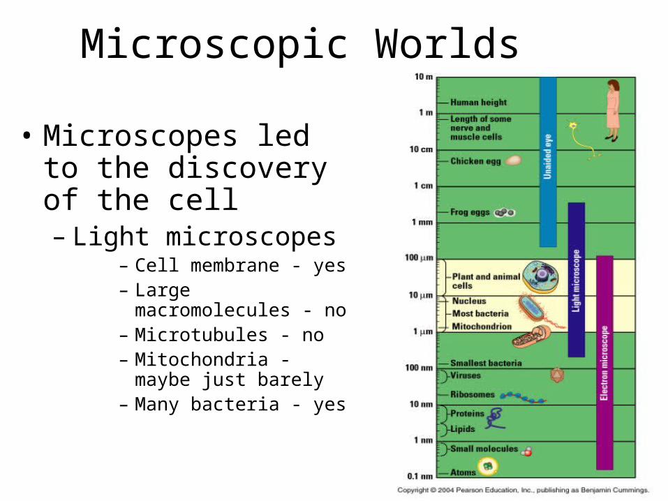
• Microscopes led to the discovery of the cell– Light microscopes
– Cell membrane - yes– Large macromolecules - no– Microtubules - no– Mitochondria - maybe just
barely– Many bacteria - yes

Microscopic Worlds
• Electron scanning microscope• Scanning electron microscope• Transmission electron microscope

Cell Size
• House DNA, protein molecules and internal structures
• Obtain nutrients and diffuse nutrients and O2
• Smaller cells have a greater surface area to volume ratio than do larger cells– Surface area is significant for diffusion and
osmosis

Surface area : Volume
• Volume= 30 um *30 um* 30 um=27000um • SA (large)= 6*(30um*30um)=5,400 um• SA (small)=(6*(10um*10um))*27=16,200 um
30 m 10 m
30 m 10 m
Surface areaof one large cube 5,400 m2
Total surface areaof 27 small cubes 16,200 m2

Domains of Life
• The 3 domains of life – Bacteria (prokaryotic cells)– Archaea (prokaryotic cells)– Eukarya (all other life forms)

Cells• Prokaryotic
– Bacteria & Archaea• Eukaryotic
– Protists, fungi, plants, animals
Prokaryotic cells are simpler & usually smaller than Eukaryotic cells
Prokaryotic cell
Nucleoidregion
Nucleus
Eukar yotic cell Organelles
Co
loriz
ed
TE
M 1
5,0
00

In Common
• Bounded by plasma membrane• Ribosomes• Cytoplasm• DNA as genetic material

Prokaryote• Do not have membrane
bound nucleus• Have a cell wall outside
their plasma membrane• Circular DNA strands• No membrane bound
organelles
Prokar yoticflagella
Ribosomes
Capsule
Cell wall
Plasmamembrane
Nucleoid region (DNA)
Pili

Eukaryote
• Membrane bound nucleus
• Linear DNA• Membrane
bound organelles
NucleusSmooth endoplasmicreticulumRough
endoplasmicreticulum
Ribosomes
Golgiapparatus
Plasma membrane
Mitochondrion
Flagellum
Not in mostplant cells Lysosome
Centriole
Microtubule
CytoskeletonIntermediatefilament
Microfilament
Peroxisome

Size of cell Smaller Larger
Nucleus No nuclear membrane True nucleus, consisting of nuclear membrane & nucleoli
Membrane-enclosed organelles
Absent Present
Cell wall Usually present; chemically complex
When present, chemically simple
Plasma membrane Present Present
Cytoplasm Present Present
Ribosomes Present Present
Chromosome (DNA) arrangement
Single circular chromosome; lacks histones
Multiple linear chromosomes with histones
Sexual reproduction No meiosis; transfer of DNA fragments through cell-to-cell contact
Involves meiosis

Eukaryotic Cells• A typical animal cell:
• Contains a variety of membranous organelles (underlined)
NucleusSmooth endoplasmicreticulum
Roughendoplasmicreticulum
Ribosomes
Golgiapparatus
Plasma membrane
Mitochondrion
Flagellum
Not in mostplant cells Lysosome
Centriole
Microtubule
CytoskeletonIntermediatefilament
Microfilament
Peroxisome
Figure 4.4A

Categories of Organelles• Manufacturing
– Nucleus, ribosomes, endoplasmic reticulum, Golgi apparatus
• Hydrolysis– Lysosomes (animals), vacuoles (plants),
peroxisomes• Energy processing
– Mitochondria (animal), chloroplasts (plants)• Structural support, movement, communication
– Cytoskeleton, plasma membrane, cell wall (plants)

Plasma Membrane• Forms boundary
around cell• Controls and
regulates material transport
• Phospholipid bilayer
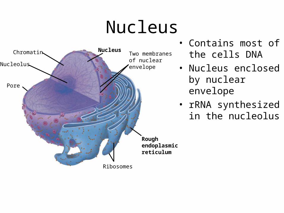
Nucleus• Contains most of the
cells DNA• Nucleus enclosed by
nuclear envelope• rRNA synthesized in the
nucleolus
NucleusChromatin
Nucleolus
Pore
Ribosomes
Roughendoplasmicreticulum
Two membranesof nuclearenvelope

Ribosomes
• Synthesize proteins• Free and bound ribosomes• Composed of 2 subunits

Endoplasmic Reticulum• Smooth lacks attached
ribosomes– Synthesis of lipids, oils,
phospholipids, and steroids
– Processes toxins and drugs in liver cells
– Stores and releases calcium ions in muscle cells
Smooth ER
Rough ER
Nuclearenvelope
Rough ERRibosomes
Smooth ER
TE
M 4
5,00
0
Figure 4.7

ER
• Makes more membrane & proteins• Rough ER has attached ribosome
– Produce proteins that are secreted, inserted into membranes, or transport ed in vesicles to other organelles

Fig. 4-9b
Transport vesiclebuds off
Secretoryproteininside trans-port vesicle
Glycoprotein
Polypeptide
Ribosome
Sugarchain
Rough ER
1
2
3
4

Golgi Apparatus
• Finishes, sorts, and ships cell products– Stacks of membranous sacs receive and modify ER
products then ship them to other organelles or the cell surface
Figure 4.9
Golgi apparatus
TE
M 1
30
,00
0
Transportvesicle fromthe Golgi“Shipping” side
of Golgi apparatus
Golgiapparatus
“Receiving” side ofGolgi apparatus
Transportvesiclefrom ER
New vesicleforming

Lysosomes
• Digestive functions in many single celled organisms
• In white blood cells, they destroy ingested bacteria
• Also recycle damaged organelles

Lysosomes
Figure 4.10AFigure 4.10A
Golgiapparatus
Plasmamembrane
“Food”
Foodvacuole
Lysosomes
2Lysosomeengulfingdamagedorganelle
5
Digestion4
3
Engulfmentof particle
Transport vesicle(containing inactivehydrolytic enzymes)
1
Rough ER

Vacuoles • Function in the general maintenance of the cell
– Plant cells contain a large central vacuole, which has lysosomal and storage functions
Chloroplast
Centralvacuole
Nucleus
Col
oriz
ed T
EM
8,7
00
Figure 4.12A

Endomembrane System• Interconnected structurally and functionally
Nucleus
Smooth ER Nuclear envelope Golgi apparatus
Lysosome
Vacuole
Plasmamembrane
Rough ERTransport vesiclefrom ER to Golgi
Transport vesicle fromGolgi to plasma membrane
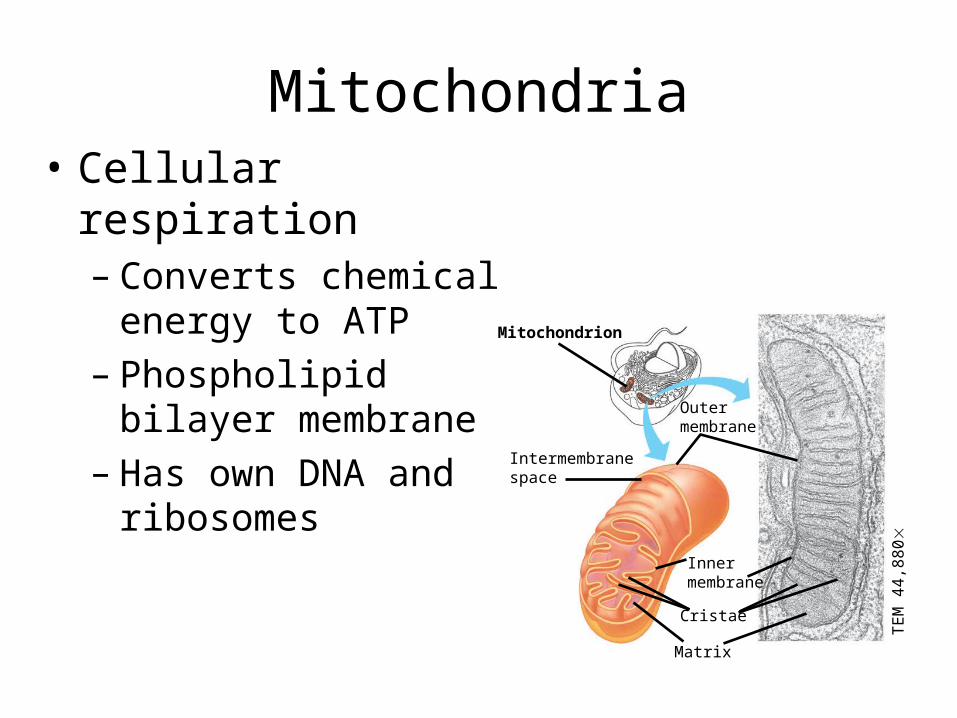
Mitochondria• Cellular respiration
– Converts chemical energy to ATP
– Phospholipid bilayer membrane
– Has own DNA and ribosomes
Mitochondrion
Outermembrane
Intermembranespace
Matrix
Innermembrane
Cristae
TE
M 4
4,8
80

Chloroplasts• Convert solar energy to chemical energy (photosynthesis)• Stroma
– Contains DNA, ribosomes and enzymes• Thylakoids
– Interconnected sacs that form stacks called granum

Endosymbosis
• Hypothesis of endosymbosis– Mitochondria and chloroplasts were once small
prokaryotes living independently– At some point, began living within larger cells

The Cytoskeleton and Related Structures
The cell’s internal skeleton helps organize its structure and activities– A network of protein fibers make up the cytoskeleton
Actin subunit
Microfilament
7 nm
Fibrous subunits
10 nm
Intermediate filament Microtubule
25 nm
Tubulin subunit

– Microfilaments (actin filiments) • Enable cells to change shape and move
– Intermediate filaments • Reinforce the cell and anchor cer tain organelles
– Microtubules give the cell rigidity• And provide anchors for organelles and act as tracks for
organelle movement
Actin subunit
Microfilament
7 nm
Fibrous subunits
10 nm
Intermediate filament Microtubule
25 nm
Tubulin subunit

MovementCilia and flagella move when microtubules bend
– Eukaryotic cilia and flagella are locomotor appendages that protrude from cer tain cells
LM
60
0
Co
loriz
ed
SE
M 4
,10
0
Figure 4.17A Figure 4.17B

Cell Junctions•Tight junctions can bind cells together into leakproof sheets•Anchoring junctions link animal cells into strong tissues•Gap junctions allow substances to flow from cell to cell
Anchoring junction
Tight junctions
Gap junctions
Extracellular matrix
Space between cells
Plasma membranes of adjacent cellsFigure 4.18B

Plants and Cell Walls•Supported by rigid cell walls made largely of cellulose•Connect by plasmodesmata
•Connecting channels
Plasma membrane
Cytoplasm
Plasmodesmata
Vacuole
Layers of one plant cell wall
Walls of two adjacent plant cells
Figure 4.18A

Fig. 4-23