certificate of attendance -...
TRANSCRIPT

Certificate of Attendance
Advanced Clinic: Shoulder Surgery CPT Coding
July 8, 2004
_____________________________________NAME
Lolita M. Jones, RHIA, CCSPresenter
The American Health Information Management Association (AHIMA) has approved this program fortwo (2) continuing education clock hours in the External Forces content area.
Retain this certificate as evidence of participation.
Advanced Clinic Skin Grafts
All CPT Codes 2002 American Medical Association* Lolita M. Jones Consulting Services 1
Advanced Clinic:
Skin Graft
Presenter:
Lolita M. Jones, RHIA, CCS
Lolita M. Jones Consulting Services
1921 Taylor Avenue
Fort Washington, MD 20744
(V) 301-292-8027
(FAX) 301-292-8244
Coding Training: www.hcprofessor.com
E-mail: [email protected]
Distributed by HCPro, Inc.
Advanced Clinic: Skin Graft
Presenter:Lolita M. Jones, RHIA, CCS
Lolita M. Jones Consulting Services1921 Taylor Avenue
Fort Washington, MD 20744(V) 301-292-8027
(FAX) 301-292-8244Coding Training: www.hcprofessor.com
E-mail: [email protected]
Distributed by HCPro, Inc.
Advanced Clinic: Shoulder Surgery
Author:Lolita M. Jones, RHIA, CCS
Lolita M. Jones Consulting Services1921 Taylor Avenue
Fort Washington, MD 20744(V) 301-292-8027
(FAX) 301-292-8244Coding Training: www.hcprofessor.com
E-mail: [email protected]

Advanced Clinic Shoulder Surgery
All CPT Codes 2003 American Medical Association 2
Table of Contents
Disclaimer 3
About Lolita M. Jones Consulting Services 4
Objective 9
I. Shoulder Surgery 10
A. Arthroscopic Heat Application 10
B. Arthroscopic Shoulder Decompression of Subacromial
Space with Partial Acromioplasty 10
C. Injection for Shoulder Arthrography 11
D. Arthroscopic Rotator Cuff Repair 11
E. Partial Acromionectomy 11
Coding Resource: Shoulder Surgery-Arthroscopy vs. Open 12
F. Postoperative Pain Management 13
II. Case Studies 15
III. Answer Key 56
Advanced Clinic Shoulder Surgery
All CPT Codes 2003 American Medical Association 3
DisclaimerAdvanced Clinic: Shoulder Surgery is designed to provide accurate and authoritative
information in regard to the subject covered. Every reasonable effort has been made to
ensure the accuracy of the information within these pages. However, the ultimate
responsibility lies with the user.
Lolita M. Jones Consulting Services and staff make no representation, guarantee or
warranty, express or implied, that this compilation is error-free or that the use of this
publication will prevent differences of opinion or disputes with Medicare or other third-
party payers, and will bear no responsibility or liability for the results or consequences of
its use.
Physician’s Current Procedural Terminology, Fourth Edition (CPT-4) is a copyrighted
coding system owned and maintained by the American Medical Association.
Please contact Lolita M. Jones, RHIA, CCS at:
(V) 301-292-8027
(Fax) 301-292-8244
Coding Training: www.hcprofessor.com
E-mail: [email protected]
©2004 Lolita M. Jones Consulting Services
All five-digit number Physician’s Current Procedural Terminology, Fourth Edition
(CPT) codes, service description, instructions and/or guidelines are 2003 American
Medical Association. All rights reserved.
All rights reserved. The author grants permission for photocopying for limited personal
use or internal use of the original purchaser. This consent does not extend to other kinds
of copying, such as for general distribution, for advertising or promotional purposes, for
creating new collective works, or for resale.
• SHOULDER

Advanced Clinic Shoulder Surgery
All CPT Codes 2003 American Medical Association 4
About Lolita M. Jones Consulting Services
HOSPITAL TRAINING PROGRAMSCoding Training: www.EzMedEd.com
(V) 301-292-8027
(FAX) 301-292-8244
E-mail: [email protected]
BIOGRAPHY:
Lolita M. Jones, RHIA, CCS, is an independent consultant specializing in hospital
outpatient and ambulatory surgery center coding, billing, reimbursement, and operations.
Ms. Jones recently launched her web-based coding program at www.EZMedEd.com.
She has over 15 years of experience in publishing, training, and auditing for the hospital
outpatient and freestanding ambulatory surgery center (ASC) markets. Ms. Jones has
earned both the Registered Health Information Administrator and Certified Coding
Specialist credentials from the American Health Information Management Association
(AHIMA) in Chicago, IL. Ms. Jones resides in Fort Washington, Maryland, and she has
developed six (6) specialty manuals for freestanding ambulatory surgery centers (ASCs)
as well as comprehensive manuals for the following ambulatory payment classification
(APC) training programs:
Basic CPT Outpatient Coding Clinic: This 6.5 hour program is designed for
(Future/Beginning/Current) Coding Specialists, Coding Managers, Reimbursement
Specialists, Compliance Auditors, Hospital-Based Clinic Managers, and ALL hospital
staff responsible for outpatient coding including emergency room, ancillary department
and hospital-based clinic staff. The contents include general guidelines, steps for coding,
and official CPT guidelines for surgical procedures that are commonly performed in the
hospital outpatient setting. Exercises based on actual ambulatory surgery operative
reports will be used to strengthen the attendees’ understanding of the guidelines
presented.
APC Institute: Impact on Emergency Services: This 3 hour program is designed for
Emergency Department: Directors, Managers, Supervisors, and Nurses; Registration
Staff, Health Information Managers, Coding Specialists, and Cast Room Technicians.
The contents include APC Grouping Logic, Mapping Logic for ED Medical Visits,
APCs for Emergency Department Services, Modifiers –25 and –27, Emergency
Screening without Treatment, Critical Care, “Clotbuster” Drugs, Tissue Adhesive Wound
Closure, and Documentation Guidelines.

Advanced Clinic Shoulder Surgery
All CPT Codes 2003 American Medical Association 5
APC Institute: Outpatient Compliance Action Plan: This 6.5 hour program is
designed for Compliance Department Staff (Corporate Officers, Directors, Managers,
Analysts, Auditors); Health Information Management Staff (Directors, Coding
Managers/Supervisors, Coding Specialists); Risk Managers, APC Coordinators,
Reimbursement Specialists, Decision Support Analysts, Outpatient Billing Supervisors,
Outpatient Billing Specialists, Software Vendor Product Managers, ALL staff responsible
for facility component outpatient coding in: Registration, Hospital-Based Clinics,
Ancillary Departments, and the Emergency Department. The contents include: Brief
Overview of APCs; CPT Surgery Coding Compliance; and APC Compliance Issues: site-
of-service billing, reason for visits, discontinued surgery, medical visits, “limited follow-
up services,” colorectal cancer screening, observation stay without recovery, critical
care, interventional radiology, modifiers, unlisted procedure codes, units of service, UB-
92 claims data, and higher level APC groups.
APC Institute: Clinical Documentation Strategies: This 6.5 hour program is designed
for nursing, utilization management, case management, and other health care
professionals responsible for health records documentation. The contents include
ambulatory payment classification (APC)-related clinical documentation requirements
and management tips for the following sites of service: Emergency Room, Observation
Beds/Unit, Ambulatory Surgery, Hospital-Based Outpatient Departments/Clinics, Pain
Management Clinic, Series/Recurring Services, Partial Hospitalization Program, Cast
Room, Ancillary Testing Areas, and Utilization Management.
APC Institute: Coding Guidelines for Hospitals - This 1 or 2 day program is designed
for all technical, clinical and managerial staff responsible for facility component
outpatient coding that will directly impact ambulatory payment classification (APC)
payments. The contents include: Ambulatory Surgery Reimbursement under APCs, APC
Data Reporting Requirements, Medicare Hospital Outpatient Edits, Outpatient Billing
Procedures and Guidelines, Ambulatory Claims Rejection Monitors, Peer Review
Ambulatory Surgery Review, Coding System Reviews, How to Use ICD-9-CM, How to
Use CPT, and CPT Coding Guidelines By Body System (Integumentary,
Musculoskeletal, Respiratory, Cardiovascular and Lymphatic, Hemic and Lymphatic,
Digestive System, Urinary, Male Genital, Laparoscopy/Hysteroscopy, Female Genital,
Endocrine, Nervous, Eye and Ocular Adnexa, Auditory).

Advanced Clinic Shoulder Surgery
All CPT Codes 2003 American Medical Association 6
Modifier Clinic: Hospital Outpatient Issues: This 6.5 hour program is designed for
coding, reimbursement, compliance, billing, database management, ancillary, and clinic
staff responsible for modifier programming, reporting, billing, and auditing. The
contents include: Modifier Reporting Requirements, Official Medicare Guidelines,
Recommended Hospital Front-End Modifier Edits, Electronic/On-Line UB-92 Reporting
of Modifiers, Coding and Billing Aborted/Discontinued Procedures, ICD-9-CM vs.
Medicare Coding Guidelines, Unsuccessful vs. Aborted/Discontinued Procedures,
Documentation of Reduced/Discontinued Procedures, Testing Potential Coders, Software
Encoder Modifier Edits, Interventional Radiology Procedures, Information System
Upgrades, Data Quality Review, Radiology Modifier Reporting Issues, Ancillary
Department Modifier Reporting for Hospitals, and Exercises/Case Studies.
APC Institute: Hospital Financial and Operational Issues: This 6.5 hour program is
designed for hospital executives, directors, chargemaster coordinators,
coding/reimbursement staff, and information system/database managers who will
implement ambulatory payment classifications (APCs). The contents include: General
Overview of APCs, APC Data Reporting Requirements, APC Policy Issues, Developing
a Plan of Action, Conducting Hospital-Wide APC Education, and Assessing Current
Outpatient Operations for: Overall Hospital, Management Information Systems, Business
Office/Patient Accounts, Health Information Management, Ancillary
Departments/Chargemaster, Emergency Room, Hospital-Based Clinics, Hospital-Owned
Satellite Facilities, Hospital-Based Physician Coding and Billing, and Utilization
Management.
APC Institute: Billing and Reimbursement Issues. This 6.5 hour program is designed
for Chief Financial Officers, Vice Presidents of Finance, Controllers, Chargemaster
Coordinators, Database Managers, Software Vendor Product Managers, Coding
Managers, Reimbursement Specialists, Director of Patient Accounts/Business Office,
Outpatient Billing Supervisor/Coordinator, Outpatient Billing Specialists. The contents
include: Durable Medical Equipment and Prosthetics, Pre-operative Registration,
Outpatient Service “Red Flags,” Chargemaster/Charge Entry, Claims Preparation, Claims
Payment, Tracking and Reviewing Medicare Billing Guidelines.

Advanced Clinic Shoulder Surgery
All CPT Codes 2003 American Medical Association 7
Lolita M. Jones Consulting Services
FREESTANDING
AMBUALTORY SURGERY CENTER
TRAINING PROGRAMS
ASC Clinic: Multi-Specialty Procedures - This 6.5 hour program is designed for
Freestanding ambulatory surgery center (ASC) Managers (Business, Nurse,
Reimbursement), Directors, Administrators, Coding Supervisors, Coding Specialists, and
Billers. The contents include: Current Freestanding ASC Structure, Proposed
Freestanding ASC Structure, Medicare Coding Requirements, Medicare Billing
Requirements, Coding Ambulatory Surgery, How To Use CPT When Coding
Ambulatory Surgery, and CPT Coding Guidelines By Body System (Integumentary,
Musculoskeletal, Respiratory, Cardiovascular and Lymphatic, Hemic and Lymphatic,
Digestive System, Urinary, Male Genital, Laparoscopy/Hysteroscopy, Female Genital,
Endocrine, Nervous, Eye and Ocular Adnexa, Auditory).
ASC Clinic: Dermatology & Plastic Surgery - This 6.5 hour program is designed for
all technical, clinical and managerial staff responsible for facility component freestanding
ASC coding and billing. The contents include: exercises based on actual outpatient
operative reports; and CPT coding guidelines for topics such as: tissue expander, pedicle
flap, pressure ulcer, skin grafts, nail avulsion and excision, scar revision, burn treatment,
lesion excisions, wound repair, adjacent tissue transfer/rearrangement, breast surgery,
free flaps with microvascular anastomosis.
ASC Clinic: Eye & Oculoplastic Surgery - This 6.5 hour program is designed for all
technical, clinical and managerial staff responsible for facility component freestanding
ASC coding and billing. The contents include: exercises based on actual outpatient
operative reports; and CPT coding guidelines for topics such as: cataracts. intraocular
lens, keratoplasty, trabeculectomy, strabismus surgery, punctum plugs, tarsorrhaphy,
trichiasis correction, retinal detachment repair, vitrectomy.
Advanced Clinic Shoulder Surgery
All CPT Codes 2003 American Medical Association 8
ASC Clinic: Gastroenterology Procedures- This 6.5 hour program is designed for all
technical, clinical and managerial staff responsible for facility component freestanding
ASC coding and billing. The contents include: exercises based on actual outpatient
operative reports; and CPT coding guidelines for topics such as: hernia repair, nasogastric
intubation, percutaneous gastrostomy tube, hemorrhoidectomy, abscess/cyst drainage,
dental procedures, covered and noncovered colorectal cancer screening, gastrointestinal
endoscopy, esophageal dilation.
ASC Clinic: Orthopaedic Surgery - This 1 or 2 day program is designed for all
technical, clinical and managerial staff responsible for facility component freestanding
ASC coding and billing. The contents include: exercises based on actual outpatient
operative reports; and CPT coding guidelines for topics such as: ganglion cyst, joint
injections, decompression fasciotomy, treatment of fractures/dislocations, skeletal
anatomy of the hand and foot, surgical knee arthroscopy, bunionectomy, toe-to-hand
transfer with microvascular anastomosis.
ASC Clinic: Urology Procedures - This 6.5 hour program is designed for all technical,
clinical and managerial staff responsible for facility component freestanding ASC coding
and billing. The contents include: exercises based on actual outpatient operative reports;
and CPT coding guidelines for topics such as: retrograde pyelogram, ureter vs. urethra,
urethral dilation, ureteral stent, urethral stent, Burch Procedure,
vesicourethropexy/urethropexy, urodynamics, chemotherapy.

Advanced Clinic Shoulder Surgery
All CPT Codes 2003 American Medical Association 9
OBJECTIVE: This program will first provide a detailed review of the shoulder surgery
CPT coding guidelines to assist the participants in their understanding of the numerous
techniques that are performed. “Real life” operative report case studies will also be
presented for many of the shoulder surgery techniques that are discussed.
Advanced Clinic Shoulder Surgery
All CPT Codes 2003 American Medical Association 10
I. Shoulder Surgery
A. Arthroscopic Shoulder Heat Application
Assign unlisted arthroscopy CPT code 29999 to report the use of heat to shrink the
capsule in the shoulder performed through an arthroscope. (Source: CPT Assistant
newsletter, August 1998, page 11.)
B. Arthroscopic Shoulder Decompression of Subacromial
Space with Partial Acromioplasty
The arthroscopic procedure involves exposing the subacromial space, bursectomy,
debridement, detaching the coracoacromial ligament and removing the undersurface of
the acromion. When subacromial decompression is performed, a flat undersurface of the
acromion and acromioclavicular joint is produced, which enlarges the supraspinatus
outlet and prevents impingement.
Coding Tip: The partial acromioplasty, arch decompression, excision of bursal tissue and
release of the coracoacromial ligament would not be reported separately, as these are
considered to be inclusive components of code 29826. [Source: May 2001 CPT Assistant
newsletter, AMA]

Advanced Clinic Shoulder Surgery
All CPT Codes 2003 American Medical Association 11
C. Injection for Shoulder Arthrography
To report an MRI of the shoulder with intra-articular contrast (MR arthrography of the
shoulder), it is appropriate to report 23350 for the shoulder joint injection. Report 76003
if fluoroscopic guidance was used to guide needle placement into the joint, and 73222 for
the MRI shoulder with contrast.
Coding Tip: There is a correct coding initiative (CCI) edit in place as a comprehensive
code pair edit for 23350 and 76003, since fluoroscopic-guided imaging is considered
included in the radiographic arthrography code (73040). Therefore, modifier –59,
Distinct procedure, should be appended to 76003 to designate the fluoroscopic guidance
as a distinct and separate procedure when radiographic arthrography is not performed.
(July 2001 CPT Assistant newsletter, AMA).
D. Arthroscopic Rotator Cuff Repair
Rotator cuff injuries are strains or tears of one or more rotator muscles or tendons, the
most common site being the supraspinatous muscle. Acute tears result from trauma, such
as falls on an outstretched hand or injuries from football throwing, baseball or softball
pitching. Racquetball serving or manipulation of a frozen shoulder. Chronic tears
originate from over-use or constant stress. Assign CPT code 23410 or 23412 for repairs
involving one or two tendons or major muscles of the rotator cuff. Assign CPT code
23420 for a repair of a complete shoulder (rotator) cuff avulsion, referring to the repair of
all three major muscles/tendons of the shoulder cuff. Source: February 2002 CPT
Assistant newsletter, AMA.
Clinical Tip: The major muscles of the rotator cuff: supraspinatus, infraspinatus and
teres minor.
. Partial Acromionectomy
23410 [Repair of musculotendinous cuff (e.g. rotator cuff); acute] includes the work
involved in performing a partial acromionectomy (23130). Therefore, it would not be
appropriate to report 23130 separately. (Source: August 2001 CPT Assistant, AMA).

Advanced Clinic Shoulder Surgery
All CPT Codes 2003 American Medical Association 12
Coding Resource:

Advanced Clinic Shoulder Surgery
All CPT Codes 2003 American Medical Association 13
F. Postoperative Pain Management
Source – 2001 Complimentary Issue CPT Assistant newsletter, AMA:
• CPT reporting of peripheral nerve/plexus nerve catheter placement is based on the :
* exclusion of other anesthesia service(s);
* performance of concomitant operative service(s) by same physician; and
* target nerve involved.
• If the catheter is placed primarily for anesthesia administration during an operative
session, then the appropriate anesthesia services code(s) should be reported.
• If placement is performed for the purpose of post-operative pain management by the
same physician at the time of another operative service (e.g., total shoulder
reconstruction), then the appropriate 64400-64450 series code should be reported
with the modifier –51 appended (if modifier –51 is acceptable by the third-party
payer).
• The appropriate nerve block code (64400-64450) should also be reported when the
catheter is placed by a physician, other than the physician performing anesthesia or
surgical services. For example, for placement of a lumbar plexus catheter, code
64449 should be reported. Similarly, a sciatic nerve catheter insertion would be
reported with the appropriate code.
• Currently, there is no specific CPT code for “daily” management of the peripheral or
plexus nerve catheter. It is not appropriate to report anesthesia code 01996 as this is
specific to epidural catheters (NOTE: anesthesia CPT codes are frequently non-
reportable by hospitals to most third-party payers).

Advanced Clinic Shoulder Surgery
All CPT Codes 2003 American Medical Association 14
October 2001 CPT Assistant newsletter, AMA:
• When general anesthesia is administered and pain management injections are
performed to provide postoperative analgesia, they are separate and distinct services
and are reported in addition to the anesthesia code. Whether the block procedure
(insertion of catheter, injection of narcotic or local anesthetic agent) occurs
preoperatively, postoperatively, or during the procedure is immaterial.
• If, on the other hand, the block procedure is used primarily for the anesthesia itself,
the service should be reported using the anesthesia code alone. In a combined
epidural/general anesthetic, the block cannot be reported separately.
Examples:
[NOTE: Many third-party payers do not accept CPT anesthesia codes from hospitals.]
• A femoral nerve block placed to provide post-operative analgesia for an anterior
cruciate ligament repair or a total knee replacement would be reported separately
from the surgical anesthesia.
• A patient undergoing a thoracotomy might receive an epidural injection of a local
anesthetic and/or narcotic (62318) for postoperative pain control in addition to the
general anesthetic, which is administered through an endotracheal tube (00540). In
this case, the epidural is not the surgical anesthetic and it would be reported
separately, as an independent procedure.
• Shoulder surgery could be performed under an interscalene brachial plexus block that
would also provide postoperative analgesia. This would be reported using the
anesthetic code (e.g., 01620). If the block were intended primarily to alleviate
postsurgical pain, and a general anesthetic was administered for the shoulder
procedure, the block would be separately reportable.
• A brachial plexus block might also provide both the anesthesia and the postoperative
pain control for an open reduction of a wrist fracture. Only the anesthesia code would
be reported.

Advanced Clinic Shoulder Surgery
All CPT Codes 2003 American Medical Association 15
II. Case Studies

Advanced Clinic Shoulder Surgery
All CPT Codes 2003 American Medical Association 16
Case Study # 1. Please assign the CPT code(s)-modifiers for
this case: _____________________________________________________________.
OPERATIVE REPORT
ASSISTANT: None.
PREOPERATIVE DIAGNOSIS: Adhesive capsulitis, large rotator cuff tear on the
right shoulder.
POSTOPERATIVE DIAGNOSIS: Adhesive capsulitis, large rotator cuff tear on the
right shoulder.
OPERATION: Manipulation under anesthesia of the right shoulder and injection of
steroids into the right glenohumeral joint.
INDICATIONS:
This is a 74-year-old female who has had persistent pain, loss of mobility in the shoulder.
She has 9 degrees at forward flexion and abduction both passively and actively. Internal
rotation is also limited to the level of L5 and she has 30 degrees of external rotation. She
has a known rotator cuff tear but she also has significant adhesive capsulitis. I will try to
immobilize the shoulder first and if we need good mobility and strength and no other
treatments really necessary for the rotator cuff tear; however, she has persistent pain and
she regains mobility then eliminating to fix the rotator cuff.
PROCEDURE/FINDINGS
The patient was placed in the supine position on the operating table. After adequate
general anesthesia was given, we gently manipulated obtaining full mobility back to
the shoulder and then placed 20 cc of 0.25% Marcaine with epinephrine and 2 cc of
Solu-Medrol. She was discharged to the recovery room in satisfactory condition. She
would start in a physiotherapy program and the CPM machine.
Advanced Clinic Shoulder Surgery
All CPT Codes 2003 American Medical Association 17
Case Study # 2. Please assign the CPT code(s)-modifiers for
this case: _____________________________________________________________.
PREOPERATIVE DIAGNOSIS: Right acromioclavicular arthritis.
POSTOPERATIVE DIAGNOSIS: Same.
OPERATION PERFORMED: Excision, right distal clavicle.
ANESTHESIA: General.
PROCEDURE: The patient was taken to the operating room and identified. General
endotracheal anesthesia was induced. The patient received prophylactic Ancef and was
placed in the beachchair position. The right shoulder was prepped and draped in the
usual sterile manner. The skin was incised over the AC joint. Bleeders were coagulated
with the Bovie. The joint was subperiosteally exposed. The micro-saw was used to
remove 1 cm of the distal clavicle. The end of the clavicle was arthritis. The wound
was irrigated. Hemostasis was reassessed. The wound was closed with 2-0 Vicryl and
staples. After closure, the area was injected with 0.5% Marcaine without epinephrine.
The patient was taken to the recovery room in stable condition.

Advanced Clinic Shoulder Surgery
All CPT Codes 2003 American Medical Association 18
Case Study # 3. Please assign the CPT code(s)-modifiers for
this case: _____________________________________________________________.
OPERATIVE REPORT
PREOPERATIVE DIAGNOSIS: 1. Right rotator cuff rupture.
2. Right acromioclavicular joint disease.
POSTOPERATIVE DIAGNOSIS: 1. Right rotator cuff rupture.
2. Right acromioclavicular joint disease.
OPERATION PERFORMED:
1. Right rotator cuff reconstruction using curve TAC instrumentation.
2. Open Mumford procedure.
ANESTHESIA:
General.
INDICATIONS: The patient is a 69-year-old white male who has a right rotator cuff
tear. He has failed nonoperative treatment. He also has been noted to have right AC
joint disease, has weakness, pain and discomfort in the shoulder. He is brought today for
definitive treatment. He is aware of possible risks and benefits.
PROCEDURE: The patient underwent general anesthesia without event. He was
sterilely prepped and draped in the usual fashion. He was placed in a semirecumbent
barber chair position.
An incision was made over the anterolateral aspect of the shoulder extending over
the AC joint. The subcutaneous was identified and incised. Dissection was carried
down to the level of the superior AC ligament. Subperiosteal dissection was performed.
Distal clavicle was identified. There was noted to be a large spur and severe degenerative
changes over the clavicle. Distal clavicle was then excised first using a saw and then a
rasp to smooth out the area. The wound was copiously irrigated out.
Attention was then placed to the lateral acromion. A subperiosteal dissection was
performed off the lateral acromion and deltoid split was performed. Bursa was
excised. There was noted to be a very complex rotator cuff tear. Complete avulsion
of supraspinatus, infraspinatus and teres minor off the greater tuberosity. Biceps
tendon was noted to be intact. There was noted to be a second tear which was separate
and a longitudinal tear between the interval between the supraspinatus and the
infraspinatus as well. This was a hypertrophic type tear. There was calcification in the
edges of the tendon in that area. The calcific deposits were then excised and the
longitudinal tear was repaired in a side-to-side fashion between the supraspinatus
and infraspinatus.

Advanced Clinic Shoulder Surgery
All CPT Codes 2003 American Medical Association 19
Case Study # 3 - continued
After completion of this, there was a longitudinal tear in the infraspinatus as well
which was appreciated. There were some calcium deposits in that as well and these
were excised as well. Using a #2 Ethibond suture, retention sutures were then placed
over the cuff and it was delivered laterally. A Cobb elevator was then used to free up
the adhesions and bring the cuff laterally, both deep and superficial to the cuff. A
rongeur was taken and a trough was made laterally. The wound was copiously
irrigated out. Hemostasis was obtained with electrocautery. Using a curved TAC
instrumentation after adequate mobilization of the cuff was completed, the cuff was
delivered in the three drill holes which had been placed laterally with a #2 Ethibond
suture utilized. The cuff was then oversewn after having been tied with the arm at the
side with good coaptation into the trough itself. The cuff was then oversewn using the
Ethibond as well as 0 Vicryl. After completion of this, the wound was copiously
irrigated out. The joint was copiously irrigated as well. Prior to closure of the joint, an
anterior-inferior acromioplasty was done at that point as well. A rasp was utilized prior
to closure of the cuff to smooth out the inferior aspect of the acromion. Hemostasis
was obtained using electrocautery. The superior AC ligament was then closed using 0
vicryl figure-of-eight. The deltoid split was repaired using 0 Vicryl figure-of-eight
suture. Subcutaneous was closed using 3-0 Vicryl and skin was closed using staples. A
sterile compressive dressing was placed. The patient tolerated the procedure well and left
the operating room in stable condition.
Advanced Clinic Shoulder Surgery
All CPT Codes 2003 American Medical Association 20
Case Study # 4. Please assign the CPT code(s)-modifiers for
this case: _____________________________________________________________.
OPERATIVE REPORT
PREOPERATIVE DIAGNOSIS:
Complete rotator cuff tear of right shoulder.
POSTOPERATIVE DIAGNOSIS:
Complete rotator cuff tear of right shoulder.
OPERATION PERFORMED:
Repair of complete rotator cuff tear right shoulder with acromioplasty.
ANESTHESIA:
General.
PROCEDURE: The patient was placed supine on the Operating Room table at which
time he was put to sleep under general anesthesia. Following this, a bump was then
placed beneath the left scapular area. The shoulder was then prepped with Betadine
Solution for a full five minute prep and draped in a sterile fashion. A saber type incision
was made over the superior aspect of the shoulder and carried down through skin and
subcutaneous tissue to the underlying tip of the acromion. The attachments of the deltoid
muscle were released and subacromial space identified. Here there is noted to be
evidence of a complete avulsion of the rotator cuff with approximately 1 cm of
retraction. This measured approximately 1.5 cm in width. In order to better visualize as
well as to decompress an oblique osteotomy cut was made across the tip of the acromion.
This was smoothed with a rasp. Next, the tear was mobilized and the bed prepared.
Two bone anchors were used to reattach the rotator cuff. Once done there was good
stability at the repair site. The area was then thoroughly irrigated with antibiotic solution
following which two drill holes were placed in the remaining tip of the acromion. #1
PDS sutures were then placed through these and the deltoid muscle was reattached.
This was oversewn with another #1 PDS suture. Subcutaneous tissues were closed with
#3-0 Vicryl. Skin was closed with #5-0 Vicryl subcuticular suture. The wound was then
dressed in a sterile fashion and a sling and swathe applied. The patient was then returned
to the Recovery Room in satisfactory condition.

Advanced Clinic Shoulder Surgery
All CPT Codes 2003 American Medical Association 21
Case Study # 5. Please assign the CPT code(s)-modifiers for
this case: _____________________________________________________________.
OPERATIVE REPORT
PREOPERATIVE DIAGNOSIS: Adhesive capsulitis, left shoulder.
POSTOPERATIVE DIAGNOSIS: Same.
PROCEDURE PERFORMED: Left shoulder arthroscopy with manipulation,
lysis of adhesions and pain capsular release.
INDICATIONS: The patient is a 58 year old with recalcitrant adhesive capsulitis. He
failed nonoperative management. He failed initial manipulation. He elected to proceed
with the above procedure and risks and benefits were discussed with him and I answered
all his questions, consents were signed and placed on the chart.
OPERATIVE TECHNIQUE: The patient was taken to the Operating Room, was
placed in the supine position and given general anesthesia per Anesthesia protocol. He
was given 1 gram of IV Kefzol and brought up in the beach chair position. Pressure
points were well padded. The arm was then manipulated and brought to a near forward
flexion. External rotation was to about 45 degrees. There was extensive release with
the manipulation. The posterior portal was established. After prepping and draping in
the usual sterile fashion, the posterior portal was established, the anterior portal was
established. A 4.5 shaver was used to debride the synovitis. Underwater
electrocautery was used for capsular release anteriorly. Once the capsular release was
complete all down to 6 o’clock from the manipulation, the remainder that needed to be
released was superiorly from 3 to 1 o’clock.
This was completed and the 4.5 shaver was used to debride the remainder capsular
tissue for this release. Once this was complete, the portals were switched and the scope
was placed in the anterior portal and a working portal was then used posteriorly.
Posterior capsular release was performed. The arm was, then again, manipulated
and the release was completed from about 5 o’clock up to a 1 o’clock position
posteriorly. The arm was then manipulated and external rotation was achieved to
about 65 degrees. Once this was complete, the joint was lavaged. The scope was
removed. A pain pump was introduced in the subacromial space. The wounds were
closed with 4-0 nylon. Sterile dressing was applied. The patient was awakened and
taken to the Recovery Room in stable condition.

Advanced Clinic Shoulder Surgery
All CPT Codes 2003 American Medical Association 22
Case Study # 6. Please assign the CPT code(s)-modifiers for
this case: _____________________________________________________________.
OPERATIVE REPORT
ASSISTANT: None.
PREOPERATIVE DIAGNOSIS: Chronic rotator cuff impingement syndrome and
acromioclavicular arthrosis, right shoulder.
POSTOPERATIVE DIAGNOSIS: Chronic rotator cuff impingement syndrome and
acromioclavicular arthrosis, right shoulder.
OPERATION: Arthroscopy right shoulder with arthroscopic subacromial
decompression and open Mumford distal claviculectomy.
ANESTHESIA: General.
INDICATIONS: The patient is a 47-year-old security guard with long history of
progressive right shoulder pain, primarily at the acromioclavicular joint. He has
responded well but only temporarily to the previous acromioclavicular and subacromial
cortisone injections. Because of chronic symptoms, which have failed conservative
management, the patient was taken to the operating room at this time to undergo
arthroscopic subacromial decompression and Mumford distal claviculectomy.
PROCEDURE/FINDINGS: After induction of awaken nasal intubation and general
anesthesia, the patient was placed in the upright sitting position in the beachchair
attachment to the operating room table. The right upper extremity was prepped and
draped free in the usual fashion. Bony landmarks were outlined with a marking pen and
a standard posterior portal was made after instilling the glenohumeral joint and
subacromial space with 0.25% Marcaine with epinephrine. The glenohumeral joint was
inspected. The glenohumeral surfaces demonstrate some minimal grade 1
chrondromalacia. There was some minimal fraying of the undersurface of the rotator cuff
but no tear. Biceps and subcapularis tendons were normal. The anterior labrum was
normal. The arthroscope was removed from the glenohumeral joint and reinserted into
the subacromial space through the posterior portal and a second portal was made laterally
along the edge of the acromion. Using a combination of a full radius shave, Arthrocare
Cautery wand and a acromionizer bur, a subacromial decompression was performed
removing soft tissue from the undersurface of the acromion, removing several millimeters
of bone and detachment of the coracoacromial ligament from the anterior edge of the
acromion. Because of the patient’s large size of over 300 pounds, and somewhat difficult
visualization, it was elected to perform an open Mumford distal claviculectomy.

Advanced Clinic Shoulder Surgery
All CPT Codes 2003 American Medical Association 23
Case Study # 6 - continued
Arthroscopic instruments were removed and incision was made overlying the Case
acromioclavicular joint and carried down to subperiosteally expose the distal clavicle.
Approximately 1.5 cm of distal clavicle was excised with a sagittal saw and the end of
the bone was rasped smooth. Periosteal and fascial tissues were closed with multiple #1
Vicryl interrupted sutures and the wound was then irrigated and infiltrated with 0.25%
Marcaine with epinephrine for postoperative pain relief. Portals and wounds were closed
in routine fashion.
An indwelling, Marcaine pump catheter was placed at the distal claviculectomy site
for postoperative pain relief. Sterile dressings and a sling were applied after reversal of
anesthesia.
The patient was transported to the recovery room in stable condition. There were no
apparent intraoperative complications.
Advanced Clinic Shoulder Surgery
All CPT Codes 2003 American Medical Association 24
Case Study # 7. Please assign the CPT code(s)-modifiers for
this case: _____________________________________________________________.
OPERATIVE REPORT
PREOPERATIVE DIAGNOSIS: Possible rotator cuff tendonitis, acromioclavicular
arthritis, and frozen shoulder, right shoulder.
POSTOPERATIVE DIAGNOSIS: Frozen shoulder, subacromial bursitis-
impingement, and acromioclavicular arthritis, right shoulder.
OPERATIVE PROCEDURE: Arthroscopic subacromial decompression with open
distal clavicle resection and manipulation under anesthesia, right shoulder.
ANESHESIA: General and block.
INDICATION FOR PROCEDURE: The patient is a 51-year-old male with reclacitrant
shoulder pain and a frozen shoulder. He had failed rehabilitative and injection treatments
and requested operative intervention.
FINDINGS AT OPERATION: Preoperative motion was 90 degrees of flexion and 30
degrees of external and internal rotation. There was significant subacromial bursitis with
very thickened coracoacromial ligament and a subacromial spur. The intra-articular
structures were normal.
DESCRIPTION OF PROCEDURE: The patient was brought to the operative suite and
general anesthesia was smoothly induced. The shoulder was examined and the above
noted limitation of motion was found. An interscalene block was placed for postop
pain control and the patient was placed in the beach chair position. The right shoulder
was manipulated with palpable and audible crepitace into 150 degrees of elevation,
external rotation was to 80 with the opposite shoulder being 90, and internal rotation
was equivalent at 70. Adduction and abduction were equivalent. The shoulder was
prepped and draped in a sterile fashion. Through anterolateral, direct lateral, and
posterolateral portals, the shoulder was examined and treated arthroscopically. The
glenohumeral joint was entered. The glenoid, humeral head, biceps tendon, and labrum
were intact. The rotator cuff was intact. The arthroscopic instruments were placed in
the subacromial space. The bursa was resected. The coracoacromial ligament was
released from the acromion with the cautery. Utilizing a bur and a shaver, the acromion
was flattened. The anterior portion was excised and the rotator cuff was found to have
a significant partial-thickness bursal side tear, but no full-thickness tear and the
arthroscopic instruments were removed. A small incision was made over the distal
clavicle. The deltotrapezial raphe was taken down in a subperiosteal fashion off of the
distal clavicle and the distal 2.5 cm of clavicle was excised with a saw. Bone wax was
placed over the cut end. The deltotrapezial raphe was closed with #1 Nurolon.
Advanced Clinic Shoulder Surgery
All CPT Codes 2003 American Medical Association 25
Case Study # 7 - continued
A small closed suction drain was placed and the wound was closed with 2-0 Vicryl and a
Monocryl for the skin. Steri-Strips were applied. The acomioplasty was checked
manually before closure. A sterile compressive dressing was applied. The patient was
awakened and taken to the recovery room in good condition. There were no
complications. Blood loss was minimal. Postoperative plans are to rehabilitate the
patient’s shoulder.

Advanced Clinic Shoulder Surgery
All CPT Codes 2003 American Medical Association 26
Case Study # 8. Please assign the CPT code(s)-modifiers for
this case: _____________________________________________________________.
OPERATIVE REPORT
PREOPERATIVE DIAGNOSIS:
1. Left shoulder rotator cuff tear with subacromial impingement.
POSTOPERATIVE DIAGNOSIS:
1. Left shoulder rotator cuff tendonitis/synovitis.
OPERATION:
Left shoulder arthroscopy, glenohumeral debridement, limited.
Subacromial decompression.
ANESTHESIA: General.
FINDINGS: The patient had an MRI which reportedly demonstrated a rotator cuff tear.
There was no tear noted either on the bursal surface or on the articular surface. There
was evidence of an impingement-type lesion on the greater tuberosity; however, the
rotator cuff insertion appeared to be intact. The rotator cuff was inspected both on the
articular and bursal surfaces. There was significant hyperemia throughout the rotator
cuff, and there was some questionable synovitis throughout the joint. A tissue sample
was sent for a pathologic evaluation, as the patient did have psoriasis. The patient
previously did undergo rheumatologic workup, which was reportedly negative. The
patient had a subacromial spur.
PROCEDURE: The patient was brought to the operating room and placed on a table in
a supine position. General anesthesia was induced. The left shoulder was prepped and
draped in the usual sterile fashion. A posterior portal was created, through which the
osteoscope was introduced. The above-noted findings were appreciated. An anterior
portal was then created with a Wissinger rod. Through this anterior portal the shaver was
introduced, and limited glenohumeral debridement was performed. Next, attention
was directed to the subacromial space, and a standard lateral portal was created through
which the cautery device and arthroscopic shaver were used to perform a
bursectomy.
Upon completion of the bursectomy, the borders of the acromion were defined. A burr
was used to perform an acromioplasty. The arthroscope was then introduced into the
lateral portal, and the burr was placed posteriorly. The burr was used to plane the
acromion. The AC joint was then visualized by using cautery and shaver to remove the
inferior capsule. There was a spur on the undersurface of the distal clavicle, and this
was coplaned. The rotator cuff was then inspected, and was found to be intact along its
bursal surface.

Advanced Clinic Shoulder Surgery
All CPT Codes 2003 American Medical Association 27
Case Study # 8 - continued
Wounds were irrigated copiously. Instruments were removed. 3.0 nylon sutures were
used to close the skin. A sterile dressing was applied. The patient was brought to the
recovery room in stable condition, having tolerated the procedure well.
Advanced Clinic Shoulder Surgery
All CPT Codes 2003 American Medical Association 28
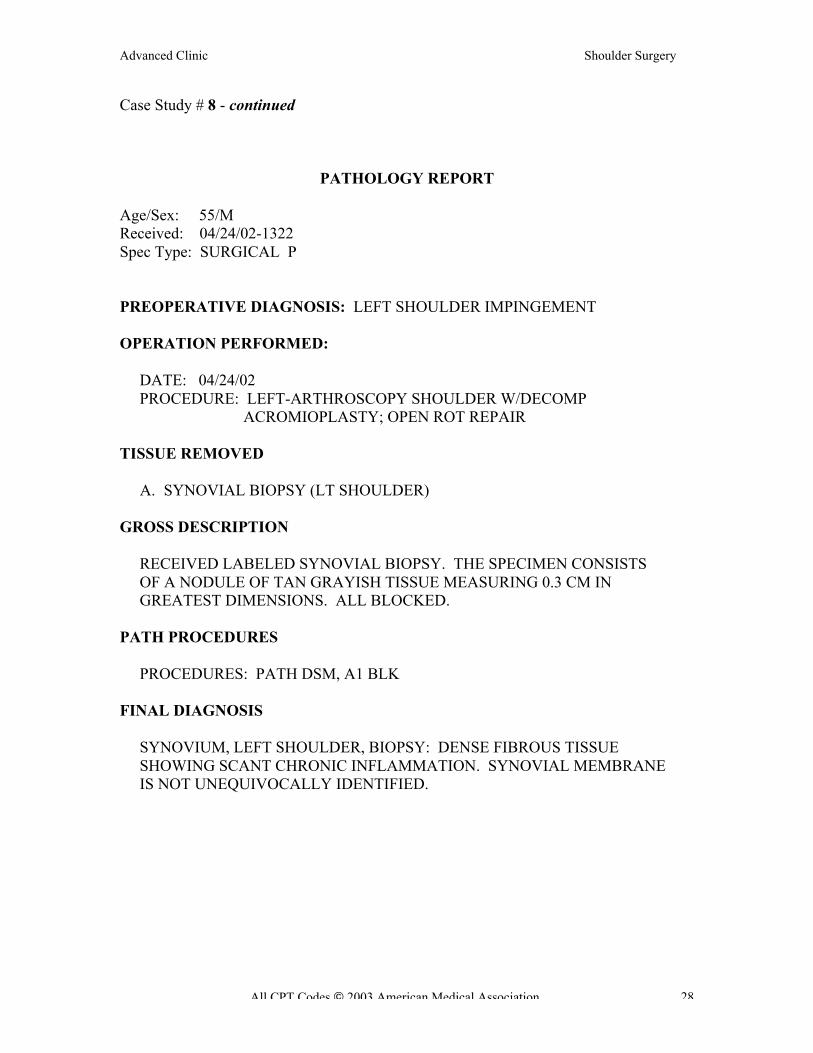
Case Study # 8 - continued
PATHOLOGY REPORT
Age/Sex: 55/M
Received: 04/24/02-1322
Spec Type: SURGICAL P
PREOPERATIVE DIAGNOSIS: LEFT SHOULDER IMPINGEMENT
OPERATION PERFORMED:
DATE: 04/24/02
PROCEDURE: LEFT-ARTHROSCOPY SHOULDER W/DECOMP
ACROMIOPLASTY; OPEN ROT REPAIR
TISSUE REMOVED
A. SYNOVIAL BIOPSY (LT SHOULDER)
GROSS DESCRIPTION
RECEIVED LABELED SYNOVIAL BIOPSY. THE SPECIMEN CONSISTS
OF A NODULE OF TAN GRAYISH TISSUE MEASURING 0.3 CM IN
GREATEST DIMENSIONS. ALL BLOCKED.
PATH PROCEDURES
PROCEDURES: PATH DSM, A1 BLK
FINAL DIAGNOSIS
SYNOVIUM, LEFT SHOULDER, BIOPSY: DENSE FIBROUS TISSUE
SHOWING SCANT CHRONIC INFLAMMATION. SYNOVIAL MEMBRANE
IS NOT UNEQUIVOCALLY IDENTIFIED.
Advanced Clinic Shoulder Surgery
All CPT Codes 2003 American Medical Association 29
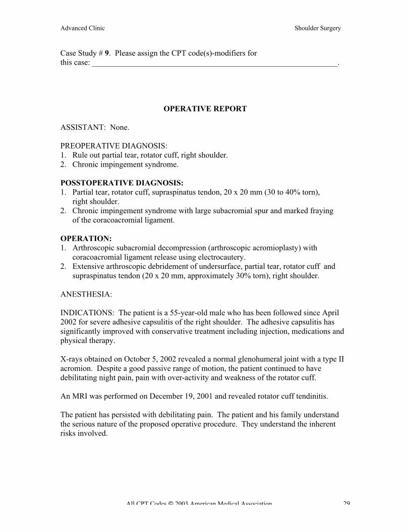
Case Study # 9. Please assign the CPT code(s)-modifiers for
this case: _____________________________________________________________.
OPERATIVE REPORT
ASSISTANT: None.
PREOPERATIVE DIAGNOSIS:
1. Rule out partial tear, rotator cuff, right shoulder.
2. Chronic impingement syndrome.
POSSTOPERATIVE DIAGNOSIS:
1. Partial tear, rotator cuff, supraspinatus tendon, 20 x 20 mm (30 to 40% torn),
right shoulder.
2. Chronic impingement syndrome with large subacromial spur and marked fraying
of the coracoacromial ligament.
OPERATION:
1. Arthroscopic subacromial decompression (arthroscopic acromioplasty) with
coracoacromial ligament release using electrocautery.
2. Extensive arthroscopic debridement of undersurface, partial tear, rotator cuff and
supraspinatus tendon (20 x 20 mm, approximately 30% torn), right shoulder.
ANESTHESIA:
INDICATIONS: The patient is a 55-year-old male who has been followed since April
2002 for severe adhesive capsulitis of the right shoulder. The adhesive capsulitis has
significantly improved with conservative treatment including injection, medications and
physical therapy.
X-rays obtained on October 5, 2002 revealed a normal glenohumeral joint with a type II
acromion. Despite a good passive range of motion, the patient continued to have
debilitating night pain, pain with over-activity and weakness of the rotator cuff.
An MRI was performed on December 19, 2001 and revealed rotator cuff tendinitis.
The patient has persisted with debilitating pain. The patient and his family understand
the serious nature of the proposed operative procedure. They understand the inherent
risks involved.

Advanced Clinic Shoulder Surgery
All CPT Codes 2003 American Medical Association 30
Case Study # 9 - continued
PROCEDURE: The patient was taken to the operating room and placed on the
operating table in the supine position. Following the induction of general anesthesia, the
patient was rotated to the left lateral decubitus position with the right side up. The right
shoulder was then prepped and draped in the usual sterile fashion.
Standard portals were used for the arthroscopic examination of the right shoulder.
Examination of the glenohumeral joint revealed normal articular surfaces. The anterior
and posterior glenoid and labra were intact; the middle and inferior glenohumeral
ligaments were intact. There was mild fraying of the biceps tendon.
The underside of the rotator cuff revealed a large underside partial tear measuring 20
x 20 mm, estimated to be 30 to 40% torn. This area was extensively debrided using the
full-radius resector. The arthroscope was then placed anteriorly and posteriorly for
further evaluation of the rotator cuff and for more extensive debridement.
The arthroscope was then placed into the subacromial space using the standard anterior,
posterior and lateral portals. Examination of the superior side of the rotator cuff revealed
marked fraying of the supraspinatus tendon. There was no complete tear identified.
The underside of the acromion revealed a large subacromial spur with marked fraying of
the coracoacromial ligament. This was transected with the use of electrocautery. The
underside of the acromion was then debrided using the Dyonics acromioplasty blade
and the Dyonics clavicularis blade; in this manner, 5 to 8 mm of bone were resected.
The arthroscope was then placed laterally and the shaver was inserted posteriorly to
further flatten the anterior hook of the acromion. The acromioclavicular joint was
visualized arthroscopically. Soft tissue was resected with the use of electrocautery.
Due to the absence of preoperative acromioclavicular joint pain, a distal clavicle
excision was not carried out. The superior side of the rotator cuff was extensively
debrided and the partial tear was estimated to be approximately 30 to 40%, involving
only the supraspinatus tendon.
The wounds were copiously irrigated throughout the procedure. Then 0.25% Marcaine
with epinephrine was instilled into the arthroscopic portals and 20 cc were placed into the
joint. A dry bulky compressive dressing was applied.
The patient tolerated the procedure well and was transferred to the recovery room in a
good condition.

Advanced Clinic Shoulder Surgery
All CPT Codes 2003 American Medical Association 31
Case Study # 10. Please assign the CPT code(s)-modifiers for
this case: _____________________________________________________________.
OPERATIVE REPORT
PREOPERATIVE DIAGNOSIS: Right rotator cuff tear.
POSTOPERATIVE DIAGNOSIS:
1. Right partial thickness rotator cuff tear.
2. Subacromial impingement.
3. Acromioclavicular joint arthritis.
OPERATION:
1. Right shoulder arthroscopy.
2. Subacromial decompression.
3. Arthroscopic Mumford (distal clavicle) resection.
4. Limited glenohumeral debridement.
ANESTHESIA: General.
OPERATIVE FINDINGS: The patient had an MRI which was reported as showing a full
thickness rotator cuff tear. She had evidence of undersurface fraying. However, there
was no evidence of a full thickness tear. A Prolene suture was placed at the articular
surface at the articular margin of the rotator cuff tendon where it appeared to have a
partial thickness tear. When this bursal surface was probed there was no evidence of a
full thickness tear. The spur was removed as was the distal clavicle.
PROCEDURE: The patient was brought to the operating room, placed on the operating
room table in the supine position. Right shoulder was prepped and draped in the usual
sterile fashion. Right shoulder was then entered through a standard posterior portal. The
above noted arthroscopic findings were appreciated. A Wissinger rod was used to create
an anterior portal for the inside-out technique. There was some fraying of the biceps
tendon at its anchor. However, there was no instability. The biceps anchor origin was
debrided. There was evidence of an undersurface fraying of the rotator cuff. This
was debrided as well in a limited fashion. Next a Prolene suture was passed through a
spinal needle in this area where the tendon appeared attenuated. It was grasped through
the anterior portal and left out at the skin as a tag suture and marking suture for later
inspection from the bursal surface. Instruments were then removed from the
glenohumeral joint. The arthroscope was placed into the subacromial space. A standard
lateral portal was created. Through this lateral portal a cautery device was used as was a
shaver to perform a bursectomy. We defined the borders of the acromion. A bone spur
was found on the undersurface of the acromion. This was resected using the
acromioblaster. Next the distal clavicle was exposed. The distal clavicle was then
resected as well.
Advanced Clinic Shoulder Surgery
All CPT Codes 2003 American Medical Association 32
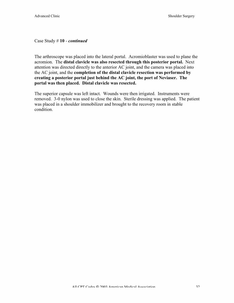
Case Study # 10 - continued
The arthroscope was placed into the lateral portal. Acromioblaster was used to plane the
acromion. The distal clavicle was also resected through this posterior portal. Next
attention was directed directly to the anterior AC joint, and the camera was placed into
the AC joint, and the completion of the distal clavicle resection was performed by
creating a posterior portal just behind the AC joint, the port of Neviaser. The
portal was then placed. Distal clavicle was resected.
The superior capsule was left intact. Wounds were then irrigated. Instruments were
removed. 3-0 nylon was used to close the skin. Sterile dressing was applied. The patient
was placed in a shoulder immobilizer and brought to the recovery room in stable
condition.
Advanced Clinic Shoulder Surgery
All CPT Codes 2003 American Medical Association 33
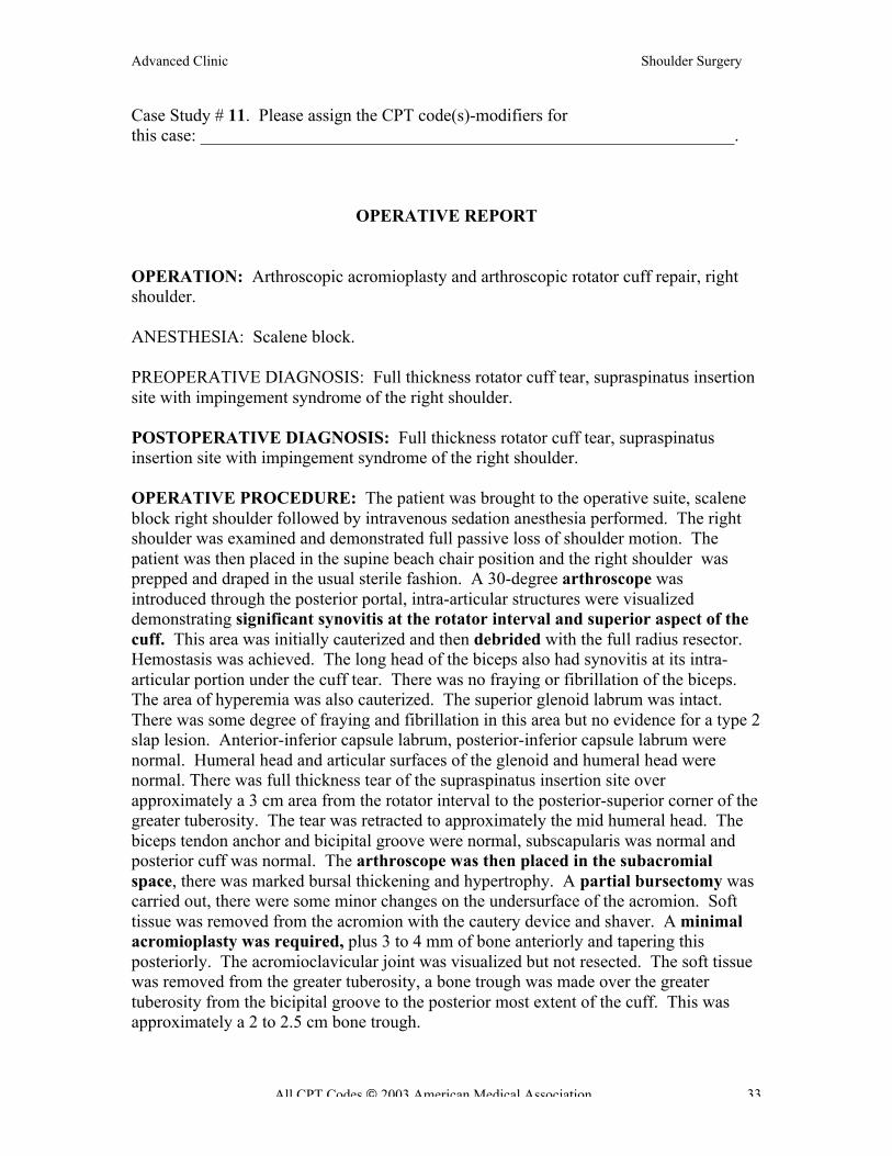
Case Study # 11. Please assign the CPT code(s)-modifiers for
this case: _____________________________________________________________.
OPERATIVE REPORT
OPERATION: Arthroscopic acromioplasty and arthroscopic rotator cuff repair, right
shoulder.
ANESTHESIA: Scalene block.
PREOPERATIVE DIAGNOSIS: Full thickness rotator cuff tear, supraspinatus insertion
site with impingement syndrome of the right shoulder.
POSTOPERATIVE DIAGNOSIS: Full thickness rotator cuff tear, supraspinatus
insertion site with impingement syndrome of the right shoulder.
OPERATIVE PROCEDURE: The patient was brought to the operative suite, scalene
block right shoulder followed by intravenous sedation anesthesia performed. The right
shoulder was examined and demonstrated full passive loss of shoulder motion. The
patient was then placed in the supine beach chair position and the right shoulder was
prepped and draped in the usual sterile fashion. A 30-degree arthroscope was
introduced through the posterior portal, intra-articular structures were visualized
demonstrating significant synovitis at the rotator interval and superior aspect of the
cuff. This area was initially cauterized and then debrided with the full radius resector.
Hemostasis was achieved. The long head of the biceps also had synovitis at its intra-
articular portion under the cuff tear. There was no fraying or fibrillation of the biceps.
The area of hyperemia was also cauterized. The superior glenoid labrum was intact.
There was some degree of fraying and fibrillation in this area but no evidence for a type 2
slap lesion. Anterior-inferior capsule labrum, posterior-inferior capsule labrum were
normal. Humeral head and articular surfaces of the glenoid and humeral head were
normal. There was full thickness tear of the supraspinatus insertion site over
approximately a 3 cm area from the rotator interval to the posterior-superior corner of the
greater tuberosity. The tear was retracted to approximately the mid humeral head. The
biceps tendon anchor and bicipital groove were normal, subscapularis was normal and
posterior cuff was normal. The arthroscope was then placed in the subacromial
space, there was marked bursal thickening and hypertrophy. A partial bursectomy was
carried out, there were some minor changes on the undersurface of the acromion. Soft
tissue was removed from the acromion with the cautery device and shaver. A minimal
acromioplasty was required, plus 3 to 4 mm of bone anteriorly and tapering this
posteriorly. The acromioclavicular joint was visualized but not resected. The soft tissue
was removed from the greater tuberosity, a bone trough was made over the greater
tuberosity from the bicipital groove to the posterior most extent of the cuff. This was
approximately a 2 to 2.5 cm bone trough.
Advanced Clinic Shoulder Surgery
All CPT Codes 2003 American Medical Association 34
Case Study # 11 - continued
The cuff was mobilized by release of the intra-articular portion of the capsule, release of
the coracohumeral ligament at the base of the coracoid. This was done with a cautery
device.
The cuff was then mobilized and pulled to the bone trough, two 5 mm Arthrex
anchors were placed in the lateral most aspect of the bone trough, and the sutures
were tied with three simple sutures and one mattress suture. An excellent anatomic
repair was achieved with the cuff being opposed to the tuberosity with firm fixation.
The arthroscopic equipment was removed from the shoulder, the portal sites were closed
with #3-0 Prolene and Steri-Strips. Sterile dressings were applied. The patient was
reversed from anesthetic and brought to the recovery room in stable and satisfactory
condition.
COMPLICATIONS: None.

Advanced Clinic Shoulder Surgery
All CPT Codes 2003 American Medical Association 35
Case Study # 12. Please assign the CPT code(s)-modifiers for
this case: _____________________________________________________________.
OPERATIVE REPORT
PREOPERATIVE DIAGNOSIS:
1. Acromioclavicular joint arthritis right shoulder.
2. Possible superior labrum anterior and posterior lesion or labrum tear right shoulder.
POSTOPERATIVE DIAGNOSIS:
1. Osteoarthritis right acromioclavicular joint.
2. Anterior superior labrum anterior and posterior lesion right shoulder.
TITLE OF THE OPERATION:
1. Arthroscopic superior labrum anterior and posterior lesion repair with one 3.0 mm
Fast-Tac suture anchor.
2. Arthroscopic distal clavicle excision right shoulder.
ANESTHESIA: General.
PREOPERATIVE NOTE: The patient is a 57-year-old gentleman with a long history of
right shoulder pain. Preoperative evaluation indicated pain emanating from an arthritic
AC joint, and we suspected a SLAP lesion as well. We did not suspect the rotator cuff or
impingement. Therefore, the above procedure was recommended.
DETAILS OF THE PROCEDURE:
Under general anesthetic, the patient was placed supine in the semi-sitting position with
the head on a Mayfield headrest. The right shoulder was scrubbed, prepped and draped in
the usual manner. The posterior viewing scrubbed, prepped and draped in the usual
manner. The posterior viewing portals were established through the glenohumeral
joint. The articular cartilage in the glenoid and humeral sides was normal. The posterior
labrum and direct superior labrum was normal. However, the anterior superior labrum
was detached. There was a SLAP lesion under the biceps anchored anteriorly coming
down to the approximately 1 o’clock. We probed this through an anterior portal in the
rotator interval and found this to be true. The biceps tendon anchor was normal except
for the anterior portion of the anterior superior labrum. The biceps tendon exited the joint
normally. The rotator cuff was normal.
Advanced Clinic Shoulder Surgery
All CPT Codes 2003 American Medical Association 36
Case Study # 12 - continued
We prepared the anterior superior glenoid neck with a shaver and a bur after using
the periosteal elevator to mobilize the soft tissue. Next, we placed a single 3.0 Bio Fast-
Tac suture anchor at approximately 12:30 on the anterior superior glenoid rim. We
used standard arthroscopic knot tying techniques to tie down the anterior superior
labrum with the anterior portion of the biceps anchor. Fortunately, the majority of
the biceps anchor was intact. We had established this using a peel-back sign
intraoperatively. Once the labrum was repaired, we probed it and found it to be stable.
Next, the subacromial space was entered. A lateral working portal was established.
We excised enough of the subacromial bursa to visualize the anterior acromion. We
denuded the anterior acromion across to the AC joint removing the inferior AC joint
ligaments. The distal clavicle was clearly identified. We removed the osteophyte on
the medial end of the anterior acromion, which was in part partial of the AC joint
osteophyte. We then did approximately an 8 mm distal clavicle excision through the
same anterior portal that we used for the labrum repair by redirecting it directly into the
AC joint. We removed all the clavicle up to, but not including the superior AC joint
ligaments.
Next, the arthroscopic instruments were removed. The portals were closed with 4-0
nylon. A sterile dressing was applied followed by a Don Joy shoulder immobilizer.
Sponge and instrument counts are correct. The patient tolerated the procedure well and
was transferred to the recovery room in satisfactory condition.
POSTOPERATIVE PLAN: The patient will be discharged home today and I will see
him in the office in a few days time for follow-up.
Advanced Clinic Shoulder Surgery
All CPT Codes 2003 American Medical Association 37
Case Study # 13. Please assign the CPT code(s)-modifiers for
this case: _____________________________________________________________.
OPERATIVE RECORD
PREOPERATIVE DIAGNOSIS: Left shoulder acromioclavicular joint arthroscopy
with possible rotator cuff tear.
POSTOPERATIVE DIAGNOSIS: Superior labrum anterior and posterior (SLAP)
lesion, left shoulder with acromioclavicular joint arthritis and impingement.
OPERATION: 1. Arthroscopy of left shoulder with repair of superior labrum
anterior and posterior (SLAP) lesion.
2. Arthroscopic Mumford procedure and subacromial decompression.
ANESTHESIA: General.
INDICATIONS: This is a 64-year-old male who has been followed for some time with
shoulder pain of the left side. He has failed conservative treatment and presents for
definitive treatment.
MRI and symptoms are consistent with AC joint arthrosis as well as possible rotator cuff
tear.
PROCEDURE: The patient was taken to the operating room and placed in the supine
position. After general anesthesia was obtained, he was placed in the beach-chair
position with all bony prominences carefully padded. The left shoulder was prepped and
draped in the usual sterile fashion.
First, a posterior portal was made, and a diagnostic arthroscopy was performed.
Immediately noted was a significant SLAP lesion on the anterior aspect of the
labrum. The rotator cuff was inspected and was noted to be completely intact. There
was some mild synovitis around the SLAP lesion as well.
Next, an anterior portal was made. The labrum was probed. Most of the labrum was well
attached to the anterior lip of the glenoid.
Next, ArthroCare wand was used to repair the SLAP lesion as well as debride it.
With this completed then, the synovectomy was completed as well. The biceps tendon
was intact as were all other ligament and structures.
Advanced Clinic Shoulder Surgery
All CPT Codes 2003 American Medical Association 38
Case Study # 13 - continued
Next, attention was paid to the subacromial space. In the subacromial space, a
subacromial bursectomy was performed with the ArthroCare wand. AC joint
arthrosis was noted. This was debrided also with the ArthroCare system. The distal
clavicle was excised with a large bur. Also, the opposing acromion was smoothed
down with the bur as well completing our subacromial decompression.
The area was copiously irrigated. The wounds were gently approximated with 4-0
undyed Vicryl. The whole area was infiltrated with 0.5% Marcaine with epinephrine
and 0.75% Marcaine plain for postoperative anesthesia. Bulky dressing was placed
and held with tape. ABD was placed under the armpit, and the arm was placed in a
regular sling.
Estimated blood loss was minimal. The IV fluid replaced: Less than 3000 cc of
crystalloid. Drains and packs: None. Complications: None.
The patient tolerated the procedure well and was taken to the recovery room in a good
postoperative condition.
Advanced Clinic Shoulder Surgery
All CPT Codes 2003 American Medical Association 39
Case Study # 14. Please assign the CPT code(s)-modifiers for
this case: _____________________________________________________________.
OPERATIVE REPORT
PREOPERATIVE DIAGNOSIS: Bilateral shoulder impingement syndrome and rotator
cuff tears with a SLAP lesion of right shoulder.
POSTOPERATIVE DIAGNOSIS: Bilateral shoulder impingement syndrome and
rotator cuff tears with a SLAP lesion of right shoulder.
OPERATIVE PROCEDURE: Arthroscopic subacromial decompression and distal
clavicle planing, both shoulders, with debridement of SLAP lesion of right shoulder
followed by mini-open repair of rotator cuff tears of both shoulders.
INDICATIONS: Mr. Scott is a 55-year-old man, who has persistent and worsening
bilateral shoulder pain, right shoulder worse than left. Because of the persistent problems
with his shoulders, he is indicated for arthroscopic inspection of the rotator cuff repair.
The patient desired to do both shoulders at same time.
DESCRIPTION OF PROCEDURE: The patient was brought to the operating room,
where a suitable general anesthetic was induced. He was initially positioned on his side,
left shoulder up so that we could work on his left arm. His left arm was placed in the
shoulder holder and the shoulder was prepped and draped in the usual sterile fashion.
The arthroscope was instilled through a posterior portal and a shoulder inspection
carried out revealing normal-appearing glenohumeral joint and biceps tendon with a
rough irregular under surface of the rotator cuff consistent with the rotator cuff tear. The
instruments were then placed in the subacromial space and a thorough subacromial
decompression was carried out. Rotatory bur, Mitek debrider and the synovial
dissector were used to debride the subacromial space. The clavicle was coplaned.
Following this thorough decompression and observation of the rotator cuff tear, a mini-
open incision was made laterally and carried down through the deltoid. The deltoid was
split at the midline raphe and the anterior of the shoulder was inspected. Two sutures of
#2 Panacryl were placed through the edge of the cuff and the SCOI fashion holding the
edge of the cuff securely. One Bio-Absorbable Arthrex suture anchor was placed at
the articular margin and two sutures were placed through the rotator cuff more
proximally. Two sutures through the edge of the cuff were tied through bone holes
using the Concept instrumentation. The four sutures were then tied, allowing for a two-
layer repair of the rotator cuff. The repair was quite secure. Once the sutures had been
tied, the deltoid was re-approximated with 0 Panacryl and subcutaneous tissues with 2-0
Monocryl and the skin with running subcuticular Prolene. Steri-Strips and sterile
dressings were placed. The patient turned with the left shoulder down, right shoulder
up, and the same procedure performed on the right. The only difference being that the
Advanced Clinic Shoulder Surgery
All CPT Codes 2003 American Medical Association 40
Case Study # 14 - continued
superior border of the labrum and the biceps tendon had a rough, irregular area
consistent with a nondisplaced SLAP lesion. This was debrided with a synovial
dissector. The rotator cuff was repaired in the same fashion with the same suture
techniques.
The repair was quite secure. A little more of the deltoid was detached at the acromion on
the right, than the left. One #2 Panacryl suture was placed through the acromion and
through the deltoid to secure the deltoid back to the acromion. Following the procedure,
the patient was placed in two slings. He was turned, awakened, and taken to recovery in
satisfactory condition.

Advanced Clinic Shoulder Surgery
All CPT Codes 2003 American Medical Association 41
Case Study # 15. Please assign the CPT code(s)-modifiers for
this case: _____________________________________________________________.
OPERATIVE REPORT
PREOPERATIVE DIAGNOSIS: 1. Right shoulder multidirectional instability.
2. Right rotator cuff impingement.
3. Right acromioclavicular joint arthrosis.
POSTOPERATIVE DIAGNOSIS: 1. Right shoulder multidirectional instability.
2. Right rotator cuff impingement.
3. Right acromioclavicular joint arthrosis.
OPERATION: 1. Right shoulder arthroscopy.
2. Arthroscopic subacromial decompression.
3. Arthroscopic distal clavicle resection.
4. Arthroscopic thermal capsulorrhaphy of the shoulder.
ANESTHESIA: General.
ESTIMATED BLOOD LOSS: Minimal.
CONDITION: To the PACU stable.
DESCRIPTION OF PROCEDURE: The patient was brought to the main operating room suite
where she was placed supine on the operating room table and underwent administration of a
general anesthetic. After which the right upper extremity and shoulder were prepped and draped
under sterile conditions. Marcaine 0.25% with epinephrine was used to infiltrate the proposed
incision sites and the anterolateral superimposed sites. The shoulder was distended with 40 cc of
saline through the posterior shoulder portal and the posterior shoulder portal was made. The
arthroscope was placed into the glenohumeral articulation where there was noted to be a
redundant axillary pouch involving the inferior glenohumeral ligament. All ligaments were
intact. The biceps were normal. The humeral head was found to have normal articular cartilage,
as was the same on the glenoid. An anterior portal was made and a thermal capsulorrhaphy
was performed with the use of the CapSure ArthroCare wand, so as to reduce and eliminate the
patient’s complaints of neurologic issues radiating into the hand, particularly given that the EMG
was negative. The arthroscope was then placed into the subacromial space, where
subacromial decompression was performed with the use of the ArthroCare wand, the 4.0
shaver, and the 5.0 burr. Thereafter the distal clavicle resection was performed, removing
approximately 9 to 10 mm of the distal clavicle.
The incisions were irrigated and thereafter closed with 3-0 Prolene sutures. The shoulder was
thereafter injected with 20 cc of 0.5% Marcaine and 5 mg of Duramorph. It was dressed with
Xeroform, 4 by 4 gauze pads, fluff, ABD, basic tape, and a basic sling. The patient was taken to
the PACU in stable condition.

Advanced Clinic Shoulder Surgery
All CPT Codes 2003 American Medical Association 42
Case Study # 15 - continued
SURGICAL PATHOLOGY REPORT
DOB: 8/9/1958 (Age: 45)
SEX: F
Specimen(s) Received
A. right distal clavicle
Final Diagnosis
Right Distal Clavicle, showing mild nonspecific reactive changes of the
osteochondrous tissue.
The bone marrow shows normocellular marrow for age with all three hematopoietic
cell lines with progressive maturation.
Clinical History
(Not Provided)
Gross Description
The specimen is labeled “right distal clavicle”.
The specimen is submitted in formalin and consists of a roughly dome-shaped portion of
bone measuring 2.0 x 1.5 x 1.0 cm. The dome portion of the bone shows attached fibrous
brown tan tissue. The opposite surface is a cut-surface showing trabeculated spongy
bone. The specimen is serially sectioned and submitted in its entirety after
decalcification.
Two sections/one cassette
Entire specimen submitted.
Advanced Clinic Shoulder Surgery
All CPT Codes 2003 American Medical Association 43
Case Study # 16. Please assign the CPT code(s)-modifiers for
this case: _____________________________________________________________.
OPERATIVE NOTE
ANESTHESIA: General, with interscalene block augmentation, right shoulder.
PREOPERATIVE DIAGNOSIS: Torn rotator cuff.
POSTOPERATIVE DIAGNOSIS: 1. Massive tear rotator cuff.
2. AC joint arthritis.
3. Dislocated biceps tendon anteriorly.
OPERATIVE PROCEDURES:
1. Open repair of massive and chronic rotator cuff tear, with acromioplasty and
CA ligament resection.
2. Open Mumford procedure (AC joint resection).
3. Biceps tenodesis.
PROCEDURE: Under a good general anesthetic, augmented by interscalene block, the
patient’s right shoulder is examined. The patient has no abnormalities on inspection.
The patient is put through a massive range of motion. With the arm abducted to 90
degrees, he can externally rotate to about 70, internal rotation is about 50 and forward
elevation is to about 160-170. There is no evidence of instability.
At this point, the patient is placed in the beach chair position and the sandbag placed
underneath the operative scapula. The right shoulder and neck are prepped and draped in
the usual fashion for right shoulder surgery. A modified anterolateral approach is made
to the shoulder. A 2-inch incision starts under the palpable AC joint and extends
distally and laterally for a couple of inches. The incision is taken through skin and
subcutaneous tissues, down to the deltoid. The deltoid is split along the raphe, separating
the anterior and middle deltoid fibers. The deltoid is subperiosteally dissected from the
anterolateral border of the acromion. The CA ligament is identified and resected. The
humeral head is depressed with a Cloverleaf retractor and an anterior inferior
acromioplasty is performed.
The undersurface of the acromion is smoothed out with a rasp. We are very happy with
our decompression. The AC joint is identified. There is inferior spurring and arthritic
changes in the AC joint, so a Mumford is performed. An oblique osteotomy is
performed, removing the distal 1 cm of the clavicle, maintaining the superior AC
ligaments. Thorough irrigation of this area is performed. Again, the undersurface is

Advanced Clinic Shoulder Surgery
All CPT Codes 2003 American Medical Association 44
Case Study # 16 - continued
rasped to a smooth surface. We are very happy with our decompression of the
subacromial arch area.
Attention is now paid to the cuff. There is an obvious massive tear involving the
supraspinatus, infraspinatus and teres minor. There is also a rotator interval split.
The biceps tendon is dislocated anteriorly. The hypertrophic bursa is resected. The
biceps tendon is relocated into its bicipital groove and tenodesed with #1 Ethibond.
The free edges of the rotator cuff are identified and 0 Vicryl is used for stay sutures. The
cuff is mobilized. We are very happy with the mobilization. A trough is made into the
greater tuberosity. The cuff is then repaired tendon-to-bone into the trough with #1
Ethibond.
We are very happy with our repair. The rotator interval is then closed with #1 Ethibond
as well. There is no undue tension with the cuff at the neutral position. We are happy
with our repair. Thorough irrigation is performed. Hemostasis is achieved where
necessary and closure started. The deltoid is reattached to the anterior acromion with #1
Ethibond. The deltoid split is closed with #1 Ethibond. The subcutaneous tissues are
closed with 2-0 Vicryl and the skin with staples. The wounds are cleaned and dried. A
sterile compression dressing is applied. The patient is put into an abductor pillow. There
were no intraoperative complications. The patient tolerated the procedure well and went
to the recovery room in good condition.

Advanced Clinic Shoulder Surgery
All CPT Codes 2003 American Medical Association 45
Case Study # 17. Please assign the CPT code(s)-modifiers for
this case: _____________________________________________________________.
OPERATIVE REPORT
PREOPERATIVE DIAGNOSIS: RECURRENT INSTABILITY AND BANKART
LESION LEFT SHOULDER.
POSTOPERATIVE DIAGNOSIS: SAME.
PROCEDURE PERFORMED: ARTHROSCPIC LEFT BANKART REPAIR AND
ELECTRICAL THERMAL SHRINKAGE OF THE CAPSULE.
ANESTHESIA: Intrascalene block.
INDICATION FOR SURGERY: This is a 52-year-old gentleman with recurrent
dislocation to his left shoulder; elected for operative intervention.
OPERATIVE PROCEDURE: The patient was properly identified, brought to the
Operating Room; had intrascalene block gently induced. The left upper extremity was
prepped and draped in the usual sterile fashion and placed in modified beach-chair
position, all bony prominences being padded and he was secured to the table. The left
upper extremity was prepped and draped in the usual sterile fashion. Posterior portal
was performed two fingerbreadths below the posterior scapula spine, two fingerbreadths
medially. Blunt for the camera sheath was introduced into the glenohumeral space and
the joint was inflated and the camera was applied. There was anterior tearing of the
labrum from the 12 o’clock to the 6 o’clock position with some chondromalacia, Grade
II, to the glenoid. There was a Hill-Sacks lesion noted. The subscapularis and the
surface of the rotator cuff biceps tendon was intact. At which point under 18 gauge
spinal needle guidance, anterior portal was performed and a disposable cannula was
placed through this. The area was shaved to abrade the anterior glenoid because
bleeding points could help the labrum heal in. A guide-wire was drilled for the
Surtak system, drilled over which. This was removed then and the Surtak system,
drilled over which. This was removed then an the Surtak anchor was then deployed.
This was done x three. A second anterior portal had to be deployed however it had to be
made to adequately deploy an appropriate anchor. Upon the three anchors being
deployed, probing showed excellent stability to the labrum. At which point the Tak-S
Oratec blade was then placed into the joint and used to electrical thermally shrink;
the collagen fibers decreased the volume in the joint. This was then removed as well as
the rest of the instrumentation. The joint was copiously irrigated with irrigant solution
and suctioned dry. The portals were closed with interrupted 4-0 nylon; 10 mg of
Duramorph was injected intra articularly. Sterile dressing with Xeroform, 4 x 4,
Combine and shoulder immobilizer was applied.

Advanced Clinic Shoulder Surgery
All CPT Codes 2003 American Medical Association 46
Case Study # 17 - continued
COMPLICATIONS: None.
DRAINS: None.
The patient returned to the recovery room in stable condition.
Advanced Clinic Shoulder Surgery
All CPT Codes 2003 American Medical Association 47
Case Study # 17 - continued
SURGICAL PATHOLOGY REPORT
DOB: 9/16/1938 (Age: 62)
SEX: M
Pathologic Diagnosis:
DEBRIDED TISSUE LT SHOULDER: Fragments of Synovium.
Nature of Specimen:
DEBRIDED TISSUE LT SHOULDER
Gross Description:
The specimen is received in formalin and consists of two cloth filters containing
approximately 4 cc in aggregate of pale gray and yellow tissue. Representative sections
are submitted. One cassette.
Advanced Clinic Shoulder Surgery
All CPT Codes 2003 American Medical Association 48
Case Study # 18. Please assign the CPT code(s)-modifiers for
this case: _____________________________________________________________.
OPERATIVE REPORT
PREOPERATIVE DIAGNOSIS: Left recurrent anterior shoulder instability.
POSTOPERATIVE DIAGNOSIS: Left recurrent anterior shoulder instability.
OPERATION: Left shoulder Bankart capsulorrhaphy, open.
INDICATIONS: The patient is a 24-year-old ruby player who presents with recurrent
anterior shoulder instability. The patient had a fairly new history of symptoms and
rehabilitation without complete resolution of his instability symptoms.
PROCEDURE/FINDINGS: The patient was brought to the main operating room and
placed supine on the operating room table. After satisfactory endotracheal intubation
general anesthetic, the patient was placed in the modified beach-chair position with the
left arm on an arm board. Her shoulder was examined under anesthesia, and it was able
to be dislocated anteriorly. The left shoulder was prepped and draped in a sterile fashion.
Subcutaneous tissue was infiltrated with 10 cc of 0.25% Marcaine with epinephrine.
Approximately an 8 cm incision was made from the coracoid process down the axillary
crease. This was carried down through soft tissue down to the deltopectoral fascia.
Medial and lateral flaps were elevated. The deltopectoral intervals were identified and
sharply and bluntly developed. The cephalic vein was retracted medially. The conjoined
tendon was then mobilized laterally and retracted medially. Soft tissues were then held
with an Innovasive self-retaining retractor. This gave us excellent visualization of the
subscapularis muscle. In the midsubstance of the subscapularis from medial to lateral,
the subscapularis was split with electrocautery down to the capsule. Superior and
inferior flaps of the subscapularis was immobilized and retracted with a Gelpi
retractor. A dural retractor was placed medially, and a medial to lateral capsulotomy was
made. A Fukuda retractor was placed holding the humeral head laterally with good
visualization of the glenoid rim. The patient had evidence of an old Bankart lesion and
had some abrasions and thinning of the anterior glenoid and appeared to have an
old Bankart lesion that at least tried to heal. He was mobilized from 6 o’clock to 9
o’clock, and two Mitek anchors were placed in the glenoid rim. These were then used
to anchor the capsule to the glenoid rim. The shoulder was then irrigated out. A #1
Mersilene and OS-1 needle were then used to do an imbrication stitch bringing the
inferior flap over the superior flap. These were tied in the shoulder in neutral position.
This reduced the capsular redundancy.
Advanced Clinic Shoulder Surgery
All CPT Codes 2003 American Medical Association 49
Case Study # 18 - continued
At this time the shoulder was irrigated again. The subscapularis muscle was
reapproximated with #1 Vicryl suture. Self-retaining retractors were taken out. The
wounds were observed for hemostasis. Hemostasis was obtained. The subcutaneous
tissue was reapproximated with 3-0 Vicryl suture, and the skin closed in a running
subcuticular 3-0 Prolene. Wound edges were infiltrated with an additional 20 cc of
0.25% Marcaine with epinephrine. Steri-Strips were used to reinforce this skin closure.
The wounds were dressed with Xeroform, plain dressing sponges, ABD pad, and foam
tape. The patient was placed in a shoulder immobilizer, extubated and brought to the
recovery room in stable condition.
Advanced Clinic Shoulder Surgery
All CPT Codes 2003 American Medical Association 50
Case Study # 19. Please assign the CPT code(s)-modifiers for
this case: _____________________________________________________________.
OPERATIVE REPORT
PREOPERATIVE DIAGNOSIS: Recurrent anterior shoulder dislocation left.
POSTOPERATIVE DIAGNOSIS: Recurrent anterior shoulder dislocation left.
OPERATION: Open capsular shift reconstruction left shoulder.
ANESTHESIA: General anesthesia.
ESTIMATED BLOOD LOSS: Less than 100 cc.
DRAINS: None.
INDICATIONS FOR SURGERY: This is a 24-year-old gentleman who is status post
open Bankart reconstruction left shoulder. He had done excellent until he had a
work-related injury June 26, 2000. His shoulder slipped out of joint and he has had
a couple of other episodes of dislocations of the shoulder which required reduction
in the emergency room, so he was admitted for elective reconstruction.
His exam under general anesthesia revealed significant anterior instability, some
subluxation posteriorly. Negative sulcus sign.
PROCEDURE/FINDINGS: The skin was prepped and draped in a sterile fashion. The
original surgical incision over the anterior left shoulder was excised. The
deltopectoral interval was developed. The cephalic vein was not encountered, I would
probably say it had been previously ligated. There was significant scar tissue adjacent to
the conjoined tendon. It was carefully separated. The underlying subscapularis muscle
was identified. The subscapularis muscle was transected 1 cm medial to its insertion on
the humerus and then carefully dissected free of the capsule and retracted so that the
capsule could be identified. The previously placed capsular stitches were still in place
and identified. The capsule was split longitudinally to the glenoid at the junction of the
proximal two-thirds and lower third of the capsule and a Bankart lesion encountered.
The labrum was excised additionally inferiorly and the glenoid rim and neck were
roughened with a rasp. Mitek SuperAnchors were placed, one at about 5:30 and the
other at approximately 8:30 on the glenoid, and then these anchors and attached
stitches were used to reattach the inferior capsule and labrum to the glenoid, pulling
it superiorly and then using previously placed #2 Ethibond sutures, the inferior
capsule was pulled and reefed superiorly underneath the superior capsule. Stitches
were placed but not tied and then with the arm in the neutral position and some slight
abduction, the stitches were tied. This gave excellent stability to the shoulder. The
superior capsule was then pulled inferiorly further reinforcing the repair.

Advanced Clinic Shoulder Surgery
All CPT Codes 2003 American Medical Association 51
Case Study # 19 - continued
At this point then soft tissues were injected with quarter-percent Marcaine with
epinephrine and the subscapularis muscle was reattached to the humerus with #2
Ethibond and some #1 Vicryl suture. Subcutaneous tissue was closed with 2-0 Vicryl
and the skin with skin clips. Sling-and-swathe were applied and the patient returned to
the recovery room in satisfactory condition.

Advanced Clinic Shoulder Surgery
All CPT Codes 2003 American Medical Association 52
Case Study # 20. Please assign the CPT code(s)-modifiers for
this case: _____________________________________________________________.
OPERATIVE REPORT
PREOPERATIVE DIAGNOSIS: Left posterior shoulder instability.
POSTOPERATIVE DIAGNOSIS: Left posterior shoulder instability.
OPERATION: Left posterior capsulorrhaphy.
INDICATIONS: The patient is 20-years-old and right hand dominant who sustained a
traumatic posterior shoulder dislocation while doing martial arts training earlier this year.
The patient has had an extensive period of rest, modified activity and physical therapy,
and continues to have functional posterior instability.
PROCEDURE/FINDINGS: The patient was brought to the main operating room and
placed supine on the operating table on a beanbag. After satisfactory endotracheal
intubation with general anesthetic, the patient is placed in the lateral decubitus position
with the left side up. The patient was secured in this position. All bony prominences
padded. The patient was then prepped and draped in a sterile fashion and placed on a
_____ stand in front of the table holding his arm in neutral rotation. Approximately an 8
cm incision was made posteriorly from approximately 2 cm medial to the posterior
corner of the acromion from the spine down to the axillary crease. This was secured
down through the soft tissues, identifying the deltoid fascia. This was incised, in line
with the skin incision. The deltoid muscles were then split over the posterior joint.
These were retracted exposing the posterior rotator cuff. Incision between his
infraspinatus and teres minor was developed and carried down through the
posterior capsule. Once this was identified, Cobb elevator was used to lift the muscle
and pin it off the posterior capsule. This gave us actually very nice visualization of the
posterior capsule. This retraction was held with an innovative self-retaining retractor.
The arm was then manipulated abduction and adduction and internal and external
rotation to further delineate the margins of the posterior capsule. A transverse incision
was then made. The capsule was very thin. Posterior labrum was inspected and there
was no evidence of a posterior Bankart lesion. The shoulder drum was irrigated out.
Then using a #1 Ethibond on an OS1, four imbrication stitches were placed
posteriorly mobilizing inferior capsule superiorly and the superior capsule
inferiorly. Then with the arm in neutral position and about 30 degrees abduction, wound
was irrigated out. Self-retaining retractor was removed. Hemostasis was adequate.
Superficial fascia with deltoid was closed with running #1 Vicryl suture, subcutaneous
tissues were reapproximated with 3-0 Vicryl suture. The skin was closed with a running
subcuticular 3-0 Prolene reinforced with Steri-Strips. Wound edges were infiltrated with
20 cc of 0.25% Marcaine with epinephrine.

Advanced Clinic Shoulder Surgery
All CPT Codes 2003 American Medical Association 53
Case Study # 20 - continued
The wound was dressed with plain dressing, sponges, ABD pads and foam tape. The
patient was then placed in an ultra sling with an abduction pillow keeping the arm in 0
degrees of rotation. The patient was then placed supine, extubated and brought to the
recovery room in stable condition.
Advanced Clinic Shoulder Surgery
All CPT Codes 2003 American Medical Association 54
Case Study # 21. Please assign the CPT code(s)-modifiers for
this case: _____________________________________________________________.
OPERATIVE REPORT
PREOPERATIVE DIAGNOSIS:
Tear of right supscapularis tendon and subluxation of long-head biceps tendon (per MRI).
POSTOPERATIVE DIAGNOSIS:
Tear of right subscapularis tendon and sublimation of long-head biceps tendon (per
MRI).
TITLE OF THE OPERATION:
Arthrotomy, right shoulder, implantation of long-head biceps tendon in humeral head and
reattachment of subscapularis tendon.
ANESTHESIA: General endotracheal.
OPERATIVE HISTORY: 58-year-old white male sustained injury to his right shoulder
with clinical impression of subscapularis tear almost five weeks earlier. He failed to
respond to therapy and symptomatic care. Hence MRI then obtained verifying the
diagnosis and also showing some subluxation of the biceps tendon. Now brought for
surgery.
OPERATIVE FINDINGS: Medial dislocation of the long-head biceps tendon, avulsion
of the subscapularis from the lesser tuberosity with only a few strands of tendonous tissue
remaining attached. The supraspinatus and infraspinatus appeared grossly intact.
There was retraction of about 2-3 cm of the subscapularis tendon and adhesion to
the underlying capsule. Articular surface that could be visualized was normal.
OPERATIVE PROCEDURE: Following administration of anesthesia, the patient in
the beach-chair position, the right shoulder area was prepped and draped in the usual
manner. Deltopectoral approach was made with skin incision starting at or just above the
level of the coracoid process and extending about 3-1/2 - 4 inches distally. The
deltopectoral grove was identified, cephalic vein retracted laterally, incision deepened
lateral to the conjoined tendon exposing the capsular tissue of the shoulder joint. This
was incised in the region of the rotator interval and the subscapularis tendon was
identified, retracted medially. There was separation of the undersurface of the
subscapularis and the capsule. Using scissor dissection, marking sutures were placed in
the subscapularis and the biceps tendon was relocated but would readily dislocate and
appeared there was no reasonable way to confine it to the groove without impingement.
Therefore, the biceps was released at its attachment to the superior labrum. The
portion of the bicipital groove was denuded using osteotome and, just above that, a 5.0
mm suture anchor, bioresorbable, was inserted with two #2 Mersilene sutures
through the eye. These were utilized to secure the tendon in what appeared to be an
Advanced Clinic Shoulder Surgery
All CPT Codes 2003 American Medical Association 55
Case Study # 21 - continued
appropriate amount of tension, and then the remaining proximal stump was curled
over and sutured through that stump, plus the long-head tendon distal to the
previous sutures and through the adjacent cuff tissue for re-enforcement. Then three #2
abraded Nylon sutures were woven through the subscapularis. Multiple holes were
drilled in the lesser tuberosity and the sutures were pulled through the prepared
drill-holes and were then tied which replace the subscapularis to the lesser tuberosity.
The repair was tested by abducting and externally rotating the shoulder, although not
taken to an extreme. The repair appeared very secure. Wound was irrigated with saline,
closed routinely, sterile bandage and shoulder immobilizer applied. The patient tolerated
the procedure well.
Advanced Clinic Shoulder Surgery
All CPT Codes 2003 American Medical Association 56
III. Answer Key
Case Study 1
23700-RT
20610
Case Study 2
23120-RT
Case Study 3
23120-RT
23420-RT
Case Study 4
23420-RT
Case Study 5
29825-LT
Case Study 6
29826-RT
23120-RT
Case Study 7
29826-RT
23120-RT
64415
Case Study 8
29826-LT
29822-59-LT
Case Study 9
29823-RT
29826-RT

Advanced Clinic Shoulder Surgery
All CPT Codes 2003 American Medical Association 57
Case Study 10
29826-RT
29824-RT
29822-59-RT
Case Study 11
29826-RT
29827-RT
Per the American Academy of Orthopaedic Surgeons’ Global Service Data for
Orthopaedic Surgery 2004 , code 29827 includes a partial synovectomy.
Case Study 12
29807-RT
29826-RT
29824-RT
Case Study 13
29807-LT
29824-LT
29826-LT
20610
Case Study 14
29826-50
23412-50
29807-RT
Case Study 15
29824-RT
29826-RT
29999-RT
Case Study 16
23420-RT
23430-59-RT
23120-RT

Advanced Clinic Shoulder Surgery
All CPT Codes 2003 American Medical Association 58
Case Study 17
29806-LT
29999
Case Study 18
23455-LT
Case Study 19
23455-LT
Case Study 20
23465-LT
Case Study 21
23450-RT
23440-RT