cerebral monitoring: jugular venous oximetry · 2013-10-18 · cerebral monitoring: jugular venous...
TRANSCRIPT

MEDICAL INTELLIGENCE
Cerebral Monitoring: Jugular Venous OximetryRandall M. Schell, MD, and Daniel J. Cole, MD
Department of Anesthesiology, Loma Linda University, Loma Linda, California
O xygenation of cerebral venous outflow has beeninvestigated as a neuromonitor for more than 50years (1–3). Currently, jugular venous oxygen
saturation (SjVO2) provides an indirect assessment ofcerebral oxygen use and is used to guide physiologicmanagement decisions in a variety of clinical para-digms (4,5). An overview of SjVO2 monitoring and anupdate on its clinical applications follow.
Placement of SjVO2 CathetersAnatomy
The internal jugular vein exits the skull and continuesits course, within the carotid sheath, beneath the ster-nocleidomastoid muscle in a posterolateral approxi-mation to the carotid artery. The jugular bulb is thedilated portion of the jugular vein just below the baseof the skull and is the preferred site for blood sam-pling (see Figure 1).
Although blood in the jugular bulb is derived fromboth cerebral hemispheres (approximately 70% ipsilat-eral and 30% contralateral) (6–8), it is generally ac-cepted that most patients have a dominant side ofvenous drainage, usually the right (9,10). The twolateral sinuses that drain to the jugular bulbs differ insize in 88% of patients (11), and mixing of cerebralvenous blood within the sinuses is incomplete (1,12)
Jugular Venous Sampling
The jugular bulb may be punctured directly by a nee-dle inserted 1 cm below and 1 cm anterior to themastoid process (1). Alternatively, an intravascularcatheter, similar to those used for central venous pres-sure monitoring, may be placed retrograde, via theinternal jugular vein, into the jugular bulb (13).
More recently, reflectance oximetry, by using a fi-beroptic catheter (see Figure 1) in a manner analogousto monitoring of mixed venous oxygen saturation inthe pulmonary artery, has allowed for continuous
SjVO2 monitoring (14–16). Fiberoptic oximetry isbased on the unique light absorption spectrum ofoxyhemoglobin. The Baxter-Edwards system (EdslabSat II, Baxter Edwards Critical Care Division, Irvine,CA) uses two wavelengths of light for reflectancespectrophotometry and is calibrated against a sampleof the patient’s blood. Conversely, Abbott’s system(Opticath Oximetrix, Abbott Critical Care System, Ab-bott Park, IL) uses three wavelengths of light and maybe calibrated in vivo (against the patient’s blood) or invitro (built-in calibration). A SjVO2 catheter containstwo optical fibers. Light is directed into the blood byone of the fibers, reflected back to the second fiber,and transmitted to a photosensor. The photosensormeasures the absorption of the reflected light at thevarious wavelengths with SjVO2 displayed as a per-centage of oxygenated hemoglobin to total hemoglo-bin. For SjVO2 catheters using two wavelengths oflight, the patient’s hemoglobin concentration must bemanually entered, with each SjVO2 value dependenton the validity of the entered value to current condi-tions. For catheters using three wavelengths, the hemo-globin concentration is calculated from the absorptionspectrum, allowing continuous, real-time monitoring ofSjVO2.
Which Side Should be Monitored?
In patients with bilateral brain injury, the catheter isusually placed in the internal jugular vein on the sideof dominant drainage, usually the right (10,17). In thepresence of a focal brain injury, it is controversial if thecatheter should be placed on the side ipsilateral tobrain injury or on the dominant side, if different.Stochetti et al. (18) noted that the proportion of head-injured patients with relevant discrepancies of SjVO2between the jugular veins is quite high. Fifteen of 32patients showed oxygen saturation differences .15%between the two jugular veins, with only 8 patientshaving consistent differences of ,5%. Beards et al. (9)proposed that asymmetry of .10%, in SjVO2 values,occurs 65% of the time.
The dominant side may be determined by compar-ing the intracranial pressure (ICP) increase caused bymanual compression of each internal jugular vein (19),by computerized tomographic assessment of jugular
Accepted for publication November 10, 1999.Address correspondence and reprint requests to Daniel J. Cole,
MD, Department of Anesthesiology, Loma Linda University, LomaLinda, CA 92350-0002. Address e-mail to [email protected].
©2000 by the International Anesthesia Research Society0003-2999/00 Anesth Analg 2000;90:559–66 559

foramen size (18), or by ultrasonography to compareinternal jugular vein size. The assumption of the com-pression technique is that a greater ICP increase onone side is the result of occlusion of a larger portion ofthe cerebral outflow, and therefore more reflective ofglobal conditions. Likewise, the assumption with im-aging studies is that there is a correlation between thelarger jugular foramen or vein and the dominant side(18).
Catheter Placement
The central approach, with a puncture sight similar tothat used for central venous catheterization, to theinternal jugular vein is often used (4,5,20–22). How-ever, contrary to central venous catheterization, theneedle, guidewire, and catheter are advanced in acephalad direction. Because of concern for vascularinjury to the jugular bulb, the Seldinger guidewireshould be J-shaped and only advanced 2–3 cm beyondthe needle insertion site, at which point the catheter isadvanced until resistance is met at the jugular bulb,usually about 15 cm. At this time, an awake patientmay note a sensation in the jaw or ear as the catheterabuts the base of the skull, indicating that the tip of thecatheter is in the jugular bulb. The catheter is thenpulled back 0.5–1.0 cm so that the catheter does not
continue to abut the roof of the jugular bulb and tominimize the cephalad vascular impact with headmovement, thereby reducing the risk of vascular in-jury. Alternatively, an oximetry catheter may be in-serted via an introducer to a distance equal to thatmeasured from the point of insertion to the level of themastoid process (approximately the level of the jugu-lar bulb) or until resistance is met.
Relative contraindications to SjVO2 monitoring in-clude a cervical spine injury or the presence of a trache-ostomy or a coagulopathy. It is important to note thatroutine maneuvers which aid placement of a catheterinserted via the internal jugular vein, such as Trendelen-berg’s position or head rotation, may be ill advised inpatients with increased ICP or cervical spine injury(21,23). Moreover, the internal jugular vein may be ab-sent, occluded, or in an unusual configuration in approx-imately 10% of patients (24,25). Therefore, some clini-cians advocate using 2-dimensional ultrasound or apercutaneous needle with Doppler probe (21) to facilitatelocalization of the internal jugular vein with the patient’shead and neck maintained in a neutral position. Com-plications of SjVO2 monitoring are uncommon and re-lated to catheter insertion. Complications include carotidartery puncture, pneumothorax, nerve injury, infection,and thrombosis. The concern that a jugular cathetermight obstruct venous return and increase ICP appearsto be unfounded (26,27).
Skull roentgenography can be used to confirmplacement (28). On a lateral neck radiograph, the cath-eter tip should be at the level of, and just medial to, themastoid process. Radiography may also detect kinksand confirm that the catheter tip is above the lowerborder of C1, which reduces the chance of extracranialcontamination.
Physiology of SjVO2 MonitoringJugular venous oxygen is an indirect assessment ofcerebral oxygen use (see Figure 2). Simplistically,when demand exceeds supply, the brain extractsgreater oxygen, resulting in decreased jugular bulboxygen saturation. If cerebral blood flow (CBF) de-creases, a point is eventually reached at which thebrain can no longer completely compensate for de-creased CBF by a further increase in oxygen extrac-tion. At this point, oxygen consumption decreases andanaerobic metabolism with lactate production ensues.When cerebral oxygen supply exceeds demand, oxy-gen saturation of jugular bulb blood is increased.
Cerebral oxygen delivery (DO2) is described by thefollowing equation (CaO2 5 arterial oxygen content):
DO2 5 CBF 3 CaO2
Figure 1. The jugular bulb is the dilated portion of the jugular veinjust below the base of the skull that contains blood with littleextracerebral contamination. Analysis may be performed by inter-mittent blood sampling via standard intravascular catheters, con-tinuous oxyhemoglobin saturation monitoring via fiberoptic oxim-etry catheters, or semicontinuous jugular Po2 monitoring viafiberoptic probes placed retrograde via the internal jugular vein inthe jugular bulb. Reprinted with permission from Atlas of Anesthesia,Vol. III.
560 MEDICAL INTELLIGENCE ANESTH ANALG2000;90:559–66
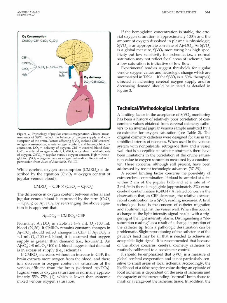
While cerebral oxygen consumption (CMRO2) is de-scribed by the equation (CjvO2 5 oxygen content ofjugular venous blood):
CMRO2 5 CBF 3 (CaO2 2 CjvO2)
The difference in oxygen content between arterial andjugular venous blood is expressed by the term (CaO22 CjvO2) or AjvDO2. By rearranging the above equa-tion it is apparent that:
AjvDO2 5 CMRO2/CBF
Normally, AjvDO2 is stable at 4–8 mL O2/100 mLblood (29,30). If CMRO2 remains constant, changes inAjvDO2 should reflect changes in CBF. If AjvDO2 is,4 mL O2/100 mL blood, it is assumed that oxygensupply is greater than demand (i.e., luxuriant). AnAjvO2 .8 mL O2/100 mL blood suggests that demandis in excess of supply (i.e., ischemia).
If CMRO2 increases without an increase in CBF, thebrain extracts more oxygen from the blood, and thereis a decrease in oxygen content or saturation of thevenous effluent from the brain (widened AjvDO2).Jugular venous oxygen saturation is normally approx-imately 55%–75% (1), which is lower than systemicmixed venous oxygen saturation.
If the hemoglobin concentration is stable, the arte-rial oxygen saturation is approximately 100% and theamount of oxygen dissolved in plasma is physiologic,SjVO2 is an appropriate correlate of AjvDO2. As SjVO2is a global measure, SjVO2 monitoring has high spec-ificity but low sensitivity for ischemia, i.e., a normalsaturation may not reflect focal areas of ischemia, buta low saturation is indicative of low flow.
Experimental studies suggest thresholds for jugularvenous oxygen values and neurologic change which aresummarized in Table 1. If the SjVO2 is , 50%, therapy(s)directed at increasing cerebral oxygen supply and/ordecreasing demand should be initiated as detailed inFigure 3.
Technical/Methodological LimitationsA limiting factor in the acceptance of SjVO2 monitoringhas been a history of relatively poor correlation of con-comitant values obtained from cerebral oximetry cathe-ters to an internal jugular venous sample analyzed by aco-oximeter for oxygen saturation (see Table 2). Theoriginal oximetry catheters were designed for use in theumbilical arteries of neonates. When used in the venoussystem with nonpulsatile, retrograde flow and a vesselwall that is susceptible to catheter abutment, there havebeen limitations in the correlation of the online satura-tion value to oxygen saturation measured by a cooxime-ter. These concerns, although still present, have beenaddressed by recent technologic advances (37–39).
A second limiting factor concerns the possibility ofextracerebral contamination. If blood is sampled at a sitewithin 2 cm of the jugular bulb and at a rate of ,2 mL/min there is negligible (approximately 3%) extra-cerebral contamination (6,40,41). A related concern is theobservation that, as CBF decreases, the relative extrace-rebral contribution to a SjVO2 reading increases. A finaltechnologic issue is the concern of catheter migrationand abutment against the vessel wall. When this occurs,a change in the light intensity signal results with a trig-gering of the light intensity alarm. Distinguishing a “de-saturation reading” as a result of a change in position ofthe catheter tip from a pathologic desaturation can beproblematic. Slight repositioning of the catheter or of thepatient’s head may be all that is needed to achieve anacceptable light signal. It is recommended that becauseof the above concerns, cerebral oximetry catheters beroutinely calibrated to a cooximeter control.
It should be emphasized that SjVO2 is a measure ofglobal cerebral oxygenation and is not particularly sen-sitive to small areas of focal ischemia. Accordingly, thelikelihood of a false negative value during an episode offocal ischemia is dependent on the area of ischemia andthe capacity of the surrounding “normal” brain tissue tomask or average-out the ischemic tissue. In addition, the
Figure 2. Physiology of jugular venous oxygenation. Clinical meas-urements of SjVO2 reflect the balance of oxygen supply and con-sumption of the brain. Factors affecting SjVO2 include CBF, cerebraloxygen consumption, arterial oxygen content, and hemoglobin con-centration. DO2 5 delivery of oxygen, CBF 5 cerebral blood flow,CaO2 5 arterial oxygen content, CMRO2 5 cerebral metabolic rateof oxygen, CjVO2 5 jugular venous oxygen content, Hgb 5 hemo-globin, SjVO2 5 jugular venous oxygen saturation. Reprinted withpermission from Atlas of Anesthesia, Vol III.
ANESTH ANALG MEDICAL INTELLIGENCE 5612000;90:559–66

recommendation regarding which jugular vein to can-nulate is not clear. As 70% of the cerebral venous blooddrains via the ipsilateral jugular veins, some cliniciansadvocate cannulating at the side of injury. However, inthe case of diffuse cerebral injury, most clinicians wouldmonitor the right side, as it is commonly dominant,whereas some clinicians would advocate monitoring theside of dominant flow in all situations (see Which Side
Should be Monitored?). More study is needed to defin-itively determine if there is an optimal side for SjVO2monitoring.
Clinical Factors Altering SjVO2Many factors affect the relationship of CMRO2 andoxygen delivery (see Figure 4). CBF can be decreasedby head injury, thromboembolism, intracranial hyper-tension, hypotension, hyperventilation, or vasospasm.If CMRO2 remains constant or increases under theseconditions, SjVO2 will decrease. Arterial hypoxia andincreased CMRO2 (e.g., febrile illness, seizures) canalso result in SjVO2 desaturation.
Causes of increased SjVO2 and luxuriant oxygen-ation are described in Figure 4. Correct interpretationof increased SjVO2 requires confirmation that the cath-eter tip is at the jugular bulb. Reduced CMRO2 (e.g.,hypothermia, sedatives), increased CBF, pathologicarterial-venous communications, and brain death mayresult in increased SjVO2.
Clinical Utility of SjVO2Monitoring-OverviewJugular venous oximetry is most often used in patientswith head injuries for neurosurgical procedures andfor cardiovascular procedures.
Head Injury
In the setting of head injury, SjVO2 monitoring providesan early diagnosis of ischemia resulting from either in-tracranial or systemic causes (42–49). Moreover, SjVO2monitoring may be useful to guide decisions for opti-mizing hyperventilation therapy (43,44,48,50,51), guid-ing fluid management and oxygenation (43,51,52),optimizing perfusion pressure (53–55), and detecting
Table 1. Summary of Data Suggesting a Correlation Between a Threshold Jugular Venous Oxygen Value and NeurologicChange
Species (Ref.) Pathologic/Physiologic Insult Jugular Venous Oxygen Value Neurologic Change
Cat (31) Increased intracranial pressure,hypocarbia
Oxygen content of 1.4 60.6 mL/100 mL
Metabolic failure (31P-magneticresonance spectroscopy)
Human (2) Nitrogen breathing SjVO2 , 33% ConfusionHuman (2) Nitrogen breathing SjVO2 ' 26% Lost consciousnessHuman (32) Nitrogen breathing SjVO2 , 40% Electroencephalographic slowingHuman (33) Carotid endarterectomy SjVO2 , 50% Neurologic deficitHuman (34) Rewarming after hypothermic
cardiopulmonary bypassSjVO2 , 50% Cerebral anaerobic metabolism
Human (35) Surgery for evacuation oftraumatic intracranialhematomas
SjVO2 , 50% Cerebral anaerobic metabolism
Human (36) Acute, severe, closed-braintrauma
SjVO2 , 30% for .10 min Decreased Glasgow Coma Scalescore
SjVO2 5 jugular venous oxygen saturation.
Figure 3. A strategy for management of low jugular venous oxygensaturation. SjVO2 5 jugular venous oxygen saturation, R/O 5 ruleout, SaO2 5 arterial oxygen saturation, CBF 5 cerebral blood flow,Paco2 5 arterial partial pressure carbon dioxide, AjvDO2 5 thedifference in oxygen content between simultaneously drawn sam-ples of arterial and jugular venous blood. Modified and reprintedwith permission from Atlas of Anesthesia, Vol III.
562 MEDICAL INTELLIGENCE ANESTH ANALG2000;90:559–66

arterial-venous fistulas (56,57). Used with a transcranialDoppler monitor, SjVO2 can help discern between hy-peremia and vasospasm. With high flow velocity de-tected by transcranial Doppler, SjVO2 is increased dur-ing hyperemia and normal or low if cerebral vasospasmis present.
Barbiturate-induced cerebral metabolic suppressionand induced hyperventilation are examples of twotherapies for head injury that may be guided by SjVO2monitoring. Cruz (58) identified a group of head-injured patients who responded to pentobarbital witha decrease in SjVO2. It was hypothesized that thevasoconstrictive effect of barbiturates resulted in in-creased cerebrovascular resistance and oligemic cere-bral hypoxia in these patients.
Routine hyperventilation after traumatic brain in-jury is not currently recommended (45). Rather, con-temporary guidelines recommend “optimal hyperven-tilation” guided by SjVO2 monitoring, thus identifyingthose head-injured patients with the potential for anischemic response to hypocarbia (45). Moreover,SjVO2 monitoring is also useful in evaluating progno-sis for head-injured patients (59). A strategy for man-agement of low SjVO2 occurring after head injury isdetailed in Figure 3.
Cardiovascular and Neurologic Surgery
Neurologic dysfunction is not uncommon after car-diac surgery with cardiopulmonary bypass and is at-tributed to the adverse effects of nonphysiologicmodes of perfusion (60). A particularly critical periodconcerns rewarming after hypothermic cardiopulmo-nary bypass. Rewarming has been related to frequentSjVO2 desaturations which are associated with in-creased postoperative neurocognitive deficits (61–63).It has been suggested that SjVO2 monitoring may have
Figure 4. Jugular venous oxygen saturation (%). Many factors alterthe relationship between cerebral oxygen consumption (CMRO2) andsupply. Simplistically, when cerebral oxygen demand exceeds supply,the brain extracts oxygen from hemoglobin, resulting in a decreasedoxygen saturation of the blood in the jugular bulb. Although somewhatarbitrary, a jugular venous oxygen saturation ,50% is considered low,and therapy directed at improving cerebral oxygen supply or decreas-ing demand should be instituted. ICP 5 intracranial pressure, Paco2 5arterial partial pressure carbon dioxide, CMRO2 5 cerebral metabolicrate of oxygen, CBF 5 cerebral blood flow, O2 5 oxygen. Modified andreprinted with permission from Atlas of Anesthesia, Vol III.
Table 2. Limitations of Jugular Venous Oximetry
Limitation Rationale Management
Incomplete mixing Venous sample may not berepresentative of the entire brain ifasymmetric venous drainage.
Cannulate dominant internal jugularvein (usually right), or considerplacing on side of the most severefocal injury.
Extracerebralcontamination
'3% of jugular blood is contaminatedby blood from scalp, meninges, andskull.
Radiograph confirmation. Location ofcatheter tip above lower border ofC1 and withdraw sample slowly(,2 mL/min).
Bohr effect Falsely high SjVO2 values may occurfrom a leftward shift of theoxyhemoglobin dissociation curveduring alkaline conditions.
Detect by measuring low jugular bulbPo2 (,27 mm Hg)
Global measure With focal cerebral injuries, SjVO2may not provide information aboutregional injury.
Measurement of arteriovenous lactatemay be helpful as an indicator ofanaerobic metabolism.
Insensitive to infratentorialflow
Brain stem and cerebellum contributelittle to the venous outflow fromthe brain.
Is of limited value for monitoringpatients with brainstem injuries.
Monitoring errors Catheter may be against wall of vein,coiled back on itself, or have fibrinformation at tip.
May need catheter repositioning,recalibration of continuousfiberoptic catheter, or heparinizedsaline flush ('3 mL/h).
SjVO2 5 jugular venous oxygen saturation.Modified and reprinted with permission from Atlas of Anesthesia, Vol III.
ANESTH ANALG MEDICAL INTELLIGENCE 5632000;90:559–66

a role in cerebral monitoring during adult (64) andpediatric (65) cardiac surgery.
The potential applications of SjVO2 monitoring dur-ing neurosurgery have been studied by Matta et al.(66). They demonstrated that the SjVO2 catheter couldbe placed quickly and detect frequent critical episodesof SjVO2 desaturation that would otherwise have beenuntreated. During intracranial aneurysm surgery,SjVO2 monitoring has been used to determine theminimal blood pressure that should be maintained toavoid hypoperfusion (67).
Systemic versus Jugular versus CerebralOxygen SaturationThe possibility of using systemic mixed venous oxy-genation as a surrogate of SjVO2 to assess cerebraloxygenation has been studied. Given that the brainreceives approximately 15% of the total cardiac outputand that it extracts a greater fraction of oxygen fromarterial blood than do other organs, it does not seemlogical that systemic mixed venous oxygenationwould be a reliable marker of cerebral oxygenation.Indeed, the lack of an association between systemicmixed venous oxygen and SjVO2 has been validatedwith limited studies (34,68–70).
A concept with some promise is the potential fornear-infrared spectroscopy (NIRS), a continuous, non-invasive monitor of brain oxygenation at the tissuelevel, to correlate with SjVO2 as a measure of cerebraloxygen use. Presently, NIRS methods in adults aresuspect primarily because of the inadequate predict-ability of extracerebral tissue to affect the NIRS signal,changes in arteriovenous partitioning, algorithm lim-itations, and variations in optical path length. It islikely that future advances in NIRS technology willresult in a better understanding of these effects, withapplication of real-time correction factors. At this time,the data do not support a correlation between SjVO2and NIRS when assessing cerebral oxygen saturationin adults (71,72).
Despite the limitations of SjVO2 monitoring, there isno better, commercially available, continuous, rela-tively low-cost, bedside monitor to assess the ade-quacy of cerebral oxygenation. Jugular venous oxy-genation provides information on global brainoxygenation and is recommended in the treatment ofpatients with head injury, especially those receivinghyperventilation therapy.
Jugular venous oxygen monitoring is often per-formed in conjunction with other monitoring and im-aging techniques and provides early detection of ce-rebral ischemia that might otherwise go unrecognized.However, the question should be asked: Do clinical
decisions impacted by SjVO2 data effect patient out-come? Although there appears to be a correlation be-tween episodes of SjVO2 desaturation and poor neu-rologic outcome, the impact of decisions influenced bySjVO2 data and their effect on long-term cerebral func-tion has yet to be proven.
References1. Gibbs EL, Lennox WG, Nims LF, Gibbs FA. Arterial and cere-
bral venous blood: arterial-venous differences in man. J BiolChem 1942;144:325–32.
2. Lennox WG, Gibbs FA, Gibbs EL. Relationship of unconscious-ness to cerebral blood flow and to anoxemia. Arch NeurolPsychiat 1935;34:1001–13.
3. Ferris E, Engel G, Stevens C, Logan M. The validity of internaljugular venous blood in studies of cerebral metabolism andblood flow in man. Am J Physiol 1946;147:517–21.
4. Feldman Z, Robertson CS. Monitoring of cerebral hemodynam-ics with jugular bulb catheters. Crit Care Clin 1997;13:51–77.
5. De Deyne C, Van Aken J, Decruyenaere J, et al. Jugular bulboximetry: review on a cerebral monitoring technique. Acta An-aesthesiol Belg 1998;49:21–31.
6. Shenkin GA, Harmel MH, Kety SS. Dynamic anatomy of thecerebral circulation. Arch Neurol Psychiatry 1948;60:240–52.
7. Gibbs EL, Lennox WG, Gibbs FA. Bilateral internal jugularblood: comparison of A-V differences, oxygen-dextrose ratiosand respiratory quotients. Am J Psychiatry 1945;102:184–90.
8. Lassen NA. Cerebral blood flow and oxygen consumption inman. Physiol Rev 1959;39:183–238.
9. Beards SC, Yule S, Kassner A, Jackson A. Anatomical variationof cerebral venous drainage: the theoretical effect on jugularbulb blood samples. Anaesthesia 1998;53:627–33.
10. Robertson CS, Narayan RK, Gokosla ZL, et al. Cerebral arterio-venous oxygen difference as an estimation of cerebral bloodflow in comatose patients. J Neurosurg 1989;70:222–30.
11. Hatiboglu MT, Anil A. Structural variations in the jugular fora-men of the human skull. J Anat 1992;180:191–6.
12. Gibbs EL, Gibbs FA. The cross section areas of the vessels thatform the torcular and the manner in which blood is distributedto the right and to the left lateral sinus. Anat Rec 1934;59:419–26.
13. Cruz J. Continuous versus serial global cerebral hemometabolicmonitoring: applications in acute brain trauma. Acta Neurochir(Wien) 1988;42(Suppl):35–9.
14. Howard L, Gopinath SP, Uzura M, et al. Evaluation of a newfiberoptic catheter for monitoring jugular venous oxygen satu-ration. Neurosurgery 1999;44:1280–5.
15. Andrews PJD, Dearden NM, Miller JD. Jugular bulbcannulation: description of a cannulation technique and valida-tion of a new continuous monitor. Br J Anaesth 1991;67:553–8.
16. Nakajima T, Ohsumi H, Kuro M. Accuracy of continuous jug-ular bulb venous oximetry during cardiopulmonary bypass.Anesth Analg 1993;77:1111–5.
17. Cowan F, Thoresen M. Ultrasound study of the cranial venoussystem in the human new-born infant and the adult. ActaPhysiol 1983;117:131–7.
18. Stochetti N, Paparella A, Bridelli F, et al. Cerebral venous oxy-gen saturation studied with bilateral samples in the internaljugular veins. Neurosurgery 1994;34:38–44.
19. Dearden NM. Jugular bulb venous oxygen saturation in themanagement of severe head injury. Curr Opin Anesthesiol 1991;4:279–86.
20. Gayle MO, Frewen TC, Armstrong RF, et al. Jugular venousbulb catheterization in infants and children. Crit Care Med1989;17:385–8.
21. Segal J. Percutaneous catheterization of the jugular bulb with aDoppler probe: technical note. Neurosurgery 1993;33:151–3.
22. Henson LC, Calalang C, Temp JA, et al. Accuracy of a cerebraloximeter in healthy volunteers under conditions of isocapnichypoxia. Anesthesiology 1998;88:58–65.
564 MEDICAL INTELLIGENCE ANESTH ANALG2000;90:559–66

23. Cruz J. Adverse effects of pentobarbital on cerebral venousoxygenation of comatose patients with acute traumatic brainswelling: relationship to outcome. J Neurosurg 1996;85:758–61.
24. Denys BG, Uretsky B. Anatomical variations of internal jugularvein location: impact on central venous access. Crit Care Med1991;19:1516–9.
25. Mallory DL, Shawker T, Evans G, et al. Effects of clinical ma-neuvers on sonographically determined internal jugular veinsize during venous cannulation. Crit Care Med 1990;18:1269–73.
26. Goetting MG, Preston G. Jugular bulb catheterization does notincrease intracranial pressure. Intensive Care Med 1991;17:195–8.
27. Stochetti N, Barbagallo M, Gordon CR, et al. Arterio-jugulardifference of oxygen and intracranial pressure in comatose,head injured patients: technical aspects and complications. Min-erva Anesthesiol 1991;57:319–26.
28. Bankier AA, Fleischman D, Windisch A, et al. Position of jugularoxygen saturation catheter in patients with head trauma: assess-ment by use of plain films. Am J Roentgenol 1995;164:437–41.
29. Souter M, Andrews P. A review of jugular venous oximetry.Intensive Care World 1996;13:32–8.
30. Ritter A, Robertson C. Cerebral metabolism. Neurosurg ClinNorth Am 1994;5:633–45.
31. Sutton LN, McLaughlin AC, Dante S, et al. Cerebral venousoxygen content as a measure of brain energy metabolism withincreased intracranial pressure and hyperventilation. J Neuro-surg 1990;73:927–32.
32. Meyer J, Gotoh F, Ebihara S, Tomita M. Effects of anoxia oncerebral metabolism and electrolytes in man. Neurology 1965;15:892–901.
33. Lyons C, Clark L, McDowell H, McArthur K. Cerebral venousoxygen content during carotid thrombintimectomy. Ann Surg1964;160:561–7.
34. Sapire KJ, Gopinath SP, Farhat G, et al. Cerebral oxygenationduring warming after cardiopulmonary bypass. Crit Care Med1997;25:1655–62.
35. Gopinath SP, Cormio M, Ziegler J, et al. Intraoperative jugulardesaturation during surgery for traumatic intracranial hemato-mas. Anesth Analg 1996;83:1014–21.
36. Cruz J. On-line monitoring of global cerebral hypoxia in acutebrain injury: relationship to intracranial hypertension. J Neuro-surg 1993;79:228–33.
37. Ritter AM, Gopinath SP, Contant C, et al. Evaluation of a re-gional oxygen saturation catheter for monitoring SjvO2 in headinjured patients. J Clin Monit 1996;12:285–91.
38. Souter MJ, Andrews PJ. Validation of the Edslab dual lumenoximetry catheter for continuous monitoring of jugular bulboxygen saturation after severe head injury. Br J Anaesth 1996;76:744–6.
39. Howard L, Gopinath SP, Uzura M, et al. Evaluation of a newfiberoptic catheter for monitoring jugular venous oxygen satu-ration. Neurosurgery 1999;44:1280–5.
40. Jakobsen M, Enevoldsen E. Retrograde catheterization of theright internal jugular vein for serial measurements of cerebralvenous oxygen content. J Cereb Blood Flow Metab 1989;9:717–20.
41. Matta BF, Lam AM. The rate of blood withdrawal affects theaccuracy of jugular venous bulb oxygen saturation measure-ments. Anesthesiology 1997;86:806–8.
42. Gupta AK, Hutchinson PJ, Al-Rawi P, et al. Measuring braintissue oxygenation compared with jugular venous oxygen sat-uration for monitoring cerebral oxygenation after traumaticbrain injury. Anesth Analg 1999;88:549–53.
43. Thiagarajan A, Goverdhan PD, Chari P, et al. The effect ofhyperventilation and hyperoxia on cerebral venous oxygen sat-uration in patients with traumatic brain injury. Anesth Analg1998;87:850–3.
44. Lewis SB, Myburgh JA, Thornton EL, et al. Cerebral oxygen-ation monitoring by near-infrared spectroscopy is not clinicallyuseful in patients with severe closed-head injury: a comparisonwith jugular venous bulb oximetry. Crit Care Med 1996;24:1334–8.
45. Guidelines for the management of severe head injury. Washing-ton, DC: Brain Trauma Foundation, 1995.
46. Murr R, Schurer L. Correlation of jugular venous oxygen satu-ration to spontaneous fluctuations of cerebral perfusion pres-sure in patients with severe head injury. Neurol Res 1995;17:329–33.
47. Fortune JB, Feustel PJ, Graca L, et al. Effect of hyperventilation,mannitol, and ventriculostomy drainage on cerebral blood flowafter head injury. J Trauma 1995;39:1091–9.
48. Fortune JB, Feustel PJ, Weigle CGM, et al. Continuous meas-urement of jugular venous oxygen saturation in response totransient elevations of blood pressure in head-injured patients.J Neurosurg 1994;80:461–8.
49. Chan KH, Dearden NM, Miller JD, et al. Multimodality moni-toring as a guide to treatment of intracranial hypertension aftersevere brain injury. Neurosurgery 1993;32:547–53.
50. Skippen P, Seear M, Poskitt K, et al. Effect of hyperventilationon regional cerebral blood flow in head-injured children. CritCare Med 1997;25:1402–9.
51. Matta BF, Lam AM, Mayberg TS. The influence of arterialoxygenation on cerebral venous oxygen saturation during hy-perventilation. Can J Anaesth 1994;41:1041–6.
52. Schaffranietz L, Heinke W. The effect of different ventilationregimes on jugular venous oxygen saturation in elective neuro-surgical patients. Neurol Res 198;20(Suppl 1):S66–70.
53. Chan KH, Miller JD, Dearden NM, et al. Multimodality moni-toring as a guide to treatment of intracranial hypertension aftersevere brain injury. Neurosurgery 1993;32:547–53.
54. Chan KH, Miller JD, Dearden NM, et al. The effect of changes incerebral perfusion pressure upon middle cerebral artery bloodflow velocity and jugular bulb venous oxygen saturation aftersevere brain injury. J Neurosurg 1992;77:55–61.
55. Cruz J. Combined continuous monitoring of systemic and cere-bral oxygenation in acute brain injury: preliminary observa-tions. Crit Care Med 1993;21:1225–32.
56. Della Corte F, Clemente A, Mignani V, et al. Diagnosis oftraumatic carotid-cavernous sinus fistula by monitoring venousoxygen saturation in the jugular bulb: report of two cases.Neurosurgery 1996;39:390–3.
57. Calon B, Freys G, Launoy A, et al. Early discovery of a traumaticcarotid-cavernous sinus fistula by jugular venous oxygen satu-ration monitoring. J Neurosurg 1995;83:910–1.
58. Cruz J. Adverse effects of pentobarbital on cerebral venousoxygenation of comatose patients with acute traumatic brainswelling: relationship to outcome. J Neurosurg 1996;85:758–61.
59. Gopinath SP, Robertson CS, Contant CF, et al. Jugular venousdesaturation and outcome after head injury. J Neurol Neuro-surg Psychiatry 1994;57:717–23.
60. Schell RM, Kern FH, Greeley WJ, et al. Cerebral blood flow andmetabolism during cardiopulmonary bypass. Anesth Analg1993;76:849–65.
61. Croughwell ND, Frasco P, Blumenthal JA, et al. Warming dur-ing cardiopulmonary bypass is associated with jugular bulbdesaturation. Ann Thorac Surg 1992;53:827–32.
62. Souter MJ, Andrews PJD, Alston RP. Jugular venous desatura-tion following cardiac surgery. Br J Anaesth 1998;81:239–41.
63. Croughwell ND, Newman MF, Blumenthal JA, et al. Jugularbulb saturation and cognitive dysfunction after cardiopulmo-nary bypass. Ann Thorac Surg 1994;58:1702–8.
64. Schell RM, Kern FH, Reves JG. The role of continuous jugularvenous saturation monitoring during cardiac surgery with car-diopulmonary bypass. Anesth Analg 1992;74:627–9.
65. Kern FH, Schell RM, Greeley WJ. Cerebral monitoring duringcardiopulmonary bypass in children. J Neurosurg Anesth 1993;5:213–7.
66. Matta BF, Lam AM, Mayberg TS, et al. A critique of the intra-operative use of jugular venous bulb catheters during neuro-surgical procedures. Anesth Analg 1994;79:745–50.
67. Moss E, Dearden NM, Berridge JC. Effects of changes in meanarterial pressure on SjO2 during cerebral aneurysm surgery. Br JAnaesth 1995;75:527–30.
ANESTH ANALG MEDICAL INTELLIGENCE 5652000;90:559–66

68. Croughwell ND, White WD, Smith LR, et al. Jugular bulb sat-uration and mixed venous saturation during cardiopulmonarybypass. J Card Surg 1995;10(4 Suppl):503–8.
69. Wohl JS, Baggs A, Lin JL, et al. Use of jugular venous blood,compared with mixed venous blood, for measurement of ve-nous oxygenation indices in a porcine model of endotoxicshock. Am J Vet Res 1997;58:910–4.
70. Newman MF, Schell RM, Reves JG. Monitoring the jugular-venous oxyghemoglobin saturation as a decision aid for normo-thermic cardiopulmonary bypass [letter]. Anesth Analg 1994;78:404.
71. Lewis SB, Myburgh JA, Thornton EL, Reilly PL. Cerebral oxy-genation monitoring by near-infrared spectroscopy is not clini-cally useful in patients with severe closed-head injury: a com-parison with jugular venous bulb oximetry. Crit Care Med1996;24:1334–8.
72. Ter Minassian A, Poirier N, Pierrot M, et al. Correlation betweencerebral oxygen saturation measured by near-infrared spectros-copy and jugular oxygen saturation in patients with severeclosed head injury. Anesthesiology 1999;91:985–90.
566 MEDICAL INTELLIGENCE ANESTH ANALG2000;90:559–66