cerebral cortex - mrsichakpchs.weebly.commrsichakpchs.weebly.com/uploads/1/1/2/3/11239671/... ·...
TRANSCRIPT

Cerebral Cortex
• Top layer of our brain.
• Contains wrinkles called fissures.
• The fissures increase surface area of our brain.
• Laid out it would be about the size of a large pizza.

Areas of the Cerebral Cortex
• Divided into eight lobes, four in each hemisphere (frontal, parietal, occipital and temporal).
• Any area not dealing with our senses or muscle movements are called association areas.

The Cerebral Cortex

Frontal Lobe
• Deals with planning, maintaining emotional control and abstract thought.

Parietal Lobes
• Located at the top of our head.
• Contains the sensory cortex.

Temporal Lobes
• Process sound sensed by ears.

Occipital Lobes
• In the back of our head.
• Handles visual input from eyes.
• Right half of each retina goes to left occipital lobe and vice versa.

The Cerebral Cortex
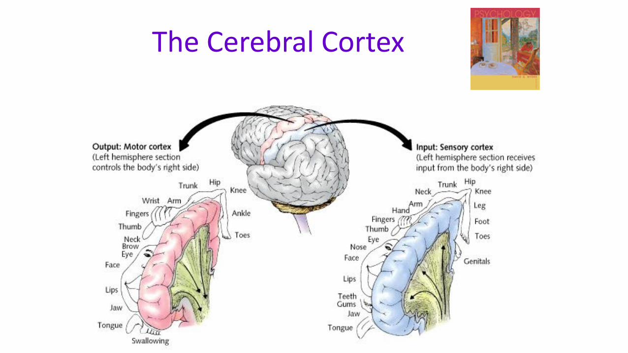
Motor Cortex
area at the rear of the frontal lobes that controls voluntary movements
Sensory Cortex
area at the front of the parietal lobes that registers and processes body sensations
The Cerebral Cortex

The Cerebral Cortex
Functional MRI scan shows the visual cortex activated as the subject looks at faces

Visual and Auditory Cortex

The Cerebral Cortex
Aphasia impairment of language, usually caused by left
hemisphere damage either to Broca’s area (impairing speaking) or to Wernicke’s area (impairing understanding)
Broca’s Area an area of the left frontal lobe that directs the
muscle movements involved in speech
Wernicke’s Area an area of the left temporal lobe involved in
language comprehension and expression

Association Areas
More intellegent animals have increased “uncommitted” or association areas of the cortex

Specialization and Integration

Specialization and Integration
Brain activity when hearing, seeing, and speaking words