cerebellopontine angle massesnams-india.in/downloads/lrm/cpa_mcvasudevan.pdf · • other cpa...
TRANSCRIPT

1
Dr. M.C.Vasudevan,Head Of Department,ALNC ‐
VHS
Management of Cerebellopontine Angle tumours

2
Introduction
• 10% of all intracranial tumors.• 78% are acoustic neuromas‐
mostly on vestibular
branch.• Other CPA masses:
– Meningiomas– Epidermoid– Other cranial nerve schwannomas– Arachnoid
cysts
– metastatic tumors– Jugular foramen tumours

3
HISTORY OF CEREBELLOPONTINE ANGLE SURGERY
• 1st successful complete removal ‐
1894 by Sir Charles Balance. The tumor was approached via a right
posterior fossa
craniectomy
and removed with the “finger”.
• H. Cushing (1917) was the first to advocate intracapsular
tumor removal and hence recurrence
was high.• W. Dandy(1925) introduced the concept of total tumor
removal‐
to prevent future reocurrences.• Olivecrona (1967) was 1st to preserve facial nerve• Leksell introduced Gamma‐knife in 1980 as a non
surgical treatment.
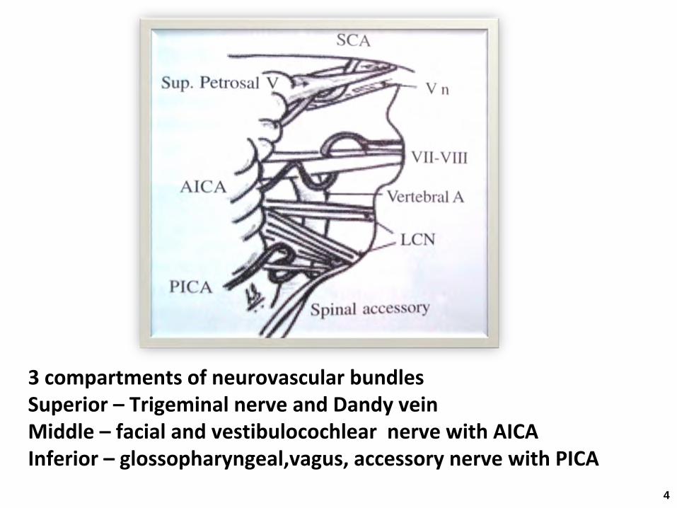
4
3 compartments of neurovascular bundlesSuperior –
Trigeminal nerve and Dandy vein
Middle – facial and vestibulocochlear
nerve with AICAInferior –
glossopharyngeal,vagus, accessory nerve with PICA

5
Acoustic Schwannomas
• 8% of intracranial tumour• The
acoustic
schwannoma
takes
origin
from
the
vestibular
component
of
the
8th
cranial nerve
near
the
internal
auditory
meatus,
at
the
transition
zone
where
the
Schwann
cells replace the oligodendroglia.

6
Symptoms & signs
• Intracanalicular:– Hearing loss (UL progressive ), tinnitus, vertigo
• Cisternal:– Worsened hearing and dysequilibrium
• Compressive:– Occasional occipital headache– CN V: reduced facial sensations, corneal hypesthesia– CN VII :loss of taste and reduced lacrimation
, LMN
facial weakness– CN VIII : progressive hearing loss,Tinnitus,vertigo– CN IX,X : swallowing difficulty, hoarseness

7
Symptoms & signs• Hydrocephalic:
– Fourth ventricle compressed and obstructed– Headache, visual changes, altered mental status– Nausea and vomiting
• Cerebellar
involvement– Incoordination
, widely based gate , tendency to fall
towards affected side
• Brainstem involvement: ‐
Ataxia, weakness and numbness of arms and legs with
exaggerated tendon reflexes.

8
1. 2.
3.4.
INTRACANALICULAR CISTERNAL
COMPRESSIVE HYDROCEPHALIC

9
(Samii and co-worker, 1995)

10
Diagnostic Tests
• Audiometric Testing.• Electrophysiologic
Testing.
• CT Brain contrast with bone cuts.• MRI brain contrast

11
Audiometric Testing
• Pure‐tone testing:– SNHL‐
most commonly high frequency (65%).
• Speech discrimination:– Scores out of proportion with pure‐tone thresholds.
• Acoustic reflex thresholds:– typically elevated or absent.– If present then reflex decay measured.– The sensitivity is 85% for detecting retrocochlear
problem.

12
Electrophysiologic
Testing
• ABR:– Most sensitive & specific audiologic
test.
• In patients with VS , the ABR is partially or completely absent , or there is a delay in latency of
wave V on the affected side.

13
BERA patterns in AN

14
Radiologic Features of vestibular schwannoma
• CT– Non‐contrast: usually isodense
to brain, calcification is
rare– IV Contrast: Over 90% of non‐treated tumors enhance
homogeneously
• MRI– T1W –
isointense
to brain, hyperintense
to CSF
– T2W –
hyperintense
to brain, iso/hypo‐intense to CSF– Gadolinium – Intense enhancement of tumor on T1W

15
CT BRAIN

16
MRI Brain
Isointense to brain, hyperintense to CSF
Hyperintense to brain, hypointense to CSF

17
CONTRAST MRI

18
NF2

19
Treatment
• Observation• Surgery
– Retrosigmoid– Translabrynthine– Middle Fossa
• Radiotherapy– Conventional radiation therapy– Stereotactic radiosurgery

20
Observation
• Indications– Advanced age – Poor health– Lack of symptoms– Non‐progression of symptoms– Only hearing ear
• Contraindications– Young patient– Healthy patient– Symptomatic progression– Compression of brainstem structures

21
BASIC REQUISITE FOR SURGERY
• CT scan brain plain and contrast• Bone cuts of the skull base with 1.5 mm cuts
to visualise the high lying jugular
• MRI scan brain plain and contrast study

22
BASIC REQUISITE FOR SURGERY
1. Microscope2. Fine dissector set3. CUSA (if available)4. Facial nerve monitor (if available)

23
Positioning

24
Positioning

25
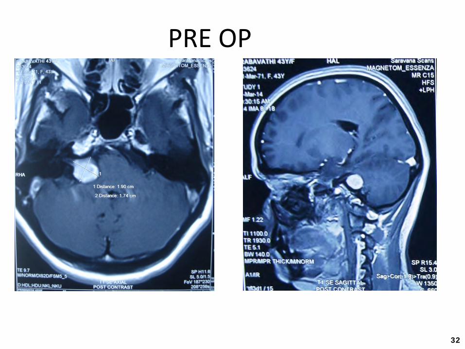
PRE OP

26
Retromastoid
suboccipital
approach

Retromastoid
suboccipital
approach

28
POST OP

29
Translabyrinthine
approach
IndicationsLesions where hearing preservation is not aimed at
1.
Acoustic neurinoma:‐
with bad preoperative hearing whatever
be the size of the tumour2.
Meningiomas
posterior to or centered to the
internal auditory canal with poor hearing3.
Epidermoids, dermoids
etc where poor
hearing is present.

30
Translabyrinthine
approach
• Contraindications:1.Only hearing ear2.Ipsilateral
CSOM

31
Translabyrinthine
approach
32
PRE OP

Translabyrinthine
approach

34
POST OP

35
Middle Fossa
• Indications– Small tumor– Intracanallicular
tumor
– Moderate CPA involvement– Adequate hearing (SRT<50 db, Disc >50%)
• Contraindications– Large tumors– Extensive CPA involvement ( > 0.5 – 1 cm)– Older patients ( > 60 yrs. may have higher rate of bleeding
or stroke)

36
37
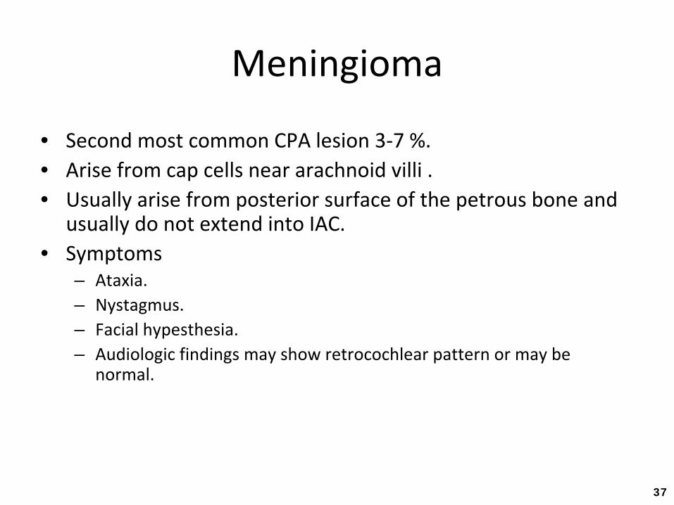
Meningioma
• Second most common CPA lesion 3‐7 %.• Arise from cap cells near arachnoid
villi
.
• Usually arise from posterior surface of the petrous
bone and usually do not extend into IAC.
• Symptoms– Ataxia.– Nystagmus.– Facial hypesthesia.– Audiologic
findings may show retrocochlear
pattern or may be
normal.
38
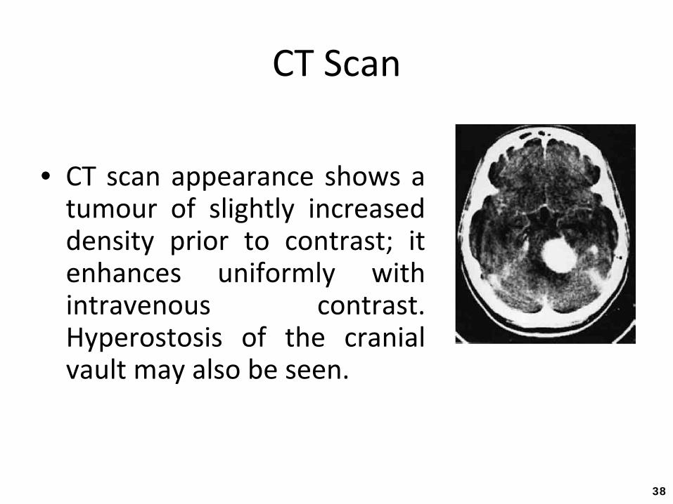
CT Scan
• CT
scan
appearance
shows
a tumour
of
slightly
increased
density
prior
to
contrast;
it enhances
uniformly
with
intravenous
contrast. Hyperostosis
of
the
cranial
vault may also be seen.

39
Meningioma
Features:
• Arise from surface of petrousbone.• Obtuse angles to petrous
bone.• Uncommonly involves the IAC.• Frequently with dural
tail.• Calcifications common.• Pial
vessel flow voids.

40
Treatment
• The treatment of choice for meningiomas
is complete excision of tumour.
• For small residual tumours, Stereotactic radiosurgery(SRS) may be advocated.

41
PRE OP

CP ANGLE MENINGIOMA

43
POST OP

44
Epidermoid
• Accounts for 2‐6% of CPA masses• Physiology:
– Congenital lesions that present in adulthood– Rests of ectodermal
tissue containing stratified squamous
lining
and keratin
• May arise within the temporal bone or in the CPA• Benign and slow growing• Symptoms
– Similar to acoustic neuroma
and meningioma– Facial nerve paresis and facial twitching may occur

45
Epidermoid
• Radiologic Features– Cistern oriented with variable shape with a
cauliflower surface appearance

46

47
Cerebellopontine
Angle Arachnoid
Cysts
• Arachnoid
cysts are intraarachnoid
masses of uncertain origin filled with CSF
• Often present with headache and ataxia. • If symptoms are few, observation is
advocated. • Symptomatic lesion require Marsupilization
of
cyst rather than excision or shunting.

48
Imaging
(a)
Axial T1‐weighted MR image shows an arachnoid
cyst with signal intensity
similar to that of CSF stretching the left seventh and eighth cranial nerve
complex (arrow).
(b)
Axial T2‐weighted MR image shows the cyst displacing the vascular structures
of the CPA (rowheads).

49
CN V Schwanoma

50
CN VII Schwanoma

51
CN X
Schwanoma

52
Glomus
Jugulare
53
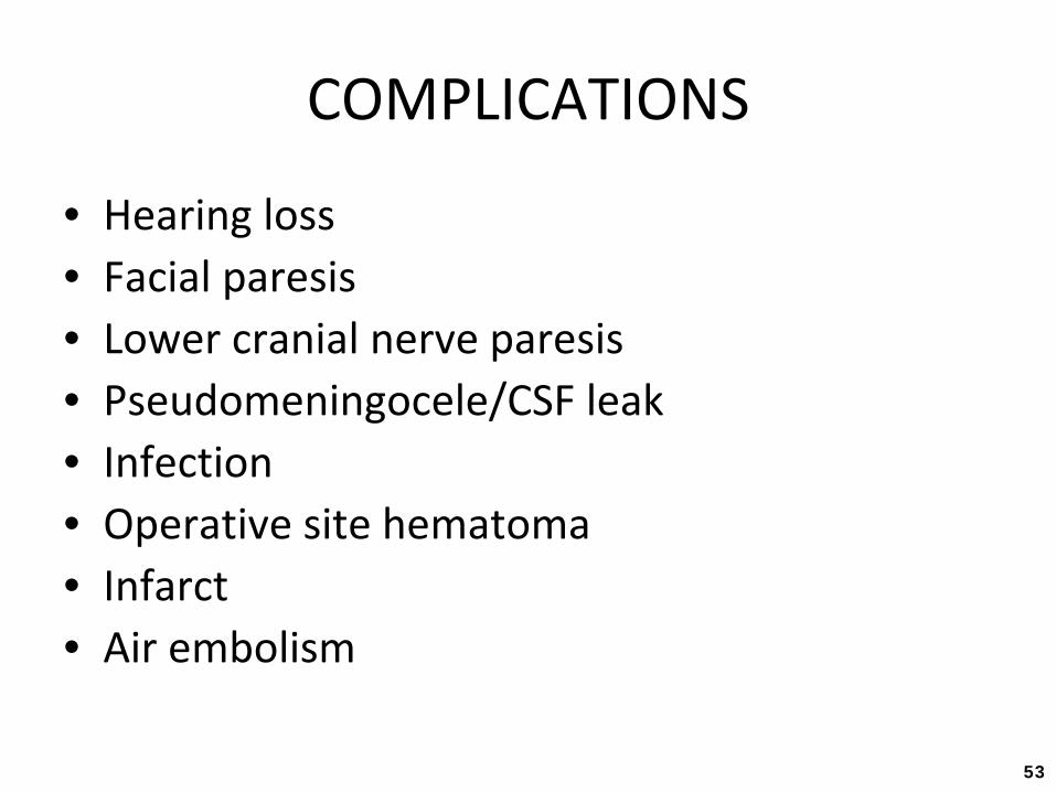
COMPLICATIONS
• Hearing loss• Facial paresis• Lower cranial nerve paresis• Pseudomeningocele/CSF leak• Infection• Operative site hematoma• Infarct• Air embolism

54

55

56
Stereotactic Radiosurgery• Indications
– Small tumors– Functional hearing– Older patients (>75)– Medically unstable patients – Small residual lesion
• Contraindications– Tumors > 3 cm– Prior radiotherapy– Tumor compressing brainstem

57
SRS

58
Thank You