cerase: a wax-decomposing enzyme in experimental tuberculosis
TRANSCRIPT

356 THE BRITISH J O U R N A L OF T U B E R C U L O S I S
CERASE
A WAX-DECOMPOSING ENZYME IN EXPERIMENTAL TUBERCULOSIS
BY MAI-IMOUD KAMAL MUYTIC
From King Faissal Hospital, Nassriyah, Iraq
IT is well known that leucocytes and other cells of the reticulo-endothelial system contain a variety of enzymes which attack the cellular structures of dead or living microbial agents. Among known enzymes one, lysozyme, causes the lysis of certain living bacteria, probably by attacking a structural component of their cell wall. There are many reports also describing the exis- tence in leucocytic extracts of fractions other than lysozyme which possess a direct bactericidal activity. Among these are the hmm compounds. It has long been known that hmms possess antibacterial activity in the free state, but this is not so when they are combined with proteins. I t is worth noting that the pigment of green pus is due to the presence in leucocytes of very high concentrations of a hmm-containing enzyme, verdoperoxidase. Contact between enzyme and substrate---in this case a living microbe--occurs naturally on the surface of the latter, that is to say, on its capsule or cell-membrane. Absorptive affinity and enzyme specificity towards cell-wall components are important factors in determining the bactericidal power of the enzyme, so that its absence may contribute to the development of a systemic infectious disease--e.g. , when bacteria can multiply within the cells, as in leprosy (Metchnikoff, I893 ) and tuberculosis (Mackaness, i954) , or when they are carried about by the cells.
Resistance of both kinds of mycobacteria seems to be associated with their acid-fastness. In x942 Rosenblatt, Fullar and Gessler observed the cell-wall of mycobacteria under an electron microscope (3o,ooo × magnified). It appears to have a uniform granular structure. Investigations into the chemical composition of the lipids of mycobacterial cell walls have been in progress for many years, and various workers have shown them to be mixtures of different esters and free long-chain fatty acids, such as phthioceric acid, phthioic acid, tuberculostearic acid, mycocerosic acid, mycolic acid isomers, etc. (Stodola and Anderson, I936; Anderson, Stodola and Lesuk, I938; Velick, i944; Ginger and Anderson, I945; and Chanley and Polgar, I95o ). This presupposes a waxy compound, so that the acid-fast membrane must be a fine waxy emulsion of high viscosity. It is well known that waxes are nearly always physiologically inert, so that their r61e is mostly protective in spite of their high caloric value. In mycobacteria they form the protective material in the cell-wall and are responsible for its acid-fastness.
Vertebrata are unable to decompose waxes either intracellularly or in the digestive apparatus, but at the beginning of this century, Metalnikov (I9o6)
(Received for publication April ~6, x 956.)
AND DISEASES OF THE CHEST 357
demonstrated the existence of wax-decomposing enzymes in wax-moth larvm (Galleria melonella). The larvae of this insect live entirely on beeswax. Metal- nikov (I 914) discovered further that Mycobacterium tuberculosis and Mycobacterium leprx can be digested very quickly by the leucocytes and other cells of these larvze, and that in their digestive tract bacteriolysis is accelerated. By adminis- tration of extracts of the larvae, he succeeded in keeping tuberculous guinea pigs alive for as long as a year after the death of the last control animal. Muftic (1949) obtained a crystalline wax-decomposing enzyme from a yeast-like fungus (Blastomyces cerolytica) which derives its energy from wax, and described its bactericidal activity against different saprophytic and pathogenic mycobac- teria. Mankiewicz (1952) obtained an active enzyme from an acetone preci- pitate of wax-moth larval extracts, which protected guinea pigs against 2 LD of tubercle bacilli (H~TRv). Kuzniecow and Wojciechowsky by means of different buffers also prepared an enzyme solution from an extract of larvae of the wax-moth. This showed a strong bactericidal action against Mycobac- terium tuberculosis.
For all enzymes which decompose waxes and wax-like substances Metal- nikov's designation (19o6) " cerase" has been adopted. The properties of the enzyme isolated by Muftic (i 955) are describedin several papers. Its composition is that of a cyclophoras-peroxidase of long carbohydrate chains, containing organic compounds of iron but not hems. Its peroxidase action is utilised for titrating its potency (measured in units), and its purity is estimated by determination of the iron content per gramme of substance. Like many enzymes containing heavy metals, cerase is thermolabile, and for this reason sterile solutions of the enzyme can be prepared only by filtration through a bacteriological filter. Cerase was not found to exert any bacteriostatic or bactericidal effect on other microbes than acid-fast ones.
In this paper we propose to describe the action of cerase in experimental tuberculosis in guinea pigs. The number of animals was limited by the small quantity of cerase available, and future experiments will be carried out on a larger scale.
MATERIALS
I. Fifty guinea pigs (male and female) weighing initially between 28o- 4oo gm.
2. MycObacterium tuberculosis hominis--H~TRv, obtained by courtesy of the Bacteriological Institute, Cairo University, and from the National Collection of Type Cultures, Medical Research Council of England. They were cultivated in Dubos' liquid medium, and before use re-inoculated on Loewenstein- Jensen's medium.
3. Cerase (wax-decomposing enzyme) solutions in ampoules containing 5 ° mg. of crystalline enzyme per ml. The enzyme was obtained in A.M.A. Laboratories Ltd. (Cairo, Heliopolis) by extraction from surface cultures of Blastomyces cerolyt#a by a special procedure described by Muftic (i955). The sterility of the ampoules was confirmed by several sub-cultures, and the activity of the enzyme estimated from its peroxidase value in an emulsion of ceryl-cerotate.

358 THE BRITISH JOURNAL OF TUBERCULOSIS
METHODS
All animals were inoculated intraperitoneally with I rag. (moist weight) of tubercle bacilli (Hs~Rv). The cxtreme Virulence of the infection was shown by the fact that the first of twenty-five untreated animals died nineteen days after being inoculated, while the last died two months after inoculation.
Cerase was administered to a second group of twenty-five guinea pigs. The administration was begun two weeks after inoculation of the animals with M. tuberculosis. About 5 ° mg. of cerase per kg. of body weight was injected intramuscularly every fourth day for ten weeks. Each of the treated animals received a total of approximately 0. 5 g. of cerase. Three of the treated animals died 7, 9 and I2 weeks respectively after inoculation; five animals were killed 6, 9, I2, 14 and 2o weeks after inoculation, and the remaining animals treated by cerase were electrocuted on the 2 ioth day.
HISTOI'ATHOLOGICAL FINDINGS Lungs
The most striking feature observed in the lungs of the first animal to be killed after cerase treatment was the paucity of giant cells in the tuberculous lesions, ill comparison with the control animals. The cells were small, with two to five nuclei. Occasionally, the tubercles were composed of an agglomeration of epithelioid cells occupying two or three alveoli. Ziehl-Neelsen's stain showed very few acid-fast bacilli in some tubercles.
For 6-12 weeks two types of lesion were distinguishable, namely, tubercles composed of epithelioid cells occupying one or many alveoli and lymphocytic islands composed of 20-40 lymphocytes. Very few giant cells were seen. The peribronchial lymph nodules were considerably swollen.
From 12-20 weeks there was a remarkable reduction in the number of loci to about one-sixth of the previous amount, and the histological picture they showed was most unusual. The inter-alveolar septa were apparently normal, but in the alveolar cavities many oval cells with multiple nuclei were seen. Although there was no proliferative tuberculosis this appearance may indicate the presence of desquamative alveolifis.
Thirty weeks after inoculation, in the treated animals, there was no sign of the pathological changes previously described. The lung tissue sections were absolutely normal.
It seems, therefore, that in the course of some 200 days the tuberculous process increased, then diminished and finally healed entirely without caseation being seen at any time. Acid-fast bacilli became increasingly rare until they disappeared after the twelfth week. The lungs of the last group of treated animals to be killed could not be distinguished from the lungs of an uninfected animal and it is difficult to believe that 200 days previously these animals had received i mg. of virulent tubercle bacilli intraperitoneally.
Liver
In the first six weeks there was occasional tubercle formation with many polynuclear and mononuclear leucocytes. There were a few emboli. Giant cells were few but large, with more than 2o nuclei, and surrounded by lympho-
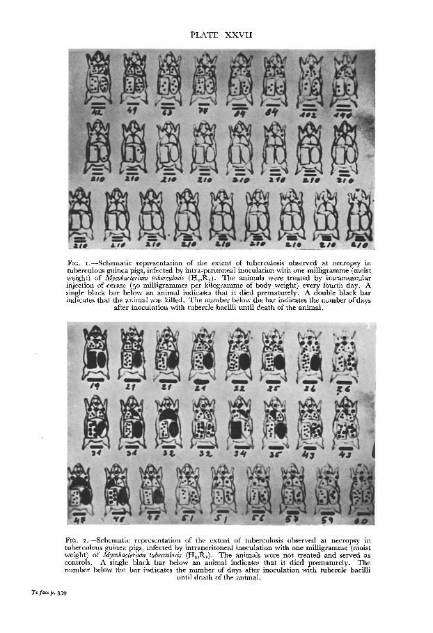
PLATE X X V I I
FIG. ~,--Schematic representation of the extent of tuberculosis observed at necropsy in tuberculous guinea pigs, infected by intra-peritoneal inoculation with one milligramme (moist weight) of Mycobacterium tuberculosis (H37Rv). The animals were treated by intramuscular injection of cerase (5 ° milligrammes per kilogramme of body weight) every fourth day. A single black bar below an animal indicates that it died prematurely. A double black bar indicates that the animal was killed. The number below the bar indicates the number of days
after inoculation with tubercle bacilli until death of the animal.
FIG. 2.--Schematic representation of the extent of tuberculosis observed at necropsy in tuberculous guinea pigs, infected by intraperitoneal inoculation with one milligramme (moist weight) of Mycobacterium tuberculosis (H37Rv), The animals were not treated and served as controls. A single black bar below an animal indicates that it died prematurely. The number below the bar indicates the number of days after inoculation with tubercle bacilli
until death of the animal.
To face p. 359
AND DISEASES OF THE CHEST 359
cytes. Plasma cells were also found. Fourteen weeks after inoculation there was considerable lymphocytic infiltration, but for the succeeding 2o weeks tubercles were extremely rare, and there was no evidence of change in the hepatic parenchyma.
Spleen In the initial 6-8 weeks there were many small giant cells in the sinuses
as well as groups of epithelial cells in the lymphoid follicles. After 14 weeks a remarkable change was seen, all the sinuses being engorged with small giant cells. Twenty to thirty weeks after inoculation, however, there were no epithelial cells left in the lymphoid follicles, and the sinuses looked normal.
Conclusion
It is evident that the lesions of these tuberculous guinea pigs, treated by wax-decomposing enzymes--cerase--did not show any tendency to caseation, and histological healing had occurred in about I4o days. Figs. I and 2 illustrate the course of the disease in the twenty-five guinea pigs treated with cerase, and the twenty-five control animals. Three animals died during treatment as a result of Pasteurella infection.
Intramuscular injection of cerase has no necrotic action on surrounding tissue, nor were any pathological changes seen affecting the kidney, heart or central nervous system. The first three injections caused a rise of temperature to 41,5 ° C. (p.r.), but following the next dose it did not reach 4 °° C. Normal, non-infected animals when injected with cerase did not show any rise in tem- perature. So far as we can determine cerase is not excreted by the kidney, but is destroyed in the organism or eliminated in some other way. Peroxidasic activity of the serum of treated animals was observed for five weeks after the last injection of enzyme.
Clearly these results must be enlarged upon by more extensive investigations, as a group of only twenty-five animals, the largest permitted by the amount of enzyme available, is too small to permit any definite conclusions being drawn regarding the pharmacological and toxicological behaviour of the enzyme preparation. I hope, however, that in the future it will be possible to study these problems on a large scale.
Discuss ion
Treatment of tuberculosis by lipolytic enzymes was introduced first by Fiessenger and Marie (I9o4). The enzymes they used were obtained from lymphatic glands of tuberculous animals. Later Fiessenger, Gajdos and Pezzangora (I935) used heptic lipase, and lipo-oxidase in tuberculous guinea pigs. They succeeded in reducing the number of foci. Corper and Sweany (I 918) observed autolysis of Mycobacterium tuberculosis after its decapsulation by organic solvents. Recently, Prina (1952) found that streptomycin possesses a lipolytic action for tributyrin, tweens and lipids from tubercle bacilli. Man- kiewicz (1952) found that cerase from larvze of Galleria melonella abolishes the acid-fastness of mycobacteria, coincidentally changing their cytochemical properties so that they become negative in the Dubos neutral red reaction, and

360 T H E B R I T I S H J O U R N A L O F T U B E R C U L O S I S
retain no Sudan Black " B " after treatment with acetone. Muftic (1955) detected the formation of fat peroxides in Mycobacterium tuberculosis after treatment with very dilute cerase solutions. The protective action of cerase in tuberculous guinea pigs was observed by Metalnikov (192o) and Mankiewicz (1952) . In our experiments the disease was better controlled by purified and highly concentrated preparations and using greater amounts of enzyme than did these two authors. I t seems that peroxide (fat) formation and decom- position of waxy substances of the cell wall of mycobacteria prevents caseation in infected animals, possibly because of changes in the antigenic structure of the bacilli. The enhanced lymphocytosis seems to be related to the benign advance- ment of the infection. The small number of giant cells suggests that there is little stimulus to the reticulo-endothelial system to attack the germs, which are already undergoing lysis. This is the same picture that Metalnikov (19o6) observed fifty years ago in the tissues of larvae of Galleria mellonella infected with tubercle bacilli.
Giant cell formation, caseation, deposition of fibrin and collagen at the site of the lesion appears to be the tissue response to invasion where tissue enzymes are incapable of destroying the invader. This incapability arises partly because the invader is protected by resistant material, which cannot be decomposed by the enzymes available in the tissues, and partly because the invader secretes substances which inhibit the enzymes. The former explanation applies in the case of acid-fast microbes. Members of the Vertebrata cannot digest wax-like substances, and the absence of wax-destroying enzymes permits the complicated tissue reactions to tuberculous infections. The problems of infection and immunity are less complex than some theories suggest. It is a question of a biological fight between host and invader, where both are substrates and both enzyme producers; the end result is digestion of one of them.
Summary Guinea pigs infected intraperitoneally by Mycobacterium tuberculosis hominis
(HsTTv) were treated with wax-decomposing enzymes--" cerase." The course of the disease was changed and no caseation was observed. About 200 days after infection the treated animals showed histological healing of lesions. The suppression of infection was striking when compared with control animals which died between 19 and 60 days after inoculation. The total dose ofcerase for one animal was about o. 5 gin. Cerase is an iron-peroxidase; its activity can be demonstrated in the serum for several weeks after injection. There was no evidence of excretion of the enzyme by the kidney. The bactericidal action of the cerase is due probably to its power in decomposing the waxy membrane of mycobacteria, which the tissues of organisms of the Vertebrata cannot do.
One part of this research was done in A.M.A. Laboratories Ltd. (Heliopolis, Cairo) at the instigation of Dr. Z. Madgachian, director. The other part was facilitated by the help of the Iraqian Ministry of Public Health.
Some necessary material was given by the Rockefeller Foundation in Cairo by courtesy of Dr. W. Macintosh. The strains of Mycobacterium tuberculosis were obtained from Prof. M. Gohar of the Bacteriological Institute, Cairo University, and from the National Collection of Type Cultures, Medical Research Council of England.
To all of them we extend our grateful thanks.
A N D D I S E A S E S O F T H E C H E S T 361
REFERENCES ANDm~SON, R. J., STODOLA, F. H., and LEsuK, A. (1938):07. biol. Chem., 126, 505 ̀ CI-IANLEY, J. D., and POL~AR, N. (I95o): .htature, 166~ 639. CORPER, M. (1., and 8WEANY, S. (1918) : 07. Bat,, 3, I~ 9. FmSSENOm% N., and MAma, P. L. (19o4) : C.R. Soc. Biol. (Paris), 67, lO 7. FmSSENOER, N., GAJDOS, A., and PEZZANGORA, F. (1935), C.R. Soc. Biol. (Paris), 120, 985 . GINQER, L. O., and ANDm~SOI% 1~. J. (1945) : 07. biol. Chem., 157, ~o3. KUZNX~.cow, A., and WOJCmCHOWSKY, E. (I95o): Med. do~w. Mikrobiol., 2, 245. MA.CKANESS, G. B. (I954): Amer. Rev. Tuberc., ~9, 479. MANKn~WIeZ, E. (x952) : Canad. 07. reed. 8ci., 30, 116. M~TALmKOV, S. (19o6) : Arch. scien, biol. de Petersburg, 12, 3oo. METALNmOV, S. (1914): Z" ImmunForsch., 22, 235. METALm~OV, S. (x92o): Ann. Inst. Past., 34, 8Io. METCHmgOFF, E. (I893) : Lectures on comparative Pathology of Inflammation, London. Mtr~TiC, M. (1949): Experientia (Basel), % 2 I 9. MtrFTIC, M. (I955): Enzymologia (Amst.), BE 339. MUFTIC, M. (I955) : Enzymologia (Amst.), Vol. XVII , Fase. 4. PRL~A, C. (I952): Boll. Soc. ital. biol. sper., 28, I5o2. I~.OSENBLATT, M. B., FULLAR, ]~. F., and G~SSL~R, E. (I942): Amer. Rev. Tuberc., 46, 587 • STODOLA, F. H., and ANDERSON, R.J . (i936) : 07. biol. Chem., 114, 467 • VELICK, S. F. (I944):07. biol. Chem., 154~ 497.