central venous catheter-related infection...
TRANSCRIPT

CENTRAL VENOUS
CATHETER-RELATED INFECTION
A thesis submitted to the Faculty of Medicine, University of the Witwatersrand,
Johannesburg, in fulfillment of the requirements for the degree of Doctor of Philosophy
Johannesburg 2012

ii
DECLARATION
I, Mervyn Mer declare that this thesis is my own unaided work. It is being submitted for the
degree of Doctor of Philosophy in the University of the Witwatersrand, Johannesburg. It
has not been submitted before for any degree or examination at this or any other
University.
……………………………………… ………………………………………
Signature Date

iii
DEDICATION
This thesis is dedicated to my parents, Morris and
Lily - special people - and to my family, Avril, Myron,
Brett and Jonty for their unconditional love, support and patience

iv
PRESENTATIONS, PUBLICATIONS AND AWARDS ARISING FROM THIS STUDY
Presentations:
Multiple presentations at Critical Care Society of Southern Africa National Congresses
and Refresher Courses, Pulmonology Congresses, Anaesthesiology Congresses and
Refresher Courses, and the Southern African Society of Thrombosis and Haemostasis
Congress. These have included presentations of the original study data, as well as
plenary sessions on the subject.
Several presentations at regional and local meetings and symposia.
22nd International Symposium on Intensive Care and Emergency Medicine, Brussels,
Belgium (“Central Venous Catheter-Related Infection”).
Publications:
Mer M. Intravascular catheter related infection. S Afr J Crit Care 1999;15(2):30-35.
Mer M. Vascular catheter related infection. Clinical Intensive Care 2001;12(2):53-60.
Mer M, Duse AG, Galpin J, Taylor R, Richards GA. Central venous catheter-related
infection. Abst. Critical Care 2002;6(S1):S94.
Mer M. Nosocomial bloodstream infection. South Afr J Epidemiol Infect 2005; 20(2):61-62.
Mer M. Intravascular catheter-related guidelines. South Afr J Epidemiol Infect 2005;
20(2):64-70.
Brink A, Feldman C, Duse A, Gopalan D, Grolman D, Mer M, Naicker S, Paget G, Perovic
O, Richards G. Guideline for the Management of Nosocomial Infections in South Africa. S
Afr Med J 2006;96(2):641-652.
Mer M. Intravascular catheter-related infection: current concepts. S Afr J Crit Care 2006;
22(1):4-12.
Mer M. Nesibopho guideline on Intravascular Catheter-related Infection – Critical Care
Society of Southern Africa endorsed and independently peer-reviewed 2007.
v
Mer M. Intravascular catheter-related infection: update and overview. CME 2008;
26(11):540-544.
Mer M, Duse AG, Galpin J, Richards GA. Central venous catheterization. A randomized
prospective study. Clin Appl Thromb Hemost 2009;15(1):19-26. (published early on line)
Awards
Best presentation by a Doctor at Critical Care Society of Southern Africa Congress held at
Sun City 2001
Best paper in a peer reviewed journal awarded by the Critical Care Society of Southern
Africa 2009 – “Central venous catheterization: A prospective randomized, double-blind
study” published in Clinical and Applied Thrombosis / Hemostasis February 2009
vi
ABSTRACT
Introduction and Background: Central venous catheters (CVCs) are extensively used
worldwide. Mechanical, infectious and thrombotic complications are well described with
their use and may be associated with prolonged hospitalisation, increased medical costs
and mortality.
CVCs account for an estimated 90% of all catheter-related bloodstream infections
(CRBSI) and a host of risk factors for CVC-related infections have been documented.
These include, most importantly, the duration of catheterisation. The duration of use of
CVCs remains controversial and the length of time such devices can safely be left in place
has not been fully and objectively addressed in the critically ill patient. Over the past few
years, antimicrobial impregnated catheters have been introduced in an attempt to limit
catheter-related infection (CRI) and increase the time that CVCs can safely be left in situ.
Recent meta-analyses concluded that antimicrobial-impregnated CVCs appear to be
effective in reducing CRI.
Materials and Methods: This was a prospective randomised double-blind study
performed in the adult multidisciplinary Intensive Care Unit (ICU) at Charlotte Maxeke
Johannesburg Academic Hospital (CMJAH) over a four year period. The study entailed a
comparison of standard triple-lumen versus antimicrobial impregnated CVCs on the rate
of CRI. The aim was to determine whether the duration of catheter insertion time could
safely be increased from the standard practice of seven days at the CMJAH adult
multidisciplinary ICU to 14 days, to assess the influence of the antimicrobial impregnated
catheter on the incidence of CRI, and to elucidate the epidemiology and risks of CRI.
Results: One hundred and eighteen critically ill patients were included in the study which
spanned 34 951.5 catheter hours (3.99 catheter years). Sixty-two patients received a
standard triple-lumen catheter and 56, a chlorhexidine-silver sulfadiazine (CSS)
impregnated triple-lumen catheter. The mean duration of placement for the full sample of
vii
118 CVCs was 12.3 days (range, 1-14). No statistically significant difference in CRI rates
between the two types of catheters could be demonstrated. The most common source of
primary CRBSI was skin, followed by hub and infusate. The site of CVC insertion (internal
jugular versus subclavian vein) and the use of parenteral nutrition were not noted to be
risk factors for catheter infection. There was no clinical evidence of catheter-related
thrombosis in either of the study groups.
Conclusion: This study was unable to demonstrate that antimicrobial catheters provided
any significant benefit over standard catheters, which it is felt, can safely be left in place
for up to 14 days with appropriate infection control measures. The most common source
of CRI was the skin. The administration of parenteral nutrition and the site of catheter
insertion (internal jugular vein versus subclavian vein) were not noted to be risk factors for
CRI. There was no clinical evidence of thrombotic complications in either of the study
groups. This study offers direction for the use of CVCs in critically ill patients and
addresses many of the controversies that exist.
Key Words: central venous catheters, infection, duration, thrombosis

viii
ACKNOWLEDGEMENTS
It is with sincere appreciation that the following persons are acknowledged:
• Professor GA Richards, for his continual encouragement, support, supervision,
editing assistance and expertise.
• Professor A Solomon for his unstinting support, mentorship, encouragement and
outstanding advice.
• Members of staff of the Pulmonology and adult multidisciplinary Intensive Care
Unit at Charlotte Maxeke Johannesburg Academic Hospital, in particular, those
who helped to care for the patients involved in the study, and who offered ongoing
support and encouragement.
• Professor A Duse and members of staff of the National Health Laboratory Service
(NHLS) for their assistance with the microbiological and laboratory aspects of the
study.
• Professor J Galpin from the Department of Statistics and Actuarial Science,
University of the Witwatersrand, for her assistance with the statistical analyses.
• Ms E Venter for all her wonderful assistance during the compilation of the thesis.

ix
TABLE OF CONTENTS
x
DECLARATION ......................................................................................................................
DEDICATION .........................................................................................................................
PRESENTATIONS, PUBLICATIONS AND AWARDS ARISING FROM THIS STUDY ..........
ABSTRACT ............................................................................................................................
ACKNOWLEDGEMENTS ......................................................................................................
TABLE OF CONTENTS .........................................................................................................
LIST OF FIGURES .................................................................................................................
LIST OF TABLES ...................................................................................................................
LIST OF ABBREVIATIONS ....................................................................................................
CHAPTER 1 INTRODUCTION ...............................................................................
CHAPTER 2 BACKGROUND AND HISTORY ........................................................
CHAPTER 3 MAGNITUDE OF THE PROBLEM
AND EPIDEMIOLOGY .......................................................................
3.1 Nosocomial Infections ..................................................................................
3.2 Intravascular catheters, central venous catheters and catheter-related infections .............................................................................
3.3 Relevance .....................................................................................................
3.4 Developing countries ....................................................................................
CHAPTER 4 DEFINITIONS ...................................................................................
CHAPTER 5 PATHOGENESIS OF CATHETER-RELATED INFECTIONS ...........
5.1 Overview of the pathogenesis ......................................................................
5.2 Biofilm ............................................................................................................
5.3 Catheter-related thrombosis and infection .....................................................
CHAPTER 6 MICROBIOLOGY ...............................................................................
6.1 Overview ........................................................................................................
Page
ii
iii
iv
vi
viii
ix
xiv
xv
xvi
1
3
5
5
5
7
7
12
13
13
13
14
17
17
xi
6.2 Catheter-related Staphylococcus aureus infection ........................................
6.3 Catheter-related infections caused by coagulase-negative Staphylococcus .............................................................................................
6.3.1 Microbiology and differentiation from Staphylococcus aureus ...........
6.3.2 Epidemiology .....................................................................................
6.3.3 Resistance ..........................................................................................
6.4 Fungal infections of catheters ........................................................................
6.5 Catheter-related infections caused by Enterococci .......................................
6.6 Catheter-related infections due to Gram-negative organisms .......................
CHAPTER 7 DIAGNOSIS OF CATHETER-RELATED INFECTION ......................
7.1 Introduction ...................................................................................................
7.2 Clinical aspects .............................................................................................
7.3 Laboratory aspects ........................................................................................
7.4 Blood cultures ...............................................................................................
7.4.1 Peripherally collected blood ..............................................................
7.4.2 Paired quantitative cultures ...............................................................
7.4.3 Differential time to positivity ...............................................................
7.4.4 Catheter-drawn quantitative blood cultures ........................................
7.5 Catheter culture ............................................................................................
7.5.1 Semi-quantitative roll-plate method ....................................................
7.5.2 Quantitative catheter culturing techniques .........................................
7.5.3 Cultures of the insertion site and catheter hub ..................................
7.5.4 Other techniques ...............................................................................
7.6 Newer techniques .........................................................................................
7.6.1 The endoluminal brush ......................................................................
7.6.2 The Gram stain and AOLC test ..........................................................
7.7 Novel techniques ...........................................................................................
7.8 Other tests .....................................................................................................
7.9 Current suggestions ......................................................................................
18
20
21
22
22
23
25
26
31
31
31
32
32
33
33
34
34
35
35
36
37
37
37
37
38
38
39
39
xii
CHAPTER 8 PREVENTION OF CENTRAL VENOUS CATHETER-RELATED INFECTION IN THE ICU ...................................................................
8.1 Staff education .............................................................................................
8.2 Infusion therapy teams .................................................................................
8.3 Maximum sterile barriers ...............................................................................
8.4 Skin antisepsis ..............................................................................................
8.5 Antibiotic prophylaxis .....................................................................................
8.6 Luminal antimicrobial flushes and lock solutions ...........................................
8.7 Tunnelling of CVCs .......................................................................................
8.8 Silver-chelated subcutaneous collagen cuffs ................................................
8.9 Antiseptic hubs ..............................................................................................
8.10 Catheter insertion site ...................................................................................
8.11 Ultrasound guided placement of CVCs .........................................................
8.12 Catheter site dressings ..................................................................................
8.13 Needleless connectors ..................................................................................
8.14 Topical antimicrobials ....................................................................................
8.15 In-line filters ...................................................................................................
8.16 Peripherally inserted central venous catheters (PICCs) ................................
8.17 Catheter material ...........................................................................................
8.18 Catheter lumens ............................................................................................
8.19 Catheter and venous line maintenance .........................................................
8.20 Guidewire exchanges ....................................................................................
8.21 Catheter securement .....................................................................................
8.22 Antimicrobial catheters ..................................................................................
8.23 Heparin ..........................................................................................................
8.24 Novel, innovative and other approaches to the prevention of catheter-related infection ...............................................................................
44
44
45
45
46
48
49
50
51
51
51
53
54
56
56
57
57
59
59
60
61
61
62
62
62

xiii
CHAPTER 9 ANTIMICROBIAL CENTRAL VENOUS CATHETERS ......................
9.1 Introduction ....................................................................................................
9.2 Chlorhexidine silver sulfadiazine catheters ..................................................
9.3 Minocycline-rifampin catheters .....................................................................
9.4 Silver, platinum and carbon catheters ...........................................................
9.5 Cost-effectiveness .........................................................................................
9.6 Guidelines .....................................................................................................
CHAPTER 10 GUIDELINES ...................................................................................
CHAPTER 11 PRINCIPLES OF TREATMENT OF CATHETER-RELATED
INFECTION .......................................................................................
11.1 Catheter removal and rationale .....................................................................
11.2 Antimicrobials ................................................................................................
11.3 Other approaches ..........................................................................................
11.4 Catheter salvage therapy ..............................................................................
11.5 Complications ...............................................................................................
11.6 Special situations .........................................................................................
CHAPTER 12 STUDY PROTOCOL .......................................................................
12.1 Aims of this study .........................................................................................
12.2 Materials and methods .................................................................................
12.3 Collection and processing of specimens ......................................................
12.3.1 Collection and processing of CVC pre-insertion and pre-removal specimens for quantitative culture of the skin insertion site ..............
12.3.2 Collection and processing of specimens from catheter hubs ...........
12.3.3 Collection and processing of infusates .............................................
12.3.4 Collection and processing of blood cultures ......................................
12.3.5 Collection and processing of catheter segments for culture ..............
12.4 Statistics .......................................................................................................
12.5 Ethics Approval and International Protocol Registration ..............................
65
65
67
68
68
69
69
71
75
75
77
78
79
79
80
82
82
82
84
84
84
84
85
85
86

xiv
CHAPTER 13 RESULTS .......................................................................................
CHAPTER 14 DISCUSSION ..................................................................................
CHAPTER 15 CONCLUSION ................................................................................
REFERENCES .........................................................................................................
STUDY PAPER ........................................................................................................
APPENDIX 1 ............................................................................................................ APPENDIX 2 ............................................................................................................
86
87
93
98
99
153
154
155

xv
LIST OF FIGURES
Page
Figure 1 Pathogenesis of catheter-related infections ........................................ 16
Figure 2 The most common organisms associated with catheter-related infections ............................................................................................. 17
Figure 3 Candida involving retina ...................................................................... 32
Figure 4 Approach to the management of patients with short-term central venous catheter-related bloodstream infection ........................ 81

xvi
LIST OF TABLES
Page
TABLE 1 Impact of catheter-related bloodstream Infections in critically ill patients ............................................................................. 9
TABLE 2 Central venous catheter-related bloodstream infection rates in ICUs in developing countries ............................................................ 10
TABLE 3 Central line-associated bloodstream infection: extra mortality in developing countries ......................................................................... 11
TABLE 4 Definitions for catheter-related infections .......................................... 12
TABLE 5 CRBSI - most common pathogens and mortality rates .................... 29
TABLE 6 Gram-negative organisms and short-term CVCs, a compilation from several studies .......................................................................... 30
TABLE 7 Microbiological diagnostic methods of catheter-related bloodstream infections ...................................................................... 40
TABLE 8 Methods of catheter-tip culture ......................................................... . 41
TABLE 9 Methods of skin and hub cultures ..................................................... 42
TABLE 10 Method of the Gram stain and acridine-orange leucocyte cytospin (AOLC) test ........................................................ 43
TABLE 11 Replacement of intravenous tubing associated with central venous catheters .................................................................. 64 TABLE 12 Examples of commercially available antimicrobial intravascular
catheters .......................................................................................... 70
TABLE 13 Selected recommendations for the prevention catheter-related infections: comparison of three national guidelines ......................... 73
TABLE 14 Guidelines for the prevention of intravascular catheter-related infections, 2011 ................................................................................ 74
TABLE 15 Definitions for identifying the source of a catheter-related bloodstream infection ....................................................................... 90
TABLE 16 Characteristics of 118 critically ill patients enrolled in the study ....... 91
TABLE 17 Colonisation and infection data comparing standard catheter vs CSS catheter ................................................................................ 92

xvii
LIST OF ABBREVIATIONS
AAP: Accumulation-associated protein
AOLC: Acridine orange leucocyte cytospin
AtiE: Autolycin
BSI: Bloodstream infection
CAPD: Central Auditory Processing Disorder
CFU: Colony forming unit
CI: Confidence interval
Clf: Clumping factor
CMJAH: Charlotte Maxeke Johannesburg Academic Hospital
CoNS: Coagulase-negative Staphylococcus
Cps: Capsular polysaccharides
CRBSI: Catheter-related bloodstream infection
CRI: Catheter-related infection
CSS: Chlorhexidine silver sufadiazine
CVC: Central venous catheter
EDTA : Edetic acid
ELISA: Enzyme linked immunosorbent assay
EPIC: European Prevalence of Infection in Intensive Care
HICPAC: Healthcare Infection Control Practices Advisory Committee
HR: Hazard ratio
ica: Intercellular adhesion
ICU: Intensive care unit
IDSA: Infectious Diseases Society of America
IgG: Immunoglobulin G
INICC: International Nosocomial Infection Consortium
JK: Jeikeium

xviii
MBC: Minimum bacterial concentration
MIC: Minimum inhibitory concentration
MR: Minocycline-rifampin
MRSA: Methicillin-resistant Staphylococcus aureus
MSSA: Methicillin-susceptible Syaphylococcus aureus
NEMIS: National Epidemiology of Mycosis Survey
NNIS: National Nosocomial Infection Surveillance
OR: Odds ratio
PIA: Polysaccharide intercellular adhesion
PIA: Polysaccharide Intercellular Adhesion
PICC: Peripherally inserted central venous catheter
PMMA: Polymethylmethacrylate
PNB: Polymixin-neomycin-bacitracin
PVC: Polyvinyl chloride
RKI: Robert Koch Institute
RR: Relative risk
S aureus: Staphylococcal aureus
SCOPE: Surveillance and Control of Pathogens of Epidemiological Importance
SICU: Surgical intensive care unit
SPC: Silver platinum/carbon
™: Trademark
UK: United Kingdom
US: United States
USA: United States of America
vs: versus

1
CHAPTER 1
INTRODUCTION
Intravascular devices are an integral component of modern-day medical practice, particularly
in intensive care units 1. They are used to administer intravenous fluids, medications, blood
products and parenteral nutrition. In addition, they serve as a useful monitor of the
haemodynamic status of critically ill patients 2,3,4,5.
Infection is one of the leading complications of intravascular catheters and is associated with
increased mortality, prolonged hospitalisation and increased medical costs 6-19. Central
venous catheters (CVCs) account for the vast majority of all catheter-related bloodstream
infections (CRBSI) 2,3,5,20-24.
Several risk factors have been documented for CVC-related infections which include very
importantly, the duration of catheterisation 25-30, location of the catheter (internal jugular vein
greater than subclavian vein), the use of parenteral nutrition, multilumen catheters (increased
manipulation), experience of personnel inserting the device, less stringent barrier precautions
during placement, type of dressing, the presence of sepsis, catheter care after placement,
and the presence of CVC-related thrombi 29-32.
The duration of use of CVCs remains controversial and the length of time they can safely be
left in place has not been fully and objectively addressed in the critically ill patient 33,34.
Over the past few years antimicrobial impregnated catheters have been introduced in an
attempt to limit catheter-related infection (CRI) and increase the time that CVCs can safely be
left in situ. A recent meta-analysis concluded that chlorhexidine silver sulfadiazine (CSS)
CVCs appear to be effective in reducing CRI 35 and two other meta-analyses have reached
similar conclusions 36,37.

2
The topic remains extremely controversial with differing viewpoints appearing in the literature
in recent years 38,39,40.

3
CHAPTER 2
BACKGROUND AND HISTORY
Just over a century ago, no means of vascular access existed for the life-sustaining support
of critically ill patients. In the late 1800s steel needles became available, and with the
advancing knowledge of electrolyte physiology, the therapeutic use of intravenous fluid
became established 2,3,4,5,41. In 1945, following the advent of penicillin and the need for
multiple intravenous injections, plastic catheters for ‘continuous’ vascular access were
described 42,43. Further technological advance took place in 1967 when the placement of long
nylon catheters into central veins to limit medication-associated phlebitis was described in
oncology patients 44. These catheters were initially inserted by peripheral cut-down
techniques and later via percutaneous approaches into the subclavian and jugular veins.
Over the past two decades the focus of research and development has been on the
physicochemical properties of catheters, looking at such aspects as improved catheter
materials, tensile strength, rupture resistance, biocompatibility and the creation of catheter
micro-environments hostile to invading organisms. Research on polymers has lead to the
development of materials such as silicone, polyvinyl chloride, Teflon and polyurethane 45,46.
The advent and evolution of intravascular devices have represented a major advance in
terms of patient comfort and care, but with them has come the burden of complications
including a variety of local and systemic infectious complications. This includes local site
infection, CRBSI, septic thrombophlebitis, endocarditis and other metastatic infections, e.g.
lung abscess, brain abscess, osteomyelitis and endophthalmitis. In general, intravascular
devices can be divided into those used for short-term (temporary) vascular access and those
used for long-term (indwelling) vascular access. Examples of long-term devices include those
described by Hickman, Broviac and others 47,48. Long-term intravascular devices usually

4
require surgical insertion while short-term devices can be inserted percutaneously. The main
focus in this review relates to short-term catheters.

5
CHAPTER 3
MAGNITUDE OF THE PROBLEM AND EPIDEMIOLOGY
3.1 Nosocomial Infections
Nosocomial infections are a leading cause of morbidity and mortality among hospitalised
patients. These infections now involve 5% to 15% of hospitalised patients and can lead to
complications in 25% to 50% of those admitted to intensive care units (ICUs) 49,50.
Preventable adverse events in the United States of America (USA), including nosocomial
infections, are responsible for 44,000 to 98,000 deaths annually and represent a cost of $17
to $29 billion 51. Comparable data published in the United Kingdom (UK) 52 suggests that at
least 100,000 infections are acquired in hospitals in England each year. These infections may
be responsible for at least 5000 deaths annually, with cost estimates as high as £1.2 billion 53.
It has been repeatedly shown that intravascular devices are among the most significant risk
factors for the development of nosocomial infection 50,54-57.
3.2 Intravascular catheters, central venous catheters and catheter-related infections
Specific statistics are unavailable in many countries, but currently clinics and hospitals in the
USA purchase more than 150 million intravascular devices annually 3,58. This includes more
than five million central venous catheters. In the USA, 15 million CVC days occur in intensive
care units each year 1,59. Globally, CRI remains among the top three causes of hospital-
acquired infections 2,3,4,5.
Intravascular catheters are the leading source of bloodstream infection in critically ill patients
60. Data regularly reported by the National Nosocomial Infection Surveillance (NNIS) system,
evaluating only ICUs, indicates that most nosocomial bloodstream infections are associated
with the use of intravascular access, with rates substantially higher among patients with

6
central venous catheters than among those with peripheral lines 61,62. More than 85% of
primary bacteraemia are considered catheter-associated 50,63-68.
Bloodstream infections represented 12% of all nosocomial infections reported in 10,038
patients from 1417 ICUs in the European Prevalence of Infection in Intensive Care (EPIC)
Study 54.
Central venous catheters account for an estimated 90% of all CRBSI 2,3,4,5,20-24. These devices
are inserted in at least half of intensive care unit patients 60. In multidisciplinary ICUs in
Germany, 82% of patients were noted to have had central venous catheters placed 69.
It is estimated that at least 80 000 CVC-related bloodstream infections occur in ICUs in the
USA each year 15,59,60,70, with the number increasing to 400,000 if entire hospitals are
assessed rather than ICUs exclusively 1,71-73.
In addition to being a common cause of hospital-acquired infection, catheter-related
bloodstream infections are potentially lethal with a mortality of up to 35%, result in prolonged
hospitalisation (on average > 1 week), as well as increased medical costs 1-12,15,19,59,60,73-75
(see Table 1). It has been suggested that in the USA alone, treating such infections each
year could cost up to $2.3 billion, with an average cost of care per patient of $45,000 1,15,71,76.
Therefore, by several analyses the cost of CVC-associated BSI is substantial, both in terms
of morbidity and in terms of financial resources expended.
Reported rates of CRBSI vary, with ranges from 1 to 60 per 1000 central catheter days. In
general, lower rates are reported from established units in developed countries as compared
to facilities in developing countries 2,3,4,5,17,59,60,70,77-79.

7
3.3 Relevance
Given the magnitude and seriousness of the problem of CRI, it has been advocated that all
healthcare workers involved with their use have a clear understanding of the subject and of
new developments in the field 5, as most of these infections can be reversed with appropriate
diagnosis and treatment and, most importantly, many can be prevented 5,77.
3.4 Developing countries
The use of CVCs and CRBSI is of particular relevance to practice in South Africa and its
geographic region, based on the findings of the recently completed and published
International Nosocomial Infection Consortium (INICC) study, as well as a review on central-
line associated bloodstream infections in resource-limited countries 5,17,77.
The INICC study evaluated device-associated infections in 55 intensive care units in eight
developing countries and compared the results with pooled data from the USA. There was a
significant increase in the number of central venous catheter-associated bloodstream
infections in so-called developing countries as compared with units in the USA (approximately
a 4-fold increase) 5,80.
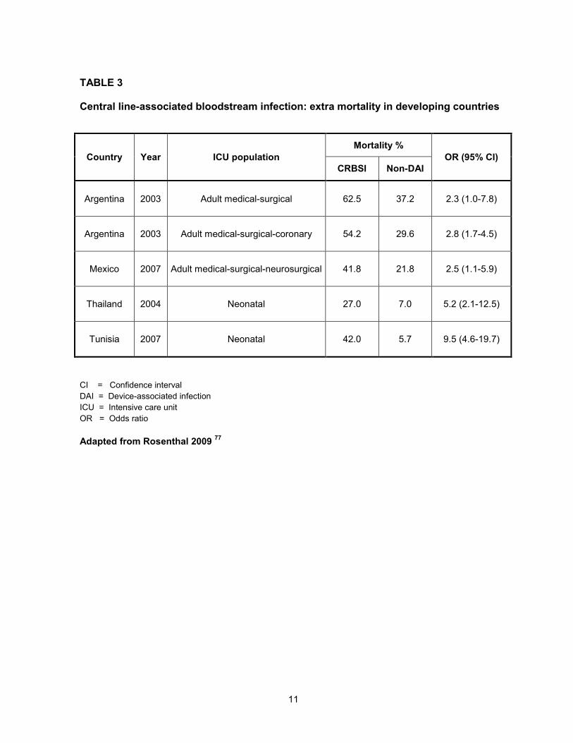
A review of the literature in resource-limited countries revealed central venous origin CRBSI
rates of up to 44.6 cases per 1,000 central line days in adult intensive care units and up to 60
cases per 1,000 central line days in neonatal ICUs 77 (see Table 2). These infections were
associated with significant extra mortality 77,80, the odds ratio ranging from 2.8 to 9.5.
Interestingly, these rates allude to hospitals in which some form of organisational health care
structure exists 77,80 (see Table 3).
Countries evaluated in this review included Argentina, Brazil, Colombia, India, Iran, Mexico,
Peru, Thailand, Tunisia and Turkey.

8
Low-income and middle-income economies, often referred to as developing countries, low-
resource countries, or emerging countries, represent 145 (69.0%) of 210 countries of the
world and more than 75% of the world population.
The World Bank classifies countries into four economic strata according to 2007 gross
national income per capita per year. These groups are as follows: low income, US$ 935 or
less; lower middle income US$ 936 - US$ 3705; upper middle income US$ 3606 - US$ 11455
and high income US$ 11456 or more 77,81.
Guidelines for the management and prevention of nosocomial infections in South Africa,
including intravascular infections, have recently been published 20,82,83 and are in the process
of being updated.

9
Table 1 Impact of Catheter-Related Bloodstream Infections in Critically Ill Patients
a Los: length of stay
b Differences are non-significant c Based on billing database
Mortality Attributable
Author & Reference Crude Attributable Los a (day) Costs (US$)
Pittet 84 45% 25% 6.5 29,000
Soufir 73 50% 29% - -
Rello 13 22% 13% b 20.0 4,000
Renaud & Brun-Buisson 85 39% 12% b 14.0 -
Dimick 76 56% 35% 20.0 71 443 c
Warren 19 51% - 2.41 11,971

10
TABLE 2 Central Venous Catheter-related Bloodstream Infection Rates in ICUs in Developing Countries
Country & reference Year Type of ICU CRBSI rate (95% CI)
Argentina 86 2003 Medical-Surgical 44.6 (32.2 - 60.0).
Argentina 87 2004 Medical-Surgical 30.3 (25.4 - 35.7)
Brazil 88 2003 Burns 34.0 (21.8 - 35.5)
Brazil 89 2002 Neonatal 60.0 (36.5 - 92.3)
Brazil 90 2003 Paediatric 10.2 - 7.2 - 16.7)
Argentina 87 2004 Coronary 14.2 (9.0 - 21.2)
Iran 91 2003 Burns 17.0 (11.6 - 24.5)
Mexico 92 2006 Medical-Surgical- Neurosurgical 23.1 (19.5 - 22.1)
Tunisia 93 2007 Neonatal 14.8 (9.2 - 20.5)
Turkey 94 2009 Medical-Surgical 17.6 (15.8 - 19.3)
India 95 2011 Tertiary Care Hospital 8.75
Adapted from Rosenthal 2009 77
11
TABLE 3
Central line-associated bloodstream infection: extra mortality in developing countries
CI = Confidence interval DAI = Device-associated infection ICU = Intensive care unit OR = Odds ratio
Adapted from Rosenthal 2009 77
Country Year ICU population Mortality %
OR (95% CI) CRBSI Non-DAI
Argentina 2003 Adult medical-surgical 62.5 37.2 2.3 (1.0-7.8)
Argentina 2003 Adult medical-surgical-coronary 54.2 29.6 2.8 (1.7-4.5)
Mexico 2007 Adult medical-surgical-neurosurgical 41.8 21.8 2.5 (1.1-5.9)
Thailand 2004 Neonatal 27.0 7.0 5.2 (2.1-12.5)
Tunisia 2007 Neonatal 42.0 5.7 9.5 (4.6-19.7)
12
CHAPTER 4
DEFINITIONS
Definitions relating to intravascular catheter infection have been put forward by various
workers, but many have complicated matters and been confusing 2,3,4,5. In part, this has been
because definitions used for surveillance and research purposes have differed from those
used for clinical diagnosis. The Centers for Disease Control and Prevention have suggested
sensible definitions 1,4,96 that incorporate both clinical and laboratory evidence of catheter
infection. These should be universally used in the definitions of intravascular catheter
infection and are documented in modified form in Table 4 2,3,4,5.
Table 4 Definitions for catheter-related infections
Item Definition
Catheter colonisation Growth of ≥15 colony-forming units (semiquantitative culture) or ≥103 colony-forming units (quantitative culture) from a proximal or distal catheter segment in the absence of local or systemic infection.
Local infection Erythema, tenderness, induration or purulence within 2cm of the skin insertion site of the catheter.
Infusate-related BSI Concordant growth of the same organism from the infusate and blood cultures (preferably percutaneously drawn) with no other identifiable source of infection.
Catheter-related bloodstream infection
Isolation of the same organism (i.e. the identical species as per antibiogram from cultures, semiquantitative or quantitative) of a catheter segment and from the blood of a patient with accompanying clinical symptoms and signs of bloodstream infection and no other apparent source of infection.

13
CHAPTER 5
PATHOGENESIS OF CATHETER-RELATED INFECTIONS
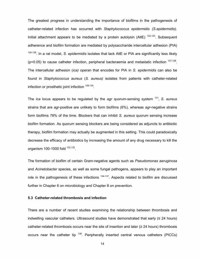
5.1 Overview of the pathogenesis (see Figure 1)
The skin around the insertion site is the most common portal of entry 4,60,97-99. Following
placement, a fibrin sheath develops around the catheter which promotes the adherence of
pathogens and is known as the biofilm layer. Skin organisms then migrate from the insertion
site along the external surface of the catheter to colonise the distal intrasvascular tip and
ultimately cause bloodstream infection 69,97,100,101.
Contamination of the catheter during the manipulation by medical and nursing personnel is
the second most common portal of entry of micro-organisms 4,98,102-108.
Less common causes include haematogenous dissemination from a distal infectious focus,
administration of contaminated infusates, and contaminated transducer kits, disinfectants and
infusion lines 107-110.
5.2 Biofilm
The importance of biofilm formation in vascular catheter infections is becoming increasingly
apparent. Microbial colonisation and biofilm formation occurs shortly after the catheter
insertion 111 which may subsequently cause bacteraemia. The composition of biofilm, includes
various blood and tissue proteins, fibrin, fibronectin, fibrinogen, collagen, elastin,
thrombospondin, laminin, vitronectin and von Willebrand’s factor, in combination with
exopolysaccharide material produced by colonising pathogens (slime, glycocalyx) 112-120. The
complex matrix or biofilm protects the bacteria or other pathogens from chemotherapeutic
agents and opsonophagocytosis 121.
14
The greatest progress in understanding the importance of biofilms in the pathogenesis of
catheter-related infection has occurred with Staphylococcus epidermidis (S.epidermidis).
Initial attachment appears to be mediated by a protein autolysin (AtlE) 122,123. Subsequent
adherence and biofilm formation are mediated by polysaccharide intercellular adhesion (PIA)
124-126. In a rat model, S. epidermidis isolates that lack AtlE or PIA are significantly less likely
(p<0.05) to cause catheter infection, peripheral bacteraemia and metastatic infection 127,128.
The intercellular adhesion (ica) operan that encodes for PIA in S. epidermidis can also be
found in Staphylococcus aureus (S. aureus) isolates from patients with catheter-related
infection or prosthetic joint infection 129,130.
The ica locus appears to be regulated by the agr quorum-sensing system 131, S. aureus
strains that are agr-positive are unlikely to form biofilms (6%), whereas agr-negative strains
form biofilms 78% of the time. Blockers that can inhibit S. aureus quorum sensing increase
biofilm formation. As quorum sensing blockers are being considered as adjuncts to antibiotic
therapy, biofilm formation may actually be augmented in this setting. This could paradoxically
decrease the efficacy of antibiotics by increasing the amount of any drug necessary to kill the
organism 100-1000 fold 132,133.
The formation of biofilm of certain Gram-negative agents such as Pseudomonas aeruginosa
and Acinetobacter species, as well as some fungal pathogens, appears to play an important
role in the pathogenesis of these infections 134-137. Aspects related to biofilm are discussed
further in Chapter 6 on microbiology and Chapter 8 on prevention.
5.3 Catheter-related thrombosis and infection
There are a number of recent studies examining the relationship between thrombosis and
indwelling vascular catheters. Ultrasound studies have demonstrated that early (≤ 24 hours)
catheter-related thrombosis occurs near the site of insertion and later (≥ 24 hours) thrombosis
occurs near the catheter tip 138. Peripherally inserted central venous catheters (PICCs)
15
appear to have a higher risk of thrombosis than centrally inserted catheters 139. Some studies
suggest that femoral catheters carry a greater risk of thrombosis as compared to subclavian
catheters which themselves carry a greater risk than internal jugular catheters 140,141. Tip
position appears to even further modify the risk of thrombosis for central venous catheters,
with location in the axillosubclavian-innominate veins or right atrium, posing a greater risk
than superior vena cava location 142-145. Catheter material may affect the risk with silicone
having up to a threefold greater risk than other materials 146. Data suggests that antibodies to
certain coagulation factors, such as inhibitors to factors V and VIII, may increase the risk of
catheter-related thrombosis through as yet, unclear mechanisms 147,148. Bone marrow
harvesting appears to induce a short-term hypercoagulable state 149. There are some data in
paediatric oncology patients that suggest that genetic risk factors such as factor V Leiden and
others, may increase the risk of catheter-related thrombosis 150,151 but this seems less clear in
adults 152,153.
An ongoing speculation is whether there is a link between catheter-related infection and
catheter-related thrombosis, in particular, which comes first 154-157. Such speculations have
been amplified by a number of studies with heparin-coated central venous catheters, which
have shown a reduction in both catheter-related thrombosis and catheter-related infection
158,159. In these studies however, heparin is attached to the catheter via benzalkonium chloride
which has antibacterial properties and may have contributed to the reduction in risk of
infection. Whether the risk of thrombosis was reduced in these studies by the bound heparin,
the benzalkonium chloride, or both, has not been determined. The association has assumed
even greater importance following reports of a greater incidence of deep venous thrombosis
associated with femoral catheterisation 140,160, which may be prevented by using heparin-
bonded catheters 158,159. In vitro studies have demonstrated that surface manipulations of
polyurethane can lead to differences in protein and platelet deposition with associated
16
differences in bacterial adherence 161. No in vivo data exist to show whether such
observations will have clinical significance.
Figure 1. Pathogenesis of catheter-related infections
HCW = health care worker Adapted from Mer 2008 5

17
CHAPTER 6
MICROBIOLOGY
6.1 Overview
The most frequent causative organisms for CRBSI are coagulase-negative Staphylococci
(CoNS), S. aureus, Enterococci and Candida species 5,13,101,162-166 (see Figure 2 and Table 5).
Contaminants from the hands of medical and nursing personnel are frequently responsible for
infection with such organisms as Pseudomonas aeruginosa, Acinetobacter species, and
Stenotrophomonas maltophilia 5,167,100.
Emerging pathogens reported to have caused CRI include species of Micrococcus,
Achromobacter, rapidly growing mycobacteria such as Mycobacterium fortuitium and
Mycobacterium chelonei, and fungal organisms such as Malassezia furfur, Rhodotorula
species, Fusarium species, Trichosporin species and Hansenula anomala 2,3,4,5,98,163,168.
Figure 2
The most common organisms associated with catheter-related infections
(Adapted from Mer 2008 5 )
Coagulase-negative Staphylococcus Staphylococcus aureus
Candida species Enterococcus species
Pseudomonas aeruginosa Klebsiella species
Enterobacter species Serratia marcescens Citrobacter freundi
Bacillus & Corynebacterium species, especially jeikeium (JK) strains
18
Other organisms associated with catheter-related infections include Bacillus species and
Corynebacterium species (especially JK strains). JK bacteraemia occurs almost exclusively in
severely immune-suppressed patients who are, or have been, receiving broad-spectrum
antibiotics and who have indwelling intravascular devices 2,3.
A concerning trend over the past few years has been the increasing rate of multidrug
resistant organisms causing CRBSI, such as methicillin-resistant S. aureus (MRSA),
fluconazole-resistant Candida species, vancomycin-resistant Enterococcus faecium and
Gram-negative rods including Klebsiella pneumoniae, Escherichia coli, Pseudomonas
aeruginosa and Acinetobacter species, which are no longer susceptible to such agents as
third and fourth generation cephalosporins, ureidopenicillins, flouroquinolones and
carbapenems 165,166,101.
6.2 Catheter-related S. aureus infection
S. aureus is a virulent pathogen that continues to cause significant morbidity and mortality in
the antimicrobial era 169. Infection of a vascular catheter by this organism may cause severe
illness, with bloodstream infection and haematogenous spread to other sites resulting in
serious secondary infections such as osteomyelitis, epidural abscess and endocarditis.
Patients with relatively minor predisposing illness occasionally succumb to such lethal
infection.
Vascular catheters have been reported to be implicated in nosocomial S. aureus
bacteraemias in several studies 170-173. One third to one half of all S. aureus nosocomial
bacteraemias have been attributed to vascular catheters in these studies.
A study of nosocomial endocarditis, which excluded cases related to prosthetic valves, found
that S. aureus accounted for 62% of cases. Causation was related to vascular catheters in
86% of cases 174.

19
The virulence of S. aureus has also been recognised in many studies of catheter-related
infections 175-184.
A review of 25 studies of catheter-related S. aureus bacteraemia found that approximately
half of the deaths in these studies were attributed to the catheter infection by the authors of
the individual studies 185.
A meta-analysis of the case fatality rate associated with catheter-related infections, which
included 187 studies and 3569 catheter infections, found an overall case fatality rate of 14.0%
186. For cases of S. aureus catheter-related infection included in this meta-analysis, the
overall case fatality rate was 18.2%. The proportion of deaths attributed to catheter infection
was significantly higher for S. aureus than for other bacterial agents (OR 3.81, 95% CI: 2.70-
5.41). A prospective epidemiologic study in a Danish hospital demonstrated a case fatality
rate of 38% for nosocomial bacteraemia overall, but the case fatality rate for S. aureus was
65%, with 25% being attributed to catheter infection 187.
Increasing numbers of MRSA infections have been reported in recent years 54,61,162,188,189.
Knowledge about the frequency and resistance patterns of S. aureus is crucial as it has both
therapeutic and prognostic implications.
A recent meta-analysis summarised the results of many studies of the mortality associated
with MRSA, as opposed to methicillin-susceptible S. aureus (MSSA) bacteraemia. It found
that there was significantly higher mortality with MRSA after adjusting for co-morbidity and
severity of illness (OR 1.93, 95% CI: 1.54 - 2.42, p<0.001) 185. This has again been most
recently shown in a paper evaluating the mortality associated with MRSA infection in the ICU
as part of the EPIC II study. In this point-prevalence study of infection, 13,796 adult patients
in 1265 participating ICUs from 75 countries were evaluated. There were 494 patients with
MRSA infections and 505 patients with MSSA infections. MRSA infection was associated with
an almost 50% higher likelihood of hospital death compared with MSSA infection 190.
20
6.3 Catheter-related infections caused by coagulase-negative Staphylococcus
Coagulase-negative Staphylococcus (CoNS), particularly S. epidermidis, is the species most
frequently isolated from infections associated with the use of vascular catheters. 1,191-196.
Traditionally CoNS have been considered avirulent microorganisms that form a major
component of the cutaneous microflora. Since the 1980s, however, it has been recognised
that these organisms are readily able to colonise and infect various devices used for
diagnostic and therapeutic procedures such as intravascular catheters, cerebrospinal fluid
shunts, prosthetic heart valves, orthopaedic devices, pacemakers, peritoneal dialysis
catheters, vascular grafts and ventricular assist devices 197. CoNS are now the leading
organism for nosocomial bacteraemia 198 and it has been suggested that most of these cases
are a consequence of infection of intravascular catheters 198,199. Bacteraemia due to CoNS is
associated with considerable hospital expenditures, morbidity and also an increased mortality
rate. 199-201. For problematic infections treatment options are increasingly narrowed by
emerging resistance against previously active antimicrobials 202. Over the last decades,
important insight into the pathogenesis of these low-virulence microorganisms has been
gained, particularly with respect to their interaction with polymer surfaces. It has now become
clear that the intimate multifactorial interaction with the artificial surface is the basis for the
role of these organisms in catheter-related infection 203.
6.3.1 Microbiology and differentiation from Staphylococcus aureus
Coagulase-negative Staphylococci are members of the family of Micrococcaceae.
These Gram-positive, cluster-forming microorganisms are differentiated from S. aureus
primarily by the lack of the exoenzyme coagulase (which in S. aureus forms fibrin by
activation of thrombin). Additional markers used in the clinical microbiology workup to
differentiate S. aureus from CoNS, are the lack of the immunoglobulin G (IgG)-binding
protein A (Spa), as well as the lack of certain cell wall adhesins recognising matrix and
21
plasma molecules, such as the clumping factor (Clf). Moreover, CoNS do not posses
certain capsular polysaccharides (Cps) specific for S. aureus such as the type 5/8 Cps
associated with S. aureus invasive disease. Accordingly, for diagnostic purposes, the
group of CoNS is differentiated from S. aureus using rabbit plasma and performing
either the slide clumping test or the tube coagulase test. Several rapid commercial latex
agglutination tests examining the presence of Clf, Spa and Cps are also available.
Furthermore, biochemical reactions (such as mannitol fermentation) may allow
laboratory technicians to distinguish between S. aureus and CoNS. With the use of
nucleic acid amplification techniques, additional markers such as the gene for the S.
aureus thermonuclease (alone or in conjunction with other markers such as the
coagene) have been used for rapid discrimination of CoNS 192-197,204.
CoNS have been subdivided into 32 species, 15 of which have been associated with
human disease. Biochemical characterisation has been the mainstay for species
identification 205 and miniaturised biochemical patterns are now available for use in
manual, semi-automatic, or automatic identification systems. Most CoNS involved in
polymer-associated infections belong to the S. epidermidis group (S. epidermidis sensu
stricto, Staphylococcus hominis, Staphylococcus haemolyticus, Staphylococcus warneri
and Staphylococcus capitis). Within the S. epidermidis group, S. epidermidis sensu
stricto accounts for about two-thirds of all strains 191. In addition to members of the S.
epidermidis group, Staphylococcus schleiferi and Staphylococcus lugdunensis 206 have
also been implicated with clinical disease.
6.3.2 Epidemiology
Approximately 30-40% of central venous catheter-related infections are caused by
coagulase-negative staphylococcal species 1,194. Others have reported that S.
epidermidis accounts for up to 75% of the cultured microorganisms 196. This proportion
22
is irrespective of the type of catheter and includes long-term implanted as well as short-
term inserted catheters, Swan-Ganz catheters, catheters used for specific purposes
such as plasmapheresis or haemodialysis, or hyperalimentation catheters which have
all been shown to yield CoNS, particularly S. epidermidis as the leading microorganism
195. CoNS have also been increasingly isolated from the blood of hospitalised patients.
This increase is thought to be in part due to an increase in line-associated CoNS
infections 198.
6.3.3 Resistance
CoNS from nosocomial infections, particularly S. epidermidis are typically resistant to
multiple antimicrobials, particularly the β-lactams. While almost all CoNS produce
penicillinase, most clinical isolates are resistant to penicillinase-resistant penicillins as
well as to all other β-lactams due to expression of mecA analogues conferring
resistance to methicillin by production of a penicillin-binding protein (PBP2a or PBP2’)
with reduced affinity to β-lactams. On the basis of homology studies it has been
suggested that the mecA gene in S. aureus arose from a mecA analogue in
Staphylococcus sciuri 207. Many clinical CoNS isolates are also resistant to macrolides,
chloramphenicol, tetracyclines, flouroquinolones, aminoglycosides, and cotrimoxazole.
CoNS species were the first Staphylococci with reduced susceptibility to glycopeptides
such as vancomycin 208.
6.4 Fungal infections of catheters
During the past decade, the overall incidence of nosocomial fungaemia has continued to
increase, with most cases involving Candida species and many such cases being related to
the use of intravascular catheters.

23
Candida species have become the third most common cause of bloodstream infections
among patients in intensive care units 209-211 and also represent the most common fungi
isolated from intravascular devices 194. The EPIC II Study revealed that 19% of all infections
in the ICU were caused by fungi, mainly Candida species 212. The type of candida species
documented is geographically and regionally determined and may vary amongst ICUs even
within the same area 209,212-218. Although overall, Candida albicans remains the most
dominant pathogen, an increasing number of non-albicans species have been noted over the
past 15-20 years 212,214,219.
Mortality due to fungal nosocomial bloodstream infection is significant and is greater than that
due to non-fungal pathogens 214,219-223.
Attributable mortalities of 35-50% are well documented 214,219,137,223.
A National Nosocomial Infections Surveillance (NNIS) analysis 221 showed that patients with
fungaemia were more likely to die during hospitalisation [954 (29%) of 3256 patients] than
were patients with bloodstream infection due to non-fungal pathogens [659 (17%) of 3882;
relative risk (RR) 1.8; 95% CI: 1.7 - 1.9; p<0.001]
A relationship between candidaemia and indwelling vascular catheters has been recognised
for decades. In 1962, it was reported 224 that 23 of 29 patients who developed systemic
candidiasis had indwelling vascular catheters. In four of these patients, cultures from the skin
around apparently uninfected cutdown sites taken at the onset of fungaemia, grew species of
Candida that were identical to those found in the blood.
Subsequently a review of 27 cases of candidaemia revealed that 25 of the patients had
indwelling central venous catheters 225. More significantly, the authors found that 89% of
these 25 patients had developed positive cultures after the central venous line had been in
place for 2 weeks. CRBSI associated with Candida species is well documented in ICUs, burn

24
units, haematology-oncology patients/units, general hospitals, neonatal units and in patients
receiving total parenteral nutrition 226-234. Central venous catheterisation has been shown to
be an independent risk factor for candidaemia in several studies 229,230,235.
The National Epidemiology of Mycosis Survey (NEMIS) was a prospective, multicentre study
conducted at six geographically dispersed academic centres to determine rates of, and risk
factors for, the development of candidal BSI among patients in surgical and neonatal ICUs.
Four thousand two hundred and seventy six patients were evaluated. Candida species
accounted for 9.2% of the total number of BSIs. More than half the Candida isolates
recovered from the blood were non-albicans species. The mortality rate was significantly
higher among patients who developed candidal BSI than among other patients (41% vs 8%;
OR 7.52; 95% CI: 3.9 - 14.6; p<0.001). Of 42 patients who developed candidal BSIs 41 had a
central venous catheter in place during their ICU stay prior to the development of infection. A
multivariate model that included only patients who underwent surgery (n = 3201) identified an
association between increased risk of candidal BSI and having had a triple-lumen catheter
placed (RR 5.4; 95% CI: 1.2 - 23.6; p = 0.03) 236.
Various workers have helped add to the understanding of the capability of Candida species to
adhere to plastic materials and cause catheter-related fungaemia. This relates largely to
Candida biofilm and the ability, particularly of Candida parapsilosis, to proliferate and produce
large amounts of slime. Glucose-containing solutions may be an important adjunct in the
pathogenesis with work demonstrating a strong relationship between Candida parapsilosis
fungaemia or systemic infection and hyperalimentation using intravascular devices 136,237-239.
The relevance of catheter-related fungaemia has been the subject of much debate recently
particularly in the light of emerging fluconazole resistance to various isolates, as well as
optimal catheter management 101,240-246.
25
There is the possibility that infected or colonised catheters may seed organisms to various
body sites resulting in a great variety of clinical presentations depending on the affected
organs. When Candida is disseminated, multiple organs are usually affected, including the
kidney, brain, myocardium and eye (see Figure 3, Chapter 7).
Current opinion in critically ill patients is that all infected catheters should be removed where
feasible 137.
Because patients with catheter-associated candidaemia who have been treated with catheter
removal but without systemic antifungal therapy have developed complications such as
vertebral osteomyelitis and endophthalmitis, resulting in permanent loss of vision, antifungal
therapy is advocated in all cases of vascular-catheter candidaemia 247.
Other reported complications include fungal thrombophlebitis 194,248,249 and right atrial septic
thrombosis 250.
6.5 Catheter-related infections caused by Enterococci
During the past decade the prevalence and importance of infections caused by Enterococci
has increased significantly. In a prospective, observational study of 110 patients with serious
enterococcal infections, such as endocarditis, bacteraemia, cholangitis, pancreatitis,
osteomyelitis, pneumonia and empyema, catheter-related bacteraemia was the single most
common infection accounting for 28% of all infections 251. In infants, all infections were
catheter-related bacteraemia. Overall 78% of the enterococcal isolates were identified as
Enterococcus faecalis, 20% as Enterococcus faecium and 1% as Enterococcus gallinarum
and Enterococcus casseliflavus, respectively. Enterococcus faecalis was the most common
species accounting for relapse. Glycopeptide resistance among Enterococcus faecium is an
emerging problem.

26
Enterococcus casseliflavus, a motile organism which forms yellow pigmented colonies on
blood agar and reported to be associated with central venous catheter-related infection, is
intrinsically resistant to low levels of Vancomycin 252,253.
6.6 Catheter-related infections due to Gram-negative organisms
Gram-negative bloodstream infections associated with intravascular catheters has received
less attention in the literature than those associated with Gram-positive infections 254.
Similarly, the vast majority of studies investigating the pathogenesis of foreign-body infections
have focused on Gram-positive organisms 255.
There is, however, an ever-increasing number of Gram-negative species causing infections
related to indwelling devices 163. Most of these are intravascular catheter-related infections
caused by three major groups of Gram-negative organisms namely, members of the family
Enterobacteriaceae, Pseudomonas aeruginosa and Acinetobacter species. CRIs have also
been reported to be caused by other Gram-negative organisms, such as rare
Enterobacteriaceae, non-aeruginosa pseudomonads and other non-fermenting Gram-
negative bacilli 25,26,165,167,256-259. The results of several studies are summarised in Table 6.
In a prospective study Gram-negative organisms were found in 22 of 41 (54%) of colonised
central venous catheters and in 6 of 11 (55%) associated bacteraemias 26. Other investigators
reported Gram-negative bacilli implicated in central venous catheter-related infection to range
between 12% and 42% 25,165,256-258.
In a study evaluating 400 ICU patients with non-tunnelled CVCs, 25% of CRBSIs were
caused by Gram-negative bacteria 259. In a prospective survey to determine the rate of CRBSI
among cases of primary BSI in febrile, neutropenic cancer patients with short-term non-
tunnelled catheters, quantitative paired blood cultures from central venous catheters as well
as peripheral vein and Bactec™ blood culture bottles were obtained to determine the

27
differential time to positivity 260. Twenty seven percent of the organisms recovered were
Gram-negative.
These organisms have also been implicated in the majority of hospital outbreaks of infusion-
related bacteraemia. Enterobacter species, Klebsiella species. and Serratia species have
been most frequently involved in these instances 261-266.
Gram-negative pathogens have additionally been associated with infections and
bacteraemias emanating from pressure transducers used for monitoring 266,267.
Factors involved in the pathogenesis of Gram-negative infections and CRI are becoming
increasingly understood.
Usually, Gram-negative bacteria adhere less readily to polymer surfaces with the exception of
Pseudomonas aeruginosa and Acinetobacter species 268.
As with CoNS, the adherence properties and the formation of biofilm of Pseudomonas
aeruginosa and Acinetobacter species appears to play an important role in the pathogenesis
of these infections 268.
Like most environmental bacteria, Pseudomonas aeruginosa lives predominantly in biofilms
adherent to available surfaces, from which it periodically releases planktonic (free-swimming)
cells. Pseudomonas aeruginosa possesses a vast array of virulence factors that have been
extensively reviewed 134. Growth in biofilms protects the organism’s cells from antibacterial
factors produced by the host as well as from antibiotics, and may account for survival and
extended persistence on foreign devices. Pseudomonas aeruginosa embedded in thick
biofilm has been seen in a variety of transcutaneous medical devices such as vascular
catheters 269, peritoneal catheters 270 and urinary catheters 271. It has been suggested that
polymer catheters made of PVC or silicone may favour survival and growth of Pseudomonas
aeruginosa 272.

28
Scanning electron microscope studies suggest that colonisation of polyurethane catheters
with different Acinetobacter species may occur in a similar time frame and to an extent
comparable to CoNS 268.
Possible complications of CRI due to Gram-negative organisms include sustained
bacteraemia, infective endocarditis and suppurative thrombophlebitis 273 (which is most often
caused by Gram-negative organisms). An emerging problem is the development of many
Gram-negative agents that are multidrug resistant pathogens.

29
TABLE 5 CRBSI - most common pathogens and mortality rates
Pathogen
Percentage of CRBSI (Rank) Crude Mortality %
Total (n = 20,978)
ICU (n = 10,515)
Non-ICU Ward
(n = 10,442) Total ICU
Non-ICU
Ward
Coagulase-negative Staphylococcus 31.3 (1) 35.9 (1) 26.6 (1) 20.7 25.7 13.8
Staphylococcus aureus 20.2 (2) 16.8 (2) 23.7 (2) 25.4 34.4 18.9
Enterococcus species 9.4 (3) 9.8 (4) 9.0 (3) 33.9 43.0 24.0
Candida species 9.0 (4) 10.1 (3) 7.9 (4) 39.2 47.1 29.0
Escherichia coli 5.6 (5) 3.7 (8) 7.6 (5) 22.4 33.9 16.9
Klebsiella species 4.8 (6) 4.0 (7) 5.5 (6) 27.6 37.4 20.3
Pseudomonas aeruginosa 4.3 (7) 4.7 (5) 3.8 (7) 38.7 47.9 27.6
Enterobacter species 3.9 (8) 4.7 (6) 3.1 (8) 26.7 32.5 18.0
Serratia species 1.7 (9) 2.1 (9) 1.3 (10) 27.4 33.9 17.1
Acinetobacter baumannii 1.3 (10) 1.6 (10) 0.9 (11) 34.0 43.4 16.3
Adapted from the Surveillance and Control of Pathogens of Epidemiological Importance (SCOPE) Study 165. Data
from a nationwide, concurrent surveillance study in 49 US hospitals over a 7-year period.

30
TABLE 6 Gram-negative organisms and short-term CVCs, a compilation from several studies
*ref = reference
Study and Reference
Haslett *(ref 258)
Yeung *(ref 257)
Gil *(ref 26)
Eyer *(ref 256)
Richet *(ref 25)
Sherertz *(ref 167)
Seifert *(ref 260)
Wispling-hof)
*(ref 165)
Total number of isolates reported 76 42 41 43 123 1032 22 20 928
ORGANISM PERCENTAGE OF ISOLATES
Gram-negative bacilli 22 12 55 42 26 28 27 31
Escherichia coli
0 0 5 6 0 14 17 6
Enterobacter species 29 0 10 33 6 16 17 3.9
Klebsiella species
12 40 14 17 16 6 17 5
Serratia species
6 20 19 0 6 7 0 17
Pseudomonas aeruginosa 29 0 29 22 41 50 17 4.3
Acinetobacter species 24 20 19 5 13 4 0 1.3

31
CHAPTER 7
DIAGNOSIS OF CATHETER-RELATED INFECTION
7.1 Introduction
The diagnosis of CRBSI remains a major challenge 274,275 and involves both clinical and
laboratory components 2,3,4,5,60,275.
Over 75% of CVCs that are removed because of suspicion of CRI are removed unnecessarily
since the septic focus may be found in another anatomic site 276. Usually, when CRBSI is
suspected, the common practice in the ICU is to remove the CVC and replace it at a new site.
However, only around 15% to 25% of CVCs so removed proved infected upon quantitative-tip
culture 2,3,4,5,60,275-278.
7.2 Clinical aspects
The clinical features of CRI are generally non-specific and include fever, rigors, hypotension
and confusion. If there is no apparent source of sepsis in a patient with an intravascular line,
especially a central venous catheter, and if the sepsis appears to be refractory to
antimicrobial therapy, or is of abrupt onset or associated with shock, the possibility of CRI
needs to be considered 2,3,4,5,275-278. Fundoscopy should always form part of the clinical
examination, as focal retinal lesions are common in patients with CVC-derived Candida
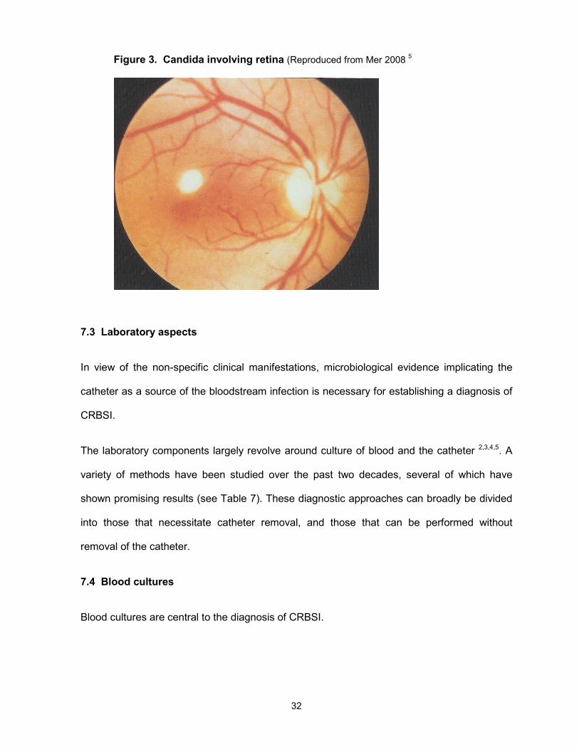
infection, even when blood cultures are negative 2,3,4,5 (see Fig 3) 5. Contamination or
purulence at the catheter insertion site is seen in less than half of the cases 2,3,4,5. It is also not
predictive of CRBSI with short-term non-cuffed CVCs 279.
32
Figure 3. Candida involving retina (Reproduced from Mer 2008 5
7.3 Laboratory aspects
In view of the non-specific clinical manifestations, microbiological evidence implicating the
catheter as a source of the bloodstream infection is necessary for establishing a diagnosis of
CRBSI.
The laboratory components largely revolve around culture of blood and the catheter 2,3,4,5. A
variety of methods have been studied over the past two decades, several of which have
shown promising results (see Table 7). These diagnostic approaches can broadly be divided
into those that necessitate catheter removal, and those that can be performed without
removal of the catheter.
7.4 Blood cultures
Blood cultures are central to the diagnosis of CRBSI.

33
7.4.1 Peripherally collected blood
Traditionally the routine is for two to three 10ml samples, ideally from separate
peripheral venepuncture sites, to be sent to the laboratory. Probable CRBSI can be
diagnosed by one or more positive blood cultures obtained from a peripheral vein,
when there is no apparent source for the bloodstream infection except the catheter
2,3,4,5,274,279.
Several other techniques have been described and advocated more recently.
7.4.2 Paired quantitative cultures
Paired quantitative cultures involve taking blood simultaneously from both the CVC
and a peripheral site. Although several quantitative blood culture methods are
available, one of the most widely used techniques involves placing a 10ml blood
sample in an isolator tube (Isolator 10, Wampole, Granbury NJ, USA) for quantitative
culturing by lysis centrifugation 280. When the quantitative blood culture drawn from the
CVC yields a colony count that is several-fold higher than that obtained from
simultaneously drawn blood of the peripheral vein, the result is considered to be
predictive of CRBSI. Since studies have reported different cut-off points for a positive
diagnosis ranging from two-fold to ten-fold 281-285, a midpoint level whereby a colony
count that is five-fold or greater from blood culture drawn from the CVC versus
peripheral vein, is regarded as indicative of CRBSI 2,3,4,5. This value has been
endorsed by the IDSA (Infectious Diseases Society of America). Simultaneous
quantitative blood cultures were found to be the most accurate test for diagnosis of
CRBSI in a meta-analysis of studies of diagnostic tests 286. Its use has been limited
however, because it is labour intensive and expensive.
34
7.4.3 Differential time to positivity
This technique involves simultaneous qualitative blood cultures drawn through the
catheter and a peripheral vein 260,287-292. Regular blood cultures are routinely placed in
an automatic culture detector that records culture positivity every 15 minutes
according to changes in fluorescence related to microbial growth. Several studies
indicated that definite diagnosis of CRBSI is established when the blood culture drawn
from the CVC becomes positive at least 2 hours earlier than the blood culture drawn
from the peripheral vein 287-289.
A meta-analysis found the pooled sensitivity and specificity for this method of
diagnosing CRBSI in short-term catheters to be 89% and 87% respectively, compared
with 90% and 72% respectively for long-term catheters 286. Automated radiometric
blood culture systems, in which blood cultures are continuously monitored for
microbial growth, are now widely used. As a consequence, adding differential time to
positivity as a routine evaluation has been advocated, as there is no additional cost or
labour required 287-289.
Interpretation of this diagnostic method may be compromised if antibiotics are being
administered at the time blood is drawn through the catheter. This may result in a
false negative result 289.
7.4.4 Catheter-drawn quantitative blood cultures
Catheter-Drawn Quantitative Blood Cultures in which a single quantitative blood
culture is drawn through the CVC without an accompanying peripheral blood culture
can also be used to diagnose CRBSI. The threshold required for a positive diagnosis
is at least 100 colony-forming units (CFU)/ml 282,293.

35
This method is unable to distinguish between CRBSI and high-grade bacteraemia,
particularly in immuno-compromised patients with severe sepsis. Further studies are
required to verify this method.
Practical and potential disadvantages to quantitative blood cultures include difficulty in
aspirating blood from the catheter 294 and the dilemma of multiple lumen CVCs as
each lumen may represent a source of infection. In one recent study, sampling of one
out of three lumens of the catheters missed 37.3% of the CRBSI 295.
7.5 Catheter culture (see Table 8)
7.5.1 Semi-quantitative roll-plate method
The most widely used laboratory technique for culturing the catheter is the semi-
quantitative roll-plate method 2,3,4,5,100 in which the catheter is removed, the distal
segment of the CVC is cut and rolled against a blood agar plate at least four times,
before the plate is incubated overnight 100,286,296. Upon examination of the plate, a
colony count of 15 CFU/ml, or more, is suggestive of catheter colonisation. Diagnosis
of CRBSI is confirmed if catheter colonisation is associated with a positive peripheral
blood culture revealing the same organisms 277,297-303. This method is better at
detecting colonisation of the external surface of the catheter rather than intra-luminal
colonisation, and is therefore less sensitive for long-term catheters in which luminal
colonisation more frequently leads to bloodstream infection 297,105. This may also
pertain to first generation antiseptic catheters where only the external surface of the
CVC is impregnated.
However, pooled sensitivity and specificity for roll-plate catheter culture in 14 trials
involving short-term CVCs were calculated to be 84% and 85% respectively 286.
Another study calculated sensitivity and specificity values for this technique of more
than 90% for both short-term and long-term CVCs. However, long-term CVCs in this

36
study were defined as those with only seven days of dwell time or more 304. For short-
term catheter-tip cultures, the roll-plate technique is recommended for routine clinical
microbiological analysis 305.
7.5.2 Quantitative catheter culturing techniques
Quantitative techniques for culturing the catheter include the sonication and vortexing
methods, which involve extracting microorganisms from the catheter surface into a
medium for culturing. This entails either flushing out the catheter segment and
immersion in culture medium or placement of the segment in culture medium with
sonication 167,277,306,307.
A colony count of more than 100 CFU per catheter segment is regarded as positive.
The advantage of sonication or vortexing is that these methods help release
microorganisms from both the external and internal surfaces of the CVC. The
disadvantage is that these methods release biofilm organisms that might not be
clinically relevant, whereas relevant planktonic organisms might be killed. The
sonication and vortexing methods have been shown to have similar sensitivity and
specificity to the roll-plate method in diagnosing CRBSI in patients with short-term
CVSs 299,308. For long-term CVCs, the sonication method was found to have higher
sensitivity than the roll-plate method 105. The pooled sensitivity and specificity of
quantitative catheter segment culture for short-term catheters were 82% and 89%
respectively, and 83% and 97% for long-term catheters, respectively 100,286,296. Many
microbiology laboratories do not perform quantitative blood cultures 309.
37
7.5.3 Cultures of the insertion site and catheter hub (see Table 9)
Various quantitative culture methods of the insertion site or catheter hub have been
reported but have been associated with a limited specificity and positive predictive
value 310-314.
7.5.4 Other techniques
Other techniques such as Gram stain of the catheter surface and culture of the tip in
broth have been associated with high false-positive rates 3 and are now not widely
used 286,296.
7.6 Newer techniques
Newer diagnostic methods of CRBSI infections include that of the endoluminal brush 4,5,315,316
and the Gram stain and acridine orange leucocyte cytospin test (AOLC) 4,5,20,317,318.
7.6.1 The endoluminal brush is a tapered nylon brush on a steel wire that is passed
through the catheter hub and lumen, withdrawn, and immediately placed in a buffered
container. This container undergoes sonication and vortexing after which the solution
is cultured onto blood agar plates. Counts of greater than 100 CFU/ml are deemed
positive 315. This technique is based on the premise that bacteria adhere to the fibrin
sheath on the inner surface of the CVC and fibrin becomes enmeshed in the brush’s
bristles. High sensitivities and specificities have been reported with this technique 315
which does not require sacrifice of the catheter, but there is still a delay before culture
results are known 4. There is also a concern that the process of brushing may lead to
embolisation of biofilm as well as other side effects such as arrhythmias 286,296,319.
The place of the endoluminal brush in clinical practice is still to be fully determined but
at present is not widely used and further studies are required 4.

38
7.6.2 The Gram stain and AOLC test (see Table 10) 320 is a recently described
method for rapidly diagnosing CRBSI without catheter removal within one hour. The
test is performed on blood samples drawn from the CVC and has been reported to
have high sensitivities and specificities. A negative predictive value of 97% and a
positive predictive value of 91% have been reported for the diagnosis of bacteraemic
CRI 315. This promising technique compares favourably with other diagnostic methods,
particularly those that require removal of the catheter and may permit early targeted
antimicrobial therapy, or conversely avoid unnecessary use of antibiotics 4,5. Although
cost-efficient and simple, this test has not been widely used at present, but may
represent a promising way of establishing the diagnosis of CRI in the future. The
theoretical risk of toxicity of acridine-orange, an intercalating agent, may be a factor
that has resulted in some precautions in microbiology laboratories.
7.7 Novel techniques
Potentially innovative techniques include a serological test for the diagnosis of CRI due to
coagulase-negative staphylococci 321.
In a study evaluating this test, the authors compared 67 patients suspected of having CRI
due to CoNS and 67 control patients with a CVC, but without CRI. Ten millilitres of blood
were obtained in both groups and serum antibody levels of both IgG and IgM against a short-
chain lipoteichoic acid antigen isolated from CoNS were determined, using an enzyme linked
immunosorbent assay (ELISA) technique. Significant differences between the mean IgG and
IgM titres of the test group and the control group were observed. Using an IgG titre of 20,000,
the test had a sensitivity of 70% and a specificity of 90%. Similar results have subsequently
been reported in a study published by the same group 322. Further studies are however
needed before such serological tests can be proposed as routine. The usefulness of
serological methods for the diagnosis of CRI remains to be confirmed.
39
7.8 Other tests
Various other tests that have been proposed, such as C-reactive protein and introblue
tetrazolium tests, have not proved to be useful in the diagnosis of CRI 283,323,324.
7.9 Current suggestions
In addition to appropriate clinical evaluation, one of the following microbiological methods to
confirm the diagnosis of CRBSI is currently suggested 305,325.
1. Positive semi-quantitative or quantitative culture of the catheter,
2. Simultaneous quantitative blood cultures drawn through the CVC and peripheral
vein with a ratio of 5:1 or more (CVC versus peripheral), or
3. Differential time to positivity.

40
TABLE 7 Microbiological diagnostic methods of catheter-related bloodstream infections
Diagnostic criteria
Accuracy
Comments Sensitivity Specificity
Techniques without CVC removal
Simultaneous quantitative blood culture
Quantitative blood culture drawn through CVC yields CFU count five-fold higher or more than CFU count from simultaneously drawn blood from peripheral vein
93% 286 97-10% 286 Found to be most accurate test for diagnostic CRBSI in a meta-analysis 286
Differential time to positivity
Blood culture drawn from CVC becomes positive ±2 h before simultaneously drawn blood culture from peripheral vein
89-90% 286 72-87% 286 Concurrent antibiotic administration may interfere with result. Cost effective
CVC-drawn quanti-tative blood culture
Quantitative blood culture from CVC is >100 CFU/ml
81-86% 286 85-96% 286 Cannot differentiate between CRBSI and high-grade bacteraemia
Acridine orange leucocyte cytospin followed by Gram stain
Presence of any bacteria 96% if followed by Gram stain 317
92% if followed by Gram stain 317
Promising
Endoluminal brush Quantitative culture with >100 CFU/ml 95% 315 84% 315
May induce bacteraemia, arrhythmia, embolisation
Techniques requiring CVC removal
Semiquantitative CVC tip culture, roll plate
≥15 CFU/ml from CVC tip 45-54% 286,297,105 85% 286,297,105
Recommended test for short-term catheter tip cultures for routine clinical, microbiological analysis
Quantitative CVC culture centrifugation, vortexing, sonication
≥103 CFU from CVC tip 52-83% 286 89-97% 286 Not performed in many microbiology laboratories
CVC = central venous catheter CFU = colony forming units CRBSI = catheter-related bloodstream infection

41
TABLE 8 Methods of catheter-tip culture
Semi-quantitative catheter-tip culture method 100
At the time of catheter removal, any antimicrobial ointment or blood present on the skin around the catheter should be removed. The catheter is withdrawn with sterile forceps, the externalised portion being kept directed upward and away from the skin surface. For short peripheral catheters, the entire length is aseptically amputated and cultured. For longer catheters, the distal 5 to 7 cm catheter segment is sectioned and cultured. Catheter segments are transported to the laboratory in a sterile tube. In the laboratory, the catheter-tip segment is transferred to the surface of a 10 cm, 5% sheep-blood agar plate for semi-quantitative culturing. While downward pressure is exerted with a flamed forceps, the catheter is rolled back and forth across the surface at least four times. Plates are subsequently incubated at 37ºC. All colony types appearing on the plate are enumerated, and all organisms recovered are fully identified.
Quantitative tip-flush culture technique 306
The intradermal segment is separated from the intravascular catheter segment. A needle is inserted into the proximal end of the intravascular segment, which is immersed in 2 ml or 10 ml of trypticase soy broth, depending on the size of the insert, and flushed three types. The broth is serially diluted 100-fold, and 0.1 ml of the dilution is streaked onto blood agar. After incubation, the number of cfu is calculated by multiplying the number of colonies by 10 times the dilution factor, and dividing by the volume of broth in which the insert has been immersed.
Quantitative culture technique after catheter vortexing 326
After removal, the distal 5 to 6 cm catheter segment is sectioned in a sterile tube. One millilitre of sterile water is dripped onto the catheter and the tube is vortexed for 1 minute, then 0.1 ml of the suspension is sampled and plated over a 5% horse-blood agar plate. The plate is incubated at 37ºC and examined daily for 5 days. All colony types are identified by colony morphology, Gram stain, and standard microbiologic techniques. The colonies are enumerated, and the counts are corrected for the initial 1/10 dilution.
Quantitative culture technique after catheter ultrasonication 167
After removal, the catheter tip is placed in 10 ml of tryptic soy broth, sonicated for 1 minute (55,000 Hertz, 125 Watt), and then vortexed for 15 seconds. A 0.1 ml sample of the broth is added to either 0.9 ml (1:10 dilution) or 9.9 ml (1:100 dilution) of saline and vortexed. Then, 0.1 ml of these dilutions and 0.1 ml of the sonicated broth are surface-plated, using a wire loop on blood agar.

42
TABLE 9 Methods of skin and hub cultures
Culture of the skin at the catheter insertion site 299,327,328,329
Cutaneous specimens may be obtained with a dry or a moistened swab, after
removal of the dressing. No antiseptic agent is needed. The cotton swab may
be moistened with 0.01 M phosphate-buffered saline (PBS), using the blister of
the device. The swab may then be rubbed on the skin in two perpendicular
directions on a predefined area surrounding the insertion point of the catheter
(eg. 2 x 2 cm, or 6 x 4 cm; a template may be used). Other authors propose to
swab the sterile cotton applicator from top to bottom in 10 back-and-forth
strokes, in the predefined area, followed by a second set of 10 back-and-forth
strokes at right angles to the first. The cotton swab is then pulled back into the
protective tube which contains 1.0 ml of PBS, and vortexed for 90 seconds. In
the Culturette® system, 0.5 ml of a Stuart medium is used. Finally, a 0.1 ml
sample of this solution (and eventually a 1:100 solution of this solution) is
plated on blood agar, and colonies are counted after incubation for 24 and 48
hours at 35ºC.
Culture of the catheter hub 103,299,313,328
The catheter is clamped in order to avoid any blood contamination, and the luer
lock is removed aseptically. After cleaning the outside of the hub with a
disinfectant, the inner hub sample is taken using a swab that is introduced into
the hub and rubbed repeatedly against its interior surface. The hub is then
streaked onto an agar plate for semi-quantitative culture.
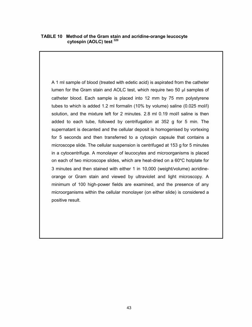
43
TABLE 10 Method of the Gram stain and acridine-orange leucocyte
cytospin (AOLC) test 320
A 1 ml sample of blood (treated with edetic acid) is aspirated from the catheter lumen for the Gram stain and AOLC test, which require two 50 µl samples of
catheter blood. Each sample is placed into 12 mm by 75 mm polystyrene
tubes to which is added 1.2 ml formalin (10% by volume) saline (0.025 mol/l)
solution, and the mixture left for 2 minutes. 2.8 ml 0.19 mol/l saline is then
added to each tube, followed by centrifugation at 352 g for 5 min. The
supernatant is decanted and the cellular deposit is homogenised by vortexing
for 5 seconds and then transferred to a cytospin capsule that contains a
microscope slide. The cellular suspension is centrifuged at 153 g for 5 minutes
in a cytocentrifuge. A monolayer of leucocytes and microorganisms is placed on each of two microscope slides, which are heat-dried on a 60ºC hotplate for
3 minutes and then stained with either 1 in 10,000 (weight/volume) acridine-
orange or Gram stain and viewed by ultraviolet and light microscopy. A
minimum of 100 high-power fields are examined, and the presence of any
microorganisms within the cellular monolayer (on either slide) is considered a
positive result.
44
CHAPTER 8
PREVENTION OF CENTRAL VENOUS CATHETER-RELATED INFECTION IN THE ICU
Because of the difficulties in treatment of catheter-related infections, their impact on morbidity
and mortality, and significant additional costs, prevention of CRI remains a major goal in
modern medicine. In most cases, CRBSIs can be prevented or limited.
Strict adherence to handwashing and aseptic technique remains the cornerstone of
prevention of CRI 2,3,4,5,20,21,82. Several other measures have been reported to confer
additional protection, some of which need to be considered in the preventive strategy. These
are described below.
8.1 Staff education
Education and training of health-care providers who insert and maintain CVCs has been
shown to be successful in preventing catheter-related infection, improving patient outcomes
and reducing healthcare costs 330. The experience of the operator is important with the risk of
infectious complications being inversely proportional to experience and operator skills.
Catheterisation by less-experienced providers is associated with a higher risk of infection 331.
Education regarding appropriate catheter insertion technique has been shown to significantly
improve patient outcomes, and simulation-based training programmes have also been
demonstrated to be useful 332,333. Programmes focusing on nurses’ catheter care have been
associated with a reduction of catheter-related infection in the USA 334. Educational initiatives
that have included interns, ICU nurses, residents and consultants have also been associated
with a sustained reduction in CRBSI in both surgical and medical ICUs.
A didactic education programme and “hands on” demonstration of insertion of both CVCs and
arterial lines to beginner physicians in training resulted in a steady and significant reduction in

45
CRBSI and primary bloodstream infection over time 333. Such educational interventions
appear to be lacking or are not adequately adhered to in limited-resource countries 77.
Although educational programmes can be helpful in reducing CRBSI, compliance to individual
elements of the programme may wane over time and continuous and repetitive re-
enforcement of the principles is usually necessary 15,335,336.
Nursing staff reductions below a critical level may contribute to increased catheter-related
infection by making adequate catheter care difficult 337. A four-fold greater risk of catheter
infection has been reported when the patient-to-nurse ratio was doubled 338. The replacement
of regular nurses by floating or agency nurses has also been shown to increase the risk of
device-related infections 339. These studies highlight the need for appropriately trained nurses
in sufficient numbers and ongoing and repetitive education is necessary for optimal catheter
care in the ICU 337.
8.2 Infusion therapy teams
A number of studies have shown that catheter insertion and catheter maintenance by a team
of specially trained physicians, technicians and nurses is associated with a lower risk of CRI.
Such infusion therapy teams have been associated with reductions of CRBSI of up to 8-fold
and also limit overall costs 334,340-344. Similarly, strict adherence to protocols for catheter
insertion in the ICU, wards and operating theatre also decreases the rates of CRI 3,4,5,20.
8.3 Maximum sterile barriers
Careful hand washing together with full barrier precautions including the use of mask, cap,
sterile gloves, gown and large sterile drape have been shown to significantly reduce the risk
of CRI and CRBSI 97,345. This practice has been associated with a greater than 6-fold
decrease in CVC-related sepsis and cannot be over-emphasised 5.
46
Strict asepsis at the time of insertion is a crucial step for effective infection control. The use of
maximum sterile barrier precautions has been shown to be cost-effective 345. The standard
practice in the 1980s and early 1990s included only the use of sterile gloves and small drape
when inserting CVCs.
The benefit of using maximal sterile barrier precautions to reduce the risk of CRI has been
elucidated in two studies. In one study it was shown that the use of masks, caps, sterile
gloves, gowns and a large drape that covers the patient during insertion of non-tunnelled non-
cuffed central venous catheters leads to a greater than 6-fold reduction in CRBSI, compared
to a control group in which only sterile gloves and small sterile drapes were used 345. In the
other study, on Swan-Ganz pulmonary artery catheters, the application of such barrier
precautions was associated with a 2-fold lower risk of infection 97.
Other observational studies have supported these findings. It is concluded that these
measures are cost-effective, beneficial to the patient and thus strongly recommended 346.
8.4 Skin antisepsis
The density of microorganisms at the catheter insertion site is a major risk factor for CRI and
cutaneous antisepsis is regarded as one of the most important measures for preventing CRI
and CRBSI 5,75. A variety of skin antiseptics, most notably 10% povidone iodine, 70%
isopropyl alcohol, and alcoholic or aqueous chlorhexidine (2%, 1%, 0.5%, 0.25%) have been
studied. Their respective efficacy in preventing catheter colonisation and bloodstream
infections has been compared in various trials.
A meta-analysis of eight randomised trials comparing chlorhexidine to aqueous povidone
iodine and involving 4143 short-term catheters, the bulk of which were CVCs, revealed that
aqueous or alcoholic chlorhexidine solutions were superior to aqueous povidone iodine and
significantly reduced catheter colonisation CRBSI 337,347. Catheter insertion sites and duration
47
of catheterisation were comparable between the two groups. Chlorhexidine solutions were
either an aqueous solution of 2% chlorhexidine (2 trials), a 70% alcoholic solution of 0.5%
chlorhexidine (4 trials), an alcoholic solution of 1% chlorhexidine (1 trial) or a combination of
0.25% chlorhexidine, 0.025% benzalkonium chloride and 4% benzyl alcohol (1 trial).
Chlorhexidine preparations were associated with a reduction of CRBSI by approximately 50%
(RR:0.51 [95% CI, 0.27 - 0.97]). Extrapolation of this data suggests that for every 1000
catheter sites disinfected with chlorhexidine solutions, 71 episodes of CVC colonisation and
11 episodes of infection would be prevented.
Similar findings with an alcoholic formulation of 2% chlorhexidine for skin antisepsis with
CVCs has also been reported 348. The use of chlorhexidine for skin antisepsis also appears to
be cost-effective. In a study evaluating the economic benefits of chlorhexidine gluconate
compared with povidone iodine for vascular catheter site care, the use of a chlorhexidine skin
preparation resulted in an economic saving of $113 per catheter 349.
The superiority of chlorhexidine in the bulk of these studies has been explained, at least in
part, by a synergistic effect with alcohol, even for low chlorhexidine concentrations 337. This
synergistic effect has also been demonstrated with povidone iodine, and is based on a
randomised multicenter crossover trial comparing the effectiveness of two pre-insertion
cutaneous antiseptic protocols using aqueous 10% povidone iodine or a solution of 5%
povidone iodine in 70% ethanol 350. Catheter colonisation and CRI rates were significantly
lower in patients who were managed using the alcohol povidone iodine solution compared
with the aqueous povidone iodine solution protocol (no significant effect was seen on
bloodstream infections but the study was underpowered to explore this issue).
A recent trial has compared a chlorhexidine-based solution to 5% alcoholic povidone iodine
for central venous catheter care. A total of 538 catheters were randomised, of which 481
(89.4%) produced evaluable culture results 351. The chlorhexidine-based solution was

48
associated with a significant reduction in the incidence of catheter colonisation (50%) (11.6%
vs 22.2%, p=0.002; incidence density 9.7 vs 18.3 per 1000 catheter days). The use of the
chlorhexidine-based solution was also associated with a trend toward lower rates of catheter-
related bloodstream infection (1.7% vs 4.2%, p=0.09; incidence denity 1.4 vs 3.4 per 1000
catheter days).
Chlorhexidine-based solutions currently appear to be more effective than povidone iodine,
even in alcoholic formulation, and should be the recommended first-line antiseptic for CVC
care.
The potential role of antiseptic combination has been tested in one monocentre randomised
controlled trial. A 1-minute application of propanol/chlorhexidine followed by a 1-minute
application of povidone iodine (4.7%) was found to significantly decrease catheter
colonisation as compared with 2-minute povidone iodine (24.4%) or 2-minute
propanol/chlorhexidine (30.8%). The potential benefit of this combination requires further
study 352.
It is the practice in the multidisciplinary adult ICU at Charlotte Maxeke Johannesburg
Academic Hospital (CMJAH) to use a chlorhexidine gluconate-containing solution for skin
preparations 5.
Tolerance to chlorhexidine-based solutions is generally excellent. Contact dermatitis and,
exceptionally rarely, anaphylactic reactions have been reported (less than 100 cases
globally).
8.5 Antibiotic prophylaxis
No studies have demonstrated a reduction in CVC infection rates with oral or parenterally
administered antibacterial or antifungal agents given at the time of catheter placement 140.
49
Several studies have, however, reported that antibiotic administration in patients with a CVC
in situ reduces the risk of catheter colonisation and CRBSI 140.
Systemic antibiotic prophylaxis should not be used during catheter placement or maintenance
for the purpose of preventing catheter infection 337.
8.6 Luminal antimicrobial flushes and lock solutions
Luminal antimicrobial flushes and lock solutions have also been used in selected cases in
some units with variable success, but are currently not routinely recommended 60.
Many antimicrobials such as vancomycin, teicoplanin, daptomycin, gentamicin,
cephalosporins, minocycline-EDTA and alcohol (at a concentration ranging between 25% and
40% ethanol), ciprofloxacin, linezolid, tigecycline, and taurolidine have been tested for lock
therapy, with interesting results 5,60,353-362.
Antimicrobial lock solutions consist of 2ml of an antimicrobial drug often mixed with an
anticoagulant, which is needed to fill the lumen of the catheter. A few studies have
independently shown a significant reduction of CRBSI associated with the use of vancomycin
and heparin lock solution 363,364 although others have failed to show significant benefit 365,366.
In view of the limited activity of vancomycin against staphylococci embedded in biofilm,
further data derived from large prospective studies are required to address concerns
regarding vancomycin resistance associated with vancomycin-containing catheter lock
solutions 358. Chelators such as EDTA (edetic acid) or citrate have an anticoagulant activity
similar to heparin and have been found to enhance the activity of antimicrobial drugs against
organisms embedded in biofilm 354,355,356,367. Heparin has been used widely as an
antithrombotic agent in catheters and the reduction in thrombus formation has been reported
to reduce catheter infection 31. More recently, it has been shown that saline is as effective as
50
heparin in maintaining catheter patency 368,369. Of note, recent work has shown that heparin,
and to a lesser extent saline, enhances staphylococcal biofilm formation 370.
Ethanol lock solutions have been evaluated in several studies. Two hours of exposure to 70%
ethanol is sufficient to kill established biofilm of gram-positive, gram-negative and Candida
species in vitro 371, and has been used to treat persistent bacteraemia in long-term
intravascular devices 372. It is effective at concentrations greater than 20% and inhibits the
biofilm formation even if left in place for a period of just two minutes (>50% ethanol) 373. No
interactions with catheter structure have been noted with these concentrations. An initial
randomised controlled trial comparing a daily instillation of lock for a two-hour dwell time of
prophylactic 70% ethanol lock versus heparinised saline in haematological patients, found a
statistically significant reduction of CRBSIs (p=0.003) 374. A very recent subsequent
randomised controlled trial involving 379 adult haematology patients failed to confirm these
encouraging results. This study utilised a 15 minute-daily 70% ethanol lock versus control.
Various side-effects were noted with the flush of the ethanol lock including dizziness (50%),
altered taste (40%) and facial flushing (40%) 375. However, a small retrospective study in
paediatric patients receiving total parenteral nutrition, revealed that a 70% ethanol lock
regimen instilled three times per week significantly decreased the rate of CVC infections. No
serious adverse reactions were noted during ethanol lock therapy 376.
A large French randomised double-blind, controlled trial comparing a two minute 60% ethanol
lock to saline on haemodialysis catheters in ICU patients is currently underway. The study will
involve twelve hundred patients. Eight hundred patients have already been enrolled 377.
8.7 Tunnelling of CVCs
This involves placing the proximal segment of the catheter under the skin at a distance from
the point of entry to the vein. A meta-analysis of randomised controlled trials demonstrated
that tunnelling decreased catheter colonisation by 30% and bloodstream infection by 44%,

51
compared to non-tunnelling 378. A subsequent study in critically ill patients reported a lower
rate of CRBSI 379. Despite these results, routine tunnelling of CVCs for short term use is not
widely practiced.
8.8 Silver-chelated subcutaneous collagen cuffs
These cuffs may be attached to percutaneously inserted CVCs and are designed to act as
both a mechanical barrier to the migration of microorganisms as well as an antimicrobial
deterrent (through the effects of silver ions). They have been shown to lower the risk of
catheter colonisation and CRBSI in critically ill patients 380,381. The anti-infective effect is short-
lived, however, as the collagen to which the silver ions are chelated is biodegradable. Other
drawbacks include cost and the need for specialised training 4.
8.9 Antiseptic hubs
These have been designed to protect against hub colonisation. Initial work demonstrated a
four-fold decrease in catheter-related sepsis with their use 382. A major limitation, however, is
that protection is only conferred against organism migration along the internal surface of the
catheter. They do not protect against migration of skin organisms along the external surface.
A subsequent randomised trial involving 130 catheters failed to show a protective effect 383.
8.10 Catheter insertion site
The location at which a catheter is inserted has been reported to influence the subsequent
risk of catheter-related infection, largely related to the differences in density of local skin flora
as well as the risks of thrombophlebitis 337.
A significant number of randomised prospective trials and observational studies indicate that
the use of the femoral insertion site is associated with a higher risk of catheter colonisation
and CRBSI as compared to the jugular and subclavian sites 101. A randomised study involving
270 catheters inserted in the femoral or subclavian veins of ICU patients, found that the

52
femoral catheters had a much higher rate of significant colonisation compared to CVCs in the
subclavian position (RR: 6.4 [95% CI: 1.9-21.2]). In addition, there was a trend toward a
higher rate of CRBSI in the patients instrumented with a femoral catheter 140. Most
prospective observational studies have reported similar findings 384. Some of the increased
risk of infection may be related to body habitus 385. Use of the femoral site is also associated
with a greater risk of thrombus 386, and the association between thrombus and infection has
been well documented 155 and previously alluded to.
A meta-analysis of three prospective non-randomised studies compared catheters inserted in
the internal jugular (n = 275) and subclavian (n = 429) veins. The use of the internal jugular
vein was associated with an increase in the risk of bloodstream infections compared to the
subclavian route (RR: 2.24 [95% CI: 0.2-22.1]). Multivariate analysis of several prospective
studies has shown more frequent infectious complications when using femoral or internal
jugular access 387. The subclavian site of insertion is reported to be associated with a lower
risk of colonisation and infection compared to the internal jugular and femoral sites 388,389,390. A
prospective observational study evaluating internal jugular vein versus subclavian vein
catheters revealed a two-fold increase in CRBSI when catheters were inserted in the internal
jugular vein 391. Post hoc analyses using multivariate regression of randomised prospective
studies evaluating the effectiveness of anti-infective catheters, have shown that the
subclavian insertion site is associated with the lowest rate of significant catheter colonisation
392,393. As a consequence of this data, the subclavian site is often suggested as the preferred
site for catheter insertion in adults, including in many guidelines 1,394-396.
Factors to consider when choosing the anatomic location for CVC placement include the
potential for mechanical complications such as pneumothorax, risk of subclavian vein
stenosis, location of pre-existing catheters, presence of anatomic deformities, risk of bleeding
and familiarity with the procedure (catheter-operator skill) 30,32. In paediatric patients, the

53
femoral route has been associated with fewer mechanical complications and an infection rate
equivalent to that of non-femoral access 397,398.
In a recently published study from Johannesburg in critically ill patients, the site of CVC
insertion (internal jugular vein versus subclavian vein) was not noted to be a risk factor for
catheter infection 21.
8.11 Ultrasound guided placement of CVCs
The use of ultrasound is gaining widespread popularity in the critical care setting for a number
of indications, including guidance for CVC placement, particularly in the internal jugular vein
position. In this technique an ultrasound probe is used to localise the vein and to measure the
depth beneath the skin. An introducer needle is then guided through the skin and into the
vessel. The location of the vein with ultrasound decreases the number of puncture failures
and complications, such as arterial puncture, and reduces time for catheter insertion 399. In a
meta-analysis of eight studies, the use of bedside ultrasound for the placement of catheters in
the internal jugular vein substantially reduced mechanical complications compared with the
standard landmark placement technique (RR: 0.22; [95% CI: 0.10 - 0.45]) 400. Data for
subclavian and femoral veins are encouraging but limited 337.
Fewer mechanical complications and reduced number of attempts at catheterisation may
translate into a lower risk of CRBSI. In a randomised study with 900 ICU patients, ultrasound-
guided placement resulted in a reduction in bloodstream infection (10.4% vs 16.0%, p <0.01)
401. Where ultrasound equipment is available and physicians have been formally trained,
ultrasound guidance should be utilised before CVC placement is attempted. This technique
appears to be a useful adjunct to limit mechanical complications and infection 402.

54
8.12 Catheter site dressings
There has been an ongoing debate concerning the best method of catheter dressing. This
has essentially revolved around the relative merit of gauze versus transparent films. In a
meta-analysis of catheter dressing regimens, CVCs on which a transparent dressing was
used were associated with a significantly higher incidence of catheter tip colonisation but a
non-significant increase in CRBSI 403. Accumulation of moisture under these dressings has
been associated with increased microbial contamination at the insertion site 404-406.
The development of new transparent, polyurethane dressings with high permeability and
increased water vapour transmission rates, thus allowing no penetration of moisture and
microbes from the outside, but permitting the evaporation of water vapour, has reduced the
risk of moisture build-up under the dressings 407. These newer transparent dressings have
become a popular way of dressing catheter insertion sites because they allow continuous
visual inspection of the site, allow patients to have baths, and to shower without saturating
the dressing. There is, however, no evidence whether the newer transparent dressings
provide better protection against infection as compared to gauze dressings. Gauze dressings
have been advocated as possibly being preferable if blood is oozing from the catheter
insertion site or if the patient has profuse perspiration 402.
Various guidelines recommend the use of either transparent or gauze dressings 1,394,395.
Others prefer sterile, transparent, semi-permeable polyurethane dressings 402 or gauze
dressings 20,83. Early work with a chlorhexidine-impregnated hydrophilic polyurethane foam
dressing has been reported to be associated with a reduction in CVC-related infection 4,5,408.
The Biopatch™ antimicrobial dressing (Johnson & Johnson, USA) is a hydrophilic
polyurethane foam dressing impregnated with chlorhexidine gluconate, designed to release
chlorhexidine and to inhibit microbial growth at the catheter entry site for at least seven days.

55
These patches are affixed around the newly inserted catheters, pressed firmly onto the skin
and covered with a transparent dressing. A recent meta-analysis 409 that included eight
randomised controlled trials demonstrated that chlorhexidine-impregnated sponge dressings
are associated with a reduction in vascular and epidural catheter exit site colonisation (14.8%
vs 26.9%, OR 0.47, 95% CI: 0.34-0.65; overall 14.3% vs 27.2%, OR 0.40, 95% CI: 0.26-0.61;
p<0.0001) but no significant reduction in CRBSI (2.2% vs 3.8%, OR 0.58, 95% CI: 0.29-1.14,
p=0.11).
The protective effect may be more pronounced in patients that are immunosuppressed. In a
study involving 601 oncology patients receiving chemotherapy, the incidence of CRBSI was
reduced in patients receiving the chlorhexidine-impregnated sponge compared to standard
dressing (p=0.016, RR 0.54, CI 0.31-0.94) 410.
In a prospective, randomised blinded study performed in seven ICUs and involving 1636
patients and 3778 catheters, the chlorhexidine-impregnated sponge was associated with a
lower rate of catheter colonisation and CRBSI even when the background rate of infection
was low 411. There was no evidence of increased bacterial resistance at the site of insertion
and the chlorhexidine-sponge dressing was well tolerated. Potential drawbacks of the
chlorhexidine-impregnated sponge include the risk of local contact dermatitis, and that it may
obscure visual inspection of the catheter entry site.
Currently, the preference in the multidisciplinary adult ICU at CMJAH is to use an adhesive
gauze dressing with a central non-adherent pad following prior appropriate administration of a
chlorhexidine gluconate-containing solution to the insertion area. This form of dressing does
not stick to the catheter and therefore limits manipulation of the catheter when the dressing is
removed. It also provides excellent absorptive and adsorptive properties, minimising moisture
retention around the catheter insertion site and the potential for microbial growth 4,5,20. The
optimal frequency for routinely changing catheter dressings is unknown. Replacement of the
56
dressing is suggested by most authorities as being necessary if it has become damp,
loosened or soiled 1. More recently, it has been suggested that there may be little benefit to
changing the dressing before seven days 411.
It is currently the policy in the CMJAH to change dressings daily. This allows for inspection of
the insertion site, which is cleaned with a chlorhexidine gluconate-containing solution in a
sterile fashion. Cleaning includes removal of old blood clots, exudates and crusts. A
chlorhexidine gluconate-soaked piece of sterile gauze is then applied to the insertion site for
approximately 30 seconds, before drying and dressing the area. Any signs of local infection
(red, hot, painful, purulence) is reported and documented 2,3,4,5,20.
8.13 Needleless connectors
Needleless intravascular catheter systems were introduced in the early 1990s to reduce the
risk of needlestick injuries to health care workers and biological contamination. A concern is
that, if not handled properly, these connectors have been shown to increase the rate of
CRBSI 402. When these devices are used, it has been suggested that the risk of
contamination should be minimised by decontaminating the access port before and after use
with a 70% alcoholic solution or a chlorhexidine gluconate solution 101,402. Among the available
devices, split system valve designs are preferred over positive pressure mechanical valves
because they are associated with a lower risk of CRBSI 412,413.
8.14 Topical antimicrobials
In an attempt to minimise the risk of microbial colonisation spreading from the insertion site
along the catheter surface, topical antimicrobials have been applied. A lower rate of CRBSI
has been associated with the use of PNB (polymixin-neomycin-bacitracin) at the skin entry
site. However the overall protective effect is offset by a higher risk of fungal colonisation and
infection 2,3,414.
57
Mupirocin which is mainly applied for the eradication of methicillin-resistant S. aureus in nasal
carriers, has also shown some success in the reduction of catheter-related infection, used
either as local treatment or to eradicate nasal staphylococcal carriage in haemodialysis
patients 415-417. However, prolonged use of mupirocin ointment may lead to the development
of mupirocin resistance in both CoNS and S. aureus 418,419.
Studies with other ointments have demonstrated conflicting results 420,421. In general, the
routine application of topical antimicrobials on the catheter site is not recommended 402.
8.15 In-line filters
To minimise the risk of catheter related infection due to extrinsic contamination by the
infusate, in-line filters with a pore size of 0.22 - 0.45µg have been proposed as a means of
retaining bacteria and endotoxins 422,423. They have also been associated with a reduction in
the incidence of infusion-related phlebitis 424,425. Most however, are not able to prevent the
passage of endotoxins. They have to be changed frequently and may become blocked,
leading to an even higher risk of contamination. In view of their doubtful benefit in minimising
catheter related infection and relatively high cost associated with their use, they are not
currently recommended for routine use to prevent CRI 1,61,394.
8.16 Peripherally inserted central venous catheters (PICCs)
PICCs provide an alternative to subclavian or jugular vein catheterisation and are inserted
into the superior vena cava or right atrium via the cephalic and basilic veins of the antecubital
fossa by “blind” percutaneous cannulation 3,4,5, or in the mid-arm by ultrasound guided
cannulation of the basilic, brachial or cephalic vein. The results of ultrasound technique are
optimal if used in conjunction with the micro-introducer technique 402. PICCs are 50-60cm in
length and usually made of silicone and polyurethane. Compared with other non-tunnelled

58
short-term CVCs, they have traditionally been associated with few mechanical complications,
an apparent lower rate of infection, and decreased cost 4,20,426,427.
PICCs are apparently associated with a lower risk of infection, most probably because of the
exit site on the arm, which is less prone to be contaminated by nasal and oral secretions 401.
In a recent multicentre study analysing 2101 central venous catheters inserted in critically ill
patients, PICCs were associated with a significantly lower rate of bloodstream infection than
standard CVCs 428. No randomised control study has yet proven this.
A meta-analysis including 48 papers published between 1979 and 2004, did not find
conclusive evidence that PICCs are superior to CVCs in acute care settings 429. In this meta-
analysis infectious complications did not significantly differ between PICCs and CVCs. All
papers included in this analysis reported experience with PICC inserted with the “blind”
technique, and not by ultrasound guidance, which is the preferred method for PICC insertion
429. Additional recent work has demonstrated that PICCs are associated with a rate of
CRBSIs similar to that of conventional CVCs placed in the internal jugular or subclavian veins
5,430.
It has been suggested that it is reasonable to consider PICC insertion for patients with severe
anatomical abnormalities of neck and thorax which may be associated with difficult
positioning and nursing of a centrally placed CVC, in patients with very low platelet counts,
possibly in those with tracheostomy and in patients who are candidates for home parenteral
nutrition for limited periods of time 401,402. PICCs are not advisable in patients with renal failure
and impending need for dialysis, in whom preservation of upper-extremity veins is needed for
fistula or graft implantation. The assumption that PICCs are safer than conventional CVCs
with regard to the risk of infection is in question and a larger, adequately powered
randomised trial that assesses peripheral vein thrombophlebitis, PICC-related thrombosis,
premature dislodgement and CRBSI has been advocated 430.

59
8.17 Catheter material
Teflon or polyurethane catheters have been associated with fewer infectious complications
than catheters made of polyvinyl chloride or polyethylene 431-434. A study evaluating the
contribution of vascular catheter material to the pathogenesis of infection, demonstrated an
increased risk for infection also with silicone catheters 45. The majority of catheters sold in the
developed world are no longer made of polyvinyl chloride or polyethylene.
8.18 Catheter lumens
There is a controversy as to whether the number of lumens in central venous catheters may
impact on the incidence of CRBSI. Traditionally, multi-lumen central venous catheters have
been felt to be associated with a higher risk for catheter-related infection as compared to
single-lumen catheters 257,402,435,436. Multi-lumen central venous catheters offer the benefit of
simultaneous administration of incompatible drugs and may separate the administration of
vasopressors and parenteral nutrition, factors which are particularly useful in the ICU.
More recently papers have questioned the contention of increased rate of infection with multi-
lumen CVCs. Two recent systematic review and quantitative meta-analyses have focused on
the risk of CRBSI and catheter colonisation in multi-lumen catheters compared with single-
lumen catheters. The first 435 concluded that multi-lumen catheters are not a significant risk
factor for increased CRBSI or local catheter colonisation compared with single-lumen
devices. The second review 437 concluded that there is some evidence from five randomised
controlled trials with data on 530 catheterisations - that for every 20 single-lumen catheter
inserted, one CRBSI will be avoided which would have occurred had multi-lumen catheters
been used. In another study involving 1396 CVCs that were prospectively studied in 1162
patients, number of lumens was noted to be an independent risk factor for CRBSI. Each
additional lumen increased the risk (HR 4.4; 95% CI: 2.5-7.7; p<0.001) whereas the
60
permanent blocking of additional lumens was protective (HR 0.3; 95% CI: 0.1-0.7; p=0.006)
438.
Several guidelines advocate the use of single-lumen CVCs where feasible 1,394,395,402, with
provision made for multiple ports if essential for the management of the patient. If a multi-
lumen catheter is used, it is recommended that a single dedicated lumen be used for any
parenteral nutrition being administered 5,20,402.
8.19 Catheter and venous line maintenance
The optimal time interval for routine replacement of intravenous administration sets has been
studied in various well controlled trials 104,439-443. These have been put into formalised form in
recent reviews and guidelines, and are now standard practice in the multidisciplinary adult
ICU at CMJAH as well as various other ICUs in South Africa and abroad 3,4,5,20. These
recommendations are shown in Table 11.
Aseptic technique is of vital importance not only during placement of CVCs but also for
maintenance and when accessing the system. Catheter, tubing or syringe manipulations
should be performed only after appropriate hand cleansing and optimally, the use of
disposable gloves 2,3,4,5,20,21. Hubs should be disinfected with chlorhexidine-based antiseptic
solutions before accessing the extension port lumens 444. During prolonged catheterisation,
infection risk is strongly connected to the duration of catheter stay 101,337 and frequent catheter
hub access increases catheter-related infection risk from colonised catheter hubs 61,194. The
number of manipulations of the central venous catheter, especially when aseptic technique is
not respected, increases the risk of catheter-related bloodstream infection 5,20,21.
The continued need for the catheter should be assessed on a daily basis and removal
considered when the catheter is no longer essential for medical management. A frequently
arising question in catheter care and still a matter of debate, is how long a particular catheter
61
can be left in place if no infection is suspected 445. Catheter replacement at scheduled time
intervals remains controversial and although still practiced in some units, is not advocated in
many guidelines 1,394,395. The duration that CVCs can reasonably be left in place with
adequate infection control measures has recently been addressed in a prospective
randomised trial 21. Based on the results of this study a fourteen day period in conjunction
with infection control measures was felt to represent a safe time for short-term CVCs, unless
removal was indicated beforehand or the catheter was no longer required 21. This policy has
now been adopted as standard practice in the multidisciplinary adult ICU at CMJAH, as well
as several other centres in South Africa and some abroad.
8.20 Guidewire exchanges
In view of the increasing risk of CRBSI with longer catheter dwell times, scheduled guidewire
exchanges of catheters have been previously proposed. However, a meta-analysis of 12
prospective randomised trials evaluating CVC replacement strategies revealed that guidewire
exchanges were associated with greater risk of CRI but fewer mechanical complications than
new site replacement 446. Based on these data, guidewire exchanges should be avoided
unless the risk of mechanical complications associated with the insertion of a CVC at a new
site exceeds the risk of infectious complications associated with a guidewire exchange. If
guidewire exchange is used, meticulous aseptic technique is necessary. The procedure
should not be performed in the setting of confirmed or clinically suspected sepsis 2,3,4,5,20. In
the multidisciplinary adult ICU at CMJAH, guidewire exchanges are not practiced.
8.21 Catheter securement
The technique for the stabilisation of CVCs has received attention over the past decade.
Suturing a CVC to the patient’s skin potentially carries the risk to healthcare workers of
needlestick injuries and exposure to blood-borne pathogens. In addition, evidence has

62
accumulated that the traditional securing of the catheter with sutures may be associated with
a high risk of contamination of the exit site 402.
The use of sutureless catheter securement devices not only mitigates the risk of needlestick
injury but has been recognised as an intervention to decrease the risk of phlebitis, catheter
migration and dislodgement. These devices avoid disruption around the catheter entry site
and may decrease the degree of microorganism colonisation and thus CRBSI 447.
8.22 Antimicrobial catheters
In recent years antimicrobial substances have been effectively bonded to catheters in an
attempt to reduce CRI, the rationale being that impregnation or coating of the catheter
polymer surface with antimicrobial agents might reduce the number of microorganisms
colonising the catheter surface and thus CRBSI 2,3,4,5,20,21,101,275,337. Antimicrobial central
venous catheters are discussed in further detail in Chapter 9.
8.23 Heparin
There is no definite evidence that heparin reduces the incidence of CRBSI, but this may
reflect a heterogeneity of heparin concentration used and its modality of administration 402.
Others have suggested that prophylactic heparin reduces the risk of thrombosis around the
catheter. In view of the notion that thrombi and fibrin deposits on catheters may constitute a
nidus for microbial colonisation of intravascular catheters, and thus promote infection, a meta-
analysis of randomised controlled trials suggested that anticoagulant therapy may have a role
in prevention 31. Moreover these agents are also indicated in the management of patients with
multiple risk factors for venous thrombosis 448-450.
8.24 Novel, innovative and other approaches to the prevention of catheter-related infection
A limited number of studies have reported potential innovative approaches to the prevention
of foreign-body infection and CRI. Most of these address the issue of interference with biofilm

63
formation, a prerequisite for CRI. A suggested approach to prevent biofilm (as a prerequisite
for CRI) involved the application of an external stimulus in the form of an electric field in
conjunction with antibiotics. This combination was associated with an enhanced killing of
biofilm embedded bacteria 451. Related approaches aimed at eradication of biofilms include
the combined use of ultrasound together with antibiotics 452,453 and extracorporeal shock
waves which have been shown to have a bactericidal effect on S. aureus 454. Active
immunisation of rabbits with a polysaccharide [(polysaccharide-adhesin factor (PS/A)]
isolated from a S. epidermidis strain that appears to be involved in adhesion to synthetic
polymers such as silicone, led to reduced bacteraemia from catheters contaminated with the
specific S. epidermidis strain 455.
In a further study, S. epidermidis prosthetic valve endocarditis was successfully prevented by
active and passive immunisation of the rabbits 456. Monoclonal antibodies against fibronectin,
and other mediators of bacterial adherence to inhibit the specific adherence process, have
also been proposed 457, as has the blocking of the gene operon detected in S. epidermidis
which is responsible for autoaggregation and biofilm formation 458. It has been shown that
antibodies directed against the accumulation-associated protein (AAP) are able to prevent S.
epidermidis biofilm formation in vitro 459. Interference with quorum sensing, a cell-to-cell
communication process which is also responsible for the regulation of biofilm formation, has
also been advocated by various workers 460-462.

64
TABLE 11 Replacement of intravenous tubing associated with central
venous catheters (Adapted from Mer M 20013, 200520, 20064, 2008 5 )
• Lines used for the administration of blood products must be replaced within 24 hours.
• Lipid-containing parenteral nutrition solutions should be completed within a 24-hour
period. • Parenteral nutrition must be administered via a single dedicated port with the admini-
stration line being replaced at 24-hour intervals (performed as a sterile procedure). • Administration sets such as those used for the delivery of inotropes and antibiotics
should be replaced at 72-hour intervals, or before if clinically indicated.
• Bridges and their attached lines, transducers and continuous flush devices can be replaced at 7-day intervals, provided there is strict adherence to aseptic technique.
• Aseptic technique also extends to care of ports and caps attached to intravascular
devices and includes the spraying of a chlorhexidine-gluconate solution with manipulations.

65
CHAPTER 9
ANTIMICROBIAL CENTRAL VENOUS CATHETERS
9.1 Introduction
The loading of medical polymers with antimicrobial substances, either for therapeutic or
preventive purposes, has a long tradition. Well known anti-infective, polymeric drug delivery
systems include the polymethylmethacrylate (PMMA)-gentamicin bone cement and the
PMMA-gentamicin beads (Septopal™) used for treatment of bone and soft tissue infections
463,464. Vascular prostheses made of Dacron™ have also been treated with various antibiotic
substances to create infection-resistant grafts 465-467.
In recent years, intravascular catheters or parts of the catheter system have been coated with
antimicrobial agents. The main principle of such devices is that an antimicrobial substance
(antibiotic, disinfectant or metal ion) is bound superficially to the catheter, either directly or by
means of a carrier, or incorporated into the interior of the polymer. When such devices come
into contact with an aqueous environment, the drug is released into the near vicinity. The
amount of the antimicrobial substance released is influenced by the processing parameters,
loading dose, applied technique, molecular size of the drug, and the physicochemical
properties of the polymeric device. A high antimicrobial concentration is reached (at least
initially) in the very near vicinity of the device surface, mostly exceeding the minimum
inhibitory concentration (MIC) and minimum bacterial concentration (MBC) of susceptible
organisms. Most such materials exhibit a release pattern according to first-order kinetics, with
an initially high drug release and afterwards an exponential decrease of the released drug.
More sophisticated drug-release systems with defined release kinetics have also been
developed.
66
Whether such devices are capable of inhibition of microbial adherence per se is not clear, but
elimination of already adherent microorganisms should potentially be achieved for the
duration that the antimicrobial compound is being released in the necessary concentrations. It
is on this basis that antimicrobial substances have been deemed suitable to prevent CRI in
short-term catheters which originates from contamination during the insertion or from hub
colonisation 61,194,468-475.
A host of studies exist evaluating the bonding of antimicrobials to biomaterials used in the
manufacture of intravascular devices. Agents involved include ampicillin, 6-aminopenicillanic
acid, dicloxacillin, clindamycin, fusidic acid, teicoplanin, cefazolin, minocycline, rifampicin,
cefamandole nafate and the anti-fungal miconazole (used alone or in combination) 473,474,476-
482.
A range of antiseptics such as Irgasan™, iodine, benzalkonium chloride, chlorhexidine and
silver sulfadiazine have also been studied 468,483,484. Among metals with antimicrobial activity,
silver has raised the interest of many investigators because of its good antimicrobial action
and low toxicity. In addition there is no cross-resistance to antibiotics 465,485-488. Further
interest with silver has resulted in the development and study of devices in which the silver is
distributed in the form of nanoparticles, or in combination with other elements such as carbon
and platinum 489-492.
Various antimicrobial catheters are now commercially available (see Table 12). Antiseptic,
antibiotic-coated and silver impregnated catheters have all been approved in the USA for
patent use. The best known commercial catheters that are also in clinical use are the
chlorhexidine silver sulfadiazine (CSS), minocycline-rifampin (MR) and silver in a
carbon/platinum (SPC) matrix.

67
9.2 Chlorhexidine silver sulfadiazine catheters
CSS catheters have been available since the mid 1990s. A synergistic effect of chlorhexidine
and sulfadiazine has been shown in vitro 493. The catheter is polyurethane-based and
impregnated with minute amounts of chlorhexidine and silver sulfadiazine. The first
generation CSS catheters are coated on the external surface and according to the
manufacturers, exhibit antimicrobial properties for approximately 15 days. Several million of
these catheters have been sold worldwide since its introduction and a considerable number
of studies and randomised clinical trials performed 99,393,494-507 and reported with this type of
catheter. Most have shown a reduction in catheter colonisation. Two trials reported a
significant reduction in CRBSI 99,504. A recent meta-analysis comparing first generation CSS
catheters to standard catheters reported a decreased risk of catheter colonisation and
bloodstream infection 508.
In one study with longer catheter dwelling times (mean duration 20 days), no difference in the
incidence of CRBSI was observed, probably reflecting less antimicrobial efficacy over time
due to a loss of activity to 25% of the baseline value after 10 days in situ 500.
Second generation chlorhexidine silver sulfadiazine catheters which are impregnated on both
the external and internal surfaces, have also been developed. These catheters also exhibit
enhanced chlorhexidine activity. Three multicentre studies have evaluated these catheters
compared to uncoated catheters in prospective randomised trials and have failed to
demonstrate any effect in reducing CRBSI, although colonisation rates were lower. Such
results may be related, in part, to a low baseline risk of infection as a result of the
implementation of other strategies to reduce CRBSI or a lack of study power 509-511.
Development of resistance to chlorhexidine has been demonstrated in vitro among Gram-
negative isolates, including Pseudomonas stutzeri 512. However, in vivo resistance associated
with the chlorhexidine silver sulfadiazine catheters has not been reported.
68
Anaphylactoid reactions, probably due to chlorhexidine, have been reported from Japan and
from the United Kingdom 513.
9.3 Minocycline-rifampin catheters
The minocycline-rifampin catheters are coated on the inner and outer surfaces with
minocycline and rifampin, which act either synergistically or additively or in combination.
These catheters have been reported to be associated with a decrease in colonisation and
bloodstream infection compared to standard catheters 508,514.
In a large multicentre trial comparing the MR catheter with first generation CSS catheters, it
was found that the MR catheter was three-fold less likely to be colonised and 12-fold less
likely to lead to CRBSI 515. This difference has been explained by the fact that the MR
catheters are coated internally and externally in contrast to the first generation CSS catheter,
that the combination of minocycline and rifampicin shows better surface activity than
chlorhexidine and finally, that the MR catheters retain surface antimicrobial activity longer in
situ 37. No study has compared MR catheters with second generation CSS catheters.
Although resistance against minocycline and rifampin could not be detected in clinical trials,
this remains a concern as in vitro development of resistance has been demonstrated 516,517,
and small increases in the minimum inhibitory concentration of MR catheters have been
observed with S. epidermidis 517.
9.4 Silver, platinum and carbon catheters
Several randomised clinical trials have shown that silver, platinum and carbon catheters,
which use the principle of oligodynamic iontophoresis allowing topical silver ion release, are
associated with a significant decrease in catheter colonisation 490,518. However, two
prospective randomised controlled trials failed to show any benefit of these catheters in
reducing catheter colonisation and CRBSI 392,519. A multicentre randomised study evaluated

69
catheters impregnated with ionic silver in 577 ICU patients and 627 CVCs. Compared to
standard catheters, impregnated catheters had no effect on colonisation or bloodstream
infection prevention 520.
A meta-analysis evaluating the pooled results from available trials indicated that silver alloy
coated, silver-impregnated and silver-iontophoretic CVCs (SPC-CVC) did not reduce catheter
tip colonisation or CRBSI. These types of catheters were also reported to be inferior to first-
generation CSS and MR CVCs. The same analysis concluded that both the first-generation
CSS CVCs and MR CVCs significantly reduce catheter tip colonisation and CRBSI,
compared to uncoated catheters 521.
9.5 Cost-effectiveness
The cost-effectiveness of antimicrobial catheters has also been advocated. Despite the
additional acquisition cost, the use of such catheters has been reported to potentially
decrease hospital costs 99,522,523,524.
9.6 Guidelines
The USA guidelines suggest that antimicrobial and anti-infective catheters be considered for
use in adults with an expected catheter duration time of greater than five days in settings
where CRBSI rates are high 1,396. The UK guideline recommends considering the use of an
antimicrobial impregnated central venous catheter for adult patients who require short-term
(less than 10 days) central venous catheterisation and who are at high risk for CRBSI 395. In
the German guideline 394 the use of antimicrobial catheters is regarded as a controversial
issue and is still a matter of debate. No recommendations are given for the use of these
catheters in paediatric patients in any of the three guidelines.

70
TABLE 12 Examples of commercially available antimicrobial intravascular
catheters
Catheter Trade Name Manufacturer Principle
ArrowGard a
ArrowGard Plus b
Cook Spectrum
AMC Thromboshield
Vantex Oligon
Logicath AgTive
Arrow International, USA
Cook Critical Care, USA
Baxter, USA
Edwards Life Sciences, USA
Smiths, UK
Chlorhexidine +
Silversulfadiazine
Minocycline + Rifampin
Benzalkoniumchloride-Heparin
Silver, Carbon, Platinum
Silver nano-sized particles
a Only externally coated b Impregnated on external and internal surfaces

71
CHAPTER 10
GUIDELINES
Over the past decade various international guidelines have been published. These include
the United States (US) Guideline for the prevention and control of intravascular catheter-
related infections 1 which replaced the former guideline 525, the German guideline provided by
the Commission for Hospital Hygiene and Infection Control at the Robert Koch Institut (RKI)
394 and the United Kingdom (UK) guidelines 395.
The US guideline 1 was intended to provide evidence-based recommendations for the
prevention of CRI and to address medical personnel involved in catheter insertion and
catheter care, as well as persons responsible for surveillance and control of infections in
health care facilities. The guideline was prepared by a group of expert professionals from
various fields including critical care, infectious diseases, health-care infection control, surgery,
anaesthesiology, interventional radiology, pulmonary medicine, nutrition, oncology,
paediatrics and nursing. Categorised recommendations are given to inform the user on the
strength of the respective recommendations.
The German guideline 394 is evidence-based and also uses categorised recommendations. A
separate chapter is provided for each different catheter type and for infusion therapy in
general.
The UK guideline is part of the general guidelines for preventing hospital-acquired infections
395. The evidence of the recommendations is graded with all the recommendations endorsed
equally and none regarded as optional. They are divided into seven distinct interventions
which include selection of catheter type and insertion site, aseptic technique during catheter
insertion, cutaneous antisepsis, catheter and catheter site care, catheter replacement
strategies and antibiotic prophylaxis.
72
A comparison of these national guidelines is summarised in Table 13.
Guidelines for the management and prevention of nosocomial infections in South Africa,
including intravascular infections, have also been published 20,82,83. Most recently, an update
on intravascular catheter infection prevention guidelines has been published by the Centers
for Disease Control and Prevention (CDC) and the Healthcare Infection Control Practices
Advisory Committee (HICPAC) 396,526.
A summary of specific recommendations from the CDC and HICPAC is included in Table 14.

73
TABLE 13 Selected recommendations for the prevention catheter-related
infections: comparison of three national guidelines 395,1,394
Preventive measure USA UK Germany
Catheter insertion: Education of medical personnel
Yes
-
Yes
Catheter type and material
Preferential use of single lumen catheters Yes Yes Yes
Preference of PUR and Teflon catheters - - Yes
Use of antimicrobial catheters in high-risk adult population when other preventive measures have not decreased infection rates
Yes
Yes
Unresolved Issue
Catheter insertion site:
Choice of subclavian rather than jugular or femoral vein for CVCs Yes Yes Yes
Preparation of insertion site with chlorhexidine-containing antiseptics
Yes
Yes
No preference of
chlorhexidine
Use of maximum sterile barriers during insertion of CVCs Yes Yes Yes
Use of specially educated personnel (IV team) Yes - -
Antimicrobial prophylaxis No No No
Catheter care:
Catheter site dressings Exchange of damp, dirty, or loosened dressings
Use of either gauze or transparent dressings
Yes
Yes
Yes
Yes
Yes
Yes
Use of topical antimicrobials
No
No
No
Replacement of catheters and administration sets
Exchange of peripheral IV catheters every 72-96 h Yes - No routine exchange
Removal of a catheter no longer needed Yes Yes Yes
Exchange of administration sets every 72 h Yes Yes Yes
Exchange of administration sets within 24h if lipids or blood (products) are administered
Yes Yes Infusion time for lipids 12 h, blood 6 h
Use of in-line filters
No
-
No
Surveillance
Daily inspection of catheter site Yes - Yes
Recording of operator, date, and time of catheter insertion, dressing changes and removal
Yes
-
-
Documentation of infection rates of CRBSI according to a standardised protocol with accepted definitions for CRI following inter-hospital comparison
Yes
-
-
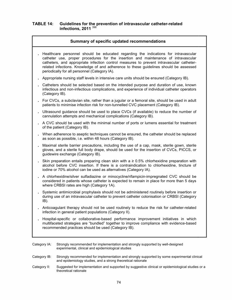
74
TABLE 14: Guidelines for the prevention of intravascular catheter-related infections, 2011 396
Category IA: Strongly recommended for implementation and strongly supported by well-designed
experimental, clinical and epidemiological studies
Category IB: Strongly recommended for implementation and strongly supported by some experimental clinical and epidemiology studies, and a strong theoretical rationale
Category II: Suggested for implementation and supported by suggestive clinical or epidemiological studies or a theoretical rationale
Summary of specific updated recommendations
. Healthcare personnel should be educated regarding the indications for intravascular
catheter use, proper procedures for the insertion and maintenance of intravascular catheters, and appropriate infection control measures to prevent intravascular catheter-related infections. Knowledge of and adherence to these guidelines should be assessed periodically for all personnel (Category IA).
. Appropriate nursing staff levels in intensive care units should be ensured (Category IB).
. Catheters should be selected based on the intended purpose and duration of use, known infectious and non-infectious complications, and experience of individual catheter operators (Category IB).
. For CVCs, a subclavian site, rather than a jugular or a femoral site, should be used in adult patients to minimise infection risk for non-tunnelled CVC placement (Category IB).
. Ultrasound guidance should be used to place CVCs (if available) to reduce the number of cannulation attempts and mechanical complications (Category IB).
. A CVC should be used with the minimal number of ports or lumens essential for treatment of the patient (Category IB).
. When adherence to aseptic techniques cannot be ensured, the catheter should be replaced as soon as possible, i.e. within 48 hours (Category IB).
. Maximal sterile barrier precautions, including the use of a cap, mask, sterile gown, sterile gloves, and a sterile full body drape, should be used for the insertion of CVCs, PICCS, or guidewire exchange (Category IB).
. Skin preparation entails preparing clean skin with a ≥ 0.5% chlorhexidine preparation with alcohol before CVC insertion. If there is a contraindication to chlorhexidine, tincture of iodine or 70% alcohol can be used as alternatives (Category IA).
. A chlorhexidine/silver sulfadiazine or minocycline/rifampicin-impregnated CVC should be considered in patients whose catheter is expected to remain in place for more than 5 days where CRBSI rates are high (Category 1A).
. Systemic antimicrobial prophylaxis should not be administered routinely before insertion or during use of an intravascular catheter to prevent catheter colonisation or CRBSI (Category IB).
. Anticoagulant therapy should not be used routinely to reduce the risk for catheter-related infection in general patient populations (Category II).
. Hospital-specific or collaborative-based performance improvement initiatives in which multifaceted strategies are “bundled” together to improve compliance with evidence-based recommended practices should be used (Category IB).

75
CHAPTER 11
PRINCIPLES OF TREATMENT OF CATHETER-RELATED INFECTION
Treatment depends on the stage of infection and the pathogen 2,3,4,5,20. As a general rule,
when CRBSI is suspected, the common practice in the ICU is to remove the CVC and replace
it at a new site if ongoing use of a CVC is required 2,3,4,5,20,60.
As many catheters may be removed unnecessarily, some authors/workers have suggested a
more conservative approach of “watchful waiting” with appropriate investigations being
performed and ongoing clinical evaluation 60,527.
Figure 4 summarises the current approach to the management of patients with short-term
central venous catheter-related bloodstream infection.
11.1 Catheter removal and rationale
Although some authorities have suggested that it may be acceptable to retain the catheter in
situ and treat with appropriate antimicrobial therapy when the CRBSI is caused by coagulase-
negative staphylococci, failure to remove the CVC is associated with a substantial chance of
recurrence 2,3,4,5,20,60. Three observational prospective studies have shown that removal of the
CVC in the setting of S. aureus CRBSI, including uncomplicated cases, is associated with a
more rapid response to therapy and a lower relapse rate 178,528,529. In the setting of
uncomplicated S. aureus CRBSI, the catheter should be removed and at least 2 weeks of
parenteral antibiotics given. There is a high relapse rate if these are given for a shorter time
2,3,4,5,20,179,529.
Failure to remove the catheter with CRBSIs due to Gram-negative bacilli such as Klebsiella
pneumoniae, Enterobacter species, Pseudomonas species, Acinetobacter species and
Stenotrophomonas maltophilia, has been reported to be associated with a higher rate of
treatment failure, relapse and bacteraemic recurrence 530. In view of this, it has been
76
suggested that Gram-negative bacillary CRBSI should be managed with removal of the CVC
in conjunction with an appropriate course of antibiotics 530,531.
At least five prospective studies have shown that CVC removal is associated with improved
outcome in patients with candidaemia 532. These include large prospective studies in which
catheter retention proved to be a significant factor for the persistence of candidaemia, or was
associated with higher mortality. In a study prospectively analysing the risk factors for death
in 145 patients with nosocomial candidaemia, catheter retention was found to be an
independent variable for increased risk of death in multivariate analysis, independent of
persistent neutropenia 242. Recent controversial but challenging data in candidaemia
evaluating patients treated with echinocandins or liposomal Amphotericin B, which have
biofilm penetrating properties, has suggested CVCs could possibly remain in situ if these
agents were used in this setting 240. This approach is currently neither a widely accepted
policy, nor is it practice at the multidisciplinary ICU at Charlotte Maxeke Johannesburg
Academic Hospital. Retention of an infected catheter has most recently been shown in a
meta-analysis involving seven randomised trials and 1915 patients in the setting of invasive
candidiasis, to be associated with increased mortality 137.
In cases of septic shock or severe sepsis of undetermined origin, or when frank local signs of
infection are found, the catheter should always be removed 60,305,309,402.
When conservative strategies have been adopted, the decision for catheter removal depends
on the microorganisms and on the evolution of the patient’s state during the first 48 hours. If
blood cultures recover S. aureus, enterococci, gram-negative bacilli or fungi, the catheter
should be removed 60,275,305. Overall, conservative strategies are deemed to be risky in
critically ill patients 60. When such an approach is embarked upon patients must be
continuously monitored.

77
11.2 Antimicrobials (see Figure 4)
When a CRBSI is associated with severe sepsis or shock, antimicrobial therapy should be
administered timeously together with catheter replacement 305. Broad spectrum antimicrobial
cover is normally advised and should be based on local epidemiology as well as antimicrobial
susceptibility patterns. This treatment should then be de-escalated according to the results of
catheter tip cultures and blood cultures 305.
In cases of CRBSI, the duration of treatment should be at least 14 days for uncomplicated S.
aureus and Candida species 305. For CRBSI-associated with Pseudomonas species and
Acinetobacter species, a similar duration of time should be considered 305. For other
microorganisms and with uncomplicated catheter-related infection, antimicrobial therapy
should not exceed 7 days if the catheter has been removed. Uncomplicated CRBSI refers to
regression of septic signs and bacteraemia within 3 days and no persistent infectious site.
Staphylococcus lugdunesis should be treated in a similar fashion to S. aureus 305.
Vancomycin is generally recommended for empirical therapy in health care settings with an
elevated prevalence of MRSA. For institutions in which the preponderance of MRSA isolates
have Vancomycin minimum inhibitory concentrations (MIC) values ≥1.5µg/ml (and probably
≥1µg/ml), alternative agents such as daptomycin are advocated 395,533. Linezolid
administration is not advocated for empirical therapy (ie. patients suspected but not proven to
have CRBSI) 305. Empiric coverage for Gram-negative bacilli should be based on local
antimicrobial susceptibility data and the severity of the disease. Examples of agents include
fourth-generation cephalosporins, carbapenems, or β-lactam / β-lactamase combinations,
with or without an aminoglycoside 305.
For empirical treatment of suspected catheter-related candidaemia, use of an echinocandin,
or in selected patients fluconazole, is recommended. Fluconazole could be considered for
patients without recent azole exposure and in health care settings where the risk of Candida

78
krusei or Candida glabrata infection is very low 305. Systemic antifungal therapy (together with
removal of the catheter) should be given in all cases of catheter-related candidaemia in view
of the potentially significant sequelae 2,3,4,5,20,534. Deoxychelate-Amphotericin B has previously
been used in this setting 3,4,5,20,535. Newer agents are, however, felt to be less toxic. Agents
such as Voriconazole which covers Candida albicans as well as non-albicans species, and
lipid formulations of Amphotericin B have also been used in this setting 4,5. Initiation of
empirical therapy for suspected catheter-related candidaemia is recommended for septic
patients who are already on broad-spectrum antibiotics, where there is no evidence of overt
sepsis elsewhere, and with any of the following risk factors: total parenteral nutrition,
prolonged use of broad-spectrum antibiotics, haematologic malignancy, receipt of bone
marrow or solid-organ transplant, and femoral catheterisation or colonisation due to Candida
species at multiple sites 60,305,309.
Empiric combination antibiotic coverage for multi-drug-resistant (MDR) Gram-negative bacilli,
such as Pseudomonas aeruginosa is suggested when CRBSI is suspected in neutropenic
patients, severely ill patients with sepsis, or patients known to be colonised with such
pathogens until the culture and susceptibility data are available and the antibiotic regimen can
be de-escalated 305,309.
11.3 Other approaches
Some workers have suggested that although antimicrobial therapy is frequently administered
and advocated, many of the infectious complications may be self-limited and resolve after
removal of the catheter. This may be applicable to cases associated with CoNS and
Enterobacteriaceae 2,3,4,5,20,60. Antimicrobials should, however, be administered if there is
persistent sepsis despite catheter removal in immunosuppressed patients, or if there is
evidence of complications 2,3,4,5,20,60.

79
11.4 Catheter salvage therapy
In some rare circumstances, catheter salvage therapy has been advocated in the ICU 305,309
with the use of antibiotic lock therapy. This treatment should only be considered in the
absence of severe sepsis and when Candida species, and S. aureus (and probably
Pseudomonas species and Acinetobacter baumannii) are not the responsible organisms 60.
An additional drawback in the ICU is that the catheter needs to be unused during the
antibiotic lock process, thus further limiting the potential usefulness. For patients with CRBSI
in whom catheter salvage is attempted, blood cultures should be obtained, and the catheter
should be removed if 2 sets of blood cultures on a given day (one set is acceptable in
neonates) remain positive for microorganisms when drawn 72 hours after initiation of
appropriate therapy 305,309.
11.5 Complications
Complications of CRBSI include septic thrombosis of the great veins 532,536, endocarditis and
metastatic foci of infection 2,3,4,5,20,536.
The strongest predictor of complicated S. aureus bacteraemia is the persistence of fever
and/or bacteraemia for more than 72 hours after catheter removal and initiation of antibiotics
179,537. In patients with S. aureus CRBSI and persistant fever or bacteraemia,
transoesophageal echocardiography should be performed to rule out infective endocarditis
305,309,325,537.
Four to 6 weeks of antibiotic therapy is advocated in patients with persistent fungaemia or
bacteraemia after catheter removal (ie. occurring beyond 72 hours after line removal), in
patients who are found to have infective endocarditis, suppurative thrombophlebitis, and in
paediatric patients with osteomyelitis. Six to 8 weeks of therapy should be used for the
treatment of osteomyelitis in adults 305,309.

80
11.6 Special situations
For CRBSI due to less virulent microbes that are difficult to eradicate such as Bacillus
species, Micrococcus species or Propionibacterium, catheters should generally be removed
after blood culture contamination is ruled out on the basis of multiple positive culture results,
with at least one blood culture sample drawn from a peripheral vein 305.

81
Figure 4: Approach to the management of patients with short-term central venous catheter-related bloodstream infection
82
CHAPTER 12
STUDY PROTOCOL
12.1 Aims of this study
The aims of this study were to determine whether the duration of central venous catheter
insertion time could safely be increased from the standard practice of seven days in the adult
multidisciplinary ICU at CMJAH, to fourteen days, to assess the influence of an antimicrobial
impregnated catheter on the incidence of CRI, as well as to elucidate the epidemiology and
various risks of CRI in a population of critically ill patients, including clinically evident catheter-
related thrombosis.
12.2 Materials and methods
This was a prospective, randomised, double-blind study performed in the adult
multidisciplinary intensive care unit at CMJAH, South Africa over a four year period. The
study included 118 critically ill medical, surgical, trauma and obstetric/gynaecological
patients, and entailed comparison of a 14 day placement of standard triple lumen (ARROW
Standard Triple Lumen Catheter, Arrow International Inc., Reading, Pa, US) versus
antimicrobial impregnated (CSS; ARROWgardTM Triple Lumen Catheter, Arrow International
Inc., Reading, PA, US) CVCs on the rate of CRI.
The two types of catheters were of identical appearance and were subsequently differentiated
by a numerical code which was broken only on completion of the study.
The randomisation protocol involved equal numbers of the two types of non-distinguishable
catheters being mixed in consignments and then selected in a consecutive fashion for
placement in study candidates.

83
Exclusion criteria for the study were age less than 18 years, white blood cell count on
admission of less than 4x10⁹/L, skin burns and a history of allergy to sulpha-containing
preparations. No guidewire exchanges were performed.
Standard infection control measures were practiced with catheter insertion. This included
handwashing, the use of sterile gowns, sterile gloves, full sterile drapes, masks and caps.
The catheters were placed by the ICU medical staff, including intensivists, fellows and
registrars in training, into the right or left subclavian or internal jugular veins as judged most
appropriate by clinical evaluation.
Skin swabs were taken for culture prior to cleansing and catheter insertion. The skin insertion
site was cleansed with a 0.5% chlorhexidine-gluconate in 70% alcohol solution. Catheters
were inspected and dressed daily and a clinical assessment undertaken for any evidence of
catheter-related thrombosis. The catheters were studied for colonisation and CRBSI at
removal. The origin of each CRBSI was sought by culturing all potential sources (skin,
catheter segments, hubs and infusate). A semi-quantitative culture was performed on the
catheters using the roll-plate technique as described by Maki et al.100. DNA-molecular typing
was employed to assist in microbiological analyses. All relevant clinical data was collected
and evaluated.
CRI was defined according to the criteria proposed by the Centers for Disease Control and
Prevention 96. Definitions used for identifying the source of an isolate causing a CRBSI are
shown in Table 15.
Catheters remained in place until they were no longer required, a specific event necessitated
removal, or for 14 days, whichever occurred first. All existing intravascular catheters were
removed prior to insertion of the study catheters.
Any parenteral nutrition administered was delivered via a single dedicated port.

84
12.3 Collection and processing of specimens
12.3.1 Collection and processing of CVC pre-insertion and pre-removal specimens
for quantitative culture of the skin insertion site
Ten square centimeters of skin about the catheter site was sampled. A sterile template
was applied to the site and the exposed area cultured with a cotton-tipped applicator pre-
moistened with Stuart’s transport medium by vigorously scrubbing the entire area four
times, twice in one direction and twice more in a direction perpendicular to the first. In the
laboratory, the tip of the applicator was immersed in 1 ml of sterile saline and agitated
vigorously on a Vortex mixer.
Serial dilutions were inoculated onto blood agar plates (done in triplicate) and incubated
aerobically at 350C overnight. All colony types were enumerated using routine laboratory
methods.
12.3.2 Collection and processing of specimens from catheter hubs
Catheter hubs were cultured aseptically by inserting a smaller, sterile, cotton-tipped
applicator into the hub and whilst exerting gentle axial pressure, rotating the applicator.
The tips of each applicator were cultured in a fashion similar to that described for skin
cultures.
12.3.3 Collection and processing of infusates
A 10 ml sample of each infusate that was administered to the patient via the CVC was
collected aseptically and sent to the laboratory in a sterile universal container. On receipt
in the laboratory, the infusate was centrifuged and the deposit cultured onto a segment of
a blood agar plate (incubated at 350C for 72 hours) and in 10 ml of brain heart infusion
85
broth and incubated at 350C for 7 days, before being terminally sub-cultured and
discarded.
12.3.4 Collection and processing of blood cultures
In the setting of local or systemic sepsis, a set of blood cultures was obtained both
percutaneously from a single venepuncture site, as well as through the catheter (patients
with fever, other sites of infection, or inflammation of the insertion site). In the absence of
local or systemic sepsis, a set of blood cultures was routinely taken from the catheter at
the time of catheter removal. Each collected “set” of blood culture bottles consisted of an
aerobic Bactec® bottle, an anaerobic Bactec® bottle and a fungal blood culture tube.
Clinicians were advised to collect no less than 5 ml of blood in each Bactec® bottle. Blood
cultures were processed at 350C for 7 days by the BACTEC-460® radiometric method.
Fungal blood cultures were processed using conventional methods.
12.3.5 Collection and processing of catheter segments for culture
To prevent contamination by skin organisms, the skin around the insertion site was
cleansed with 0.5% chlorhexidine gluconate in 70% alcohol and allowed to dry prior to
removal. Catheters were removed aseptically keeping the externalised portion directed
upward and away from the skin surface to minimise contamination by skin organisms.
The distal intravascular tip and the proximal transcutaneous segments of the catheter
were submitted to the laboratory for bacteriological processing after being cut with a
sterile scapel into 5 cm portions. The two segments of the catheter were sent to the
laboratory in sterile universal containers and cultured within 2 hours of collection to
prevent dessication of micro-organisms. A semi-quantitative culture was performed on
the catheters using the method described by Maki 100 with colony types subsequently
enumerated and micro-organisms identified.

86
12.4 Statistics
Statistical analysis was performed using logistic regression, the Wilcoxan Mann-Whitney test
and a chi-squared contingency table test 538-541.
A chi-squared contingency table test was used when testing for differences between the
catheter types versus categorical variables such as sex, and yes/no variables such as site,
systemic sepsis, parenteral nutrition, ventilation and reason for admission.
Where the test was between the catheter types and continuous variables such as age and
number of bacteria around the insertion site (either prior to insertion or at the time of
removal), a Wilcoxon-Mann-Whitney test was used.
The reason for use of a nonparametric test rather than a t-test or an Analysis of Variance is
that the data used cannot be assumed to be from a normal distribution, i.e. the data are
skewed. This is also the reason why the median and range of the ages was reported (median
47, range 18-83)
In order to investigate possible confounding due to covariates, logistic regression was used.
This allowed testing for a difference between the catheter types once a covariate or multiple
covariates is/are taken into account.
A p-value less than 0.05 was considered significant.
12.5 Ethics Approval and International Protocol Registration
The study was approved by the Committee for Research on Human Subjects of the
University of the Witwatersrand (M941129) and the protocol registered with ClinicalTrials.gov
protocol registration system (NCT00533988). Informed consent was obtained for all
participants.
87
CHAPTER 13
RESULTS
Sixty two patients received a standard triple-lumen catheter and 56 patients a CSS
impregnated triple-lumen catheter. The study spanned 34,951.5 catheter hours (≡1,456
catheter days, ≡3.99 catheter years).
Patient demographics and risk factors for CRI were similar in both groups (see Table 16).
However, significantly more trauma patients received a standard catheter [20/62 (32%) vs.
9/56 (16%)] p=0.0449. There was no significant difference in infections between the
catheters, after taking trauma into account (p=0.8464).
Following catheter insertion, bloodstream infection was diagnosed in 18 patients, one of
whom died. Primary CRBSI occurred in 15 patients and in three cases the CVC was
contaminated following haematogenous seeding. Five patients demonstrated catheter
colonisation. Ninety-five catheters (81%) (49 standard, 46 CSS) had neither evidence of
colonisation nor bloodstream infection. The mean duration of placement of these 95 CVCs
was 12.4 days.
Thirty-three catheters (17 standard, 16 CSS) were removed prior to 14 days (suspected
sepsis, accidental displacement, death, no longer required).
Of the CVCs left in place for 14 days, 84% had no evidence of catheter colonisation or
bloodstream infection (71/85). The mean duration of placement for the full sample of 118
CVCs was 12.3 days (range 1 – 14). No statistically significant differences between the two
catheters could be demonstrated in terms of number of days of catheter placement
(p=0.3341) and colonisation or bloodstream infection (13/62 vs. 10/56) vs. no infection
(p=0.6704) (see Table 16).
88
Furthermore, no statistically significant difference in CRBSI or colonisation between the two
catheters could be demonstrated for removal at or before 7 days (p=0.6003) and after 7 days
(p=1.000).
The site of CVC insertion (internal jugular vein versus subclavian vein), the administration of
parenteral nutrition and bloodstream infection at the time of catheter insertion were not noted
to be risk factors for catheter infection (p=0.1617, 0.5449 and 0.4575). In addition, no
statistical difference could be demonstrated between the two catheters for these variables
(p=0.3320, 0.5868 and 0.1861). A significantly greater number of patients had an internal
jugular vein insertion site (95 vs. 23; p<0.0001).
The most common source of primary CRBSI was skin, followed by hub and infusate. There
was no statistical difference between the two catheters.
The most common microorganism isolated as a cause of CRBSI was CoNS. Other
microorganisms associated with CRBSI included S. aureus and various gram-negatives such
as Serratia marcescens, Klebsiella pneumoniae, Enterobacter aerogenes, Acinetobacter
baumanii, Providencia species and Alkaligenes faecalis. A yeast was identified in one case
(CSS catheter). Primary CRBSI occurred in nine cases with a standard catheter and in six
cases with a CSS catheter. There were no significant differences between the catheters in
terms of the nature of the micro-organisms causing CRBSI.
Similarly, with respect to colonisation, there were no significant differences in the nature of
the colonising bacteria between the two catheters. CoNS were the most commonly isolated
coloniser involving three catheters (two standard catheters, one CSS), with Enterobacter
cloacae (standard CVC) and Bacillus cereus (CSS CVC) being the other two colonisers.
No statistically significant differences were noted in the number of bacteria colonising the skin
around the CVC insertion site both prior to catheter insertion and at the time of catheter
89
removal on analysis of colony forming units of isolates (p=0.08). CoNS constituted the
majority of 161 skin colonisers that were isolated (60%). Other bacteria isolated from the skin
cultures in descending order of frequency included Enterobacteriaceae (10%), Bacillus
species (7.5%), Acinetobacter baumanii (5%), S. aureus (4%), enterococci (4%), diptherioids
(4%), Pseudomonas species (3%) and Streptococcus species (2.5%). For each of these
microorganisms, there were no statistically significant differences between isolates when
comparing the two types of catheter (p> 0.05).
No significant non-infectious complications such as pneumothorax related to catheter
placement were documented. There was no clinical evidence of catheter-related thrombosis
in either of the study groups.

90
TABLE 15 Definitions for identifying the source of a catheter-related bloodstream infection
Infusate-related infection: same organism present in the infusate,
central venous catheter hubs and central venous catheter distal
segment. Skin cultures negative.
Hub-related infection: same organism recovered from the central
venous catheter hub, the central venous catheter distal segment and
blood cultures. Cultures of infusates negative. Skin cultures negative,
or positive for different cultures.
Skin-related infection: same microorganism isolated from the skin,
the central venous catheter transcutaneous segment, the central
venous catheter distal segment and blood cultures.
Hematogenous seeding of the central venous catheter: same
microorganism isolated from blood cultures, the central venous
catheter distal segment and a distant source of infection. Cultures of
infusate/s, central venous catheter hubs and skin negative.

91
TABLE 16 Characteristics of 118 critically ill patients enrolled in the study
CSS = Chlorhexidine silver sulfadiazine CVC = Central venous catheter
Patient demographics Standard catheter CSS catheter
Number of patients 62 56
Median age in years (range) 47 (18 – 83) 43 (18 – 78)
Sex M:F 38 : 24 (61%) : (39%)
34 : 22 (61%) : (39%)
Systemic sepsis at time of CVC insertion 36 (58%) 33 (59%)
Parenteral nutrition 33 (53%) 27 (48%)
Ventilation 54 (87%) 55 (98%)
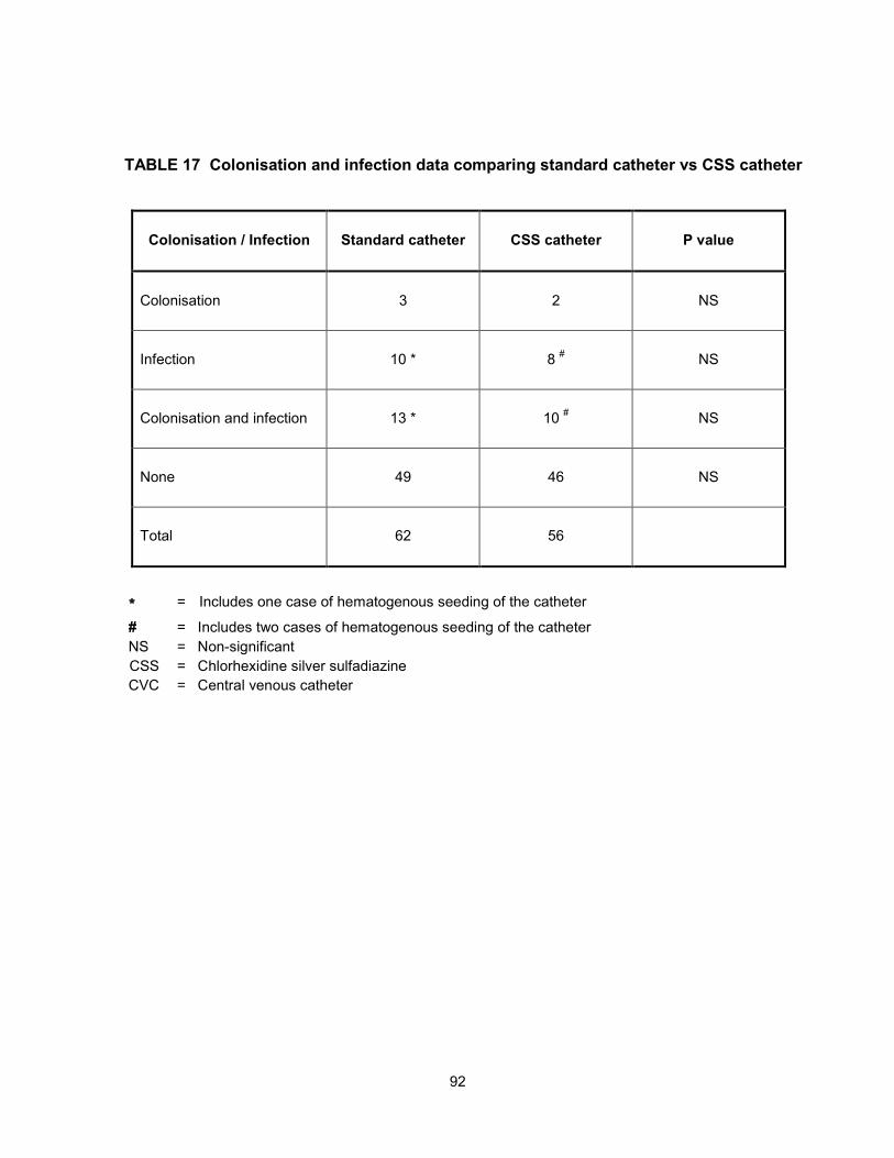
92
TABLE 17 Colonisation and infection data comparing standard catheter vs CSS catheter
∗ = Includes one case of hematogenous seeding of the catheter
# = Includes two cases of hematogenous seeding of the catheter NS = Non-significant CSS = Chlorhexidine silver sulfadiazine CVC = Central venous catheter
Colonisation / Infection Standard catheter CSS catheter P value
Colonisation 3 2 NS
Infection 10 * 8 # NS
Colonisation and infection 13 * 10 # NS
None 49 46 NS
Total 62 56
93
CHAPTER 14
DISCUSSION
This study examined the influence of antimicrobial impregnated central venous catheters on
the incidence of catheter-related infection, whether CVCs can safely be left in place for a
period of up to 14 days, as well as elucidating the epidemiology and risks (such as insertion
site, clinical evidence of thrombosis and the use of parenteral nutrition) of catheter-related
infection in a population of critically ill patients.
Exactly how long non-cuffed short term CVCs can safely be left in place, particularly in
critically ill patients, has not been previously assessed 33,34. In general, most studies that have
evaluated duration of placement as a risk factor have shown that prolonged placement
significantly increases the cumulative risk of infection, particularly if longer than 5 - 7 days
26,256,436,542,543,. Indeed, a critical review of well designed published studies of risk factors for
CVC-related bloodstream infections revealed that duration beyond 7 days was associated with
a significantly increased risk of CRBSI 33. Despite suggestions that routine replacement of
CVCs at short term intervals such as every 4 - 5 or 7 days may not significantly reduce the risk
of CVC-related BSI in patients requiring prolonged central access, this has not been
conclusively established 30. While some studies report no decline in the incidence with routine
replacement, most have not had sufficient statistical power to answer this question 33. Because
the issue of “safe” duration of catheter retention has remained controversial and unanswered
33, scheduled replacement remains widely practiced in many ICUs, despite currently available
guidelines and recommendations. In a study in mainland Britain where 165 ICUs were
surveyed, catheters were routinely replaced in the majority, the mean time being 6.5 days 544.
It is felt that this study with almost 35,000 hours of catheter dwell time provides an ample basis
to advocate “safe” CVC dwell time and offers suitable direction. The need for an intravascular
94
catheter should however, be frequently assessed and the device should be removed as soon
as the intended use is over 21,545.
Over the past several years, antimicrobial impregnated CVCs have been introduced in an
attempt to limit catheter-related infection and increase the time that CVCs can safely be left in
situ. Several studies and meta-analyses have reported the benefit of these CVCs in the
prevention or reduction of CRI 36,37,39,99,393,494-498,500-507,515. Cost efficacy has also been claimed
99,522-524,546.
On the basis of this data, various advisory panels and authorities have recommended the use
of antimicrobial impregnated CVCs in selected clinical settings 1,39,40,59,395,396,542,547-549.
However, their role remains controversial with various workers suggesting that more work is
required to support or refute the hypothesis that they may reduce the rate of, or prevent
CRBSI 38,550.
A recent review of eleven randomised studies failed to demonstrate any significant clinical
benefit associated with the use of antimicrobial impregnated CVCs for the purpose of
reducing CRBSI or improving patient outcome 550. Concern about the emergence of
antimicrobial resistance with long term use of these catheters has also been expressed
38,517,547,548,550-552.
This study was unable to demonstrate any benefit of an antimicrobial impregnated catheter
(CSS) over standard catheters in critically ill patients. Of interest, there was no difference in
colonisation or bloodstream infection rate between the two catheters prior to or after 7 days,
which appears to refute the contention that antimicrobial impregnated catheters may be of
particular value. Furthermore, a significantly greater number of trauma patients received a
standard catheter, a variable recognised to potentially increase the rate of CRI.
95
Placement of CVCs in the internal jugular or femoral vein rather than the subclavian vein has
been associated with a significantly increased risk of CRBSI 33,388,389,390,391. The subclavian
vein is currently recommended as the preferred site of placement of CVCs in several
guidelines and reviews 33,39,396,553. Several studies, including one randomised clinical trial,
have found that percutaneous placement of a CVC in an internal jugular or femoral vein is
associated with a substantially higher risk of CRSBI than subclavian vein placement 25,33,97,554-
556. It has been postulated that internal jugular vein placement may be associated with
greater rates of CRI due to possible contamination from oro-nasal and airway secretions, a
higher incidence of CVC-related thrombosis, as well as difficulty in adequately securing the
catheter.
In this study, we were unable to demonstrate any difference in catheter infection related to
site of CVC insertion when comparing internal jugular vein versus subclavian vein placement.
This was despite the fact that a significantly greater number of patients had an internal
jugular vein insertion site. There was also no statistical difference when comparing the
standard versus CCS impregnated catheters.
Similarly, the use of parenteral nutrition has also been well documented to be a risk factor for
CRBSI with non-cuffed percutaneously inserted CVCs 33,557. In this study, the administration
of parenteral nutrition via a single dedicated port was not noted to be a risk factor for catheter
infection and again, there was no statistical difference between the two types of catheters.
Patients with CVCs in situ have previously been reported to be at high risk for catheter-
related thrombosis, especially if the catheter has been in place for one week or more 29,30,558.
Of particular relevance, is that the risk of CRI has been strongly correlated to the presence of
catheter-related thrombi 29,30,31,32. The risk of catheter-related thrombosis appears to be
greatest with femoral venous catheters, followed by those placed in the internal jugular vein,
with the least risk reported to involve subclavian venous catheters 140,156,559. The risk of

96
thrombosis is documented to be four-fold greater with internal jugular lines as compared to
subclavian vein catheters 156. Recognised predisposing factors to venous thrombosis include
catheter-related factors (indwelling time, diameter, CVC material), procedural factors (difficult
or traumatic insertion) and patient factors (insertion site, blood viscosity, activation of
coagulation factors due to the underlying disease) 32,559.
In this study, there was no clinical evidence of catheter-related thrombosis in any of the
patients, despite the majority of the patients having an internal jugular vein insertion site, a
minimum of one of the predisposing factors, and a mean dwell time well beyond one week.
The administration of subcutaneous low-molecular-weight heparin as part of the routine
management of these patients may have decreased the risk of clinically apparent thrombus
formation.
Experience with insertion technique has been identified as an important risk for CVC-related
BSI 33,331. Studies have shown that intensified training and educational programs can
substantially reduce the baseline risk of CRBSI 560,561. This study was performed in an
academic training unit with a large turnover of junior staff, many of whom were involved in
catheter placement. This in conjunction with the background severity of illness of the patients
involved may have been contributing factors to the documented CRIs. All the patients in this
study had at least one major organ failure and were initially assessed to be likely to spend a
substantial amount of time in ICU and hospital and thus be potential candidates for 14 day
catheter dwell time.
Subsequent to this study a dedicated policy regarding the insertion, maintenance and use of
CVCs has been developed. The basic principle revolves around strict adherence to aseptic
technique at all times and has been described in recent reviews 3,4,20. It is currently the unit
policy in the multidisciplinary adult ICU at CMJAH to leave standard CVCs in situ for up to 14
days in critically ill patients after which time the device is replaced and resited if it is deemed

97
necessary. Since the introduction of this practice and with ongoing repetitive education it is
now the exception that a CVC is removed for suspected CRI.
Epidemiologically, the most common source of primary CRBSI was skin followed by hub and
infusate. No difference could be demonstrated between the two catheters. These findings are
similar to those previously reported.

98
CHAPTER 15
CONCLUSION
In this study, no significant difference in CRI rates between standard and CSS impregnated
CVCs could be demonstrated. Standard CVCs can safely be left in situ in critically ill patients
for up to 12 days and probably 14 days, with appropriate infection control measures. The
most common source of CRI was the skin. Administration of parenteral nutrition and the site
of catheter insertion (subclavian vein versus internal jugular vein), were not noted to be risk
factors for CRI. Furthermore, there was no clinical evidence of catheter-related thrombosis in
any of the patients.
The data and observations presented in this study are important and offer direction for the
use of CVCs in critically ill patients, irrespective of location, particularly in light of the many
controversies that exist, and also in view of the suggestions by various workers in the field
that more data on the subject is required and that there is room for improvement in central
venous catheter care 562,563.
99
REFERENCES
1. O’Grady NP, Alexander M, Dellinger EP, Gerberding JL, Heard SO, Maki DG, et
al. Guidelines for the prevention of intravascular catheter-related infections.
Centers for Disease Control and Prevention. MMWR Recomm Rep 2002;51(RR-
10):1-29.
2. Mer M. Intravascular catheter sepsis. S Afr J Crit Care 1999;15(2);30-35.
3. Mer M. Vascular catheter related infection. Clinical Intensive Care 2001;12(2):53-
60.
4. Mer M. Intravascular catheter related infection: current concepts. S Afr J Crit Care
2006;22(1):4-12.
5. Mer M. Intravascular catheter-related infection: update and overview. CME 2008;
26(11):540-544.
6. Smith RL, Meixler SM, Simberkoff MS. Excess mortality in critically ill patients with
nosocomial bloodstream infections. Chest 1991;100:164-167.
7. Martin MA, Pfaller MA, Wenzel RP. Coagulase-negative staphylococcal bac-
teremia. Mortality and hospital stay. Ann Intern Med 1989;110:9-16.
8. Haley RW, Schaberg DR, Von Allmen SD, McGowen JE Jr. Estimating the extra
charges and prolongation of hospitalization due to nosocomial infections: a
comparison of methods. J Infect Dis 1990;141:248-257.
9. Pittet D, Tarara D, Wenzel RP. Nosocomial bloodstream infection in critically ill
patients. Excess length of stay, extra costs and attributable mortality. JAMA
1994;271:1598-1601.
10. Arnow PM, Quimosing EM, Beach M. Consequences of intravascular catheter
sepsis. Clin Infect Dis 1993;16:778-784.
11. Heiselman D. Nosocomial bloodstream infection in the critically ill. JAMA
1994;272:1819-1820.
12. Collignon PJ. Intravascular catheter associated sepsis: a common problem. The
Australia Study on Intravascular Catheter Associated Sepsis. Med J Aust 1994;
161:374-378.
100
13. Rello J, Ochagavia A, Sabanes E, Roque M, Mariscal D, Reynaga E, et al.
Evaluation of outcome of intravenous catheter-related infections in critically ill
patients. Am J Respir Crit Care Med 2000;162:1027-1030.
14. Rosenthal VD, Guzman S, Migone O, Crnich CJ. The attributable cost, length of
hospital stay, and mortality of central line-associated bloodstream infection in
intensive care departments in Argentina: a prospective matched analysis. Am J
Infect Control 2003;31:475-480.
15. Pronovost P, Needham D, Berenholtz S, Sinopoli D, Chu H, Cosgrove S, et al. An
intervention to decrease catheter-related bloodstream infections in the ICU. N Engl
J Med 2006;355:2725-2732.
16. Wenzel RP, Edmond MB. Team-based prevention of catheter-related infections. N
Engl J Med 2006;355:2781-2783.
17. Rosenthal VD, Maki DG, Salamao R, Moreno CA, Mehta Y, Higuera F, et al.
International Nosocomial Infection Control Consortium. Device-associated
nosocomial infections in 55 intensive care units of 8 developing countries. Ann
Intern Med 2006;145:582-591.
18. Laupland KB, Lee H, Gregson DB, Manns BJ. Cost of intensive care unit-acquired
bloodstream infections. J Hosp Infect 2006;63:124-132.
19. Warren DK, Quadir WW, Hollenbeak CS, Elward AM, Cox MJ, Fraser VJ.
Attributable cost of catheter-associated bloodstream infections among intensive
care patients in a nonteaching hospital. Crit Care Med 2006;34:2084-2089.
20. Mer M. Intravascular catheter-related guidelines. S Afr J Epidemiol Infect 2005;
20(2):64-70.
21. Mer M, Duse GA, Galpin JS, Richards GA. Central venous catheterization: a
prospective randomized, double-blind study. Clin Appl Thomb Hemost 2009;
15(1):19-26.
22. File TM Jr, Abell VL. Prevention of bloodstream infections: basics and beyond. Crit
Care Med 2009;37:375-376.
23. Wang H, Huang T, Jing J, Jin J, Wang P, Yang M, et al. Effectiveness of different
central venous catheters for catheter-related infections: a network meta-analysis. J
Hosp Infect 2010;76:1-11.
101
24. Maki DG. Infections due to infusion therapy. In: Bennett JV, Brachman PS, eds.
Hospital Infections. 3rd ed. Boston, MA: Little, Brown and Co. 1992.
25. Richet H, Hubert B, Nitemberg, Andremont A, Buu-Hoi A, Ourbak P, et al.
Prospective multicenter study of vascular catheter-related complications and risk
factors for positive central-catheter cultures in intensive care unit patients. J Clin
Microbiol 1990;28:2520-2525.
26. Gil RT, Kruse JA, Thill-Baharozian MC, Carlson RW. Triple vs. single lumen
central venous catheters. A prospective study in a critically ill population. Arch
Intern Med 1989;149:1139-1143.
27. Miller JJ, Venus B, Mathru M. Comparison of the sterility of long-term central
venous catheterization using single lumen, triple lumen and pulmonary artery
catheters. Crit Care Med 1984;12:634-637.
28. Ullman RF, Gurevich I, Schoch PE, Cunha BA. Colonization and bacteremia
related to duration of triple-lumen intravascular catheter placement. Am J Infect
Control 1990;18:201-207.
29. Polderman KH, Girbes AR. Central venous catheter use. Part 2: infectious
complications. Intensive Care Med 2002;28:18-28.
30. Girbes AR, Polderman KH. Mechanical and infectious complications of central
venous catheters. Minerva Anestesiol 2003;69:330-332.
31. Randolph AG, Cook DJ, Gonzales CA, Andrew M. Benefit of heparin in central
venous and pulmonary artery catheters: a meta-analysis of randomized controlled
trials. Chest 1998;113:165-171.
32. Polderman KH, Girbes AR. Central venous catheter use. Part 1: Mechanical
complications. Intensive Care Med 2002;28:1-17.
33. Safdar N, Kluger DM, Maki DG. A review of risk factors for catheter-related
bloodstream infection caused by percutaneously inserted noncuffed central venous
catheters: implications for preventive strategies. Medicine (Baltimore) 2002;81:
466-479.
34. Norwood S, McAuley CE. Vascular catheter-related infection. In: Fink MP,
Abraham E, Vincent JL, eds. Textbook of Critical Care 5th ed. Philadelphia, PA:
Elsevier Saunders; 2005:1239-1248.
102
35. Veenstra DL, Saint S, Saha S, Lumley T, Sullivan SD. Efficacy of antiseptic-
impregnated central venous catheters in preventing catheter-related bloodstream
infection: a meta-analysis. JAMA 1999;281:261-267.
36. Marin MG, Lee JC, Skurnick JH. Prevention of nosocomial bloodstream infections:
effectiveness of antimicrobial-impregnated and heparin-bonded central venous
catheters. Crit Care Med 2000;28:3332-3338.
37. Crnich CJ, Maki DG. The promise of novel technology for the prevention of
intravascular device-related bloodstream infection. I. Pathogenesis and short-term
devices. Clin Infect Dis 2002;34:1232-1242.
38. McConnel SA, Gubbins PO, Anaissie EJ. Are antimicrobial-impregnated catheters
effective? Replace the water and grab your washcloth, because we have a baby to
wash. Clin Infect Dis 2004;39:1829-1833.
39. Crnich CJ, Maki DG. Are antimicrobial-impregnated catheters effective? Don’t
throw out the baby with the bathwater. Clin Infect Dis 2004;38:1287-1292.
40. Crnich CJ, Maki DG. Are antimicrobial-impregnated catheters effective? When
does repetition reach the point of exhaustion? Clin Infect Dis 2005;41:681-685.
41. Cosnett JE. The origins of intravenous fluid therapy. Lancet 1989;1:768-771.
42. Zimmerman B. Intravenous tubing for parenteral therapy. Science 1945;101:567-
568.
43. Myers L. Intravenous catheterization. Am J Nursing 1945;45:930-931.
44. Roy RB, Wilkinson RH, Bayliss CE. The utilization of long nylon catheters for
prolonged intravenous infusions. Can Med Assoc J 1967;96:94-97.
45. Sherertz FJ, Carruth WA, Marosok RD, Espeland MA, Johnson RA, Solomon DD.
Contribution of vascular catheter material to the pathogenesis of infection: the
enhanced risk of silicone in vivo. J. Biomed Mater Res 1995;29:635-645.
46. Brown JM. Polyurethane and silicone: myths and misconceptions. J Intravenous
Nurs 1995;18:120-122.
47. Hickman RO, Buckner CD, Clift RA, Sanders JE, Stewart P, Thomas ED. A
modified right atrial catheter for access to the venous system in marrow transplant
recipients. Surg Gynecol Obstet 1979;148:871-875.

103
48. Broviac JW, Cole JJ, Scribner GH. A silicone rubber atrial catheter for prolonged
parenteral alimentation. Surg Gynecol Obstet 1973;136:602-606.
49. Bates DW, Miller EB, Cullen DJ, Burdick L, Williams L, Laird N et al. Patient risk
factors for adverse drug events in hospitalized patients. ADE Prevention Study
Group. Arch Intern Med 1999;159:2523-2560.
50. Eggimann P, Pittet D. Infection control in the ICU. Chest 2001;120:2059-2093.
51. Kohn L, Corrigan J, Donaldson M. eds: To Err is Human: Building a Safer Health
System. Washington DC: Institute of Medicine, 1999.
52. Mayor S. Hospital acquired infections kill 5000 patients a year in England. Br Med
J 2000;321:1370.
53. The Committee of Public Account for the United Kingdom Parliament. The
management and control of hospital acquired infection in acute NHS trusts in
England.
http://www,publications.parliament.uk/pa/cm199900/cmselect/cmpubacc/306/3060
3.htm. Accessed 1st August 2002.
54. Vincent JL, Bihari DJ, Suter PM, Bruining HA, White J, Nicolas-Chanoin MH et al.
The prevalence of nosocomial infection in intensive care units in Europe. Results
of the European Prevalence of Infection in Intensive Care (EPIC) study. JAMA
1995;274:639-644.
55. Pittet D, Harbarth S, Ruef C, Francioli P, Sidre P, Pétignat C et al. Prevalence and
risk factors for nosocomial infections in four university hospitals in Switzerland.
Infect Control Hosp Epidemiol 1999;29:37-42.
56. Harbarth S, Ruef C, Francidi P, Widmer A, Pittet D. Nosocomial infection in Swiss
university hospitals: a multi-centre survey and review of the published experience.
Swiss-Noso Network. Schweiz Med Wochenschr 1999;129;1521-1528.
57. Kollef MH, Sherman G, Ward S, Fraser VJ. Inadequate antimicrobial treatment of
infections: a risk factor for hospital mortality among critically ill patients. Chest
1999;115:462-474.
58. Maki DG. Infection Caused by Intravascular Devices: Pathogenesis, Strategies for
prevention. Royal Society of Medicine Services Limited: London, 1991.
104
59. Mermel LA, Prevention of intravascular catheter-related infections. Ann Intern Med
2000;132:391-402.
60. Timsit JF, Dubois Y, Minet C, Bonadona A, Lugosi M, Ara-Somohano C et al. New
challenges in the diagnosis, management and prevention of central venous
catheter-related infections. Semin Respir Crit Care Med 2011;32:139-150.
61. Maki DG, Mermel LA. Infections due to infusion therapy. In: Bennett JV,
Brachmann PS, eds. Hospital Infections, Vol 44, 4th ed. Philadelphia: Lippincott-
Raven, 1998;689-724.
62. Monitoring hospital-acquired infections to promote patient safety - United States
1990-1999. Morb Mortal Wkly Rep 2000;49:149-153.
63. Garner JS, Jarvis WR, Emoni TG, Toran TC, Hughes JM. CDC definitions for
nosocomial infections. Am J Infect Central 1988;16:128-140.
64. Richards MJ, Edwards JR, Culver DH, Gaynes RP. Nosocomial infections in
Coronary Care Units in the United States. National Nosocomial Infection
Surveillance System. Am J Cardiol 1998;82:789-793.
65. Richards MJ, Edwards JR, Culver DH, Gaynes RP. Nosocomial infections in
pediatric intensive care units in the United States. National Nosocomial Infections
Surveillance System. Pediatrics 1999;103:39-45.
66. Richards MJ, Edwards JR, Culver DH, Gaynes RP. Nosocomial infections in
medical intensive care units in the United States. National Nosocomial Infections
Surveillance System. Crit Care Med 1999;27:887-892.
67. Richards MJ, Edwards JR, Culver DH, Gaynes RP. Nosocomial infections in
combined medical-surgical intensive care units in the United States. Infect Contrpl
Hosp Epidemiol 2000;21:510-515.
68. Eggimann P, Harbarth S, Constantin MN, Touveneau S, Chevrolet JC, Pillet D.
Impact of a prevention strategy targeted at vascular-access care on incidence of
infections acquired in intensive care. Lancet 2000;355:1864-1868.
69. Safdar N, Maki DG. The pathogenesis of catheter-related bloodstream infection
with non-cuffed short-term central venous catheters. Intensive Care Medicine
2004;30:62-67.

105
70. National Nosocomial Infections Surveillance System. National Nosocomial
Infections Surveillance (NNIS) System Report, data summary from January 1992
through June 2004, issued October 2004. Am J Infect Control 2004;32(8):470-485.
71. National Nosocomial Infections Surveillance System. National Nosocomial
Infections Surveillance (NNIS) System Report, data summary from October 1986 –
April 1998, issued June 1998. Am J Infect Control 1998;26:522-533.
72. Kluger DM, Maki DG. The relative risk of intravascular device related bloodstream
infections in adults (abstract). In: Abstracts of the 39th Interscience Conference on
Antimicrobial Agents and Chemotherapy. San Francisco, CA: American Society
for Microbiology;1999:514.
73. Soufir L, Timsit JF, Mahe C, Carlet J, Regnier B, Chevret S. Attributable morbidity
and mortality of catheter septicemia in critically ill patients: a matched, risk-
adjusted, cohort study. Infect Control Hosp Epidemiol 1999;20(6):396-401.
74. Burke JP. Infection control - a problem for patient safety. N Engl J Med 2003;
348:651-656.
75. O’Grady NP, Alexander M, Dellinger EP, Gerberding JL, Heard SO, Maki DG et al.
Guidelines for the prevention of intravascular catherer-related infections. The
Hospital Infection Control Practices Advisory Committee, Center for Disease
Control and Prevention, US. Pediatrics 2002;110:e51.
76. Dimick JB, Pelz RK, Consunji R, Swoboda SM, Hendrix CW, Lipsett PA. Increased
resource use associated with catheter-related bloodstream infection in the surgical
intensive care unit. Arch Surg 2001;136:229-234.
77. Rosenthal VD. Central line-associated bloodstream infections in limited-resource
countries: A review of the literature. Clin Infect Dis 2009;49:1899-1907.
78. Suetens C, Morales I, Savey A, Palomar M, Hiesmayr M, Lepape A, et al.
European surveillance of ICU-acquired infections (HELICS-ICU): Methods and
main results. J. Hosp Infect 2007;65 (Suppl 2):171-173.
79. Centers for Disease Control and Prevention (CDC). Vital signs: Central line-
associated blood stream infections - United States, 2001, 2008 and 2009. MMWR
Morb Mortal Wkly Rep 2011;60(8):243-248.
106
80. Rosenthal VD. Device-associated nosocomial infections in limited-resource
countries: findings of the International Nosocomial Infection Control Consortium
(INICC). Am J Infect Control 2008;36:S171.e7-12
81. World Bank classification of economics. 2007. Available at:
http://www.worldbank.org/WBSITE/EXTERNAL/.../0,,contentMDK:20420458~menu
PK:64133156~pagePK:64133150~piPK:64133175~theSitePK:239419,00.html.
Accessed 5 October 2008.
82. Mer M. Nosocomial bloodstream infection. S Afr J Epidemiol Infect 2005;20:61-62.
83. Brink A, Feldman C, Duse A, Gopalan D, Grolman D, Mer M, et al. Guideline for
the management of nosocomial infections in South Africa. S Afr Med J 2006;
96(2):641-652.
84. Pittet D, Wenzel RP. Nosocomial bloodstream infection in the critically ill. JAMA
1994;272:1819-1820.
85. Renaud B, Brun-Buisson C. Outcomes of Primary and Catheter-related
Bacteremia. A cohort and case control study in critically ill patients. Am J Respir
Crit Care Med 2001;163:1584-1590.
86. Rosenthal VD, Guzmán S, Orellano PW. Nosocomial infections in medical-
surgical intensive care units in Argentina: attributable mortality and length of stay.
Am J Infect Control 2003;31:291-295.
87. Rosenthal VD, Guzmán S, Crnich C. Device-associated nosocomial infection rates
in intensive care units of Argentina. Infect Control Hosp Epidemiol 2004;25:251-
255.
88. Santucci SG, Gobara S, Santos CR, Fontana C, Levin AS. Infections in a burn
intensive care unit: experience of seven years. J Hosp Infect 2003;53:6-13.
89. Nagata E, Brito AS, Matsuo T. Nosocomial infections in a neonatal intensive care
unit: incidence and risk factors. Am J Infect Control 2002;30:26-31.
90. Abramczyk ML, Carvalho WB, Carvalho ES, Medeiros EA. Nosocomial infection in
a pediatric intensive care unit in a developing country. Braz J Infect Dis
2003;7:375-380.

107
91. Askarian M, Gooran NR. National nosocomial infection surveillance system-based
study in Iran: additional hospital stay attributable to nosocomial infections. Am J
Infect Control 2003;31:465-468.
92. Ramirez Barba EJ, Rosenthal VD, Higuera F, Oropeza MS, Hernandez HT, López
MS, et al. Device-associated nosocomial infection rates in intensive care units in
four Mexican public hospitals. Am J Infect Control 2006;34:244-247.
93. Ben Jaballah ND, Bouziri A, Mnif K, Hamdi A, Khaldi A, Kchaou W. Epidemiology
of hospital-acquired bloodstream infections in a Tunisian pediatric intensive care
unit: a 2-year prospective study. Am J Infect Control 2007;35:613-618.
94. Leblebicioglu H, Rosenthal VD, Arikan OA, Ozgültekin A, Yalcin AN, Koksal I, et al.
Device-associated hospital-acquired infection rates in Turkish intensive care units.
Findings of the International Nosocomial Infection Control Consortium (INICC). J
Hosp Infect 2007;65:251-257.
95. Parameswaran R, Sherchan JB, Varma DM, Mukhopadhyay C, Vidyasagar S.
Intravascular catheter-related infections in an Indian tertiary care hospital. J Infect
Dev Ctries 2011;5(6):452-458.
96. Pearson ML. Hospital Infection Control Practices Advisory Committee. Guideline
for prevention of intravascular device related infections. Infect Control Hosp
Epidemiol 1996;17:438-473.
97. Mermel MA, McCormick RD, Springman SR, Maki DG. The pathogenesis and
epidemiology of catheter-related infection with pulmonary artery Swan-Ganz
catheters: a prospective study utilizing molecular subtyping. Am J Med 1991;
91(3B):197S-205S.
98. Raad I. Intravascular-catheter related infections. Lancet 1998;351:893-898.
99. Maki DG, Stolz SM, Wheeler S, Mermel LA. Prevention of central venous catheter-
related bloodstream infection by use of an antiseptic-impregnated catheter: a
randomized controlled trial. Ann Intern Med 1997;127:257-266.
100. Maki DG, Weise CE, Sarafin HW. A semiquantitative culture method for identifying
intravenous catheter-related infection. N Engl J Med 1977;296:1305-1309.
101. Walz JM, Memtsoudis SG, Heard SO. Prevention of central venous catheter
bloodstream infections. J Intensive Care Med 2010;25:131-138.
108
102. Salzman MB, Isenberg HD, Shapiro JF, Lipsitz PJ, Rubin LG. A prospective study
of the catheter hub as the portal of entry for microorganisms causing catheter-
related sepsis in neonates. J Infect Dis 1993;167:487-490.
103. Liñares J, Sitges-Serra A, Garau J, Pérez JL, Martin R. Pathogenesis of catheter
sepsis: a prospective study with quantitative and semiquantitative cultures of
catheter hub and segments. J Clin Microbiol 1985;21:357-360.
104. Sitges-Serra A, Liñares J, Pérez JL, Jaurrieta E, Lorente L. A randomized trial on
the effect of tubing changes on hub contamination and catheter sepsis during
parenteral nutrition. J Parenter Enteral Nutr 1985;9:322-325.
105. Raad I, Costerton W, Sabharwal U, Sacilowski M, Anaissie E, Bodey GP.
Ultrastructural analysis of indwelling vascular catheters: a quantitative relationship
between luminal colonization and duration of placement. J Infect Dis 1993;
168:400-407.
106. Dobbins BM, Catton JA, Kite PP, McMahon MJ, Wilcox MH. Each lumen is a
potential source of central venous catheter-related bloodstream infection. Crit Care
Med 2003;31:1688-1690.
107. Anaissie E, Samonis G, Kontoyiannis D, Costerton J, Sabharwal U, Bodey G, et al.
Role of catheter colonization and infrequent hematagenous seeding in catheter-
related infections. Eur J Clin Microbiol Infect Dis 1995;14:134-137.
108. Raad I, Hanna HA, Awad A, Alrahwan A, Bivins C, Khan A et al. Optimal frequency
of changing intravenous administration sets: is it safe to prolong use beyond 72
hours? Infect Control Hosp Epidemiol 2001;22:136-139.
109. Maki DG, Martin WT. Nationwide epidemic of septicemia caused by contaminated
infusion products. J Infect Dis 1975;131:267-272.
110. Maki DG, Anderson RL, Shulman JA. In-use contamination of intravenous infusion
fluid. Appl Microbiol 1974;28:778-784.
111. Vaudaux P, Francois P, Lew DP, Waldvogel FA. Host factors predisposing to and
influencing therapy of foreign body infections. In: Waldvogel FA, Bisro AL, eds.
Infections Associated with Medical Devices. Washington DC: ASM Press, 2000:1-
26.
109
112. Dankert J, Host AH, Feijen J. Biomedical polymers: bacterial adhesion,
colonization and infection. In: Williams DF, ed. Critical Reviews in Biocompatibility.
Boca Raton, FL: CRC Press, 1986:219-301.
113. Jansen B, Peters G, Pulverer G. Mechanisms and clinical relevance of bacterial
adhesion to polymers. J Biomater Appl 1988;2(4):520-543.
114. Jansen B, Schumacher-Perdreau F, Peters G, Pulverer G. New aspects in the
pathogenesis and prevention of polymer-associated foreign-body infections
caused by coagulase-negative staphylococci. J Invest Surg 1989;2(4):361-380.
115. Peters G, Schumacher-Perdreau F, Jansen B, Bey M, Pulverer G. Biology of
Staphylococcis epidermidis slime. In: Pulvere G, Quie P, Peters G, eds. Clinical
Significance and Pathogenicity of Coagulase-negative Staphylococci. Stuttgart:
Gustav Fischer Verlag, 1987:15-32.
116. Gristina AG. Biomaterial-centred infection: microbial adhesion versus tissue
integration. Science 1987;237:1588-1595.
117. Herrmann M, Vaudaux PE, Pittet D, Auckenthaler R, Lew PD, Schumacher-
Perdreau F, et al. Fibronectin, fibrinogen, and laminin act as mediators of
adherence of clinical staphylococcal isolates to foreign material. J Infect Dis
1988;158(4):693-701.
118. Vaudaux P, Suzuki R, Waldvogel FA, Morgenthaler JJ, Nydegger UE. Foreign
body infection: role of fibronectin as a ligand for the adherence of Staphylococcus
aureus. J Infect Dis 1984;150(4):546-553.
119. Herrmann M, Lai QJ, Albrecht RM, Mosher DF, Proctor RA. Adhesion of
Staphylococcus aureus to surface-bound platelets: role of fibrinogen/fibrin and
platelet integrins. J Infect Dis 1993;167(2):312-322.
120. Herrmann M, Suchard SJ, Boxer LA, Waldvogel FA, Lew PD. Thrombospondin
binds to Staphylococcus aureus and promotes staphylococcal adherence to
surfaces. Infect Immun 1991;59(1):279-288.
121. Stewart PS, Costerton JW. Antibiotic resistance of bacteria in biofilms. Lancet
2001;358:135-138.
122. Heilmann C, Gerke C, Perdreau-Remington F, Götz F. Characterization of Tn917
insertion mutants of Staphyloccus epidermidis affected in biofilm formation. Infect
Immun 1996;64:227-282.
110
123. Heilmann C, Hussain M, Peters G, Götz F. Evidence for autolysin-mediated
primary attachment of Staphyloccus epidermidis to a polystyrene surface. Mol
Microbiol 1997;24:1013-1024.
124. Mack D, Fischer W, Krokotsch A, Leopold K, Hartmann R, Egge H, et al. The
intercellular adhesion involved in biofilm accumulation of Staphylococcus
epidermidis is a linear β-1,6-linked glucosaminoglycan: purification and structural
analysis. J Bacterial 1996;178:175-183.
125. Mack D, Nedelmann M, Krokotsch A, Schwarzkopf A, Heesemann J, Laufs R.
Characterization of transposon mutants of biofilm-producing Staphyloccus
epidermidis impaired in the accumulative phase of biofilm production: genetic
identification of a hexosamine-containing polysaccharide intercellular adhesion.
Infect Immun 1994;62:3244-3253.
126. Mack D, Siemssen N, Laufs R. Parallel induction by glucose of adherence and a
polysaccharide antigen specific for plastic-adherent Staphylococcus epidermidis:
evidence for functional relation to intercellular adhesion. Infect Immun
1992;60:2048-2057.
127. Rupp ME, Ulphani JS, Fey PD, Mack D. Characterization of Staphyloccus
epidermidis polysaccharide intercellular adhesion/hemagglutinin in the
pathogenesis of intravascular catheter-associated infection in a rat model. Infect
Immun 1999;67:2656-2659.
128. Rupp ME, Fey PD, Heilmann C, Götz F. Characterization of the importance of
Staphyloccus epidermidis autolysin and polysaccharide intercellular adhesion in
the pathogenesis of intravascular catheter-associated infection in a rat model. J
Infect Dis 2001;183:1038-1042.
129. Arciola CR, Baldassarri L, Montanaro L. Presence of icaA and icaD genes and
slime production in a collection of staphylococcal strains from catheter-associated
infections. J Clin Microbiol 2001;39:2151-2156.
130. Fowler VG, Fey PD, Reller LB, Chamis AL, Corey ER, Rupp ME. The intercellular
adhesion locus ica is present in clinical isolates of Staphylococcus aureus from
bacteremic patients with infected and uninfected joints. Med Microbiol Immunol
2001;189:127-131.

111
131. Vuong C, Saenz HL, Götz F, Otto M. Impact of the agr quorum-sensing system on
adherence to polystyrene in Staphylococcus aureus. J Infect Dis 2000;182:1688-
1693.
132. Sherertz RJ. Pathogenesis of vascular catheter infections. In: Waldvogel FA, Bisno
AL, eds. Infections associated with indwelling medical devices. ASM Press
2000:111-125.
133. Sherertz RJ. Pathogenesis of vascular catheter-related infections. In: Seifert H,
Jansen B, Farr BM, eds. Catheter-Related Infections. New York: Marcel Dekker
Inc, 1997:1-30.
134. Woods DE, Vasil ML. Pathogenesis of Pseudomonas aeruginosa infections. In:
Baltch AL, Smith RP, eds. Pseudomonas aeruginosa infections and treatment.
New York: Marcel Dekker, 1994:21-50
135. Seifert H. Acinetobacter species as a cause of catheter-related infections. Zentralbl
Bakteriol 1995 Dec;283(2):161-8.
136. Pfaller MA, Messer SA, Hollis RJ. Variations in DNA subtype, antifungal
susceptibility, and slime production among clinical isolates of Candida parapsilosis.
Diagn Microbiol Infect Dis 1995;21(1):9-14.
137. Andes DR, Safdar N, Baddley JW, Playford G, Reboli AC, Rex JH, et al. Mycoses
Study Group. Impact of treatment strategy on outcomes in patients with
candidemia and other forms of invasive candidiasis: a patient-level quantitative
review of randomized trials. Clin Infect Dis 2012;54:1110-1122.
138. Everitt NJ, Krupowicz DW, Evans JA, McMahon MJ. Ultrasonographic
investigation of the pathogenesis of infusion thrombophlebitis. Br J Surg 1997;
84:642-645.
139. Kuriakose P, Colon-Otero G. Paz-Fumagalli R. Risk of deep venous thrombosis
associated with chest versus arm central venous subcutaneous port catheters: a 5-
year single-institution retrospective study. J Vasc Interv Radiol 2002;13:179-184.
140. Merrer J, De Jonghe B, Golliot F, Lefrant JY, Raffy B, Barre E, et al. French
Catheter Study Group in Intensive Care. Complications of femoral and subclavian
venous catheterization in critically ill patients: a randomized controlled trial. JAMA
2001;286(6):700-707.
112
141. Trerotola SO, Kuhn-Fulton J, Johnson MS, Shah H, Ambrosius WT, Kneebone PH.
Tunneled infusion catheters: increased incidence of symptomatic venous
thrombosis after subclavian versus internal jugular access. Radiol 2000;217:89-
93..
142. Kearns JP, Coleman S, Wehner JH. Complications of long arm-catheters: a
randomized trial of central vs peripheral tip location. JPEN 1996;20:20-24.
143. Cohn DE, Mutch DG, Rader JS, Farrell M, Awantang R, Herzog TJ. Factors
predicting subcutaneous implanted central venous port function: the relationship
between catheter trip location and port failure in patients with gynecological
malignancies. Gyn Oncol 2001;83:533-536.
144. Luciani A, Clement O, Halimi P, Goudot D, Portier F, Bassot V, et al. Cathether-
related upper extremity deep venous thrombosis in cancer patients: a prospective
study based on Doppler US. Radiol 2001;220:655-660.
145. Gilon D, Schechter D, Rein AJ, Gimmon Z, Or R, Rozenman Y, et al. Right atrial
thrombi are related to indwelling central venous catheter position: insights into time
course and possible mechanism of formation. Am Heart J 1998;135:457-462.
146. Mhic Iomhair M, Lavelle SM. The antithrombotic effect of some EUROBIOMAT
project test polymers in vivo. Technol Health Care 1996;4:385-388.
147. Sands JJ, Nudo SA, Moore KD, Ortel TL. Antibodies to prothrombin, factor V, and
B2-glycoprotein I and vascular access thrombosis. ASAIO J 2001;47(5):507-510.
148. Collins PW, Khair KS, Liesner R, Hann IM. Complications experienced with central
venous catheters in children with congenital bleeding disorders. Br J Haematol
1997;99:206-208.
149. Sletnes KE, Holte H, Halvorsen S, Jakobsen E, Wisløff F, Kvaløy S. Activation of
coagulation and deep vein thrombosis after bone marrow harvesting and insertion
of a Hickman-catheter in ABMT patients with malignant lymphoma. Bone Marrow
Transplant 1996;17:577-581.
150. Wermes C, von Depka Prondzinski M, Lichtinghagen R, Barthels M, Welte K,
Sykora KW. Clinical relevance of genetic risk factors for thrombosis in paediatric
oncology patients with central venous catheters. Eur J Pediatr 1999;158 Suppl
3:S143-146.

113
151. Knöfler R, Siegert E, Lauterbach I, Taut-Sack H, Siegert G, Gehrisch S, et al.
Clinical importance of prothrombotic risk factors in pediatric patients with
malignancy - impact of central venous lines. Eur J Pediatr 1999:158:S147-S150.
152. Riordan M. Weiden PL. Factor V Leiden mutation does not account for central
venous catheter-related thrombosis. Am J Hematol 1998;58:150-152.
153. Leebeek FWG, Stadhouders NA, van Stein D, Gómez-García EB, Kappers-Klunne
MC. Hypercoagulability states in upper-extremity deep venous thrombosis. Am J
Hematol 2001;67:15-19.
154. Press OW, Ramsey PG, Larson EB, Fefer A, Hickman RO. Hickman catheter
infections in patients with malignancies. Med 1984;63:189-200.
155. Raad II, Luna M, Khalil SA, Costerton JW, Lam C, Bodey GP. The relationship
between the thrombotic and infectious complications of central venous catheters.
JAMA 1994:271:1014-1016.
156. Timsit JF, Farkas JC, Boyer JM, Martin JB, Misset B, Renaud B, et al. Central vein
catheter-related thrombosis in intensive care patients: incidence, risk factors, and
relationship with catheter-related sepsis. Chest 1998;114:207-213.
157. Eastman ME, Khorsand M, Maki DG, Williams EC, Kim K, Sondel PM et al. Central
venous device-related infection and thrombosis in patients treated with moderate
dose continuous-infusion interleukin-2. Cancer 2001;91:806-814.
158. Krafte-Jacobs B, Sivit CJ, Mejia R, Pollack MM. Catheter-related thrombosis in
critically ill children: comparison of catheters with and without heparin-bonding. J
Pediatr 1995;126:50-54.
159. Pierce CM, Wade A, Mok Q. Heparin-bonded central venous lines reduce
thrombotic and infective complications in critically ill children. Intensive Care Med
2000;26:967-972.
160. Mian NZ, Bayly R, Schreck DM, Besserman EB, Richmand D. Incidence of deep
venous thrombosis associated with femoral venous catheterization. Acad Emerg
Med 1997;4:1118-1121.
161. Baumgartner JN, Cooper SL. Bacterial adhesion on polyurethane surfaces
conditioned with thrombus components. ASAIO J 1996:42:M476-M479.

114
162. Richards MJ, Edwards JR, Culver DH, Gaynes RP. Nosocomial infections in
medical intensive care units in the United States. National Nosocomial Infections
Surveillance System. Crit Care Med 1999;27:887-892.
163. Kiehn TE, Armstrong D. Changes in the spectrum of organisms causing
bacteremia and fungemia in immunocompromised patients due to venous access
devices. Eur J Clin Microbiol Infect Dis 1990;9:869–872.
164. Rupp ME, Archer GL. Coagulase-negative Staphylococci: pathogens associated
with medical progress. Clin Infect Dis 1994; 19:231-243.
165. Wisplinghoff H, Bischoff T, Tallent SM, Seifert H, Wenzel RP, Edmond MB.
Nosocomial bloodstream infections in US hospitals: analysis of 24,179 cases from
a prospective nationwide surveillance study. Clin Infect Dis 2004;39(3):309-317.
166. Gaynes R, Edwards JR. Overview of nosocomial infections caused by gram-
negative bacilli. Clin Infect Dis 2005;41(6):848-854.
167. Sherertz RJ, Raad II, Belani A, Koo LC, Rand KH, Pickett DL, et al. Three-year
experience with sonicated vascular catheter cultures in a clinical microbiology
laboratory. J Clin Microbiol 1990;28:76-82.
168. Raad I. CVC-associated fungaemia in cancer patients. Clinical Guide of Fungal
Infection 1995;6:5-8.
169. Sheagren JN. Staphylococcus aureus: the persistent pathogen. N Engl J Med
1984;310:1368-1373.
170. Steinberg JP, Clark CC, Hockman BO. Nosocomial and community-acquired
Staphylococcus aureus bacteremias from 1980 to 1993: impact of intravascular
devices and methicillin resistance. Clin Infect Dis 1996;23(2):255-259.
171. McGowan JE, Parrott PL, Duty VP. Nosocomial bacteremia. Potential for
prevention of procedure-related cases. JAMA 1977;237:2727-2729.
172. Libman H, Arbeit RD. Complications associated with Staphylococcus aureus
bacteremia. Arch Intern Med 1984;144:541-545.
173. Mylotte JM, McDermott C. Staphylococcus aureus bacteremia caused by infected
intravenous catheters. Am J Infect Control 1987;15:1-6.

115
174. Liñares MP, Núñez FM, Cordero LL, Pereira SS, Romero PE, Moure CR.
Nosocomial infective endocarditis in patients without hear prosthesis. Revista
Clinica Espanola 1997;197(12):814-818.
175. Cheesborough JS, Finch RG, Burden RP. A prospective study of the mechanisms
of infection associated with hemodialysis catheters. J Infect Dis 1986;154:579-589.
176. Watanakunakorn C, Baird IM. Staphylococcus aureus bacteremia and endocarditis
associated with a removable infected intravenous device. Am J Med 1977;63: 253-
256.
177. Mylotte JM, McDermott C, Spooner JA. Prospective study of 114 consecutive
episodes of Staphylococcus aureus bacteremia. Rev Infect Dis 1987;9:891-907.
178. Dugdale DC, Ramsey PG. Staphylococcus aureus bacteremia in patients with
Hickman catheters. Am J Med 1990;89:137-141.
179. Raad I, Sabbagh MF. Optimal duration of therapy for catheter-related
Staphylococcus aureus bacteremia: A study of 55 cases and review. Clin Infect Dis
1992;14:75-82.
180. Rahal JJ, Chan YK, Johnson G. Relationship of Staphylococcal tolerance, teichoic
acid antibody and serum bactericidal activity to therapeutic outcome in
Staphylococcus aureus bacteremia. Am J Med 1986;81:43-52.
181. Bentley DW, Lepper MH. Septicemia related to indwelling venous catheter. JAMA
1968;206:1749-1752.
182. Ryan JA Jr, Abel RM, Abbott WM, Hopkins CC, Chesney TM, Colley R, et al.
Catheter complications in total parenteral nutrition. A prospective study of 200
consecutive patients. N Engl J Med 1974;290:757-761.
183. Bryan CS, Kirkhart B, Brenner ER. Staphylococcal bacteremia: current patterns in
nonuniversity hospitals. South Med J 1984;77:693-696.
184. Collignon PJ, Munro R, Sorrell TC. Systemic sepsis and intravenous devices. A
prospective survey. Med J Aust 1984:141:345-348.
185. Cosgrove S, Sakoulas G, Perencevich E, Schwaber M, Karchmer AW, Carmeli Y.
Comparison of mortality with methicillin-resistant and methicillin susceptible
Staphylococcus aureus (MSSA) bacteremia: a meta-analysis. Clin Infect Dis
2003;36:53-59.
116
186. Byers KE, Adal KA, Anglim AM, Farr BM. Case fatality rate for catheter-related
bloodstream infections (CRBSI): A meta-analysis. Infect Control Hosp Epidemiol
1995;16(Part 2, suppl):23.
187. Eliasen K, Nielsen PB, Espersen E. A one-year survey of nosocomial bacteremia
at a Danish university hospital. J Hyg 1986;97:471-478.
188. National Nosocomial Infections Surveillance (NNIS) System Report, Data
Summary from January 1992 – June 2001, issued August 2001. Am J Infect
Control 2001;29:404-421.
189. Vos A, Milatovic D, Wallrauch Schwarz C, Rosdahl VT, Braveny I. Methicillin-
resistant Staphylococcus aureus in Europe. Eur J Clin Microbiol Infect Dis
1994;13:50-55.
190. Hanberger H, Walther S, Leone M, Barie PS, Rello J, Lipman J, et al. EPIC II
Group of Investigators. Increased mortality associated with methicillin-resistant
Staphylococcus aureus (MRSA) infection in the intensive care unit: results from the
EPIC II study. Int J Antimicrob Agents 2011;38(4):331-335.
191. Peters G, Schumacher-Perdreau F, Pulverer G. Adherence of coagulase-negative
staphylococci to polymers. In: Medical Microbiology. London: Academic Press,
1986:209-226.
192. Peters G. Adherence and proliferation of bacteria on artificial surfaces. In: Jackson
GG, Schlumberger HD, Zeiler HJ, eds. Perspectives in anti-infective therapy.
Braunschweg, Wiesbaden: Friedrich Vieweg & Sohn, 1988:209-215.
193. Rupp ME, Archer GL. Coagulase-negative Staphylococci: pathogens associated
with medical progress. Clin Infect Dis 1994;19:231-243.
194. Widmer AF. IV-Related infections. In: Wenzel RPB, ed. Prevention and Control of
Nosocomial Infections. Philadelphia: Williams and Wilkins, 1993:556-579.
195. Archer GL. Staphylococcus epidermidis and other coagulase-negative
staphylococci. In: Mandell GL, Bennett JE, Dolin R eds. Principles and Practice of
Infectious Diseases. Philadelphia: Churchill Livingstone, 2000:2092-2100.
196. Kloos WE, Bannerman TE. Update on clinical significance of coagulase-negative
staphylococci. Clin Micobiol Rev 1994;7:117-140.

117
197. Vuong C, Otto M. Staphylococcus epidermidis infections. Microbes Infect
2002;4:481-489.
198. Schaberg DR, Culver DH, Gaynes RP. Major trends in the microbial etiology of
nosocomial infection. Am J Med 1991;91(3B):72S-75S.
199. Finkelstein R, Fusman R, Oren I, Kassis I, Hashman N. Clinical and epidemiologic
significance of coagulase-negative staphylococci bacteremia in a tertiary care
university Israeli hospital. Am J Infect Control 2002;30:21-25 .
200. Lark RL, Chenoweth C, Saint S, Zemencuk JK, Lipsky BA, Plorde JJ. Four year
prospective evaluation of noso-comial bacteremia: epidemiology, microbiology,
and patient outcome. Diagn Microbiol Infect Dis 2000;38:131-140.
201. Thylefors JD, Harbarth S, Pittet D. Increasing bacteremia due to coagulase-
negative staphylococci: fiction or reality? Infect Control Hospital Epidemiol
1998;19:581-589.
202. Raad I, Alrahwan A, Rolston K. Staphylococcus epidermidis: emerging resistance
and need for alternative agents. Clin Infect Dis 1998;26:1182-1187.
203. Dunne WM Jr. Bacterial adhesion: seen any good biofilms lately? Clin Microbiol
Rev 2002;15:155-166.
204. Maki DG. Infections caused by intravascular devices used for infusion therapy:
Pathogenesis, prevention, and management. In Bisno AL, Waldovogel FA eds.
Infections Associated with Indwelling Medical Devices. Washington DC: American
Society for Microbiology, 1994:155-212.
205. Kloos WE, Schleifer KH. Simplified scheme for routine identification of human
Staphylococcus species. J Clin Microbiol 1975;1:82-88.
206. Vandenesch F, Etienne J, Reverdy ME, Eykyn SJ. Endocarditis due to
Staphylococcus lugdunensis: report of 11 cases and review. Clin Infect Dis
1993;17:871-876.
207. Wu S, Piscitelli C, de Lencastre H, Tomasz A. Tracking the evolutionary origin of
the methicillin resistance gene: cloning and sequencing of a homologue of mecA
from a methicillin susceptible strain of Staphylococcus sciuri. Microb Drug Resist
1996;2:435-441.
118
208. Schwalbe RS, Stapleton JT, Gilligan PH. Emergence of vancomycin resistance in
coagulase-negative staphylococci. N Engl J Med 1987;316:927-931.
209. Pfaller MA, Diekema DJ. Epidemiology of invasive candidiasis: a persistent public
health problem. Clin Microbiol Rev 2007 Jan;20(1):133-63.
210. Darouiche RO. Candida in the ICU. Clin Chest Med. 2009;30:287-293.
211. Muskett H, Shahin J, Eyres G, Harvey S, Rowan K, Harrison D. Risk factors for
invasive fungal disease in critically ill adult patients: a systematic review. Crit Care
2011;15:R287.
212. Vincent JL, Rello J, Marshall J, Silva E, Anzueto A, Martin CD, et al. EPIC II Group
of Investigators. International study of the prevalence and outcomes of infection in
intensive care units. JAMA 2009 Dec 2;302:2323-2329.
213. Rüping MJ, Vehreschild JJ, Cornely OA. Patients at high risk of invasive fungal
infections: when and how to treat. Drugs 2008;68:1941-1962.
214. Mer M. Fungal infection in critically ill patients. S Afr J Crit Care 2003;19:20-27.
215. Zaoutis TE, Argon J, Chu J, Berlin JA, Walsh TJ, Feudtner C. The epidemiology
and attributable outcomes of candidemia in adults and children hospitalized in the
United States: a propensity analysis. Clin Infect Dis 2005;41:1232-1239.
216. Chow JK, Golan Y, Ruthazer R, Karchmer AW, Carmeli Y, Lichtenberg D, et al.
Factors associated with candidemia caused by non-albicans Candida species
versus Candida albicans in the intensive care unit. Clin Infect Dis 2008;46:1206-
1213.
217. Leroy O, Gangneux JP, Montravers P, Mira JP, Gouin F, Sollet JP, et al.
AmarCand Study Group. Epidemiology, management, and risk factors for death of
invasive Candida infections in critical care: a multicenter, prospective,
observational study in France (2005-2006). Crit Care Med 2009;37:1612-1618.
218. Horn DL, Neofytos D, Anaissie EJ, Fishman JA, Steinbach WJ, Olyaei AJ, et al.
Epidemiology and outcomes of candidemia in 2019 patients: data from the
prospective antifungal therapy alliance registry. Clin Infect Dis 2009;48:1695-1703.
219. Mer M. Antifungal Pocket Guide for Systemic Fungal Infections 2006. Pfizer
Laboratories.
119
220. Miller PJ, Wenzel RP. Etiologic organisms as independent predictors of death and
morbidity associated with bloodstream infections. J Infect Dis 1987;156:471-477.
221. Beck-Sagué CM, Jarvis R and the National Nosocomial Infections Surveillance
System. Secular trends in the epidemiology of nosocomial fungal infections in the
United States, 1980-1990. J Infect Dis 1993;167:1247-1251.
222. Klotz SA, Chasin BS, Powell B, Gaur NK, Lipke PN. Polymicrobial bloodstream
infections involving Candida species: analysis of patients and review of the
literature. Diagn Microbiol Infect Dis 2007;59:401-406.
223. Kett DH, Azoulay E, Echeverria PM, Vincent JL, Extended Prevalence of Infection
in ICU Study (EPIC II) Group of Investigators. Candida bloodstream infections in
intensive care units: analysis of the extended prevalence of infection in intensive
care unit study. Crit Care Med 2011;39:665-670.
224. Louria DB, Stiff DP, Bennet B. Disseminated moniliasis in the adult. Medicine
1962;41:317-333.
225. Williams RJ, Chandler JG, Orloff MJ. Candida septicaemia. Arch Surg 1971;103:8-
11.
226. Moro ML, Maffei C, Manso E, Morace G, Polonelli L, Biavasco F. Nosocomial
outbreak of systemic candidosis associated with parenteral nutrition. Infect Control
Hosp Epidemiol 1990;11:27-35.
227. Franceschi D, Gerding RL, Phillips G, Fratianne RB. Risk factors associated with
intravascular catheter infections in burned patients: a prospective, randomized
study. J Trauma 1989;29:811-816.
228. Siegman-Igra Y, Golan H, Schwartz D, Cahaner Y, De-Mayo G, Orni-Wasserlauf
R. Epidemiology of vascular catheter-related bloodstream infections in a large
university hospital in Israel. Scand J Infect Dis 2000;32:411-415.
229. Bross J, Talbot GH, Maislin G, Hurwitz S, Strom BL. Risk factors for nosocomial
candidemia: a case-control study in adults without leukemia. Am J Med 1989;
87:614-620.
230. Karabinis A, Hill C, Leclercq B, Tancrède C, Baume D, Andremont A. Risk factors
for candidemia in cancer patients: a case-control study. J Clin Microbio. 1988;
26:429-432.

120
231. Groeger JS, Lucas AB, Thaler HT, Friedlander-Klar H, Brown AE, Kiehn TE, et al.
Infectious morbidity associated with long-term use of venous access devices in
patients with cancer. Ann Intern Med 1993;119:1168-1174.
232. Armstrong CW, Mayhall CG, Miller KB, Newsome HH Jr, Sugerman HJ, Dalton
HP, et al. Clinical predictors of infection of central venous catheters used for total
parenteral nutrition. Infect Control Hosp Epidemiol 1990;11:71-78.
233. Banerjee SN, Emori TG, Culver DH, Gaynes RP, Jarvis WR, Horan T, et al.
Secular trends in nosocomial primary bloodstream infections in the United States,
1980-1989. National Nosocomial Infections Surveillance System. Am J Med
1991;91(3B):86S-89S.
234. Solomon SL, Alexander H, Eley JW, Anderson RL, Goodpasture HC, Smart S, et
al. Nosocomial fungemia in neonates associated with intravascular pressure-
monitoring devices. Pediatr Infect Dis 1986;5:680-685.
235. Wey SB, Mori M, Pfaller MA, Woolson RF, Wenzel RP. Risk factors for hospital-
acquired candidemia. A matched case-control study. Arch Intern Med 1989;
149:2349-2353.
236. Blumberg HM, Jarvis WR, Soucie JM, Edwards JE, Patterson JE, Pfaller MA, et al.
National Epidemiology of Mycoses Survey (NEMIS) Study Group. Risk factors for
candidal bloodstream infections in surgical intensive care unit patients: the NEMIS
prospective multicenter study. The National Epidemiology of Mycosis Survey. Clin
Infect Dis 2001;33:177-186.
237. Weems JJ, Candida parapsilosis: epidemiology, clinical manifestations, and
antimicrobial susceptibility. Clin Infect Dis 1992;14:756-766.
238. Branchini ML, Pfaller MA, Phine-Chalberg J, Frempong T, Isenberg HD. Genotypic
variation and slime production among blood and catheter of Candida parapsilosis.
J Clin Microbiol 1994;32:452-456.
239. Ref 26 Wey Chapter 9 Fungi: Critchley IA, Douglas LJ. Differential adhesion of
pathogenic Candida species to epithelial and inert surfaces. FEMS Microbiol Lett
1985;28:199-203.
240. Nucci M, Anaissie E, Betts RF, Dupont BF, Wu C, Buell DN, et al. Early removal of
central venous catheters in patients with candidemia does not improve outcome:
121
analysis of 842 patients from 2 randomized clinical trials. Clin Infect Dis 2010;
51:295-303.
241. Nguyen MR, Peacock JE Jr, Tanner DC, Morris AJ, Nguyen ML, Snydman DR, et
al. Therapeutic approaches in patients with candidemia: evaluated in a multicenter
prospective observational study. Arch Intern Med 1995;155:2429-2435.
242. Nucci M, Colombo AL, Silveira F, Richtmann R, Salomao R, Branchini ML, et al.
Risk factors for death in patients with candidemia. Infect Control Hosp Epidemiol
1998;19:846-850.
243. Hung CC, Chen YC, Chang SC, Luh KT, Hsieh WC. Nosocomial candidemia in a
university hospital in Taiwan. J Formos Med Assoc 1996;95:19-28.
244. Rex JH, Bennett JE, Sugar AM, Pappas PG, Serody J, Edwards JE, et al.
Intravascular catheter-exchange and duration of candidemia. NIAID Mycoses
Study Group and the Candidemia Study Group. Clin Infect Dis 1995;21:994-996.
245. Karlowicz MG, Hashimoto LN, Kelly RE, Buescher ES. Should central venous
catheters be removed as soon as candidemia is detected in neonates? Pediatrics
2000;106:E63.
246. Raad I, Hanna H, Boktour M, Girgawy E, Danawi H, Mardani M, et al.
Management of central venous catheters in patients with cancer and candidemia.
Clin Infect Dis 2004;38:1119-1127.
247. Mermel LA, Farr BM, Sherertz RJ, Raad II, O’Grady N, Harris JS, et al. Guidelines
for the management of intravascular catheter-related infections. Infect Control
Hosp Epidemiol 2001;22:222-242.
248. Walsh TJ, Bustamente GI, Vlahov HC, Standiford HC. Candidal suppurative
peripheral thrombophlebitis: recognition, prevention and management. Infect
Control 1986;7:16-22.
249. Fry DE, Fry RV, Borzotta AP. Nosocomial blood-borne infection secondary to
intravascular devices. Am J Surg 1994;167:268-272.
250. Paut O, Kreitmann B, Silicani MA, Wernert I, Broin P, Viard L, et al. Successful
treatment of fungal right atrial thrombosis complicating central venous
catheterization in a critically ill child. Intensive Care Med 1992; 18:375-376.

122
251. Patterson JE, Sweeney AH, Simms M, Carley N, Mangi R, Sabetta J, et al. An
analysis of 110 serious enterococcal infections. Epidemiology, antibiotic
susceptibility, and outcome. Medicine (Baltimore) 1995;74:191-200.
252. Ruoff KL, de la Maza L, Murtagh MJ, Spargo JD, Ferraro MJ. Species identities of
enterococci isolated from clinical specimens. J Clin Microbiol 1990; 28:435-437.
253. Van Goethem GF, Louwagie BM, Simoens MJ, Vandeven JM, Verhaegen JL,
Boogaerts MA. Enterococcus casseliflavus septicaemia in a patient with acute
myeloid leukaemia. Eur J Clin Microbiol Infect Dis 1994;13:519-520.
254. Diekema DJ, Pfaller MA, Jones RN, Doern GV, Winokur PL, Gales AC, et al.
Survey of bloodstream infections due to gram-negative bacilli: frequency of
occurrence and antimicrobial susceptibility of isolates collected in the United
States, Canada, and Latin America for the SENTRY Antimicrobial Surveillance
Program, 1997. Clin Infect Dis 1999;29:595-607.
255. Goldmann DA, Pier GB. Pathogenesis of infections related to intravascular
catheterization. Clin Microbiol Rev 1993;6:176-192.
256. Eyer S, Brummitt C, Crossley K, Siegel R, Cerra F. Catheter-related sepsis:
prospective randomized study of three methods of long-term catheter
maintenance. Crit Care Med 1990;18:1073-1079.
257. Yeung C, May J, Hughes R. Infection rate for single lumen v triple lumen
subclavian catheters. Infect Control Hosp Epidemiol 1988;9:154-158.
258. Haslett TM, Isenberg HD, Hilton E, Tucci V, Kay BG, Vellozzi EM. Microbiology of
indwelling central venous catheters. J Clin Microbiol 1988;26:696-701.
259. Gowardman JR, Montgomery C, Thirlwell S, Shewan J, Idema A, Larsen PD, et al.
Central venous catheter-related bloodstream infections: an analysis of incidence
and risk factors in a cohort of 400 patients. Intensive Care Med 1998;24:1034-
1039.
260. Seifert H, Cornely O, Seggewiss K, Decker M, Stefanik D, Wisplinghoff H, et al.
Bloodstream infection in neutropenic cancer patients related to short-term
nontunnelled catheters determined by quantitative blood cultures, differential time
to positivity, and molecular epidemiological typing with pulsed-field gel
electrophoresis. J Clin Microbiol 2003;41:118-123.
123
261. Maki DG. Nosocomial bacteremia. An epidemiologic overview. Am J Med
1981:70:719-732.
262. Archibald LK, Ramos M, Arduino MJ, Aguero SM, Deseda C, Banerjee S, et al.
Enterobacter cloacae and Pseudomonas aeruginosa polymicrobial bloodstream
infections traced to extrinsic contamination of a dextrose multidose vial. J Pediatr
1998;133:640-644.
263. Bennett SN, McNeil, MM, Bland LA, Arduino MJ, Villarino ME, Perrotta DM, et al.
Postoperative infections traced to contamination of an intravenous anesthetic,
propofol. N Engl J Med 1995;333:147-154.
264. Ostrowsky BE, Whitener C, Bredenberg HK, Carson LA, Holt S, Hutwagner L, et
al. Serratia marcescens bacteremia traced to an infused narcotic. N Engl J Med
2002;346:1529-1537.
265. Buchholz DH, Young VM, Friedman NR, Reilly JA, Mardiney MR Jr. Bacterial
proliferation in platelet products stored at room temperature. Transfusion-induced
Enterobacter sepsis. N Engl J Med 1971; 285:429-433.
266. Beck-Sague CM, Jarvis WR. Epidemic bloodstream infections associated with
pressure transducers: a persistent problem. Infect Control Hosp Epidemiol
1989;10:54-59.
267. Mermel LA, Maki DG. Epidemic bloodstream infections from hemodynamic
pressure monitoring: signs of the times. Infect Control Hosp Epidemiol 1989;10:47-
53 .
268. Peters G, Locci R, Pulverer G. Microbial colonization of prosthetic devices. II.
Scanning electron microscopy of naturally infected intravenous catheters. Zentralbl
Bakteriol Mikrobiol Hyg B 1981;173:293-239.
269. Kowalewska-Grochowska K, Richards R, Moysa GL, Lam K, Costerton JW, King
EG. Guidewire catheter change in central venous catheter biofilm formation in a
burn population. Chest 1991 Oct;100(4):1090-1095.
270. Dasgupta MK, Costerton JW. Significance of biofilm-adherent bacterial
microcolonies on Tenckhoff catheters of CAPD patients. Blood Purif 1989;7(2-
3):144-155.
271. Nickel JC, Downey JA, Costerton JW. Ultrastructural study of microbiologic
colonization of urinary catheters. Urology 1989 Nov;34(5):284-291.

124
272. Martinez-Martinez L, Pascal A, Perea EJ. Effect of three plastic catheters on
survival and growth of Pseudomonas aeruginosa. J Hosp Infect 1990;16:311-318.
273. Topiel MS, Bryan RT, Kessler CM, Simon GL. Treatment of silastic catheter-
induced central vein septic thrombophlebitis. Am J Med Sci 1986;291(6):425-428.
274. Mermel LA, Farr BM, Sherertz RJ, Raad II, O’Grady N, Harris JS, et al. Guidelines
for the management of intravascular catheter-related infections. Clin Infect Dis
2001;32:1249-1272. 275. Intravascular catheter-related infections: advances in diagnosis, prevention and
management. Lancet Infect Dis 2007;7:645-657.
276. Ryan JA Jr, Abel RM, Abbott WM, Hopkins CC, Chesney TM, Colley R, et al.
Catheter complications in total parenteral nutrition. A prospective study of 200
consecutive patients. N Engl J Med 1974;290:757-761.
277. Raad II, Sabbagh MF, Rand KH, Sherertz RJ. Quantitative tip culture methods and
the diagnosis of central venous catheter-related infections. Diagn Microbiol Infect
Dis 1992;15:13-20.
278. Moyer MA, Edwards LD, Farley L. Comparative culture methods on 101
intravenous catheters. Routine, semi-quantitative and blood culture. Arch Intern
Med 1983;143:66-69.
279. Safdar N, Maki DG. Inflammation of the insertion site is not predictive of catheter-
related bloodstream infection with short-term noncuffed central venous catheters.
Crit Care Med 2002;30:2632-2635.
280. Dorn GI, Land GA, Wilson HE. Improved blood culture technique based on
centrifugation: clinical evaluation. J Clin Microbiol 1979;9:391-396.
281. Douard MC, Clementi E, Arlet G, Marie O, Jacob L, Schremmer B, et al. Negative
catheter-tip culture and diagnosis of catheter-related bacteraemia. Nutrition 1994;
10:397-404.
282. Capdevila JA, Planes AM, Palomar M, Gasser I, Almirante B, Pahissa A, et al.
Value of differential quantitative blood cultures in the diagnosis of catheter-related
sepsis. Eur J Clin Microbiol Infect Dis 1992;11:403-407.

125
283. Douard MC, Arlet G, Longuet P, Troje C, Rouveau M, Ponscarme D, et al.
Diagnosis of venous access port-related infections. Clin Infect Dis 1999;29:1197-
1202.
284. Flynn PM, Shenep JL, Barrett FF. Differential quantitation with a commercial blood
culture tube for diagnosis of catheter-related infection. J Clin Microbiol 1988;
26:1045-1046.
285. Raucher HS, Hyatt AC, Barzilai A, Harris MB, Weiner MA, LeLeiko NS, et al.
Quantitative blood cultures in the evaluation of septicemia in children with Broviac
catheters. J Pediatr 1984;104:29-33.
286. Safdar N, Fine JP, Maki DG. Meta-analysis: methods for diagnosing intravascular
device-related bloodstream infection. Ann Intern Med 2005;142:451-466.
287. Blot F, Schmidt E, Nitenberg G, Tancrède C, Leclercq B, Laplanche A et al. Earlier
positivity of central-venous versus peripheral-blood cultures is highly predictive of
catheter-related sepsis. J Clin Microbiol 1998;36:105-109.
288. Blot F, Nitenberg G, Chachaty E, Raynard B, Germann N, Antoun S et al.
Diagnosis of catheter-related bacteraemia: a prospective comparison of the time to
positivity of hub-blood versus peripheral-blood cultures. Lancet 1999;354:1071-
1077.
289. Raad I, Hanna HA, Alakech B, Chatzinikolaou I, Johnson MM, Tarrand J.
Differential time to positivity: a useful method for diagnosing catheter-related
bloodstream infections. Ann Intern Med 2004;140:18-25.
290. Malgrange VB, Escande MC, Theobald S. Validity of earlier positivity of central
venous blood cultures in comparison with peripheral blood cultures for diagnosing
catheter-related bacteremia in cancer patients. J Clin Micribiol 2001;39:274-278.
291. Rijnders BJ, Verwaest C, Peetermans WE, Wilmer A, Vandecasteele S, Van
Eldere J, et al. Difference in time to positivity of hub-blood versus nonhub-blood
cultures is not useful for the diagnosis of catheter-related bloodstream infection in
critically ill patients. Crit Care Med 2001;39:1399-1403.
292. Gaur AH, Flynn PM, Giannini MA, Shenep JL, Hayden RT. Difference in time to
detection: a simple method to differentiate catheter-related from non-catheter-
related bloodstream infection in immunocompromised pediatric patients. Clin Infect
Dis 2003;37(4):469-475.
126
293. Siegman-Igra Y, Anglim AM, Shapiro DE, Adal EA, Strain BA, Farr RM. Diagnosis
of vascular catheter-relate bloodstream infection: a meta-analysis. J Clin Microbiol
1997;35:928-936.
294. Catton JA, Dobbins BM, Kite P, Wood JM, Eastwood K, Sugden S, et al. In situ
diagnosis of intravascular catheter-related bloodstream infection: a comparison of
quantitative culture, differential time to positivity, and endoluminal brushing. Crit
Care Med 2005;33(4):787-791.
295. Guembe M, Rodrigues-Creixems M, Sanchez-Carrillo C, Pérez-Parra A, Martin-
Rabadán P, Bouza E. How many lumens should be cultured in the conservative
diagnosis of catheter-related bloodstream infections? Clin Infect Dis 2010;
50(12):1575-1579.
296. Hanna R, Raad II. Diagnosis of catheter-related bloodstream infection. Curr Infect
Dis Rep 2005;7:413-419.
297. Rello J, Gatell JM, Almirall J, Campistol JM, Gonzalez J, Puig de la Bellacasa J.
Evaluation of culture techniques for identification of catheter-related infection in
hemodialysis patients. Eur J Clin Microbiol Infect Dis 1989;8:620-622.
298. Gutiérrez J, Leon C, Matamoros R, Nogales C, Martin E. Catheter-related
bacteremia and fungemia. Reliability of the methods for catheter culture. Diagn
Microbiol Infect Dis 1992;15:575-578.
299. Cercenado E, Ena J, Rodríguez-Créixems M, Romero I, Bouza E. A conservative
procedure for the diagnosis of catheter-related infections. Arch Intern Med
1990;150:1417-1420.
300. Rello J, Coll P, Pratts G. Laboratory diagnosis of catheter-related bacteraemia.
Scand J Infect Dis 1991;23:583-588.
301. Aufwerber E, Ringertz S, Ransjö U. Routine semiquantitative cultures and central
venous catheter-related bacteremia. APMIS 1991;99:627-630.
302. Collignon PJ, Soni N, Pearson IY, Woods WP Munro R, Sorrell TC. Is
semiquantitative culture of central vein catheter-tips useful in the diagnosis of
catheter-associated bacteremia? J Clin Microbiol 1986;29:532-535.
303. Widmer AF, Nettleman M, Flint K, Wenzel RP. The clinical impact of culturing
central venous catheters. A prospective study. Arch Intern Med 1992; 10:1299-
1302.

127
304. Bouza E, Alvarado N, Alcalá I, Sanchez-Conde M, Perez MJ, Muñoz P, et al. A
prospective, randomized, and comparative study of 3 different methods for the
diagnosis of intravascular catheter colonization. Clin Infect Dis 2005;40:1096-1100.
305. Mermel LA, Allan M, Bouza E, Craven DE, Flynn P, O’Grady NP, et al. Clinical
practice guidelines for the diagnosis and management of intravascular catheter-
related infection: 2009 Update by the Infectious Diseases Society of America. Clin
Infect Dis 2009;49:1-45.
306. Cleri DJ, Corrado ML, Seligman SJ. Quantitative culture of intravenous catheters
and other intravascular inserts. J Infect Dis 1980;141(6):781-786.
307. Sherertz RJ, Heard SO Raad II. Diagnosis of triple-lumen catheter infection:
comparison of roll plate, sonication and flushing methodologies. J Clin Microbiol
1997;35:641-646.
308. Widmer AF, Frei R. Diagnosis of central-venous catheter-related infection:
comparison of the roll-plate and sonication technique in 1000 catheters. 43rd
Annual Interscience Conference on Antimicrobial Agents Chemotherapy; Chicago,
IL, USA; Sept 14-17, 2003. Abstract K-2036.
309. File T. New guidelines for the management of intravascular catheter-related
infections. Infectious Diseases in Clinical Practice 2010;18(1):52-53.
310. Fortún J, Perez-Molina JA, Asensio A, Calderón C, Casado JL, Mir N, et al. Semi-
quantitative culture of subcutaneous segment for conservative diagnosis of
intravascular catheter-related infection. JPEN J Parenter Enteral Nutr 2000;
24:210-214.
311. Nielson J, Kolmos HJ, Rosdahl VT. Poor value of surveillance cultures for
prediction of septicaemia caused by coagulase-negative staphylococci in patients
undergoing haemodialysis with central venous catheters. Scand J Infect Dis
1998;30:569-572.
312. Atela I, Coll P, Rello J, Quintana E, Barrio J, March F et al. Serial surveillance
cultures of skin and catheter hub specimens from critically ill patients with central
venous catheters: molecular epidemiology of infection and implications for clinical
management and research. J Clin Microbiol 1999;35:1784-1790.
128
313. Fan ST, Teoh-Chan CH, Lau KF, Chu KW, Kwan AK, Wong KK. Predictive value
of surveillance skin and hub cultures in central venous catheters sepsis. J Hosp
Infect 1988;12:191-198.
314. Snydman DR, Gorbea HF, Pober BR, Majka JA, Murray SA, Perry LK. Predictive
value of surveillance skin cultures in total-parenteral-nutrition-related infection.
Lancet 1982;2:1385-1388.
315. Kite P, Dobbins BM, Wilcox MH, Fawley WN, Kindon AJ, Thomas D, et al.
Evaluation of a novel endoluminal brush method for in situ diagnosis of catheter-
related sepsis. J Clin Pathol 1997;50:278-282.
316. Tighe MJ, Kite P, Fawley WN, Thomas D, McMahon MJ. An endoluminal brush to
detect the infected central venous catheter in situ: a pilot study. Brit Med J
1996;313:1528-1529.
317. Kite P, Dobbins BM, Wilcox MH, MacMahon MJ. Rapid diagnosis of central-
venous-catheter-related bloodstream infection without catheter removal. Lancet
1999;354:1504-1507.
318. Bong JJ, Kite P, Ammori BJ, Wilcox MH, McMahon MJ. The use of a rapid in situ
test in the detection of central venous catheter-related bloodstream infection: a
prospective study. JPEN J Parenter Enteral Nutr 2003;27:146-150.
319. Bouza E, Burillo A, Muñoz P. Catheter-related infections: diagnosis and
intravascular treatment. Clin Microbiol Infect 2002;8:265-274.
320. Rushforth JA, Hoy CM, Kite P, Puntis JW. Rapid diagnosis of central line catheter
sepsis. Lancet 1993;342:402-403.
321. Elliott TS, Tebbs SE, Moss HA, Worthington T, Spare MK, Faroqui MH, et al. A
novel serological test for the diagnosis of central venous catheter-associated
sepsis. J Infect 2000;40:262-266.
322. Worthington T, Lambert PA, Traube A, Elliott TS. A rapid ELISA for the diagnosis
of intravascular catheter-related sepsis caused by coagulase negative
staphylococci. J Clin Pathol 2002;55:41-43.
323. Farr BM, Shapiro D. Diagnostic tests: distinguishing good tests from bad and even
ugly ones. Infect Control Hosp Epidemiol 2000;21:278-284.
129
324. Farr BM. Accuracy and cost-effectiveness of new tests for the diagnosis of
catheter-related bloodstream infections. Lancet 1999;354:1487-1488.
325. Mermel LA, Farr BM, Sherertz RJ, Raad II, O’Grady N, Harris JS, et al. Guidelines
for the management of intravascular catheter-related infections. Clin Infect Dis
2001;32:1249-1272.
326. Brun-Buisson C, Abrouk F, Legrand P, Huet Y, Larabi S, Rapin M. Diagnosis of
central venous catheter-related sepsis. Critical level of quantitative tip cultures.
Arch Intern Med 1987;147:873-877.
327. Guidet B, Nicola I, Barakett V, Gabillet JM, Snoey E, Petit JC, et al. Skin versus
hub cultures to predict colonization and infection of central venous catheter in
intensive care patients. Infection 1994;22:43-48.
328. Mahé I, Fourrier F, Roussel-Delvallez M, Martin G, Chopin C. Predictive value of
skin culture in central venous catheter colonization. Réan Urg 1998;7:17-24.
329. Raad II, Baba M, Bodey GP. Diagnosis of catheter-related infections: the role of
surveillance and targeted quantitative skin cultures. Clin Infect Dis 1995;20:593-
597.
330. Zingg W, Imhof A, Maggiorini M, Stocker R, Keller E, Ruef C. Impact of a
prevention strategy targeting hand hygiene and catheter care on the incidence of
catheter-related bloodstream infections. Crit Care Med 2009;37:2167-2173.
331. Armstrong CW, Mayhall CG, Miller KB, Newsome HH Jr, Sugerman HJ, Dalton
HP, et al. Prospective study of catheter replacement and other risk factors for
infection of hyperalimentation catheters. J Infect Dis 1986; 154(5):808-816.
332. Barsuk JH, Cohen ER, Feinglass J, McGaghie WC, Wayne DB. Use of simulation-
based education to reduce catheter-related bloodstream infections. Arch Intern
Med 2009;169:1420-1423.
333. Sherertz RJ, Ely EW, Westbrook DM, Gledhill KS, Streed SA, Kiger B et al.
Education of physicians-in-training can decrease the risk for vascular catheter
infection. Ann Intern Med 2000;132(8):641-648.
334. Soifer NE, Borzak S, Edlin BR, Weinstein RA. Prevention of peripheral venous
catheter complications with an intravenous therapy team: a randomized controlled
trial. Arch Intern Med 1998;158:473-477.

130
335. Coopersmith CM, Zack JE, Ward MR, Sona CS, Schallom ME, Everett SJ, et al.
The impact of bedside behavior on catheter-related bacteremia in the intensive
care unit. Arch Surg 2004;139(2):131-136.
336. Berenholtz SM, Pronovost PR, Lipsett PA, Hobson D, Earsing K, Farley JE, et al.
Eliminating catheter-related bloodstream infections in the intensive care unit. Crit
Care Med 2004;32(10):2014-2020.
337. Frasca D, Dahyot-Fizelier C, Mimoz O. Prevention of central venous catheter-
related infection in the intensive care unit. Crit Care 2010;14:212.
338. Fridkin SK, Pear SM, Williamson TH, Galgiani JN, Jarvis WR. The role of
understaffing in central venous catheter-associated bloodstream infections. Infect
Control Hosp Epidemiol 1996;17:150-158.
339. Alonso-Echanove J, Edwards JR, Richards MJ, Brennan P, Venezia RA, Keen J.
et al. Effect of nurse staffing and antimicrobial-impregnated central venous
catheters on the risk of bloodstream infections in intensive care units. Infect
Control Hosp Epidemiol 2003;24:916-925.
340. Faubion WC, Wesley JR, Khalidi N, Silva J. Total parenteral nutrition catheter
sepsis: impact of the team approach. JPEN J Parenter Enteral Nutr 1986;
10(6):642-645.
341. Keohane PP, Jones BJ, Attrill H, Cribb A, Northover J, Frost P, et al. Effect of
catheter tunneling and a nutrition nurse on catheter sepsis during parenteral
nutrition. A controlled trial. Lancet 1983;2(8364):1388-1390.
342. Nelson DB, Kien CL, Mohr B, Frank S, Davis SD. Dressing changes by specialized
personnel reduce infection rates in patients receiving central venous parenteral
nutrition. JPEN J Parenter Enteral Nutr 1986;10(2):220-222.
343. Tomford JW, Hershey CO, McLaren EE, Porter DK, Cohen DI. Intravenous therapy
team and peripheral venous catheter-associated complications. A prospective
controlled study. Arch Intern Med 1984;144(6):1191-1194.
344. Tomford JW, Hershey CO. The intravenous therapy team: impact on patient care
and costs of hospitalization. NITA 1985;8(5):387-389.
345. Raad II, Hohn DC, Gilbreath BJ, Suleiman N, Hill LA, Bruso PA, et al. Prevention
of central venous catheter-related infections by using maximal sterile barrier
precautions during insertion. Infect Control Hosp Epidemiol 1994;15:231-238.

131
346. Carrer S, Bocchi A, Bortolotti M, Braga N, Gilli G, Candini M, et al. Effect of
different sterile barrier precautions and central venous catheter dressing on the
skin colonization around the insertion site. Minerva Anestesiol 2005;71(5):197-206.
347. Chaiyakunapruk N, Veenstra DL, Lipsky BA, Saint S. Chlorhexidine compared with
povidone-iodine solution for vascular catheter-site care: a meta-analysis. Ann
Intern Med 2002;136:792-801.
348. Balamongkhon B, Thamlikitkul V. Implementation of chlorhexidine gluconate for
central venous catheter site care at Siriraj Hospital, Bangkok, Thailand. Am J Infect
Control 2007;35:585-588.
349. Chaiyakunapruk N, Veenstra DL, Lipsky BA, Sullivan SD, Saint S. Vascular
catheter site care: the clinical and economic benefits of chlorhexidine gluconate
compared with povidone iodine. Clin Infect Dis 2003;37(6):764-771.
350. Parienti JJ, du Cheyron D, Ramakers M, Malbruny B, Leclercq R, Le Coutour X, et
al. Alcoholic povidone-iodine to prevent central venous catheter colonization: A
randomized unit-crossover study. Crit Care Med 2004;32:708-713.
351. Mimoz O, Villeminey S, Ragot S, Dahyot-Fizelier C, Laksiri L, Petitpas F, et al.
Chlorhexidine-based antiseptic solution vs alcohol-based povidone iodine for
central venous catheter care. Arch Intern Med 2007;167:2066-2072.
352. Langgartner J, Linde HS, Lehn M, Reng M, Schölmerich J, Glück T. Combined
skin disinfection with chlorhexidine/propanol and aqueous povidone-iodine reduces
bacterial colonisation of central venous catheters. Intensive Care Med 2004;
30(6):1081-1088.
353. Safdar N, Maki DG. Use of vancomycin-containing lock or flush solutions for
prevention of bloodstream infection associated with central venous access
devices: a meta-analysis of prospective randomized trials. Clin Infect Dis 2006;
43:474-484.
354. Bleyer AJ, Mason L, Russell G, Raad II, Sherertz RJ. A randomized, controlled trial
of a new vascular flush solution (minocycline- EDTA) in temporary hemodialysis
access. Infect Control Hosp Epidemiol 2005;26:520-524.
355. Raad I, Chatzinikolaou I, Chaiban G, Hanna H, Hachem R, Dvorak T, et al. In vitro
and ex vivo activity of minocycline and EDTA against microorganisms embedded
in biofilm on catheter surfaces. Antimicrob Agents Chemother 2003;47:3580-3585.
132
356. Raad I, Hachem R, Tcholakian RK, Sherertz R. Efficacy of minocycline and EDTA
lock solution in preventing catheter-related bacteremia, septic phlebitis, and
endocarditis in rabbits. Antimicrob Agents Chemother 2002;46:327-332.
357. Chatzinikolaou I, Zipf TF, Hanna H, Umphrey J, Roberts WM, Sherertz R, et al.
Minocycline-ethylenediaminetetraacetate lock solution for the prevention of
implantable port infections in children with cancer. Clin Infect Dis 2003;36:116-119.
358. Raad I, Hanna H, Jiang Y, Dvorak T, Reitzel R, Chaiban G, et al. Comparative
activities of daptomycin, linezolid and tigecycline against catheter-related
methicillin–resistant Staphyloccocus bacteremic isolates embedded in bioflim.
Antimicrob Agents Chemother 2007;51:1656-1660.
359. Jurewitsch B, Lee T, Park J, Jeejeebhoy K. Taurolidine 2% as an antimicrobial lock
solution for prevention of recurrent catheter-related bloodstream infections. JPEN J
Parenter Enteral Nutr 1998;22:242-244.
360. Jurewitsch B, Jeejeebhoy KN. Taurolidine lock: the key to prevention of recurrent
catheter-related bloodstream infections. Clin Nutr 2005;24(3):462-465.
361. Sherertz RJ, Boger MS, Collins CA, Mason L, Raad II: Comparative in vitro
efficacies of various catheter lock solutions. Antimicrob Agents Chemother
2006;50(5):1865-1868.
362. Solomon LR, Cheesbrough JS, Ebah L, Al-Sayed T, Heap M, Millband N, et al. A
randomized double-blind controlled trial of taurolidine-citrate catheter locks for the
prevention of bacteremia in patients treated with hemodialysis. Am J Kidney Dis
2010;55(6):1060-1068.
363. Henrickson KJ, Axtell RA, Hoover SM, Kuhn SM, Pritchett J, Kehl SC et al.
Prevention of central venous catheter-related infections and thrombotic events in
immuno-compromised children by the use of vancomycin/ciprofloxacin/heparin
flush solution: A randomized, multicenter, double-blind trial. J Clin Oncol 2000;
18:1269-1278.
364. Garland JS, Henrickson KJ, Maki DG. A prospective randomized trial of
vancomycin-heparin lock for prevention of catheter-related bloodstream infection in
an NNICU. 2002 Annual Meeting of the Pediatric Academic Societies; Baltimore,
MD, USA: May 4-7, 2002. Abstract 1734.
133
365. Rackoff WR, Weinman M, Jakobowski D, Hirschl R, Stallings V, Bilodeau J, et al.
A randomized, controlled trial of the efficacy of a heparin and vancomycin solution
in preventing central venous catheter infections in children. J Pediatr 1995;
127:147-151.
366. Daghistani D, Horn M, Rodriguez Z, Schoenike S, Toledano S. Prevention of
indwelling central venous catheter sepsis. Med Pediatr Oncol 1996;26:405-408.
367. Dogra GK, Herson H, Hutchison B, Irish AB, Heath CH, Golledge C, et al.
Prevention of tunneled hemodialysis catheter-related infections using catheter-
restricted filling with gentamycin and citrate: a randomized controlled study. J Am
Soc Nephrol 2002;13:2133-2139.
368. Goode CJ, Titler M, Rakel B, Ones DS, Kleiber C, Small S, et al. A meta-analysis
of effects of heparin flush and saline flush: quality and cost implications. Nurs Res
1991;40:324-330.
369. Schallom ME, Prentice D, Sona C, Micek ST, Skrupky LP. Heparin or 0.9% sodium
chloride to maintain centrak venous catheter patency: a randomized trial. Crit Care
Med 2012;40:1820-1826.
370. Shanks RM, Donegan NP, Graber MI, Buckingham SE, Zegans ME, Cheung AL,
et al. Heparin stimulates Staphylococcus aureus biofilm formation. Infect Immun
2005;73:4596-4606.
371. Raad I, Hanna H, Dvorak T, Chaiban G, Hachem R. Optimal antimicrobial catheter
lock solution, using different combinations of minocycline, EDTA, and 25-percent
ethanol, rapidly eradicates organisms embedded in biofilm. Antimicrob Agents
Chemother 2007;51(1):78-83.
372. Onland W, Shin CE, Fustar S, Rushing T, Wong WY. Ethanol-lock technique for
persistent bacteremia in long-term intravascular devices in pediatric patients. Arch
Pediatr Adolesc Med 2006;160(10):1049-1053.
373. Balestrino D, Souweine B, Charbonnel N, Lautrette A, Aumeran C, Traore O. et al.
Eradication of microorganisms embedded in biofilm by an ethanol-based catheter
lock solution. Nephrol Dial Transplant 2009;24(10):3204-3209.
374. Sanders J, Pithie A, Ganly P, Surgenor L, Wilson R, Merriman E, et al. A
prospective double-blind randomized trial comparing intra-luminal ethanol with
heparinized saline for the prevention of catheter-associated bloodstream infection
134
in immunosuppressed haematology patients. J Antimicrob Chemother 2008;
62(4):809-815.
375. Slobbe L, Doorduijn JK, Lugtenburg PJ, El Barzouhi A, Boersma E, van Leeuwen
WB, et al. Prevention of catheter-related bacteremia with a daily ethanol lock in
patients with tunneled catheters: a randomized, placebo-controlled trial. PLoS One
2010:5(5):e10840.
376. Jones BA, Hull MA, Richardson DS, Zurakowski D, Gura K, Fitzgibbons SC, et al.
Efficacy of ethanol locks in reducing central venous catheter infections in pediatric
patients with intestinal failure. J Pediatr Surg 2010;45:1287-1293.
377. http://www.clinicaltrials.gov NCT 00 875069.
378. Randolph AG, Cook DJ, Gonzales CA, Brun-Buisson C. Tunneling short-term
central venous catheters to prevent catheter-related infection: a meta-analysis of
randomized controlled trials. Crit Care Med 1998;26:1452-1457.
379. Timsit JF, Sebille V, Farkas JC, Misset B, Martin JB, Chevret S, et al. Effect of
subcutaneous tunneling on internal jugular catheter-related sepsis in critically ill
patients: a prospective randomized multicenter study. JAMA 1996;276:1416-1420.
380. Maki DG, Cobb L, Garman JK, Shapiro JM, Ringer M. Helgerson RB. An
attachable silver-impregnated cuff for the prevention of infection with central
venous catheters: a prospective randomized multicenter trial. Am J Med
1988;85:307-314.
381. Flowers RH 3rd, Schwenzer KJ, Kopel RF, Fisch MJ, Tucker Sl, Farr BM, et al.
Efficacy of an attachable subcutaneous cuff for the prevention of intravascular
catheter-related infection. JAMA 1989:261:878-883.
382. Segura M, Alvarez-Lerma F, Tellado JM, Jiménez-Ferreres J, Oms L, Rello J, et
al. A clinical trial on the prevention of catheter-related sepsis using a new hub
model. Ann Surg 1996;223:363-369.
383. Luna J, Masdeu G, Pérez M Claramonte R, Forcadell I, Barrachina F, et al. Clinical
trial evaluating a new hub device designed to prevent catheter-related sepsis. Eur
J Clin Mircobiol Infect Dis 2000;19:655-662.
384. Deshpande KS, Hatem C, Ulrich HL, Currie BP, Aldrich TK, Bryan-Brown CW, et
al. The incidence of infectious complications of central venous catheters at the

135
subclavian, internal jugular and femoral sites in an intensive care unit population.
Crit Care Med 2005;33(1):13-20; discussion 234-235.
385. Parienti JJ, Thirion M, Mérgarbane B, Souweine B, Ouchikhe A, Polito A, et al.
Femoral vs jugular venous catheterization and risk of nosocomial events in adults
requiring acute renal replacement therapy: a randomized controlled trial. JAMA
2008;299(20):2413-2422.
386. Trottier SJ, Veremakis C, O’Brien J, Auer AI. Femoral deep vein thrombosis
associated with central venous catheterization: results from a prospective,
randomized trial. Crit Care Med 1995;23(1):52-59.
387. Ruesch S, Walder B, Tramèr MR. Complications of central venous catheters:
internal jugular versus subclavian access – a systematic review. Crit Care Med
2002;30:454-460.
388. Collignon P, Soni N, Pearson I, Sorrell T, Woods P. Sepsis associated with central
vein catheters in critically ill patients. Intensive Care Med 1988;14(3):227-231.
389. Horowitz JW, Dworkin BM, Savino JA, Byrne DW, Pecora NA. Central catheter-
related infections: comparison of pulmonary artery catheters and triple lumen
catheters for the delivery of hyperalimentation in a critical care setting JPEN J
Parenter Enteral Nutr 1990;14(6):588-592.
390. Pinilla JC, Ross DF, Martin T, Crump H. Study of the incidence of intravascular
catheter infection and associated septicemia in critically ill patients. Crit Care Med
1983:11(1):21-25.
391. Nagashima G, Kikuchi T, Tsuyuzaki H, Kawano R, Tanaka H, Nemoto H, et al. To
reduce catheter-related bloodstream infections: is the subclavian route better than
the jugular route for central venous catheterization? J Infect Chemother
2006;12(6):363-365.
392. Moretti EW, Ofstead CL, Kristy RM, Wetzler HP. Impact of central venous catheter
type and methods on catheter-related colonization and bacteraemia. J Hosp Infect
2005;61(2):139-145.
393. Heard SO, Wagle M, Vijayakumar E, McLean S, Brueggemann A, Napolitano LM,
et al. Influence of triple-lumen central venous catheters coated with chlorhexidine
and silver sulfadiazine on the incidence of catheter-related bacteremia. Arch Intern
Med 1998;158(1):81-87.

136
394. Trautmann M, Jansen B, Frey P, Hummler H, Rasche M, Scheringer I, Schwalbe
B. Prävention Gefäßkatheter-assoziierter Infektionen. Bundesgesundheitsbl
Gesundheitforsch Gesundheitsschutz 2002;45:907-924.
395. Pratt RJ. Guidelines for preventing infections associated with the insertion and
maintenance of central venous catheters. Journal of Hospital Infection 2001;
47(suppl):547-566.
396. O'Grady NP, Alexander M, Burns LA, Dellinger EP, Garland J, Heard SO, et al.
Healthcare Infection Control Practices Advisory Committee (HICPAC). Guidelines
for the prevention of intravascular catheter-related infections. Clin Infect Dis
2011;52(9):e162-193.
397. Venkataraman ST, Thompson AE, Orr RA. Femoral vascular catheterization in
critically ill infants and children. Clin Pediatr (Phila) 1997;36(6):311-319.
398. Goldstein AM, Weber JM, Sheridan RL. Femoral venous access is safe in burned
children: an analysis of 224 catheters. J Pediatr 1997;130(3):442-446.
399. Hind D, Calvert N, McWilliams R, Davidson A, Paisley S, Beverley C, et al.
Ultrasonic locating devices for central venous cannulation: meta-analysis. Br Med
J 2003;327(7411):361
400. Randolph AG, Cook DJ, Gonzales CA, Pribble CG. Ultrasound guidance for
placement of central venous catheters: a meta-analysis of the literature. Crit Care
Med 1996;24:2053-2058.
401. Karakitsos D, Labropoulos N, De Groot E, Patrianakos AP, Kouraklis G, Poularas
J, et al. Real-time ultrasound-guided catheterisation of the internal jugular vein: a
prospective comparison with the landmark technique in critical care patients. Crit
Care 2006;10(6):R162.
402. Pittiruti M, Hamilton H, Biffi R, MacFie J, Pertkiewicz M. ESPEN guidelines on
parenteral nutrition: central venous catheters (access, care, diagnosis and therapy
of complications). Clin Nutr 2009;28:365-377.
403. Hoffmann KK, Weber DJ, Samsa GP, Rutala WA. Transparent polyurethane film
as an intravenous catheter dressing: a meta-analysis of the infection risks. JAMA
1992;267:2072-2076.

137
404. Maki DG, Ringer M. Evaluation of dressing regimens for prevention of infection
with peripheral intravenous catheters. Gauze, a transparent polyurethane dressing,
and an iodophor-transparent dressing. JAMA 1987;258(17):2396-2403.
405. Ricard P, Martin R, Marcoux JA. Protection of indwelling vascular catheters:
incidence of bacterial contamination and catheter-related sepsis. Crit Care Med
1985;13(7):541-543.
406. Hoffmann KK, Western SA, Kaiser DL, Wenzel PR, Groschel DH. Bacterial
colonization and phlebitis-associated risk with transparent polyurethane film for
peripheral intravenous dressings. Am J Infect Control 1988;16(3):101-106.
407. Thomas S, Loveless P, Hay NP. Comparative review of the properties of six semi
permeable film dressings. Pharm J 1988;240:785-788.
408. Garland JS, Alex CP, Mueller CD, Otten D, Shivpuri C, Harris MC, et al. A
randomized trial comparing povidone-iodine to a chlorhexidine gluconate-
impregnated dressing for prevention of central venous catheter infections in
neonates. Pediatrics 2001;107(6):1431-1436.
409. Ho KM, Litton E. Use of chlorhexidine-impregnated dressing to prevent vascular
and epidural catheter-colonization and infection: a meta-analysis. J Antimicrob
Chemother 2006;58(2):281-287.
410. Ruschulte H, Franke M, Gastmeier P, Zenz S, Mahr KH Buchholz S, et al.
Prevention of central venous catheter related infections with chlorhexidine
gluconate impregnated wound dressings: a randomized controlled trial. Ann
Hematol 2009;88(3):267-272.
411. Timsit JF, Schwebel C, Bouadma L, Geffroy A, Garrouste-Oregas M, Pease S, et
al. Chlorhexidine-impregnated sponges and less frequent dressing changes for
prevention of catheter-related infections in critically ill adults: a randomized
controlled trial. JAMA 2009;301(12):1231-1241.
412. Rupp ME, Sholtz LA, Jourdan DR, Marion ND, Tyner LK, Fey PD, et al. Outbreak
of bloodstream infection temporally associated with the use of an intravascular
needleless valve. Clin Infect Dis 2007;44(11):1408-1414.
413. Salgado CD, Chinnes L, Paczesny TH, Cantey JR. Increased rate of catheter-
related bloodstream infection associated with use of a needleless mechanical

138
valve device at a long-term acute care hospital. Infect Control Hosp Epidemiol
2007;28(6):684-688.
414. Maki DG, Band JD. A comparative study of polyantibiotic and iodopher ointments
in prevention of vascular catheter-related infection. Am J Med 1981;70:739-744.
415. Hill RL, Casewell MW. Reduction in the colonization of central venous cannulae by
mupirocin. J Hosp Infect 1991;19(Suppl B):47-57.
416. Boelaert JR, De Baere YA, Geernaert MA, Godard CA, Van Landuyt HW. The use
of nasal mupirocin ointment to prevent Staphylococcus aureus bacteraemias in
haemodialysis patients: an analysis of cost effectiveness. J Hosp Infect
1991;19(Suppl B):41-46.
417. Sesso R, Barbosa D, Leme IL, Sader H, Canziani ME, Manfredi S, et al.
Staphylococcus aureus prophylaxis in haemodialysis patients using central venous
catheters: effect of mupirocin ointment. J Am Soc Nephrol 1998;9(6):1085-1092.
418. Zakrzewska-Bode A, Muytjens HL, Liem KD, Hoogkamp-Korstanje JA. Mupirocin
resistance in coagulase-negative staphylococci, after topical prophylaxis for the
reduction of colonization of central venous catheters. J Hosp Infect 1995:
31(3):189-193.
419. Miller MA, Dascal A, Portnoy J, Mendelson J. Development of mupirocin
resistance among methicillin-resistant Staphylococcus aureus after widespread
use of nasal mupirocin ointment. Infect Control Hosp Epidemiol 1996;17(12):811-
813.
420. Zinner SH, Denny-Brown BC, Braun P, Burke JP, Toala P, Kass EH. Risk of
infection with intravenous indwelling catheters: effect of application of antibiotic
ointment. J Infect Dis 1969;120(5):616- 619.
421. Norden CW. Application of antibiotic ointment to the site of venous catheterization
– a controlled trial. J Infect Dis 1969;120(5):611-615.
422. Baumgartner TG, Schmidt GL, Thakker KM, Sitren HS, Cerda JJ, Mahaffey SM, et
al. Bacterial endotoxin retention by inline intravenous filters. Am J Hosp Pharm
1986;43(3):681-684.
423. Freeman JB, Litton AA. Preponderance of gram-positive infections during
parenteral alimentation. Surg Gynecol Obstet 1974;139(6):905-908.
139
424. Falchuk KH, Peterson L, McNeil BJ. Microparticulate-induced phlebitis. Its
prevention by in-line filtration. N Engl Med 1985;312(2):78-82.
425. Maddox RR, John JF Jr, Brown LL, Smith CE. Effect of inline filtration on post-
infusion phlebitis. Clin Pharm 1983:2(1):58-61.
426. Ryder MA. Peripheral access options. Surg Oncol Clin N Am 1995;4:395-427.
427. Raad I, Davis S, Becker M, Hohn D, Houston D, Umphrey J, et al. Low infection
rate and long durability of non-tunneled silastic catheters. A safe, cost-effective
alternative for long-term venous access. Arch Intern Med 1993;153:1791-1796.
428. Garnacho-Montero J, Aldabós-Pallás T, Palomar-Martinez M, Vallés J, Almirainte
B, Garcés R, et al. Risk factors and prognosis of catheter related bloodstream
infection in critically ill patients: a multicenter study. Intensive Care Med
2008;34:2185-2193.
429. Turcotte S, Dubé S, Beauchamp G. Peripherally inserted central venous catheters
are not superior to central venous catheters in the acute care of surgical patients
on the ward. World J Surg 2006;30:1605-1619.
430. Safdar N, Maki DG. Risk of catheter-related bloodstream infection with peripherally
inserted central venous catheters used in hospitalized patients. Chest 2005;
128:489-495.
431. Maki DG, Ringer M. Risk factors for infusion-related phlebitis with small peripheral
venous catheters: a randomized controlled trial. Ann Intern Med 991;114:845-854.
432. Sheth NK, Franson TR, Rose HD, Buckmire FL, Cooper JA, Sohnle PG.
Colonization of bacteria on polyvinyl chloride and Teflon intravascular catheters in
hospitalized patients. J Clin Microbiol 1983;18:1061-1063.
433. Collins RN, Braun PA, Zinner SH, Kass EH. Risk of local and systemic infection
with polyethylene intravenous catheters. A prospective study of 213
catheterizations. N Engl J Med 1968;279(7):340-343.
434. Maki DG, Goldman DA, Rahme FS. Infection control in intravenous therapy. Ann
Intern Med 1973;79(6):867-887.
435. Dezfulian C, Lavelle J, Nallamothu BK, Kaufman SR, Sants S. Rates of infection
for single-lumen versus multi-lumen central venous catheters: a meta-analysis. Crit
Care Med 2003;31:2385-2390.

140
436. McCarthy MC, Shives JK, Robison RJ, Broadie TA. Prospective evaluation of
single and triple lumen catheters in total parenteral nutrition. JPEN J Parenter
Enteral Nutr 1987;11(3):259-262.
437. Zürcher M, Tramèr M, Walder B. Colonization and bloodstream infection with
single- versus multi-lumen central venous catheters: a quantitative systematic
review. Anesth and Analg 2004;99:177-182.
438. Templeton A, Schlegel, M, Fleisch F, Rettenmund G, Schöbi B, Henz S,et al. Multi-
lumen central venous catheters increase risk for catheter-related bloodstream
infection: prospective surveillance study. Infection 2008;36(4):322-327.
439. Maki DG, Botticelli JT, LeRoy ML, Thielke TS: Prospective study of replacing
administration sets for intravenous therapy at 48- vs 72-hour intervals: 72 hours is
safe and cost-effective. JAMA 1987;258:1777-1781.
440. Raad I, Hanna JA, Awad A, Alrahwan A, Bivins C, Khan A, et al. Optimal
frequency of changing intravenous administrative sets: Is it safe to prolong use
beyond 72 hours? Infect Control Hosp Epidemiol 2001;22:136-139.
441. Bard JD, Maki DG. Safety and changing intravenous delivery systems at longer
than 24-hour intervals. Ann Intern Med 1979;91:173-178.
442. Gillies D, O’Riordan L, Wallen M, Rankin K, Morrison A, Nagy S. Timing of
intravenous administration set changes: a systematic review. Infect Control Hosp
Epidemiol 2004;25:240-250.
443. Gillies D, O’Riordan L, Wallen M, Morrison A, Rankin K, Nagy S. Optimal timing for
intravenous administration set replacement. Cochrane Database Syst Rev 2005;
(4):CD003588.
444. Salzmann MB, Isenberg HD, Rubin LG. Use of disinfectants to reduce microbial
contamination of hubs of vascular catheters. J Clin Microbiol 1993;31:475-479.
445. Jansen B, Kohnen W. Prevention and control of catheter-related infections, 2nd ed,
revised and expanded. In: Seifert H, Jansen B, Farr BM, eds. Catheter-related
infections. New York: Marcel Dekker 2005:89-139.
446. Cook D, Randolph A, Kernerman P, Cupido C, King D, Soukup C, Brun-Buisson C.
Central venous catheter replacement strategies: a systematic review of the
literature. Crit Care Med 1997;25(8):1417-1424.
141
447. Yamamoto AJ, Solomon JA, Soulen MC, Tang J, Parkinson K, Lin R, et al.
Sutureless securement device reduces complications of peripherally inserted
central venous catheters. J Vasc Interv Radiol 2002;13(1):77-81.
448. Geerts WH, Bergqvist D, Pineo GF, Heit JA, Samama CM Lassen MR, et al.
Prevention of venous thromboembolism: American College of Chest Physicians
Evidence-Based Clinical Practice Guidelines (8th Edition). Chest 2008;133:381S-
453S.
449. Kearon C, Aki EA, Comerota AJ, Prandoni P, Bounameaux H, Goldhaber SZ, et al.
Antithrombotic therapy for VTE disease: Antithrombotic Therapy Prevention of
Thrombosis, 9th ed: American College of Chest Physicians Evidence-Based
Clinical Practice Guidelines. Chest 2012;141:e419S-494S.
450. Jacobson BF, Louw S, Mer M, Haas S, Büller HR, Abdul-Carim AT, et al. Venous
thromboembolism – prophylactic and therapeutic practice guideline. S Afr Med J
2009;99:467-473.
451. Khoury AE, Lam K, Ellis B, Costerton JW. Prevention and control of bacterial
infections associated with medical devices. ASAIO J 1992;33(3):M174-M178.
452. Rediske AM, Roeder BL, Brown MK, Nelson JL, Robison RL, Draper DO, et al.
Ultrasonic enhancement of antibiotic action on Escherichia coli biofilms: an in vivo
model. Antimicrob Agents Chemother 1999;43(5):1211-1214.
453. Rediske AM, Roeder BL, Nelson JL, Robison RL, Schaalje GB, Robison RA, et al.
Pulsed ultrasound enhances the killing of Escherichia coli biofilms by
aminoglycoside antibiotics in vivo. Antimicrob Agents Chemother 2000;44(3):771-
772.
454. von Eiff C, Overbeck J, Haupt G, Herrmann M, Winckler S, Richter KD, et al.
Bactericidal effect of extracorporeal shock waves on Staphylococcis aureus. J Med
Microbiol 2000;49(8):709-712.
455. Kojima Y, Tojo M, Goldmann DA, Tosteson TD, Pier GB. Antibody to the capsular
polysaccharide/adhesin protects rabbits against catheter-related bacteremia due to
coagulase-negative staphylococci. J Infect Dis 1990;162(2):435-441.
456. Takeda S, Pier GB, Kojima Y, Tojo M, Muller E, Tosteson T, et al. Protection
against endocarditis due to Staphylococcus epidermidis by immunization with
capsular polysaccharide/adhesion. Circulation 1991;84(6):2539-2546.

142
457. Vandaux P, Yasuda H, Velazco MI, Huggler E, Ratti I, Waldvogel FA et al. Role of
host and bacterial factors in modulating staphylococcal adhesion to implanted
polymer surfaces. J Biomater Appl 1990;5(2):134-153.
458. Ziebuhr W, Heilmann C, Götz F, Meyer P, Wilms K, Straube E, et al. Detection of
the intercellular adhesion gene cluster (ica) and phase variation in Staphylococcus
epidermidis blood culture strains and mucosal isolates. Infect Immunol 1997;
65(3):890-896.
459. Hussain M, Herrmann M, von Eiff C, Perdreau-Remington F, Peters G. A 140-
kilodalton extracellular protein is essential for the accumulation of Staphylococcus
epidermidis strains on surfaces. Infect Immunol 1997;65(2):519-524.
460. Pérez-Giraldo C, Rodriguez-Benito A, Morán FJ, Hurtado C, Blanco MT, Gómez-
Garcia AC. Influence of N-acetylcysteine on the formation of biofilm by
Staphylococcus epidermidis. J Antimicrob Chemother 1997;39(5):643-646.
461. Balaban N, Giacometti A, Cirioni O, Gov Y, Ghiselli R, Mocchegiani F, et al. Use of
the quorum-sensing inhibitor RNAIII-inhibiting peptide to prevent biofilm formation
in vivo by drug-resistant Staphylococcus epidermidis. J Infect Dis 2003;187(4):625-
630.
462. Danese PN. Antibiofilm approaches: prevention of catheter colonization. Chem
Biol 2002;9(8):873-880.
463. Marcinko DE. Gentamicin-impregnated PMMA beads: an introduction and review.
J Foot Surg 1985;24(2)116-121.
464. Welch A. Antibiotics in acrylic bone cement. In vitro studies. J Biomed Mater Res
1978;12(5):679-700.
465. Moore WS, Chvapil M, Seiffert G, Keown K. Development of an infection-resistant
vascular prosthesis. Arch Surg 1981;116(11):1403-1407.
466. Powell TW, Burnham SJ, Johnson G Jr. A passive system using rifampin to create
an infection-resistant vascular prosthesis. Surgery 1983;94(5):765-769.
467. McDougal EG, Burnham SJ, Johnson G Jr. Rifampin protection against
experimental graft sepsis. J Vasc Surg 1986;4(1):5-7.
468. Tebbs SE, Elliott TS. Modification of central venous catheter polymers to prevent
in vitro microbial colonisation. Eur J Clin Microbiol Infect Dis 1994;13(2):111-117.

143
469. Jansen B, Schareina S, Steinhauser H, Peters G, Schumacher-Perdreau F,
Pulverer G. Development of polymers with anti-infective properties. Polym Mater
Sc Eng 1987;57:43-46.
470. Jansen B. New concepts in the prevention of polymer-associated foreign body
infections. Zentralbl Bacteriol 1990;272(4):401-410.
471. Kohnen W, Jansen B. Changing material surface chemistry for preventing bacterial
adhesion. In: An YH, Friedman RJ, eds. Handbook of bacterial adhesion. Totowa
NJ: Humana Press, 2000:581-589.
472. Jansen B, Peters G. Modern strategies in the prevention of polymer-associated
infections. J Hosp Infect 1991;19(2):83-88.
473. Raad I, Darouiche R, Hachem R, Sacilowski M, Bodey GP. Antibiotics and
prevention of microbial colonization of catheters. Antimicrob Agents Chemother
1995;39(11):2397-2400.
474. Donelli G, Francolini I, Piozzi A, Di Rosa R, Marconi W. New polymer-antibiotic
systems to inhibit bacterial biofilm formation: a suitable approach to prevent central
venous catheter-associated infections. J Chemother 2002;14(5):501-507.
475. Sioshansi P. New processes for surface treatment of catheters. Artif Organs
1994;18(4):266-271.
476. Solovskij MV, Ulbrich K, Kopecek J. Synthesis of N-(2-hydroxypropyl) methacryl-
amide copolymers with antimicrobial activity. Biomaterials 1983;4(1):44-48.
477. Sherertz RJ, Carruth WA, Hampton AA, Byron MP, Solomon DD. Efficacy of
antibiotic-coated catheters in preventing subcutaneous Staphyloccocus aureus
infection in rabbits. J Infect Dis 1993;167(1):98-106.
478. Jansen B, Jansen S, Peters G, Pulverer G. In-vitro efficacy of a central venous
catheter (“Hydrocath”) loaded with teicoplanin to prevent bacterial colonization. J
Hosp Infect 1992;22(2):93-107.
479. Jansen B. Beschichtung von Kathetern undIimplantaten mit Teicoplanin zur
Prävention von Ffremdkörperinfektionen. Chemotherapie Journal 1996;11 (suppl):
42-44.

144
480. Jansen B, Ruiten D, Pulverer G. In-vitro activity of a catheter loaded with silver
and teicoplanin to prevent bacterial and fungal colonization. J Hosp Infect
1995;31(3):238-241.
481. Kamal GD, Pfaller MA, Rempe LE, Jebson PJ. Reduced intravascular catheter
infection by antibiotic bonding. A prospective, randomized, controlled trial. JAMA
1991;265(18):2364-2368.
482. Schierholz JM, Fleck C, Beuth J, Pulverer G. The antimicrobial efficacy of a new
central venous catheter with long-term broad-spectrum activity. J Antimicrob
Chemother 2000;46(1):45-50.
483. Kingston D, Seal DV, Hill ID. Self-disinfecting plastics for intravenous catheters
and prosthetic inserts. J Hyg (Lond) 1986;96(2):185-198.
484. Jansen B, Kristinsson KG, Jansen S, Peters G, Pulverer G. In-vitro efficacy of a
central venous catheter complexed with iodine to prevent bacterial colonization. J
Antimicrob Chemother 1992;30(2):135-139.
485. McClean RJ, Hussain AA, Sayer M, Vincent PJ, Hughes DJ, Smith TJ.
Antibacterial activity of multilayer silver-copper surface films on catheter material.
Can J Microbiol 1993;39(9):895-899.
486. Davenas J, Thévenard P, Phillippe F, Arnaud MN. Surface implantation treatments
to prevent infection complications in short term devices. Biomol Eng 2002;19:263-
268.
487. Gatter N, Kohnen W, Jansen B. In vitro efficacy of a hydrophilic central venous
catheter loaded with silver to prevent microbial colonization. Zentralbl Bakteriol
1998;287(1-2):157-169.
488. Jansen B, Rinck M, Wolbring P, Strohmeier A, Jahns T. In vitro evaluation of the
antimicrobial efficacy and biocompatibility of a silver-coated central venous
catheter. J Biomater Appl 1994;9(1):55-70.
489. Böswald M, Lugauer S, Regenfus A, Braun GG, Martus P, Geis C, et al. Reduced
rates of catheter-associated infection by use of a new silver-impregnated central
venous catheter. Infection 1999;27(1 Suppl):S56-S60.
490. Ranucci M, Isgrò G, Giomarelli PP, Pavesi M. Luzzani A, Cattabriga I, et al. Impact
of oligon central venous catheters on catheter colonization and catheter-related
bloodstream infection. Crit Care Med 2003;31(1):52-59.

145
491. Liu WK, Tebbs SE, Byrne PO, Elliott TS. The effects of electric current on bacteria
colonising intravenous catheters. J Infect 1993;27(3):261-269.
492. Raad I, Hachem R, Zermeno A, Dumo M, Bodey GP. In vitro antimicrobial efficacy
of silver iontophoretic catheter. Biomaterials 1996;17(11):1055-1059.
493. Quesnel LB, Al-Najjar AR, Buddhavudhikrai P. Synergism between chlorhexidine
and sulphadiazine. J Appl Bacterial 1978;45(3):397-405.
494. Ramsay J, Nolte F, Schwarzmann S. Incidence of catheter colonization and
catheter-related infection with an antiseptic impregnated triple lumen catheter
(abstract). Crit Care Med 1994;22:A115.
495. Trazzera S, Stern G, Rakesh B, Sinna S, Reiser P. Examination of antimicrobial-
coated central venous catheters in patients at high risk for catheter related
infections in a medical intensive care unit and leukemia/bone marrow transplant
unit (abstract). Crit Care Med 1995;23:A152.
496. Bach A, Schmidt H, Böttiger B, Schreiber B, Böhrer H, Motsch J, et al. Retention of
antibacterial activity and bacterial colonization of antiseptic-bonded central venous
catheters. J Antimicrob Chemother 1996;37:315-322.
497. Ciresi DI, Albrecht RM, Volkers PA, Scholten DJ. Failure of antiseptic bonding to
prevent central venous catheter-related infection and sepsis. Am Surg
1996;62:641-646.
498. Hannan M, Juste RN, Shankar U, Nightingale C, Azadian B, Soni N. Colonization
of triple lumen catheters: a study on antiseptic-bonded and standard catheters
(abstract). Clin Intensive Care 1996;7:56.
499. Pemberton LB, Ross V, Cuddy P, Kremer H, Fessler T, McGurk E. No difference in
catheter sepsis between standard and antiseptic central venous catheters: a
prospective randomized trial. Arch Surg 1996;131:986-989.
500. Logghe C, Van Ossel C, D’Hoore W, Ezzedine J, Wauters G, Haxhe JJ. Evaluation
of chlorhexidine and silver-sulfadiazine impregnated central venous catheters for
the prevention of bloodstream infection in leukaemic patients: a randomized
controlled trial. J Hosp Infect 1997;37:145-156.
501. George SJ, Vuddamalay P, Boscoe MJ. Antiseptic-impregnated central venous
catheters reduce the incidence of bacterial colonization and associated infection in
immunocompromised transplant patients. Eur J Anaesthesiol 1997;14:428-431.

146
502. Tennenberg S, Lieser M, McCurdy B, Boomer G, Howington E, Newman C, et al.
A prospective randomized trial of an antibiotic- and antiseptic-coated central
venous catheter in the prevention of catheter-related infections. Arch Surg
1997;132:1348-1351.
503. Collin GR. Decreasing catheter colonization through the use of an antiseptic-
impregnated catheter: a continuous quality improvement project. Chest 1999;
115:1632-1640.
504. Hannan M, Juste RN, Umasanker S, Glendenning A, Nightingale C, Azadian B, et
al. Antiseptic–bonded central venous catheters and bacterial colonisation.
Anaesthesia 1999;54:868-872.
505. Hanley EM, Veeder A, Smith P, Drusano G, Currie E, Venezia RA. Evaluation of
an antiseptic triple-lumen catheter in an intensive care unit. Crit Care Med
2000;28:366-370.
506. Sheng WH, Ko WJ, Wang JT, Chang SC, Hsueh PR, Luh KT. Evaluation of
antiseptic-impregnated central venous catheters for prevention of catheter-related
infection in intensive care unit patients. Diagn Microbiol Infect Dis 2000;38:1-5.
507. Dünser MW, Mayr AJ, Hinterberger S, Flörl CL, Ulmer H, Schmid S, et al. Central
venous catheter colonization in critically ill patients: a prospective, randomized,
controlled study comparing standard with two antiseptic-impregnated catheters.
Anesth Analg 2005;101:1778-1784.
508. Ramritu P, Halton K, Collignon P, Cook D, Fraenkel D, Battistutta D, et al. A
systematic review comparing the relative effectiveness of antimicrobial-coated
catheters in intensive care units. Am J Infect Control 2008;36:104-117.
509. Brun-Buisson C, Doyon F, Sollet JP, Cochard JF, Cohen Y, Nitenberg G.
Prevention of intravascular catheter-related infection with newer chlorhexidine-
silver sulfadiazine-coated catheters: a randomized controlled trial. Intensive Care
Med 2004;30:837-843.
510. Rupp ME, Lisco SJ, Lipsett PA, Perl TM, Keating, K, Civetta JM, et al. Effect of a
second-generation venous catheter impregnated with chlorhexidine and silver
sulfadiazine on central catheter-related infection: a randomized, controlled trial.
Ann Intern Med 2005;143:570-580.
147
511. Ostendorf T, Meinhold A, Harter C, Salwender H, Egerer G, Geiss HK, et al.
Chlorhexidine and silver-sulfadiazine coated central venous catheters in
haematological patients – a double-blind, randomized, prospective, controlled trial.
Support Care Cancer 2005;13:993-1000.
512. Tattawasart U, Maillard JY, Furr JR, Russel AD. Development of resistance to
chlorhexidine diacetate and cetylpyridinium chloride in Pseudomonas stutzeri and
changes in antibiotic susceptibility. J Hosp Infect 1999;42(3):219-229.
513. Oda T, Hamasaki J, Kanda N, Mikami K. Anaphylactic shock induced by an
antiseptic-coated central venous (correction of nervous) catheter. Anesthesiology
1997;87(5):1242-1244.
514. Raad I, Darouiche R, Dupuis J, Abi-Said D, Gabrielli A, Hachem R, et al. Central
venous catheters coated with minocycline and rifampin for the prevention of
catheter-related colonization and bloodstream infections. A randomized double-
blind trial. The Texas Medical Center Catheter Study Group. Ann Intern Med
1997;127(4):267-274.
515. Darouiche RO, Raad II, Heard SO, Thornby JI, Wenker OC, Gabrielli A, et al. A
comparison of two antimicrobial-impregnated central venous catheters. Catheter
Study Group. N Engl J Med 1999;340(1):1-8.
516. Tambe SM, Sampath L, Modak SM. In vitro evaluation of the risk of developing
bacterial resistance to antiseptics and antibiotics used in medical devices. J
Antimicrob Chemother 2001;47(5):589-598.
517. Sampath LA, Tambe SM, Modak SM. In vitro and in vivo efficacy of catheters
impregnated with antiseptics or antibiotics: evaluation of the risk of bacterial
resistance to the antimicrobials in the catheters. Infect Control Hosp Epidemiol
2001;22(10):640-646.
518. Corral L, Nolla-Salas M, Ibañez-Nolla J, León MA, Diaz RM, Cruz Martin M, et al.
A prospective, randomized study in critically ill patients using the Oligon Vantex
catheter. J Hosp Infect 2003;55:212-219.
519. Bong JJ, Kite P, Wilco MH, McMahon MJ. Prevention of catheter related
bloodstream infection by silver iontophoretic central venous catheters: A
randomised controlled trial. J Clin Pathol 2003;56:731-735.

148
520. Kalfon P, de Vaumas C, Samba D, Boulet E, Lefrant JY, Eyraud D, et al.
Comparison of silver-impregnated with standard multi-lumen central venous
catheters in critically ill patients. Crit Care Med 2007;35:1032-1039.
521. Casey AL, Mermel LA, Nightingale P, Elliott S. Antimicrobial central venous
catheters in adults: a systematic review and meta-analysis. Lancet Infect Dis
2008;8(12):763-776.
522. Halton KA, Cook DA, Whitby M, Paterson DL, Graves N. Cost-effectiveness of
antimicrobial catheters in the intensive care unit: addressing uncertainty in the
decision. Crit Care 2009;13:R35.
523. Veenstra DL, Saint S, Sullivan SD. Cost-effectiveness of antiseptic-impregnated
central venous catheters for the prevention of catheter-related bloodstream
infection. JAMA 1999;282:554-560.
524. Marciante KD, Veenstra DL, Lipsky BA, Saint S. Which antimicrobial impregnated
central venous catheter should we use? Modelling the costs and outcomes of
antimicrobial catheter use. Am J Infect Control 2003;31(1):1-8.
525. Pearson ML. Guideline for prevention of intravascular device-related infections.
Part I. Intravascular device-related infections: an overview. The Hospital Infection
Control Practices Advisory Committee. Am J Infect Control 1996;24(4):262-277.
526. O'Grady NP, Alexander M, Burns LA, Dellinger EP, Garland J, Heard SO, et al.
Healthcare Infection Control Practices Advisory Committee (HICPAC)(Appendix 1).
Summary of recommendations: Guidelines for the Prevention of Intravascular
Catheter-related Infections. Clin Infect Dis. 2011;52(9):1087-1099.
527. Rijnders BJ, Peetermans WE, Verwaest C, Wilmer A, Van Wijngaerden E.
Watchful waiting versus immediate catheter removal in ICU patients with
suspected catheter-related infection: a randomized trial. Intensive Care Med 2004;
30(6):1973-1080.
528. Fowler VG Jr, Sanders LL, Sexton DJ, Kong L, Marr KA, Gopal AK, et al. Outcome
of Staphylococcus aureus bacteraemia according to compliance with
recommendations of infectious disease specialists: experience with 244 patients.
Clin Infect Dis 1998;27:478-486.

149
529. Malonski G, Samore MH, Pefanis A, Karchmer AW. Staphylococcus aureus
bacteremia. Minimal effective therapy and unusual infectious complications
associated with arterial sheath catheters. Arch Intern Med 1995;155:1161-1166.
530. Elting LS, Bodey GP. Septicemia due to Xanthomonas species and non-
aeruginosa Pseudomonas species: increasing incidence of catheter-related
infections. Medicine (Baltimore)1990;69:296-306.
531. Hanna H, Afif C, Alakech B, Boktour M, Tarrand J, Hachem R, et al. Central
venous catheter-related bacteremia due to Gram-negative bacilli: significance of
catheter-removal in preventing relapse. Infect Control Hosp Epidemiol
2004;25:646-649.
532. Abdelkefi A, Ben Romdhane N, Kriaa A, Chelli M, Torjman L, Ladeb S, et al.
Prevalence of inherited prothrombotic abnormalities and central venous catheter-
related thrombosis in haematopoietic stem cell transplant recipients. Bone Marrow
Transplant 2005;36:885-889.
533. Rehm SJ, Boucher H, Levine D, Campion M, Eisenstein BI, Vigliani GA, et al.
Daptomycin versus vancomycin plus gentamycin for treatment of bacteraemia and
endocarditis due to Staphylococcus aureus: subset analysis of patients infected
with methicillin–resistant isolates. J Antimicrob Chemother 2008;62:1413-1421.
534. Rose HD. Venous catheter-associated candidemia. Am J Med Sci 1978;275:265-
269.
535. Rex JH, Bennett JE, Sugar AM, Pappas PG, van der Horst CM, Edwards JE, et al.
A randomized trial comparing fluconazole with amphotericin B for the treatment of
candidemia in patients without neutropenia. Candidemia Study Group and the
National Institute. N Engl J Med 1994;331:1325-1330. 536. Ghanem GA, Boktour M, Warneke C, Pham-Williams T, Kassis C, Bahna P, et al.
Catheter-related Staphylococcus aureus bacteremia in cancer patients: high rate
of complications with therapeutic implications. Medicine (Baltimore) 2007;86(1):54-
60.
537. Fowler VG Jr, Olsen MK, Corey GR, Woods CW, Cabell CH, Reller LB, et al.
Clinical identifiers of complicated Staphylococcus aureus bacteremia. Arch Intern
Med 2003;163(17):2066-2072.
538. Conover WJ. Practical nonparametric statistics. 3rd ed. New York: Wiley, 1999.

150
539. Sprent P, Smeeton NC. Applied nonparametric statistical methods. 3rd ed. Boca
Raton: Chapman & Hall/CRC, 2001.
540. Christensen R. Log-linear models and logistic regression. 2nd ed. New York:
Springer, 1997.
541. Hosmer DW, Lemeshow S. Applied logistic regression. New York: Wiley, 1989.
542. Alonso-Echanove J, Edwards J, Richards M, Gaynes RP. Risk factors for central
line-associated bloodstream infections: preliminary analysis of the detailed
intensive care unit surveillance component study (abstract). In: Programs and
abstracts of the 4th Decennial International Conference on Nosocomial and
Healthcare-Associated Infections, March 5-9, Atlanta GA. Infect Control Hosp
Epidemiol 2000;21:86-174.
543. Ena J, Cercenado E, Martinez D, Bouza E. Cross-sectional epidemiology of
phlebitis and catheter-related infections. Infect Control Hosp Epidemiol 1992;
13:15-20.
544. Cyna AM, Hovenden JL, Lehmann A, Rajaseker K, Kalia P. Routine replacement
of central venous catheters: telephone survey of intensive care units in mainland
Britain. Br Med J 1998;316:1944-1945.
545. Lederle FA, Parenti CM, Berskow LC, Ellingson KJ. The idle intravenous catheter.
Ann Intern Med 1992;116:737-738.
546. Shorr AF, Humphreys CW, Helman DL. New choices for central venous catheters.
Chest 2003;124:275-284.
547. O’Grady NP, Alexander M, Dellinger EP, et al. Guidelines for the prevention of
intravascular catheter-related infections. Clin Infect Dis 2002;35:1281-1307.
548. Brun-Buisson C. New technologies and infection control practices to prevent
intravascular catheter-related infections. Am J Respir Crit Care Med 2001;
164:1157-1158.
549. Saint S. Prevention of intravascular catheter-associated infections. In: Shojania
KG, Duncan BW, McDonald KM, Wachter RM, eds. Making health care safer; a
critical analysis of patient safety practices. Rockville, MD: Agency for Healthcare
Research and Quality, 2001:163-184.
151
550. McConnel SA, Gubbins PO, Anaissie EJ. Do antimicrobial-impregnated central
venous catheters prevent catheter-related bloodstream infection? Clin Infect Dis
2003;37:65-72.
551. Archer GL, Armstrong BC. Alteration of staphylococcal flora in cardiac surgery
patients receiving antibiotic prophylaxis. J Infect Dis 1983;147:642-649.
552. Raad I, Darouiche R, Hachem R, Mansouri M, Bodey GP. The broad-spectrum
activity and efficacy of catheters coated with minocycline and rifampicin. J Infect
Dis 1996;173:418-424.
553. O’Grady NP, Dezfulian C. The femoral site as first choice for central venous
access? Not so fast. Crit Care Med 2005;33:234-235.
554. Goetz AM, Wagener MM, Miller JM, Muder RR. Risk of infection due to central
venous catheters. Effect of site placement and catheter type. Infect Control Hosp
Epidemiol 1998;19:842-845.
555. Pittet D. Intravenous catheter-related infections: Current understanding. In:
Programs and Abstracts of the 32nd Interscience Conference on Antimicrobial
Agents and Chemotherapy, Anaheim, CA, 1992.
556. Mermel LA, Maki DG. Infectious complications of Swan-Ganz pulmonary artery
catheters. Pathogenesis, epidemiology, prevention and management. Am J Respir
Crit Care Med 1994;149:1020-1036.
557. Smith T, Elia M. Artificial nutrition support in hospital: indications and
complications. Clin Med 2006;6:457-460.
558. Bernard RW, Stahl WM. Subclavian vein catheterizations: A prospective study. I.
Non-infectious complications. Ann Surg 1971;173:184-190.
559. McGee DC, Gould MK. Preventing complications of central venous catheterization.
N Engl J Med 2003;348:1123-1133.
560. Eggiman P, Harbarth S, Constantin MN, Touveneau S, Chevrolet JC, Pittet D.
Impact of a prevention strategy targeted at vascular-access care on incidence of
infections acquired in intensive care. Lancet 2000;355:1864-1868.
561. Parenti CM, Lederle FA, Impola CL, Peterson LR. Reduction of unnecessary
intravenous catheter use. Internal medicine house staff participate in a successful
quality improvement project. Arch Intern Med 1994;154:1829-1832.

152
562. Lipman J. Intensive Care Monitor 1999. ICM Comment;VI(2):33.
563. Chertow DS, O’Grady NP. Room for improvement in central venous catheter post-
insertion care. Crit Care Med 2012;40:1962-1963.
564. Higgens JPT, Altman DG. Assessing the risk of bias in included studies. In:
Higgens JPT, Green S, eds. Cochrane handbook for systematic reviews of
interventions 5.0.1. Chichester: Wiley, 2008:187-241 (updated September 2008).

153
STUDY PAPER (see appendix A)
Citations
10 citations to date (October 2012)
Study appraisal and star rating
Methodological quality evaluated by independent reviewers using methods described by
Higgins and Altman 564 in the publication:
Wang H, Huang T, Jing J, Jin J, Wang P, Yang M, Cui W, Zheng Y, Shen H. Effectiveness
of different central venous catheters for catheter-related infections: a network meta-
analysis. Journal of Hospital Infection 2010;76:1-11.
This was an assessment of all relevant trials on the topic 1996-2009 (428 articles
identified, 48 randomised controlled clinical trials included).
Four star rating (maximum 5).
Biomed Library Rating October 2010
Top 20 articles published since study publication (see appendix B)

154
APPENDIX 1

155
APPENDIX 2