central nervous system regenerative failure: role of ... · pdf filephic factor (bdnf),...
TRANSCRIPT

Central Nervous System Regenerative Failure:Role of Oligodendrocytes, Astrocytes,and Microglia
Jerry Silver1, Martin E. Schwab2, and Phillip G. Popovich3
1Department of Neurosciences, Case Western Reserve University, Cleveland, Ohio 441402Brain Research Institute, University of Zurich and Department of Health Sciences and Technology,ETH Zurich, 8057 Zurich, Switzerland
3Center for Brain and Spinal Cord Repair, Ohio State University, Columbus, Ohio 43210
Correspondence: [email protected]
Animal studies are now showing the exciting potential to achieve significant functionalrecovery following central nervous system (CNS) injury by manipulating both the inefficientintracellular growth machinery in neurons, as well as the extracellular barriers, which furtherlimit their regenerative potential. In this review, we have focused on the three major glial celltypes: oligodendrocytes, astrocytes, and microglia/macrophages, in addition to some oftheir precursors, which form major extrinsic barriers to regrowth in the injured CNS.Although axotomized neurons in the CNS have, at best, a limited capacity to regenerate orsprout, there is accumulating evidence that even in the adult and, especially after boostingtheir growth motor, neurons possess the capacity for considerable circuit reorganization andeven lengthy regeneration when these glial obstacles to neuronal regrowth are modified,eliminated, or overcome.
The failure of injured central nervous system(CNS) axons to regenerate over long dis-
tances and reestablish connections interruptedby traumatic lesions has been known for a verylong time. As early as 1890, the striking dif-ference between central axons and the oftenwell-regenerating peripheral nerves was exper-imentally studied; peripheral nerve grafts wereimplanted into different parts of the brain, ret-ina, and spinal cord. The results showed thatdenervated peripheral nerves are excellentgrowth-promoting substrates for regenerating
axons, whether of peripheral or central origin.Santiago Ramon y Cajal summarized these pio-neering studies in his seminal book, Regenera-tion and Degeneration of the Nervous System(1913 in Spanish; 1928 first English edition; Ra-mon y Cajal et al. 1991). He concluded that adultcentral neurons can be induced to grow longaxons by attractive and trophic factors originat-ing from peripheral nerves. He also speculatedthat the absence of regeneration in CNS tissuewould be because of a lack of such factors in theadult brain and spinal cord. Modern tracing
Editors: Ben A. Barres, Marc R. Freeman, and Beth Stevens
Additional Perspectives on Glia available at www.cshperspectives.org
Copyright # 2015 Cold Spring Harbor Laboratory Press; all rights reserved; doi: 10.1101/cshperspect.a020602
Cite this article as Cold Spring Harb Perspect Biol 2015;7:a020602
1
on September 28, 2016 - Published by Cold Spring Harbor Laboratory Press http://cshperspectives.cshlp.org/Downloaded from

methods and electron microscopy confirmedthe old findings in the early 1980s (Aguayo etal. 1991), but the discovery of neurotrophicactivities, for example, brain-derived neurotro-phic factor (BDNF), ciliary neurotrophic factor(CNTF), or leukemia inhibitory factor (LIF), inadult CNS tissue reopened the question aboutmolecular mechanisms. In vitro studies on theinteraction of neurons confronted with CNStissue explants or frozen sections led to a newconcept of specific neurite growth inhibitoryfactors in the adult CNS (Schwab and Thoenen1985; Carbonetto et al. 1987; Schwab and Caroni1988; Fawcett et al. 1989; Rudge and Silver 1990;Mckeon et al. 1991). Surprisingly, these factorswere enriched in CNS myelin and oligodendro-cytes, but also in scar areas and, as found later,in perineuronal nets (PNNs) (Schwab and Ca-roni 1988; Sandvig et al. 2004; Pizzorusso et al.2006; Cregg et al. 2014). Today, a detailed pictureon growth inhibitory and repulsive factors ex-pressed by different types of glial and neuronalcells at various stages of CNS development andmaturation arises (Lutz and Barres 2014; Silverand Silver 2014). This article summarizes thecontributions of astrocytes, oligodendrocytes,and microglia/macrophages, as well as some oftheir precursors to growth inhibition and regen-eration failure in the adult CNS.
Although glial cells influence the growth ofregenerating axons by soluble factors or mem-brane contacts at the level of growth cones, theirinfluence can also regulate the growth state andprograms of neurons at the transcriptional andposttranscriptional levels. Microglia and mono-cyte-derived macrophages (MDMs) recruitedinto lesioned tissue are expected to exert similareffects on axons. Thus, “extrinsic” growth reg-ulatory cues interact with and codetermine the“intrinsic” ability of injured CNS neurons toform regenerating sprouts and elongate overlong distances. Current experimental therapeu-tic approaches in animal models aim at manip-ulating all of these components, for instance, bysuppressing or neutralizing growth inhibitorysignals, supplying growth promoters, and en-hancing neuron intrinsic growth programs(Cafferty et al. 2008; Zorner and Schwab 2010;Hollis and Tuszynski 2011; Liu et al. 2011).
OLIGODENDROCYTES AND CNS MYELININHIBIT NEURITE REGENERATION,COMPENSATORY GROWTH, ANDPLASTICITY
Adult CNS Myelin Is Inhibitory for NeuriteGrowth and Regeneration
When growing dorsal root ganglion, cortical orcerebellar neurons derived from perinatal ratsand mice were confronted in culture with opticnerve explants, white matter, CNS myelin,or oligodendrocytes, growth cones collapsedshortly after contact and neurite elongationstopped (Schwab and Thoenen 1985; Carbon-etto et al. 1987; Schwab and Caroni 1988; Faw-cett et al. 1989). Clinical and experimental ob-servations suggested that the repair capacity ofthe CNS after injuries is much higher duringdevelopment than at more mature stages. Usingin vivo lesion experiments in embryonic chickenand newborn rodents, the switch from a regen-eration-permissive to a nonpermissive propertyof CNS tissue seemed to be correlated in timeand space with myelin formation (Reh and Kalil1982; Keirstead et al. 1992). For a more detailedanalysis of such effects, neurite growth inhibi-tion, antiadhesive effects, and growth cone col-lapse were subsequently used to characterize andpurify the main factors responsible for the fibergrowth inhibitoryeffects of the adult CNS tissue.In line with the observations from the in vitroencounter assays, potent neurite growth inhibi-tory factors were found to be enriched in CNSmyelin. The membrane proteins Nogo-A, mye-lin-associated glycoprotein (MAG) and oligo-dendrocyte/myelin glycoprotein (OMgp), theephrins B3 and A3, the semaphorins 4D, 5A,and 3F, as well as chondroitin sulfate proteogly-cans (CSPGs) and the myelin glycolipid sulfa-tide were all found to exert strong growth inhib-itory effects on an variety of neuronal cells invitro (Sandvig et al. 2004; Giger et al. 2010; Faw-cett et al. 2012). The molecules are active at verylow concentrations, which prompted the searchfor corresponding receptors. Today, the Nogoreceptor family, NgR 1–3, the new Nogo-A-spe-cific receptor sphingosine-1-phosphate receptor2 (S1PR2), several Eph receptors, semaphorinreceptors, and the CSPG-interacting proteins
J. Silver et al.
2 Cite this article as Cold Spring Harb Perspect Biol 2015;7:a020602
on September 28, 2016 - Published by Cold Spring Harbor Laboratory Press http://cshperspectives.cshlp.org/Downloaded from

LAR and protein tyrosine phosphatase PTPase-s have been identified as functional receptorsmediating growth cone collapse and growth in-hibition of the corresponding ligands (Liu et al.2006a,b; Shen et al. 2009; Gigeret al. 2010; Fisheret al. 2011; Kempf 2014). Like neurotrophinsor Wnt, many of the growth inhibitory ligandsseem to function by activation of multisubunitreceptor complexes (Schwab 2010).
Specific Growth Inhibitory Factors Acting viaNeuronal Receptor Complexes Are Presentin the CNS and Enriched in Myelin
Two main effects can be distinguished whengrowing neurites interact with growth inhibitoryfactors: (1) a fast local collapse of lamellipodiaand filopodia of the growth cones after contactwith many of the inhibitory factors, and (2)long-lasting cell-body-mediated growth inhibi-tion, for example, by Nogo-A (Nash et al. 2009;Schwab 2010). Growth cone collapse is largelymediated by effects on the cytoskeleton, in par-ticular, in the form of actin filament destabiliza-tion (Nash et al. 2009). Activation of the smallGTPase signal transducer Rho, of the down-stream Rho-associated protein kinase (ROCK),and actin regulators slingshot and cofilin seemto play major roles for Nogo-A-induced inhibi-tion. For ephrins and Nogo-A, endocytotic up-take of ligand/receptor complexes, followed byretrograde transport to neuronal cell bodies insignaling endosomes, has been shown (Zimmeret al. 2003; Joset et al. 2010). For Nogo-A, Rho-Aactivation and cAMP-response element-bind-ing (CREB) inactivation play crucial roles forthe subsequent long-term down-regulation ofthe neuronal growth machinery (Hannila andFilbin 2008; Joset et al. 2010). Accordingly, theseinhibitory effects of Nogo-A, MAG, or CNS my-elin can be counteracted by elevated levels ofcAMP or high concentrations of neurotrophicfactors, which, in turn, elevate cAMP and P-CREB (Hannila and Filbin 2008). Interestingly,Nogo-A also down-regulates a potent cellulargrowth regulator, mammalian target of rapamy-cin (mTOR) (Peng et al. 2011). Conversely, ex-ogenous stimulation of mTOR, either throughthe use of genetic tools or pharmacologically
(e.g., by rapamycin), leads to strong stimulationof sprouting and growth, even on inhibitorysub-strates invitro or invivo (Liu et al. 2011). Wheth-er different growth inhibitory factors convergeon signaling pathways, leading to growth conecollapse or cell-body-mediated growth suppres-sion, remains to be analyzed. A first example forsuch a convergence is shown by the recentdemonstration that the Nogo/MAG/OMgp re-ceptor, NgR1, can also function as a receptor forthe structurally very different growth inhibitoryCSPGs (Dickendesher et al. 2012).
Myelin-Associated Growth Inhibitors RestrictDevelopmental Plasticity and Stabilize theStructure of the Adult CNS
The growth inhibitory nature of CNS myelinand the expression of several different growthinhibitory factors by oligodendrocytes and inmyelin membranes was a surprising finding atfirst. During development, many of these factorsare expressed by different cell types, includingsubpopulations of neurons, and they serve re-pulsive, negative guidance functions, or anti-adhesiveormigrationmodulatory roles(Schwab2010). Myelin formation in the CNS is tract de-pendent; it starts in a given fiber tract after theaxons have reached their targets and establishedfunctional connections. Restricting any furthergrowth and axonal branching in such a tract maybe one of the important functions of myelin-associated growth inhibitory factors. A numberof findings support this concept. Structuralplasticity is very low in white matter, but higherin gray matter in the adult CNS, and highlyplastic regions are often particularly low inmyelin content. In the neocortex, a temporalcoincidence exists between myelin formationin layers IV–VI and the maturation-dependentreduction of plasticity, for example, the closureof the critical window for ocular dominanceplasticity (McGee et al. 2006). Importantly, de-velopmental levels of structural plasticity couldbe reestablished in full adult life in Nogo orNogo receptor (NgR1) knockout (KO) micein the visual and the sensorimotor cortex(McGee et al. 2006; Akbik et al. 2013). Similarresults were obtained by enzymatic removal of
CNS Regenerative Failure
Cite this article as Cold Spring Harb Perspect Biol 2015;7:a020602 3
on September 28, 2016 - Published by Cold Spring Harbor Laboratory Press http://cshperspectives.cshlp.org/Downloaded from

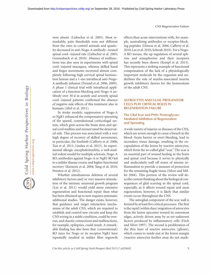
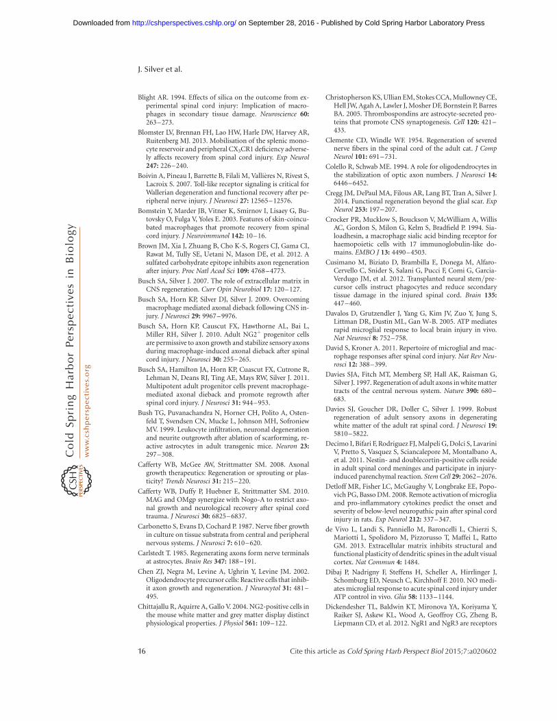
CSPGs (Pizzorusso et al. 2006). Furthermore,aberrant sprouting was observed after experi-mental or pathologic demyelination, for exam-ple, in the optic nerve (Tansey et al. 1985; Colelloand Schwab 1994; Phokeo et al. 2002). Neuritegrowth inhibitory factors expressed by oligoden-drocytes, including Nogo-A and CSPGs, there-fore, appear as specific stabilizers of the highlycomplex structure and wiring of the CNS ofhigher vertebrates (Fig. 1).
Suppression of Neurite Growth InhibitoryFactors Enhances Sprouting and Regenerationof Injured Neurites and Functional Recoveryafter CNS Injury
A variety of methods has been used to neutral-ize or delete myelin-associated inhibitory fac-tors to study axonal regeneration and repairprocesses after CNS injury. The most extensiveliterature exists for Nogo-A, for which function-
blocking antibodies or autoimmunizations,Nogo receptor–blocking peptides or fusionproteins, gene knockdowns (KOs), or receptorKOs have been used for in vivo manipulations,in particular, in the context of spinal cord injury(SCI), stroke studies, as well as autoimmunedisease models (Schwab 2004; Cafferty et al.2008; Pernet and Schwab 2012). Acute blockadeof Nogo-A, the Nogo receptor complex, or ofthe downstream Rho/ROCK pathway led toenhanced regenerative sprouting and elonga-tion over variable distances of injured cortico-spinal, rubrospinal, or aminergic axons in thespinal cord of adult rats and mice after injury.Enhanced compensatory sprouting of sparedfibers is often also observed. On the level ofbehavior, animals frequently show significantlyhigher levels of functional recovery than thecontrol reagent-treated or -untreated controls.Negative effects, which could be expected if un-directed or random growth was overstimulated,
Oligodendrocyte
Gray matter
LesionWhite matter
Sprouting and regenerating nerve fibers
Figure 1. Oligodendrocytes express neurite growth inhibitory proteins, including the membrane protein Nogo-A, on their cell surface and CNS myelin. These proteins inhibit branch formation along the mature axon in whitematter, but they also impair compensatory and regenerative fiber growth following axonal injury. In gray matter,the lower levels of these inhibitory proteins allow some structural remodeling of dendritic and axonal arbors andconnections to occur, but these processes can still be potentiated by neutralization or deletion of the neuritegrowth inhibitors in the mature CNS.
J. Silver et al.
4 Cite this article as Cold Spring Harb Perspect Biol 2015;7:a020602
on September 28, 2016 - Published by Cold Spring Harbor Laboratory Press http://cshperspectives.cshlp.org/Downloaded from
were absent (Liebscher et al. 2005). Most re-markably, pain thresholds were not differentfrom the ones in control animals and spastic-ity decreased in anti-Nogo-A antibody–treatedspinal cord–injured rats (Liebscher et al. 2005;Gonzenbach et al. 2010). Absence of malfunc-tions was also seen in experiments with spinalcord–injured macaques, whereas skilled handand finger movements recovered almost com-pletely following high cervical spinal hemisec-tion lesions and a 1-mo intrathecal anti-Nogo-A antibody infusion (Freund et al. 2006, 2009).A phase 1 clinical trial with intrathecal appli-cation of a function-blocking anti-Nogo-A an-tibody over 30 d in acutely and severely spinalcord–injured patients confirmed the absenceof negative side effects of this treatment also inhumans (Abel et al. 2011).
In stroke models, suppression of Nogo-Aor NgR1 enhanced the compensatory sproutingof the spared, contralesional corticofugal sys-tem, which grew across the brain stem and spi-nal cord midline and reinnervated the denervat-ed side. This process was associated with a veryhigh degree of recovery of skilled movements,in particular, the forelimb (Cafferty et al. 2008;Tsai et al. 2011; Lindau et al. 2013). In experi-mental allergic encephalomyelitis, a well-stud-ied rodent model for multiple sclerosis, Nogo-AKO, antibodies against Nogo-A or NgR1 KO ledto a milder disease course and higher functionalrecovery (Karnezis et al. 2004; Yang et al. 2010;Petratos et al. 2012).
Whether simultaneous deletion of severalinhibitory factors and/or very massive stimula-tion of the intrinsic neuronal growth program(Liu et al. 2011) would yield more extensiveregeneration and functional repair than whathas been obtained up to now requires systematicadditional studies. The danger exists, however,that guidance and target interaction mecha-nisms of the adult CNS, which are required toestablish and control new circuits and keep theCNS wiring in a stable condition, could be over-run, and chaotic connections and malfunctions,for example, epilepsies, could result. A remark-able finding has also been that (conventional)KO mice for Nogo or its receptor NgR1 haverepeatedly resulted in milder fiber regrowth
effects than acute interventions with, for exam-ple, neutralizing antibodies or receptor-block-ing peptides (Dimou et al. 2006; Cafferty et al.2010; Lee et al. 2010; Schwab 2010). For a Nogo-A KO mouse, the up-regulation of several eph-rins and semaphorins and their receptorshas recently been shown (Kempf et al. 2013).This represents a striking example of functionalcompensation of the lack of a physiologicallyimportant molecule by the organism and un-derlines the role of myelin-associated neuritegrowth inhibitory factors for the homeostasisof the adult CNS.
ASTROCYTES AND GLIAL PROGENITORCELLS PLAY CRITICAL ROLES INREGENERATION FAILURE
The Glial Scar and PNN: Proteoglycan-Mediated Inhibition of Regenerationand Sprouting
Awide variety of injuries or diseases of the CNS,which are severe enough to cause a breach in theblood–brain barrier or overt bleeding, lead tosecondary tissue damage, resulting in the en-capsulation of the lesion by reactive astrocytes,which form the so-called glial “scar.” The scar isan essential part of wound healing in the brainand spinal cord because it serves to physicallyand molecularly wall off zones of intense in-flammation to provide a measure of protectionfor the remaining fragile tissue (Silver and Mil-ler 2004). This portion of the review will de-scribe current thinking about the biological con-sequences of glial scarring in the spinal cord,especially, as it affects wound repair and axonregeneration; however, it is likely that similarevents occur throughout the CNS.
The astroglial component of the scar wall isformed by at least five critical processes. The firstis the rapid (within days) migration of astrocytesfrom the lesion epicenter toward its outermostedges, actively driven away by as-yet-unknownfactors produced by inflammatory cells (Fitchand Silver 1997). The second is proliferation ofthe thin layer of reactive astrocytes (gliosis),which comes to reside just at the lesion margin(reactive astrocytes further away do not mark-
CNS Regenerative Failure
Cite this article as Cold Spring Harb Perspect Biol 2015;7:a020602 5
on September 28, 2016 - Published by Cold Spring Harbor Laboratory Press http://cshperspectives.cshlp.org/Downloaded from
edly increase their proliferation rates, nor dothey migrate extensively) (Bush et al. 1999;Faulkner et al. 2004; Wanner et al. 2013). Thethird is the accumulation of intermediate fila-ment proteins, predominantly glial fibrillaryacidic protein (GFAP), vimentin, and nestin,leading to cellular hypertrophy of the astrogli-otic layer, as well as nondividing reactive astro-cytes further away (Pekny et al. 1999; Xu et al.1999; Wilhelmsson et al. 2004; Bardehle et al.2013). The fourth involves the restructuring ofthe gliotic layer into a mesh-like envelope,which changes from a radial, longitudinal ori-entation to an alignment largely perpendicularto the long axis of the cord and is, thus, highlyobstructive to any potential regrowth of the ma-jor projection axon pathways (Bardehle et al.2013; Wanner et al. 2013). In addition, thereoccurs the production of a variety of potentlygrowth inhibitory extracellular matrix (ECM)molecules, among which are the lectican familyof CSPGs (McKeon et al. 1991, 1995, 1999; Da-vies et al. 1999; Yamaguchi 2000; Busch andSilver 2007; Alilain et al. 2011; Brown et al.2012; Kawano et al. 2012; Li et al. 2013; Takeuchiet al. 2013). Thus, the scar presents a physicaland molecular constraint against the release ofintralesional inflammatory agents, but also, un-fortunately, to axon regeneration.
It is now known that this entire cascade ofevents is triggered, in part, by TGF-b bound tofibrinogen, which pores in through the leaky orhemorrhagic blood–brain barrier and activatesthe SMAD2 signaling cascade. Blocking theTGF-b receptor pathway abolishes the fibrin-ogen-induced effects on glial scar formationand, in particular, reduces proteoglycan depo-sition (Schachtrup et al. 2010). The early migra-tory response of astrocytes appears to be, at leastin part, under the control of glycogen synthasekinase-3 (GSK-3) activity because acute treat-ment with a potent GSK-3 inhibitor acceleratesmigration, resulting in better sequestration ofinflammatory cells and significantly enhancedfunctional improvement (Renault-Mihara etal. 2011). Also, the transcription factor SOX9appears to be a critical component of the path-way that leads to inhibitory matrix deposition inthe lesion because its conditional KO leads to
reduced expression of various CSPGs and im-proved locomotor function (Mckillop et al.2013). The architectural glial changes are underthe control of STAT3. When this transcriptionactivator is genetically deleted in astrocytes (orthe proliferating/gliotic astrocytes are them-selves deleted), the walling off phenomenon isseverely perturbed and inflammatory infiltratesinvade much larger regions of the cord, leadingto rampant tissue destruction and further loss offunction (Bush et al. 1999; Herrmann et al.2008; Wanner et al. 2013). Another critical mo-lecular determinant of astroglial scar building isinjury-induced glial Ca2þ signaling, which reg-ulates expression of the cell-to-cell adhesionmolecule N-cadherin. This calcium-dependenttight adhesion-forming molecule likely plays animportant role in strengthening the scar wall(Kanemaru et al. 2013). N-cadherin binds thefibroblast growth factor receptor (FGFR) andactivates a FGFR-dependent signaling cascade,which, in turn, can enhance GFAP expressionand is known to play a critical role in controllingthe polarity of astrocytes (Goldshmit et al. 2012;Lee et al. 2013; Macaya et al. 2013). In its ab-sence, N-cadherin KO mice display abnormalscar formation, leading to increased neuronaldeath (Kanemaru et al. 2013). Thus, the astro-cytic response to injury is an essential compo-nent of damage control in the CNS, and thelarge number of molecular determinants in-volved with scar formation could be potentialtherapeutic targets.
There are also reactive changes in astrocytesmuch further away from the lesion, which even-tually fill in the space vacated by dying oligo-dendrocytes and axons undergoing Walleriandegeneration (WD), but the structural changeshere take a much longer time to manifest (Silverand Miller 2004; Wanner et al. 2013). Over ex-tended periods of time, astroglial hypertrophyat the lesion edge, and in the tract beyond, leadsto very dense aggregates of cells that, at the le-sion and distally, especially near the pial surface,become obstructive to axonal regeneration (Sil-ver and Miller 2004). Interestingly, denervatedtarget regions, which are distant from the lesion,also undergo reactive glial changes, again asso-ciated with the production of sulfated proteo-
J. Silver et al.
6 Cite this article as Cold Spring Harb Perspect Biol 2015;7:a020602
on September 28, 2016 - Published by Cold Spring Harbor Laboratory Press http://cshperspectives.cshlp.org/Downloaded from

glycans that are largely contained within thePNN (Massey et al. 2006; Alilain et al. 2011;Andrews et al. 2012; Hansen et al. 2013). Themolecular triggers, which instigate up-regula-tion of these net-associated proteoglycans farfrom lesions, are largely unknown, but also ap-pear to be regulated, in part, by the SOX9 tran-scription factor pathway (Mckillop et al. 2013)as well as neuronal activity (Wang and Fawcett2012). CSPG up-regulation within the PNN isextremely important because it serves to limitpotential functional plasticity, which could oc-cur via compensatory sprouting from survivinginputs (Hockfield et al. 1990; Yamada et al.1997; Berardi et al. 2004; Massey et al. 2006;Pizzorusso et al. 2006; Gogolla et al. 2009; Gar-cıa-Alıas et al. 2011; Kwok et al. 2011; Wang andFawcett 2012; de Vivo et al. 2013; Xue et al.2014).
Oligodendrocyte Progenitor Cells and theNeuroglial 2 Proteoglycan: The Role ofthe Lesion Core in Regeneration Failure
Although the astroglial component of scar for-mation and its purported role in regenerationfailure has been suggested for more than a cen-tury (Ramon y Cajal 1928; Windle and Cham-bers 1950) and has been clearly revealed by theuse of microtransplantation experiments (Da-vies et al. 1997, 1999), the astroglial capsule isnot solely responsible for axonal regenerationfailure. When one examines, precisely, the inter-actions that occur between dystrophic axon tipsand the cells that they closely associate with overtime, it was surprising to learn that, for the mostpart, severed axons do not interact directly withreactive astrocytes, but rather with a populationof neuroglial-2-proteoglycan (NG2)–produc-ing oligodendrocyte precursor cells (OPCs)within the core of the lesion (Busch et al. 2010;Filous et al. 2010). It had long been thought thatafter SCI, severed axons would retract back tosustaining collateral (Ramon y Cajal 1928), and,thus, the free segment of remaining axon withinthe white matter would eventually be eliminat-ed. However, with the advent of modern label-ing techniques, we now know that, following thephase of axonal retraction (which is largely the
result of an aggressive attack on the dystrophicaxon tip by inflammatory blood-derived mac-rophages) (Horn et al. 2008; Busch et al. 2009;Evans et al. 2014), axotomized neurons oftensurvive (Kwon et al. 2002; Nielson et al. 2010).Eventually, the cut axon stops retracting and itsdystrophic tip can come to rest for many years(even decades) (Ruschel et al. 2013) within thepenumbra of the lesion (Li and Raisman 1995;Guest et al. 2005; Kadoya et al. 2009). Whatmaintains the dystrophic end of the axon chron-ically within the hostile environment of the glialscar? Are the mechanisms involved with long-term maintenance of the severed axon critical toregeneration failure? Although SCI results inastroglial emigration away from the lesion, in-side the core of the lesion, during the first sev-eral weeks postinjury, there is a robust recruit-ment and proliferation of a wide variety of celltypes, which all become surrounded by astro-glial scar. In addition to the vast array of acti-vated blood-derived macrophages and other in-flammatory cells, which begin to invade thelesion core within the first day (Popovich andLongbrake 2008; Kigerl et al. 2009; Evans et al.2014), the normally rarely dividing ependymalcells around the central canal become activatedand rapidly proliferate (Meletis et al. 2008).Within the first week, they also move into thecore of the lesion and, as they do so, they down-regulate their ependymal markers and begin todisplay reactive astroglial phenotypes, thus,contributing to the glial scar (Johansson et al.1999). Additionally, after penetrating injuriesthat open the dura mater, but also, importantly,after contusive or ischemic injuries that leavethe meninges largely intact, fibroblast-like stro-mal cells, which are derived from the meningesor pericytes or pericyte-like cells located aroundthe perimeter of blood vessels, divide vigorouslyand slough off from the meninges or vasculatureto help populate the lesion epicenter (Decimoet al. 2011; Goritz et al. 2011; Fernandez-Klett etal. 2013; Sabelstrom et al. 2013; Soderblom et al.2013). These cells interact with the astroglialcomponent of the scar and form a fibrotic-likelayer internal to the astroglial capsule. Via theirinteractions with astrocytes and the collage-nous/proteoglycan-rich matrices that are pro-
CNS Regenerative Failure
Cite this article as Cold Spring Harb Perspect Biol 2015;7:a020602 7
on September 28, 2016 - Published by Cold Spring Harbor Laboratory Press http://cshperspectives.cshlp.org/Downloaded from

duced, they also play a role in helping to seal thelesion, but also may play a role in blocking re-generation (Davies et al. 1999; Stichel et al. 1999;Kawano et al. 2012; Sabelstrom et al. 2013; So-derblom et al. 2013). Finally, there occurs a ro-bust proliferation of OPCs, which produce thepurportedly potently inhibitory NG2 CSPG, aswell as a cocktail of growth-promoting ECMmolecules, including laminin and fibronectin(Zai and Wrathall 2005; Lytle et al. 2006; Buschet al. 2010). Thus, the early lesion core becomesa rich oasis of cells with a mixture of growth-inhibiting and -promoting properties.
The role of NG2þ cells, both in the normalCNS and after injury, remains controversial.NG2 is a member of the CSPG family of ECMmolecules that is thought to contribute to regen-eration failure. Because NG2 is one of the mostdramatically up-regulated CSPG after CNS in-jury (Levine 1994), it has been suggested thatNG2þ cells are “the” major regeneration-block-ing cell type (Dou and Levine 1994; Fidler et al.1999; Chen et al. 2002; Tan et al. 2006). In con-trast, several studies suggest that NG2þ cells maynot be repulsive at all. Indeed, the dystrophictips of severed axons, which remain within thelesion penumbra for extended periods, resideclosely among NG2þ glia (Zhang et al. 2001;McTigue et al. 2006; Busch et al. 2010) andNG2þ cells seem to facilitate growth of develop-ing axons (Yang et al. 2006). Our laboratorysuggested that the population of stem-like,NG2-producing cells in the lesion core may con-tribute to regeneration failure by acting as a kindof “safe haven” for dystrophic axons, stabilizingthem as they are forced to retract backward intothe caudal end of the lesion by activated macro-phages (Busch et al. 2010). Indeed, severed ax-ons in the lesion appear to be “addicted” to thesurface of these cells and refuse to leave. How-ever, the mechanisms that govern this tight cell–cell interaction and, in particular, whether theNG2 CSPG is involved in this close association,remained important and unresolved questions.
Recently, we sought a better understandingof the interaction between severed sensory axonsand adult cord-derived NG2 glia after a dorsalcolumn injury (Filous et al. 2012). In our stud-ies, we observed a novel mechanism of regener-
ation failure. When combined with growth-promoting ECM molecules in critical ratios, pu-rified NG2 and other CSPGs initially constrainaxons to their territory via a GAG/LAR familyCSPG receptor-mediated interactive mecha-nism (Shen et al. 2009; Filous et al. 2010; Fisheret al. 2011; Lang et al. 2012, 2013). NG2 glia alsoconstrain early axonal outgrowth but, in ad-dition, can lead to longer lasting entrapmentof the neuron onto the glial cell surface throughan unusual neuroglia synaptoid-mediated sta-bilization, both in vitro and in vivo. Given thatneurons form synapses with NG2þOPCs underphysiological conditions throughout the CNS(Bergles et al. 2000; Chittajallu et al. 2004; Linet al. 2005), it is possible that such synaptic-likeinteractions within the damaged white matterallow for long-lasting associations between thedystrophic tips of sensory neurons and NG2þ
cells. Although these stabilizations, initially,may be beneficial to prevent further dieback(Filous et al. 2010), they may also place furtherlimitations on the forward movements of thestruggling axon tip. The idea that synaptic-likeconnections form between regenerating axonsand reactive glia, and may serve to curtail axonalregrowth after injury, had been suggested manyyears ago (Carlstedt 1985), although the impor-tance of this phenomenon in regeneration fail-ure had largely been abandoned. After a dorsalroot crush, even following a peripheral condi-tioning lesion, injured sensory axons can regen-erate rapidly within the proximal root until theyreach the dorsal root entry zone (DREZ), a tran-sitional region between the peripheral nervoussystem (PNS) and the CNS, where they abruptlyhalt their forward progress and remain (Carl-stedt 1985; Liuzzi and Lasek 1987; Di Maioet al. 2011). Early studies suggested that, as pe-ripheral axons regenerate toward the CNS, theycontact reactive astrocytes, which initiate theearly stages of so-called synaptoid formationsin close association with the astrocyte surface.Interestingly, our current studies suggest that,in addition to their wall-building job, reactiveastrocytes may also play an indirect role in sig-naling for sensory axons to begin synapse for-mation mediated, at least in part, via thrombo-spondins, which are important in regulating
J. Silver et al.
8 Cite this article as Cold Spring Harb Perspect Biol 2015;7:a020602
on September 28, 2016 - Published by Cold Spring Harbor Laboratory Press http://cshperspectives.cshlp.org/Downloaded from

neuron-to-neuron synaptogenesis (Christo-pherson et al. 2005). However, our data showclearly that dystrophic axons after DCC are ac-tually synapsing on the NG2þ cell, rather thanastrocytes, and this relationship is also likelyto occur at the DREZ. It is also possible thatreactive astrocytes can directly induce prolifera-tion of OPCs by releasing the mitogen, Sonichedgehog (SHH) into the injury environment(Amankuloret al. 2009). Activation of the SHH-Gli-signaling axis within the OPC populationresults in its dramatic expansion and the poten-tial amplification of OPC-mediated effects onsevered axons. It is also probable that inflamma-tory cells play a role in accelerating OPC mitosisas well (Miron et al. 2013).
This close interaction between NG2þ cellsand injured neurons after SCI provides a newway of thinking about how CSPGs and the coreof the scar “inhibit” axonal migration and helpsexplain how dystrophic axon tips persist withinthe scar-encased, hostile lesion environment.Thus, we hypothesize that in vivo within thescar core, CSPGs do not cause axon tips to ceasegrowing because of a lack of adhesion, but rath-er because they create dystrophy and increasingentrapment of the growth cone via abnormallystrong bonds with the substrate. Thus, the scar,with its two distinct regions (the core and thewall), can inflict a measure of inhibition thatthwarts the advancement of the regeneratingneuron (see Fig. 1).
Plasticity of Reactive Astrocytes?
The final question that I would like to speculateon is whether reactive astrocytes in the scar wallare permanently refractory to axonal regenera-tion or whether they can become plastic andpromote or, at least, allow axonal growth (asthey do during development) (Silver et al. 1982,1993; Silver and Ogawa 1983). Emerging datasuggests that they can be plastic and regenerationfailure through the scar is the result of an imbal-ance between a lack of intrinsic growth machin-ery in the neuron (Ylera et al. 2009) and extrinsicforces (some of which are discussed above) thatlimit growth. Astrocytes that contribute to thescar and are derived from the ependymal tube
appear to be slightly more “immature” than as-trocytes derived from self-duplication becausethey express less GFAP relative to vimentin(Fig. 2) (Meletis et al. 2008). It would be veryinteresting if the well-known regeneration-en-hancing functions of ependymoglial cells thatare present in robustly regenerating cold-blood-ed species (Singer et al. 1979) are retained, atleast to some extent, in the ependymal sub-population of reactive astrocytes in scar tissueof mammals (Silver and Steindler 2009). Poten-tial functional differences between astrocytepopulations in the scar may be appearing inthe rather dramatic ability of neurons to regen-erate their severed axons right across and beyondcarefully crafted lesions within the rodent spinalcord or optic nerve following PTEN/SOCS3 de-letion (Park et al. 2010; Sun et al. 2011). Indeed,the impressive regeneration, albeit across rela-tively narrow lesions, when the protein productsof these growth or cytokine regulatory genes arediminished or genetically deleted, is strictly con-fined to and dependent on astroglial bridges,which form spontaneously across the lesioncore (Filous et al. 2010; Zukor et al. 2013). Theappreciation of whether separate reactive astro-glial subpopulations exert these guidance func-tions or possibly even if gliotic astrocytes in thescar wall can be plastic and made growth per-missive or even promoting in response to thepresence of a robustly growing axon, could bevery important and therapeutically provocative(Ahmed et al. 2005). It would suggest that weconsider strategies tailored toward amplifyingor attenuating particular, functionally distinctastrocyte subpopulations or to further enhancethe plasticity of gliotic astrocytes to help maxi-mize functional recovery.
MICROGLIA AND MACROPHAGES
Origin of Macrophages in Injured CNS
In parallel with the injury-induced changes de-scribed above for oligodendrocytes and astro-cytes, a robust and long-lasting inflammatoryresponse is initiated, which is dominated bymacrophages. These cells are mostly derivedfrom two sources: (1) resident microglia, and
CNS Regenerative Failure
Cite this article as Cold Spring Harb Perspect Biol 2015;7:a020602 9
on September 28, 2016 - Published by Cold Spring Harbor Laboratory Press http://cshperspectives.cshlp.org/Downloaded from
(2) blood monocytes, that is, macrophage pre-cursors that emigrate from bone marrow or thespleen (Popovich et al. 1999; Popovich andHickey 2001; Longbrake et al. 2007; Swirskiet al. 2009; Blomster et al. 2013). Microglia orig-inate from precursor cells in the yolk sac andbecome homogeneously distributed through-out the CNS during early embryogenesis (Gin-houx et al. 2010). Like astrocytes, microglia re-spond rapidly to injury, extending cellularprocesses or migrating toward the lesion sitewhere they participate in scar formation (Dava-los et al. 2005; Dibaj et al. 2010). Surely, thisearly and rapid response serves a protectiverole, as there is no obvious evolutionary advan-
tage for blanketing the CNS with cells that,when provoked, will mobilize and destroy deli-cate nervous tissue. Indeed, blocking or pre-venting microglial activation, via either phar-macologic or genetic means, exacerbates lesionpathology and impairs recovery of function (La-lancette-Hebert et al. 2007; Hines et al. 2009).
After a delay of �2 d postinjury, monocytesbind to endothelial adhesion molecules andthen migrate into the lesioned CNS, down che-motactic gradients established by astrocytes(Pineau et al. 2010). Shortly thereafter, mono-cytes differentiate into tissue macrophages. Be-cause microglia and MDMs are both of myeloidlineage, lineage-specific markers cannot be used
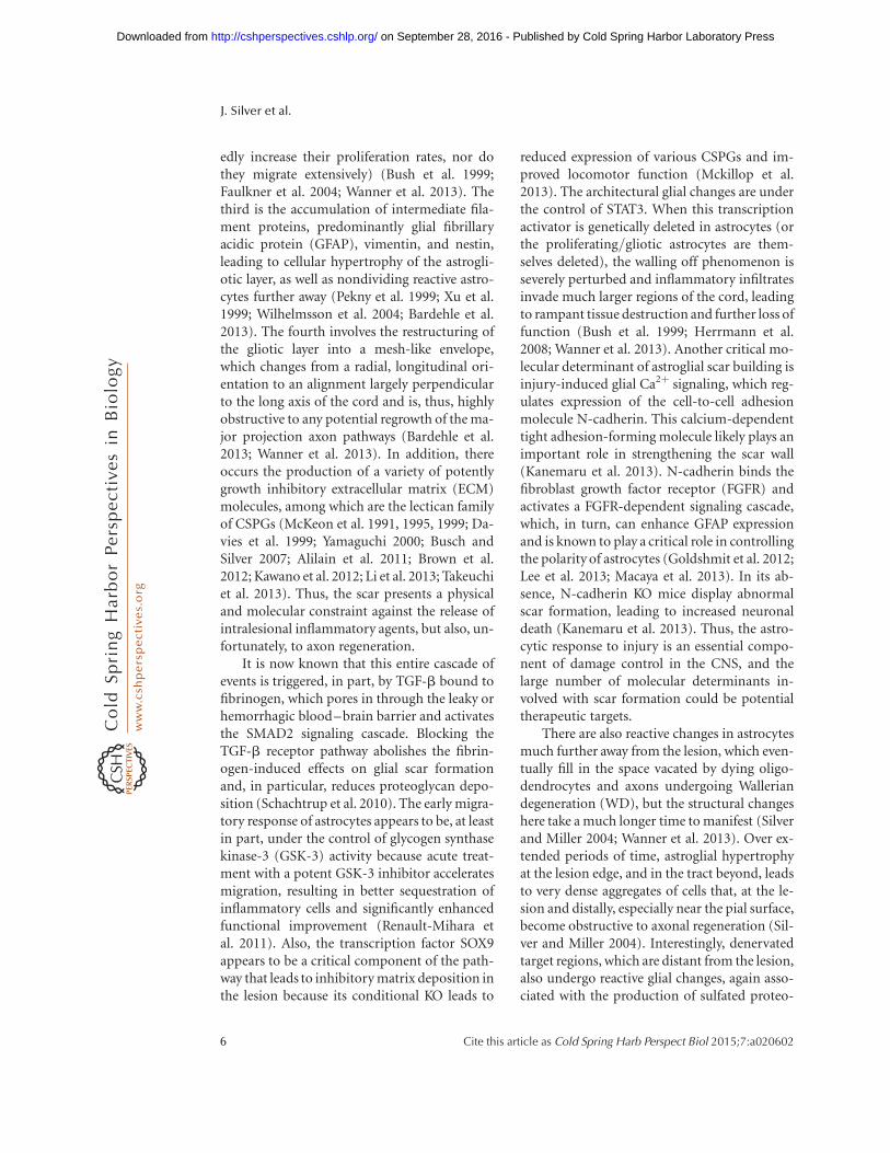
Lesion core
Lesion penumbra
Reactive astrocyte
Macrophage
MG2+ cell
Dystrophic endingStabilized ending
Conditioned ending
1
2
3
4
Figure 2. Schematic representation of the proximal end of a dorsal column crush lesion 7 d after injury. GFAPþ
astrocytes (blue) have pulled away from the lesion core, which is now populated by NG2þ cells (purple) andphagocytic ED1þmacrophages (green). The fibroblastic and ependymal cell types are not displayed, but are alsoplentiful in the lesion core. Dorsal root ganglion neurons (red) attempt to regenerate into the lesion core. (1)Typical axon with a dystrophic growth cone that has become susceptible to macrophage attack. (2) Typical axonthat has undergone macrophage-mediated retraction back to NG2þ cells and stabilized. (3) Atypical axon thathas stabilized further distally within the lesion core on a contiguous bridge of NG2þ cells. (4) Growth cone of aneuron that has been stimulated or conditioned and able to overcome macrophage-induced axonal dieback andextend into the lesion core on NG2þ cells. (From Busch et al. 2010; reprinted, with express permission, from theJournal of Neuroscience and the investigators of this review.)
J. Silver et al.
10 Cite this article as Cold Spring Harb Perspect Biol 2015;7:a020602
on September 28, 2016 - Published by Cold Spring Harbor Laboratory Press http://cshperspectives.cshlp.org/Downloaded from

to distinguish between these major CNS mac-rophage subsets. Equally ambiguous is the effectthat CNS macrophages have on neurons andaxons that survive after CNS injury.
Seemingly conflicting data implicate macro-phages, regardless of their source, as effectors ofboth tissue repair and secondary tissue damage.Although Ramon y Cajal is often recognized asthe “father of neuroscience,” he also providedsome of the earliest descriptions of neuroim-mune interactions in the injured CNS. Specifi-cally, he noted that macrophages accumulatedand persisted at sites of injury and concludedthat their primary role was as scavenger cells(Ramon y Cajal 1991):
This leukocytic invasion of the dead neuron isnot surprising. It is a general law that any mor-tified portion, no matter what is its character,becomes a pasture-ground for phagocytes. Webelieve that the protagonists of all acts of neuro-nophagy are nothing else than the granular cor-puscles which accumulate so prodigiously in thenecrotic focus of the centres and in the periph-eral stumps of degenerated nerves.
Because he did not have the benefit of modern-day techniques (e.g., radiation bone-marrowchimeras, transgenic mice, etc.), Ramon y Cajaland his contemporaries were unable to un-equivocally determine the origin of CNS mac-rophages. Regardless, he accurately predictedthat most phagocytes present in lesioned CNStissue were derived from blood, that is, mono-cytes:
. . . we believe also that the phagocytes—ourtraumatocytes—which have penetrated into theneuronal cadaver positively represent large leu-cocytes with a lobulated nucleus, which havecome from the host’s blood.
We now know that his predictions were correctand the biased accumulation of MDMs at thelesion center may have significant implicationsfor the growth or retraction (“dieback”) of in-jured axons (see below).
Macrophage Functions in Injured CNS
Some years after Ramon y Cajal’s seminal obser-vations (circa 1950), additional insight intoCNS macrophage function was gleaned from a
serendipitous discovery. Although studyingneural mechanisms of thermal regulation indogs with SCI, Windle, Clemente, and col-leagues discovered that deliberate systemic in-jection of pyrogens had the unintended benefitof enhancing neurologic recovery (Windle andChambers 1950; Clemente and Windle 1954).Postmortem analysis of dogs injected with crudepyrogens revealed markedly increased numbersof intraspinal macrophages and reduced intra-lesional scarring as compared with injured spi-nal cords of untreated dogs (Clemente andWindle 1954). Almost 30 years later, Guth andcolleagues extended Windle’s observationsshowing that systemic injections of purified en-dotoxin (i.e., lipopolysaccharide [LPS]) intospinal-injured rats enhanced intraspinal leuko-cytosis beyond that normally seen after SCI(Guth et al. 1994a). This enhanced inflammato-ry reaction was accompanied by more robustaxon growth and quantitatively superior im-provements in hindlimb locomotor function.Guth later found that the salutary effects ofLPS could be further improved by simulta-neously treating animals with anti-inflammato-ry agents, including indomethacin or steroids(Guth et al. 1994b). This combination ap-proach, although seemingly counterintuitive,was based on keen insight regarding the diver-gent functions of activated CNS macrophages.Guth realized that, during maturation, macro-phages become “primed” or partially activatedby cytokines (and other factors) present in theinjury milieu; however, to attain a greater level offunctional competency, including the ability topromote axon growth or neuroprotection, mac-rophages likely require a second distinct signal,in this case, LPS. He also recognized that onceactivated, these same cells release hydrolyzingenzymes, oxidative metabolites, and aracha-donic acid metabolites (e.g., prostaglandins),which can damage neurons and glia. Indometh-acin and steroids were used to inhibit these de-structive secretory components of activatedmacrophages.
Over the next 10–15 yr, data from severallaboratories using rabbit, guinea pig, and ratmodels of SCI showed that, in the absence of asecondary stimulus, the injurious effects of in-
CNS Regenerative Failure
Cite this article as Cold Spring Harb Perspect Biol 2015;7:a020602 11
on September 28, 2016 - Published by Cold Spring Harbor Laboratory Press http://cshperspectives.cshlp.org/Downloaded from

traspinal macrophages predominate. Regardlessof species, injury type (e.g., compression, con-tusion), or injury severity, selective inhibitionor depletion of macrophages during the first1–2 wk postinjury consistently reduces second-ary or bystander tissue injury, leading to im-proved recovery of sensory, motor, or autonom-ic functions (Giulian and Robertson 1990;Blight 1994; Popovich et al. 1999; Gris 2004).
Emerging data now indicate that in responseto different combinations of factors, which arenormally found in the extracellular milieu ofthe injured nervous system, macrophages dif-ferentiate into functionally distinct cell subsetsthat differentially affect neuron survival andaxon growth (Stout et al. 2005; Kigerl et al.2009). For example, cytokines, cell fragments,and nucleic acids promote differentiation ofmacrophages into “classically” (M1) or “alter-natively” activated (M2) cells. The canonical invitro model for promoting inflammatory M1macrophage differentiation is exposure of natıve(unstimulated) myeloid cells to LPS and inflam-matory cytokines, including interferon (IFN)-gor tumor necrosis factor (TNF)-a. Alternative-ly, to promote M2 differentiation, immaturemyeloid cells are stimulated with interleukin(IL)-4 or -13 (Gordon and Taylor 2005). AfterCNS injury, signaling pathways that polarizemacrophages toward an M1 phenotype pre-dominate (Kigerl et al. 2009; David and Kroner2011). M1 macrophages can be neurotoxic andcause axon dieback (Horn et al. 2008; Kigerlet al. 2009). Thus, the neuroprotective effectsof acute macrophage inhibition or depletionin SCI models might be explained by reducingthe burden of M1 macrophages at the injurysite. Surprisingly, these same cells also can en-hance neurite outgrowth.
In vivo injections of inflammatory stimuli(e.g., LPS, zymosan), which are needed to pro-mote an M1 macrophage phenotype in vitro,enhance regeneration of injured peripheraland central axons (Yin et al. 2003; Steinmetzet al. 2005; Boivin et al. 2007; Gensel et al.2009). In injured brain, spinal cord, and opticnerve, macrophage clusters are often associatedwith sprouting of injured axons (Fig. 3). Thisendogenous repair phenomenon is mediated by
macrophages via the release of neurotrophinsand growth factors or, indirectly, by activatingglia within the scar, which subsequently pro-duces a trophic gradient. BDNF, CNTF, and glialcell line–derived neurotrophic factor (GDNF)have been implicated in this response (Batcheloret al. 2002; Yin et al. 2006; Muller et al. 2007;Gensel et al. 2009; Benowitz and Popovich2011). The ability of transplanted microglia ormacrophages to promote neurite outgrowth indifferent models of SCI might be explained by asimilar mechanism (Prewitt et al. 1997; Rab-chevsky and Streit 1997; Rapalino et al. 1998).
Compared with M1 macrophages, M2 mac-rophages may be less destructive and better ableto repair the injured CNS. M2 macrophagespromote more robust neurite outgrowth andrecent data show that these cells release acti-vin-A, which enhances oligodendrocyte pro-genitor cell differentiation and, subsequently,remyelination (Kigerl et al. 2009; Miron et al.2013). Enhancing M2 microglia/macrophagedifferentiation in lesioned CNS tissues is asso-ciated with neuroprotection; however, limiteddata exist linking M2 macrophages with axonregeneration in vivo. Combining peripheralnerve grafts with acidic fibroblast growth factorin an injured spinal cord produces a cytokinemilieu that favors M2 macrophage differentia-tion, polyamine synthesis with improved axonregeneration (Kuo et al. 2011). Similarly, infu-sion of IL-4 (M2 cytokine) into guidance chan-nels placed into injured sciatic nerves inducesan M2 macrophage response that stimulatesSchwann cell migration with enhanced axon re-generation into the distal nerve stump (Mokar-ram et al. 2012).
Manipulating Macrophagesto Promote Axon Regeneration:Future Considerations
ProCord was an experimental cell-based therapythat was developed to treat acute SCI in humans.Clinical trials were initiated by Proneuron Bio-technologies (New York, NY) based on datashowing that autologous macrophages, whenactivated ex vivo, then injected into the injuredspinal cord, promote axon regeneration and
J. Silver et al.
12 Cite this article as Cold Spring Harb Perspect Biol 2015;7:a020602
on September 28, 2016 - Published by Cold Spring Harbor Laboratory Press http://cshperspectives.cshlp.org/Downloaded from
reduce tissue damage in two different rodent SCImodels (Rapalino et al. 1998; Bomstein et al.2003). An overview of the rationale and designfor the phase I trial was reviewed previously (Ki-gerl and Popovich 2006). Results of the phase 2randomized controlled multicenter trial, involv-ing 43 participants, showed a trend for betterrecovery in the control group relative to patientsreceiving macrophage transplants, but withoutgroup differences in the number of adverseevents (Lammertse et al. 2012). Although effica-cy was not established, future cell-based clinicaltrials for SCI (and other diseases) will benefitfrom the ProCord experience, because this trialidentified and overcame numerous logistical
and technical constraints associated with enroll-ing, preparing, and injecting into the spinal cordwithin 14 d of injury, an autologous cellulartherapy (Jones et al. 2010).
Autologous macrophage transplantation re-mains a promising therapeutic approach; how-ever, new preclinical data indicate that the phe-notype of macrophages generated ex vivo maynot persist after injection into lesioned CNS.When M2 polarized macrophages are trans-planted into lesioned spinal cord, they differ-entiate into M1 macrophages. Conversely, M2macrophages maintain their phenotype whentransplanted into intact spinal cord (Kigerlet al. 2009). Accordingly, future transplantation
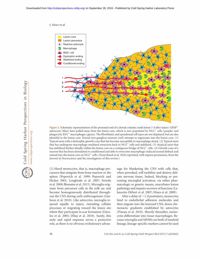
Surveyingmicroglia
Injured CSTaxons
Dystrophicaxon Effector
microglia
M1 phenotype
M2 phenotype
Undifferentiated/mixed phenotype
Infiltrating monocyte-derived macrophages
Wallerian-degeneratingsegments ofcorticospinaltracts
Astrocytes
Rostral to injury Caudal to injury
Lesion center
Oligodendrocyte progenitor cells (OPCs)
Growthcone
Figure 3. Schematic of microglia and MDM reactions elicited by SCI. After injury, the lesion center (also referredto as “epicenter” in contusion lesions) becomes filled with phagocytic macrophages derived from blood mono-cyte precursors. These cells become enlarged as they phagocytose lipid and cell debris. These and other stimuli inthe lesion prime an M1 macrophage phenotype (red). Only a subset of macrophages become “alternatively”activated (i.e., M2 macrophages, green). Some cells remain undifferentiated or adopt a heterogeneous phenotype(orange/green mix). Macrophages in the lesion center are “walled off” by reactive astrocytes, which create a scar.OPCs interdigitate between scar-forming astrocytes and are drawn toward the lesion edge by undefined factors.Complete OPC differentiation into myelinating oligodendrocytes may require factors derived from (M2) micro-glia subsets, which often lie outside the lesion microenvironment (gradient fill). Microglia exist in intact spinalcord as sentinel cells, which continuously survey the microenvironment. After injury or in response to subtlechanges in homeostasis, microglia become activated and transform morphologically and phenotypically intoeffector microglia. Depending on the composition of factors present in the microenvironment, microglia canbecome polarized to become M1 or M2 effector cells. Rostral to the site of injury, surveying and effectormicroglia colocalize with damaged axons, a subset of which are undergoing dieback, but also with a subsetthat are stabilized or attempting to grow. Caudal to the lesion, descending axons undergo Wallerian degeneration(WD). Various factors released during WD activate microglia (and macrophages). It is common to see effectormicroglia (and, presumably, a subset of MDMs) colocalized with WD axon segments. CST, corticospinal tract.
CNS Regenerative Failure
Cite this article as Cold Spring Harb Perspect Biol 2015;7:a020602 13
on September 28, 2016 - Published by Cold Spring Harbor Laboratory Press http://cshperspectives.cshlp.org/Downloaded from

protocols, whether macrophages or other celltypes, will need to incorporate measures thatmodify the lesion microenvironment. Genericimmune suppressive drugs (e.g., steroids) arenot practical in this context because these drugswill affect injurious and reparative macrophagesubsets. Generic macrophage inhibition or de-pletion strategies, including intravenous injec-tions of anti-integrin antibodies or liposome-encapsulated bisphosphonates, could be useful,especially in the acute postinjury period or ifused together with neuroprotective drugs (Po-povich et al. 1999; Gris 2004; Iannotti et al.2011; Lee et al. 2011). Neuropeptides (e.g., sub-stance P), antibodies that block cytokine signal-ing or stem cells, also could be used as each isable to modulate the injury milieu, creating anenvironment that favors polarization of endog-enous macrophages toward an M2 phenotype(Busch et al. 2011; Cusimano et al. 2012; Guer-rero et al. 2012).
In addition to macrophage transplanta-tion, targeted or “precision” immunotherapies,which inhibit or stimulate one or more pheno-typically distinct macrophage subsets, is an idealapproach. Along with the M1/M2 CNS macro-phage subsets described above, new reagentsand genetic tools have revealed the presence ofother distinct intraspinal macrophage subsets(Thawer et al. 2013). For example, variationsin the relative expression of the chemokine re-ceptor CX3CR1 or maturation markers (e.g.,Ly6) define functionally distinct CNS macro-phages (Shechter et al. 2009; Donnelly et al.2011; Saiwai et al. 2013). Antibodies and smallmolecule inhibitors can or have been designedto target these macrophages, but whether suchmanipulations will affect axon regeneration re-quires additional research. Ideally, future studieswill incorporate acute and chronic CNS lesionmodels. Although macrophages persist indefi-nitely in CNS lesions, their role in the chronicinjury milieu and nearby spared tissue is un-known.
The possibility that microglia and MDMswill have distinct effects on cell repair andaxon regeneration after CNS injury is likelyand should also be considered when designingor interpreting preclinical studies (Popovich
and Longbrake 2008; London et al. 2013). Mi-croglia and MDMs develop by discrete tran-scriptional control mechanisms from uniqueprecursor cells (Prinz et al. 2011; Schulz et al.2012). After injury, the discrete spatial distribu-tion of different macrophage subsets producesheterogeneous microenvironments that can dif-ferentially affect injured axons, nascent axonalgrowth cones, and surrounding glia. Recent datashow that signals emanating from aged braintrigger a neuroprotective transcriptomic sig-nature in microglia (Hickman et al. 2013).Whether similar neuroregenerative or neuro-toxic “sensomes” exist in microglia or MDMs,respectively, is unknown, but such profiles seemlikely, especially because the ratio of microglia toMDMs increases in regions remote from theinjury site, along with clear evidence of anatom-ical and functional plasticity or endogenousCNS repair (Zhang and Guth 1997; Popovichand Hickey 2001; Zhou et al. 2003; McTigueet al. 2006; Detloff et al. 2008; Busch et al.2010; Hansen et al. 2013).
Conversely, physical contact between axonsand macrophages within the lesion core (highratio of MDMs to microglia) causes axons toretract or “dieback” from the injury site. Bothsoluble factors and cell surface proteins are cul-pable in this degenerative response (Horn et al.2008; Busch et al. 2011). Macrophages expressnumerous membrane-bound proteins, includ-ing receptors for ephrins, siglecs (sialoadhe-sins), and integrins (Crocker et al. 1994; Sobelet al. 1995; Tang et al. 1997; Liu et al. 2006). Axongrowth and guidance may be positively or neg-atively affected when these proteins are boundby corresponding ligands found on axons. Giv-en the discrete spatiotemporal dynamics ofmacrophages and microglia, when, where, andhow much injured axons are exposed to thesecells will undoubtedly affect their ability to re-generate.
Although there is a growing appreciationthat macrophages are important contributorsto CNS regeneration failure, we have only a ru-dimentary understanding of how or whetherthese cells influence axon regeneration. Achiev-ing a greater understanding of CNS macrophag-es should improve the safety and success of
J. Silver et al.
14 Cite this article as Cold Spring Harb Perspect Biol 2015;7:a020602
on September 28, 2016 - Published by Cold Spring Harbor Laboratory Press http://cshperspectives.cshlp.org/Downloaded from

future clinical trials designed to promote regen-eration or repair of the injured CNS.
CONCLUSION
Over the past several decades, there has beensteady progress in understanding basic mole-cular mechanisms that are responsible for thepoor regenerative potential of injured centralnervous system axons. Indeed, there are limita-tions within the neuron, that is, molecularswitches that impede intrinsic regeneration ma-chinery, and there are various glial cells thatcreate lesion barricades or “extrinsic” inhibitorycues, which curtail the relatively limited regen-erative potential of injured CNS axons. In thisreview, we have focused on each of the majorglial cell types that serve as the primary extrinsicregulators of axon regeneration with an empha-sis on the injured spinal cord. We have describedhow the severed axon tip, struggling to advancea new growth cone, is collapsed by myelin-de-rived growth-inhibitory factors, made dystro-phic by proteoglycans, and further attacked bythe destructive actions of M1 macrophages,whose job, early on, is to phagocytose the nox-ious debris. The unfortunate neuron, whoseaxon was once enveloped by supportive oligo-dendrocytes and astrocytes, is left to fend foritself during the attack; oligodendrocytes dieand reactive astrocytes abandon the core of thelesion as they attempt to protect and mechani-cally stabilize the remaining fragile tissue froman expanding inflammatory reaction, creatingyet another obstacle to regeneration. But, thereis some relief, even within the eye of the storm.Once neurotoxic macrophages convert into amore reparative M2 state, and various stem-like cells, including oligodendrocyte progeni-tors, begin to thrive within the lesion core, theretracting axon can find a safe haven and evenform synaptic-like connections on the primitiveglia where, unfortunately, they remain lockedin place for decades. As we have acquired amore complete appreciation of the molecularmechanisms that control the untoward effectsof glia, new approaches are being developedthat can readily prevent axons from dying back-ward and also may allow them to robustly sprout
or sometimes regenerate beyond the scar towardnew functional synaptic targets. A major goalfor the future will be to combine the most suc-cessful glia-targeted strategies with others thatdrive the neuron’s intrinsic growth capacity tomaximize the regenerative potential that wenow know exists within the damaged adult CNS.
REFERENCES
Abel R, Baron HC, Casha S, Harms J, Hurlbert J, Kucher K,Maier D, Thietje R, Weidner N, Curt A. 2011. Therapeu-tic anti-Nogo-A antibodies in acute spinal cord injury:Safety and pharmacokinetic data from an ongoing first-in-human trial. ISCOS Meeting 2011, Washington, DC.
Aguayo AJ, Rasminsky M, Bray GM, Carbonetto S, McKer-racher L, Villegas-Prez M, Vidal-Sanz M, Carter DA.1991. Degenerative and regenerative responses of injuredneurons in the central nervous system of adult mammals.Phil Trans R Soc B 331: 337–343.
Ahmed Z, Dent RG, Leadbeater WE, Smith C, Berry M,Logan A. 2005. Matrix metalloproteases: Degradation ofthe inhibitory environment of the transected optic nerveand the scar by regenerating axons. Mol Cell Neurosci 28:64–78.
Akbik, Feras V, Sarah M, Bhagat, Pujan R, Patel, William BJ,Cafferty, Stephen M, Strittmatter. 2013. Anatomical plas-ticity of adult brain is titrated by Nogo receptor 1. Neuron77: 859–866.
Alilain WA, Horn KP, Hu H, Dick TE, Silver J. 2011. Func-tional regeneration of respiratory pathways after spinalcord injury. Nature 475: 196–200.
Amankulor NM, Hambardzumyan D, Pyonteck SM, BecherOJ, Joyce JA, Holland EC. 2009. Sonic hedgehog pathwayactivation is induced by acute brain injury and regulatedby injury-related inflammation. J Neurosci 29: 10299–10308.
Andrews EM, Richards RJ, Yin FQ, Viapiano MS, JakemanLB. 2012. Alterations in chondroitin sulfate proteoglycanexpression occur both at and far from the site of spinalcontusion injury. Exp Neurol 235: 174–187.
Bardehle S, Kruger M, Buggenthin F, Schwausch J, NinkovicJ, Clevers H, Snippert HJ, Theis FJ, Meyer-Luehmann M,Bechmann I, et al. 2013. Live imaging of astrocyte re-sponses to acute injury reveals selective juxtavascular pre-oliferation. Nat Neurosci 16: 580–586.
Batchelor PE, Porritt MJ, Martinello P, Parish CL, LiberatoreGT, Donnan GA, Howells DW. 2002. Macrophages andmicroglia produce local trophic gradients that stimulateaxonal sprouting toward but not beyond the wound edge.Mol Cell Neurosci 21: 436–453.
Berardi N, Pizzorusso T, Maffei L. 2004. Extracelluar matrixand visual cortical plasticity: Freeing the synapse. Neuron44: 905–908.
Bergles DE, Roberts JD, Somogyi P, Jahr CE. 2000. Gluta-matergic synapses on oligodendrocyte precursor cells inthe hippocampus. Nature 405: 187–191.
Benowitz LI, Popovich PG. 2011. Inflammation and axonregeneration. Curr Opin Neurol 24: 577–583.
CNS Regenerative Failure
Cite this article as Cold Spring Harb Perspect Biol 2015;7:a020602 15
on September 28, 2016 - Published by Cold Spring Harbor Laboratory Press http://cshperspectives.cshlp.org/Downloaded from
Blight AR. 1994. Effects of silica on the outcome from ex-perimental spinal cord injury: Implication of macro-phages in secondary tissue damage. Neuroscience 60:263–273.
Blomster LV, Brennan FH, Lao HW, Harle DW, Harvey AR,Ruitenberg MJ. 2013. Mobilisation of the splenic mono-cyte reservoir and peripheral CX3CR1 deficiency adverse-ly affects recovery from spinal cord injury. Exp Neurol247: 226–240.
Boivin A, Pineau I, Barrette B, Filali M, Vallieres N, Rivest S,Lacroix S. 2007. Toll-like receptor signaling is critical forWallerian degeneration and functional recovery after pe-ripheral nerve injury. J Neurosci 27: 12565–12576.
Bomstein Y, Marder JB, Vitner K, Smirnov I, Lisaey G, Bu-tovsky O, Fulga V, Yoles E. 2003. Features of skin-coincu-bated macrophages that promote recovery from spinalcord injury. J Neuroimmunol 142: 10–16.
Brown JM, Xia J, Zhuang B, Cho K-S, Rogers CJ, Gama CI,Rawat M, Tully SE, Uetani N, Mason DE, et al. 2012. Asulfated carbohydrate epitope inhibits axon regenerationafter injury. Proc Natl Acad Sci 109: 4768–4773.
Busch SA, Silver J. 2007. The role of extracellular matrix inCNS regeneration. Curr Opin Neurobiol 17: 120–127.
Busch SA, Horn KP, Silver DJ, Silver J. 2009. Overcomingmacrophage mediated axonal dieback following CNS in-jury. J Neurosci 29: 9967–9976.
Busch SA, Horn KP, Causcut FX, Hawthorne AL, Bai L,Miller RH, Silver J. 2010. Adult NG2þ progenitor cellsare permissive to axon growth and stabilize sensory axonsduring macrophage-induced axonal dieback after spinalcord injury. J Neurosci 30: 255–265.
Busch SA, Hamilton JA, Horn KP, Cuascut FX, Cutrone R,Lehman N, Deans RJ, Ting AE, Mays RW, Silver J. 2011.Multipotent adult progenitor cells prevent macrophage-mediated axonal dieback and promote regrowth afterspinal cord injury. J Neurosci 31: 944–953.
Bush TG, Puvanachandra N, Horner CH, Polito A, Osten-feld T, Svendsen CN, Mucke L, Johnson MH, SofroniewMV. 1999. Leukocyte infiltration, neuronal degenerationand neurite outgrowth after ablation of scarforming, re-active astrocytes in adult transgenic mice. Neuron 23:297–308.
Cafferty WB, McGee AW, Strittmatter SM. 2008. Axonalgrowth therapeutics: Regeneration or sprouting or plas-ticity? Trends Neurosci 31: 215–220.
Cafferty WB, Duffy P, Huebner E, Strittmatter SM. 2010.MAG and OMgp synergize with Nogo-A to restrict axo-nal growth and neurological recovery after spinal cordtrauma. J Neurosci 30: 6825–6837.
Carbonetto S, Evans D, Cochard P. 1987. Nerve fiber growthin culture on tissue substrata from central and peripheralnervous systems. J Neurosci 7: 610–620.
Carlstedt T. 1985. Regenerating axons form nerve terminalsat astrocytes. Brain Res 347: 188–191.
Chen ZJ, Negra M, Levine A, Ughrin Y, Levine JM. 2002.Oligodendrocyte precursor cells: Reactive cells that inhib-it axon growth and regeneration. J Neurocytol 31: 481–495.
Chittajallu R, Aquirre A, Gallo V. 2004. NG2-positive cells inthe mouse white matter and grey matter display distinctphysiological properties. J Physiol 561: 109–122.
Christopherson KS, Ullian EM, Stokes CCA, Mullowney CE,Hell JW, Agah A, Lawler J, Mosher DF, Bornstein P, BarresBA. 2005. Thrombospondins are astrocyte-secreted pro-teins that promote CNS synaptogenesis. Cell 120: 421–433.
Clemente CD, Windle WF. 1954. Regeneration of severednerve fibers in the spinal cord of the adult cat. J CompNeurol 101: 691–731.
Colello R, Schwab ME. 1994. A role for oligodendrocytes inthe stabilization of optic axon numbers. J Neurosci 14:6446–6452.
Cregg JM, DePaul MA, Filous AR, Lang BT, Tran A, Silver J.2014. Functional regeneration beyond the glial scar. ExpNeurol 253: 197–207.
Crocker PR, Mucklow S, Bouckson V, McWilliam A, WillisAC, Gordon S, Milon G, Kelm S, Bradfield P. 1994. Sia-loadhesin, a macrophage sialic acid binding receptor forhaemopoietic cells with 17 immunoglobulin-like do-mains. EMBO J 13: 4490–4503.
Cusimano M, Biziato D, Brambilla E, Donega M, Alfaro-Cervello C, Snider S, Salani G, Pucci F, Comi G, Garcia-Verdugo JM, et al. 2012. Transplanted neural stem/pre-cursor cells instruct phagocytes and reduce secondarytissue damage in the injured spinal cord. Brain 135:447–460.
Davalos D, Grutzendler J, Yang G, Kim JV, Zuo Y, Jung S,Littman DR, Dustin ML, Gan W-B. 2005. ATP mediatesrapid microglial response to local brain injury in vivo.Nat Neurosci 8: 752–758.
David S, Kroner A. 2011. Repertoire of microglial and mac-rophage responses after spinal cord injury. Nat Rev Neu-rosci 12: 388–399.
Davies SJA, Fitch MT, Memberg SP, Hall AK, Raisman G,Silver J. 1997. Regeneration of adult axons in white mattertracts of the central nervous system. Nature 390: 680–683.
Davies SJ, Goucher DR, Doller C, Silver J. 1999. Robustregeneration of adult sensory axons in degeneratingwhite matter of the adult rat spinal cord. J Neurosci 19:5810–5822.
Decimo I, Bifari F, Rodriguez FJ, Malpeli G, Dolci S, LavariniV, Pretto S, Vasquez S, Sciancalepore M, Montalbano A,et al. 2011. Nestin- and doublecortin-positive cells residein adult spinal cord meninges and participate in injury-induced parenchymal reaction. Stem Cell 29: 2062–2076.
Detloff MR, Fisher LC, McGaughy V, Longbrake EE, Popo-vich PG, Basso DM. 2008. Remote activation of microgliaand pro-inflammatory cytokines predict the onset andseverity of below-level neuropathic pain after spinal cordinjury in rats. Exp Neurol 212: 337–347.
de Vivo L, Landi S, Panniello M, Baroncelli L, Chierzi S,Mariotti L, Spolidoro M, Pizzorusso T, Maffei L, RattoGM. 2013. Extracellular matrix inhibits structural andfunctional plasticity of dendritic spines in the adult visualcortex. Nat Commun 4: 1484.
Dibaj P, Nadrigny F, Steffens H, Scheller A, Hirrlinger J,Schomburg ED, Neusch C, Kirchhoff F. 2010. NO medi-ates microglial response to acute spinal cord injury underATP control in vivo. Glia 58: 1133–1144.
Dickendesher TL, Baldwin KT, Mironova YA, Koriyama Y,Raiker SJ, Askew KL, Wood A, Geoffroy CG, Zheng B,Liepmann CD, et al. 2012. NgR1 and NgR3 are receptors
J. Silver et al.
16 Cite this article as Cold Spring Harb Perspect Biol 2015;7:a020602
on September 28, 2016 - Published by Cold Spring Harbor Laboratory Press http://cshperspectives.cshlp.org/Downloaded from

for chondroitin sulfate proteoglycans. Nat Neurosci 15:703–712.
di Maio A, Skuba A, Himes BT, Bhagat SL, Hyun JK, TesslerA, Bishop D, Son Y. 2011. In vivo imaging of the dorsalroot regeneration: Rapid immobilization and presynapticdifferentiation at the CNS/PNS border. J Neurosci 31:4569–4582.
Dimou L, Schnell L, Montani L, Duncan C, Simonen M,Schneider R, Liebscher T, Gullo M, Schwab ME. 2006.Nogo-A-deficient mice reveal strain-dependent differ-ences in axonal regeneration. J Neurosci 26: 5591–5603.
Donnelly DJ, Longbrake EE, Shawler TM, Kigerl KA, Lai W,Tovar CA, Ransohoff RM, Popovich PG. 2011. DeficientCX3CR1 signaling promotes recovery after mouse spinalcord injury by limiting the recruitment and activation ofLy6Clo/iNOSþmacrophages. J Neurosci 31: 9910–9922.
Dou CL, Levine JM. 1994. Inhibition of neurite outgrowthby the NG2 chondroitin sulfate proteoglycan. J Neurosci14: 7616–7628.
Evans TA, Barkauskas DS, Myers J, Hare EG, You J, HuangAY, Silver J. 2014. High-resolution intravital imaging re-veals that secondary axonal dieback in traumatic spinalcord injury is facilitated by blood derived macrophagesbut not microglia. Exp Neurol 254: 109–120.
Faulkner JR, Herrmann JE, Woo MJ, Tansey KE, Doan NB,Sofroniew MV. 2004. Reactive astrocytes protect tissueand preserve function after spinal cord injury. J Neurosci24: 2143–2155.
Fawcett JW, Rokos J, Bakst I. 1989. Oligodendrocytes repelaxons and cause axonal growth cone collapse. J Cell Sci-ence 92: 93–100.
Fawcett JW, Montani SM, Brazda N, Muller HW. 2012. De-feating inhibition of regeneration by scar and myelincomponents. Handb Clin Neurol 109: 503–522.
Fernandez-Klett F, Potas JR, Hilpert D, Blazej K, Radke J,Huck J, Engel O, Stenzel W, Genove G, Priller J. 2013.Early loss of pericytes and perivascular stromal cell-in-duced scar formation after stroke. J Cereb Blood FlowMetab 33: 428–439.
Fidler PS, Schuette K, Asher RA, Dobbertin A, Thornton SR,Calle-Patino Y, Muir E, Levine JM, Geller HM, Rogers JH,et al. 1999. Comparing astrocytic cell lines that are inhib-itory or permissive for axon growth: The major axon-inhibitory proteoglycan is NG2. J Neurosci 19: 8778–8788.
Filous AR, Miller JH, Coulson-Thomas YM, Horn KP, Ali-lain WJ, Silver J. 2010. Immature astrocytes promote CNSaxonal regeneration when combined with chondroitinaseABC. Dev Neurobiol 70: 826–841.
Filous AR, Evans TA, Lang BT, Levine J, Bai L, Miller RH,Silver J. 2012. Dystrophic axons form synapse-like con-nections on NG2þ cells after spinal cord injury. Abstr SocNeurosci 47.13/E11.
Fisher D, Xing B, Dill J, Li H, Hoang HH, Zhao Z, Yang XL,Bachoo R, Cannon S, Longo FM, et al. 2011. Leukocytecommon antigen-related phosphatase is a functional re-ceptor for chondroitin sulfate proteoglycan axon growthinhibitors. J Neurosci 31: 14051–14066.
Fitch MT, Silver J. 1997. Activated macrophages and theblood–brain barrier: Inflammation after CNS injuryleads to increases in putative inhibitory molecules. ExpNeurol 148: 587–603.
Freund P, Schmidlin E, Wannier T, Bloch J, Mir A, SchwabME, Rouiller EM. 2006. Nogo-A-specific antibody treat-ment enhances sprouting and functional recovery aftercervical lesion in adult primates. Nat Med 12: 790–792.
Freund P, Schmidlin E, Wannier T, Bloch J, Mir A, SchwabME, Rouiller EM. 2009. Anti-Nogo-A antibody treat-ment promotes recovery of manual dexterity after unilat-eral cervical lesion in adult primates—Re-examinationand extension of behavioral data. Eur J Neurosci 29:983–996.
Garcıa-Alıas G, Petrosyan HA, Schnell L, Horner PJ, BowersWJ, Mendell LM, Fawcett JW, Avranian VL. 2011. Chon-droitinase ABC combined with neurotrophin NT-3 se-cretion and NR2D expression promotes axonal plasticityand functional recovery in rats with lateral hemisection ofthe spinal cord. J Neurosci 31: 17788–17799.
Gensel JC, Nakamura S, Guan Z, van Rooijen N, Ankeny DP,Popovich PG. 2009. Macrophages promote axon regen-eration with concurrent neurotoxicity. J Neurosci 29:3956–3968.
Giger RJ, Hollis ER 2nd, Tuszynski MH. 2010. Guidancemolecules in axon regeneration. Cold Spring Harb Per-spect Biol 2: a001867.
Ginhoux F, Greter M, Leboeuf M, Nandi S, See P, Gokhan S,Mehler MF, Conway SJ, Ng LG, Stanley ER, et al. 2010.Fate mapping analysis reveals that adult microglia derivefrom primitive macrophages. Science 330: 841–845.
Giulian D, Robertson C. 1990. Inhibition of mononuclearphagocytes reduces ischemic injury in the spinal cord.Ann Neurol 27: 33–42.
Gogolla N, Caroni P, Luthi A, Herry C. 2009. Perineuronalnets protect fear memories from erasure. Science 325:1258–1261.
Goldshmit Y, Sztal TE, Jusuf PR, Hall TE, Nguyen-Chi M,Currie PD. 2012. Fgf-dependent glial cell bridges facili-tate spinal cord regeneration in zebrafish. J Neurosci 32:7477–7492.
Gonzenbach RR, Gasser P, Zorner B, Hochreutener E, DietzV, Schwab ME. 2010. Nogo-A antibodies and trainingreduce muscle spasms in spinal cord-injured rats. AnnNeurol 68: 48–57.
Gordon S, Taylor PR. 2005. Monocyte and macrophage het-erogeneity. Nat Rev Immunol 5: 953–964.
Goritz C, Dias DO, Tomilin N, Barbacid M, Shupilakov O,Frisen J. 2011. A pericyte origin of spinal cord scar tissue.Science 333: 238–242.
Gris D. 2004. Transient blockade of the CD11d/CD18 in-tegrin reduces secondary damage after spinal cord injury,improving sensory, autonomic, and motor function. JNeurosci 24: 4043–4051.
Guerrero AR, Uchida K, Nakajima H, Watanabe S, Naka-mura M, Johnson WE, Baba H. 2012. Blockade of inter-leukin-6 signaling inhibits the classic pathway and pro-motes an alternative pathway of macrophage activationafter spinal cord injury in mice. J Neuroinflammation 9:40.
Guest JD, Hiester ED, Bunge RP. 2005. Demyelination andSchwann cell responses adjacent to injury epicenter cav-ities following chronic human spinal cord injury. ExpNeurol 192: 384–393.
CNS Regenerative Failure
Cite this article as Cold Spring Harb Perspect Biol 2015;7:a020602 17
on September 28, 2016 - Published by Cold Spring Harbor Laboratory Press http://cshperspectives.cshlp.org/Downloaded from

Guth L, Zhang Z, DiProspero NA, Joubin K, Fitch MT.1994a. Spinal cord injury in the rat: Treatment with bac-terial lipopolysaccharide and indomethacin enhancescellular repair and locomotor function. Exp Neurol 126:76–87.
Guth L, Zhang Z, Roberts E. 1994b. Key role for pregneno-lone in combination therapy that promotes recovery afterspinal cord injury. Proc Natl Acad Sci 91: 12308–12312.
Hannila SS, Filbin MT. 2008. The role of cyclic AMP signal-ing in promoting axonal regeneration after spinal cordinjury. Exp Neurol 209: 321–332.
Hansen CN, Fisher LC, Deibert RJ, Jakeman LB, Zhang H,Noble-Haeusslein L, White S, Basso DM. 2013. Elevatedmmp-9 in the lumbar cord early after thoracic spinal cordinjury impedes motor relearning in mice. J Neurosci 33:13101–13111.
Herrmann JE, Imura T, Song B, Qi J, Ao Y, Nguyen TK,Korsak RA, Takeda K, Akira S, Sofroniew MV. 2008.STAT3 is a critical regulator of astrogliosis and scar for-mation after spinal cord injury. J Neurosci 28: 7231–7243.
Hickman SE, Kingery ND, Ohsumi TK, Borowsky ML,Wang L-C, Means TK, Khoury JE. 2013. The microglialsensome revealed by direct RNA sequencing. Nat Neuro-sci 16: 1896–1905.
Hines DJ, Hines RM, Mulligan SJ, Macvicar BA. 2009. Mi-croglia processes block the spread of damage in the brainand require functional chloride channels. Glia 57: 1610–1618.
Hockfield S, Kalb RG, Zaremba S, Fryer H. 1990. Expressionof neural proteoglycans correlates with the acquisition ofmature neuronal properties in the mammalian brain.Cold Spring Harb Symp Quant Biol 55: 505–514.
Hollis E 2nd, Tuszynski M. 2011. Neurotrophins: Potentialtherapeutic tools for the treatment of spinal cord injury.Neurotherapeutics 8: 694–703.
Horn KP, Busch SA, Hawthorne AL, van Rooijen N, Silver J.2008. Another barrier to regeneration in the CNS: Acti-vated macrophages induce extensive retraction of dystro-phic axons through direct physical interactions. J Neuro-sci 28: 9330–9341.
Iannotti CA, Clark M, Horn KP, van Rooijen N, Silver J,Steinmetz MP. 2011. A combination immunomodulato-ry treatment promotes neuroprotection and locomotorrecovery after contusion SCI. Exp Neurol 230: 3–15.
Johansson CB, Momma S, Clarke DL, Risling M, Lendahl U,Frisen J. 1999. Identification of a neural stem cell in theadult mammalian central nervous system. Cell 96: 25–34.
Jones LAT, Lammertse DP, Charlifue SB, Kirshblum SC,Apple DF, Ragnarsson KT, Poonian D, Betz RR, KnollerN, Heary RF, et al. 2010. A phase 2 autologous cellulartherapy trial in patients with acute, complete spinal cordinjury: Pragmatics, recruitment, and demographics. Spi-nal Cord 48: 798–807.
Joset A, Dodd DA, Halegoua S, Schwab ME. 2010. Pincher-generated Nogo-A endosomes mediate growth cone col-lapse and retrograde signaling. J Cell Biol 188: 271–285.
Kadoya K, Tsukada S, Lu P, Coppola G, Geschwind D, FlibinM, Blesch A, Tuszynski MH. 2009. Combined intrinsicand extrinsic neuronal mechanisms facilitate bridgingaxonal regeneration one year after spinal cord injury.Neuron 29: 165–172.
Kanemaru K, Kubota J, Sekiya H, Hirose K, Okubo Y, IinoM. 2013. Calcium-dependent N-cadherin up-regulationmediates reactive astrogliosis and neuroprotection afterbrain injury. Proc Natl Acad Sci 110: 11612–11617.
Karnezis T, Mandemakers W, McQualter JL, Zheng B, Ho PP,Jordan KA, Murray BM, Barres B, Tessier-Lavigne M,Bernard CCA. 2004. The neurite outgrowth inhibitorNogo A is involved in autoimmune-mediated demyeli-nation. Nat Neurosci 7: 736–744.
Kawano H, Kimura-Kurode J, Komuta Y, Yoshioka N, Li HP,Kawamura K, Li Y, Raisman G. 2012. Role of the lesionscar in the response to damage and repair of the centralnervous system. Cell Tissue Res 349: 169–180.
Keirstead HS, Hasan SJ, Muir GD, Steeves JD. 1992. Sup-pression of the onset of myelination extends the permis-sive period for the functional repair of embryonic spinalcord. Proc Natl Acad Sci 89: 11664–11668.
Kempf A, Montani L, Petrinovic MM, Schroeter A, Wein-mann O, Patrignani A, Schwab ME. 2013. Upregulationof axon guidance molecules in the adult central nervoussystem of Nogo-A knockout mice restricts neuronalgrowth and regeneration. Eur J Neurosci 38: 3567–3579.
Kempf A, Tews B, Arzt ME, Weinmann O, Obermair FJ,Pernet V, Zagrebelsky M, Delekate A, Iobbi C, ZemmarA, et al. 2014. The sphingolipid receptor S1PR2 is a re-ceptor for Nogo-A repressing synaptic plasticity. PLoSBiol 12: e1001763.
Kigerl K, Popovich P. 2006 Drug evaluation: ProCord—Apotential cell-based therapy for spinal cord injury. IDrugs9: 354–360.
Kigerl KA, Gensel JC, Ankeny DP, Alexander JK, DonnellyDJ, Popovich PG. 2009. Identification of two distinctmacrophage subsets with divergent effects causing eitherneurotoxicity or regeneration in the injured mouse spinalcord. J Neurosci 28: 13435–13444.
Kuo HS, Tsai MJ, Huang MC, Chiu CW, Tsai CY, Lee MJ,Huang WC, Lin YL, Kuo WC, Cheng H. 2011. Acid fi-broblast growth factor and peripheral nerve grafts regu-late Th2 cytokine expression, macrophage activation,polyamine synthesis, and neurotrophin expression intransected rat spinal cords. J Neurosci 31: 4137–4147.
Kwok JC, Dick G, Wang D, Fawcett JW. 2011. Extracellularmatrix and perineuronal nets in CNS repair. Dev Neuro-biol 71: 1073–1089.
Kwon BK, Liu J, Messerer C, Kobayashi NR, McGraw J,Oschipok L, Tetzlaff W. 2002. Survival and regenerationof rubrospinal neurons 1 year after spinal cord injury.Proc Natl Acad Sci 99: 3246–3251.
Lalancette-Hebert M, Gowing G, Simard A, Weng YC, Kriz J.2007. Selective ablation of proliferating microglial cellsexacerbates ischemic injury in the brain. J Neurosci 27:2596–2605.
Lammertse DP, Jones LAT, Charlifue SB, Kirshblum SC,Apple DF, Ragnarsson KT, Falci SP, Heary RF, ChoudhriTF, Jenkins AL, et al. 2012. Autologous incubated mac-rophage therapy in acute, complete spinal cord injury:Results of the phase 2 randomized controlled multicentertrial. 50: 661–671.
Lang BT, Cregg JM, Deparul MA, Filous AR, Evans TA, WengYL, Huang AY, Li S, Silver J. 2012. Peptide inhibitors ofLAR family phosphatases release CSPG mediated entrap-ment of axons and promote robust behavioral recovery
J. Silver et al.
18 Cite this article as Cold Spring Harb Perspect Biol 2015;7:a020602
on September 28, 2016 - Published by Cold Spring Harbor Laboratory Press http://cshperspectives.cshlp.org/Downloaded from

following contusive spinal cord injury. Abstr Soc Neurosci252.07/M6.
Lang BT, Cregg JM, Depaul MA, Filous AR, Tran AP, Li S,Evans TA, Busch SA, Silver J. 2013. Non-invasive systemicmodulation of the CSPG receptor PTP sigma promoteslocomotor and urinary recovery following severe contu-sive spinal cord injury. Abstr Soc Neurosci 148.03/P13.
Lee JK, Chow R, Xie F, Chow SY, Tolentino KE, Zheng B.2010. Combined genetic attenuation of myelin and sem-aphorin-mediated growth inhibition is insufficient topromote serotonergic axon regeneration. J Neurosci 30:10899–10904.
Lee SM, Rosen S, Weinstein P, van Rooijen N, Noble-Haeus-slein LJ. 2011. Prevention of both neutrophil and mono-cyte recruitment promotes recovery after spinal cord in-jury. J Neurotrauma 28: 1893–1907.
Lee Y-S, Lin C-Y, Jiang HH, Depaul M, Lin VW, Silver J.2013. Nerve regeneration restores supraspinal control ofbladder function after complete spinal cord injury. J Neu-rosci 33: 10591–10606.
Levine JM. 1994. Increased expression of the NG2 chondroi-tin-sulfate proteoglycan after brain injury. J Neurosci 14:4716–4730.
Li Y, Raisman G. 1995. Sprouts from cut corticospinal axonspersist in the presence of astrocytic scarring in long-termlesions of the adult rat spinal cord. Exp Neurol 134: 102–111.
Li H-P, Komuta Y, Kimura-Kuroda J, van Kuppevelt TH,Kawano H. 2013. Roles of chondroitin sulfate and der-matan sulfate in the formation of a lesion scar and axonalregeneration after traumatic injury of the mouse brain. JNeurotrauma 30: 413–425.
Liebscher T, Schnell L, Schnell D, Scholl J, Schneider R,Gullo M, Fouad K, Mir A, Rausch M, Kindler D, et al.2005. Nogo-A antibody improves regeneration and loco-motion of spinal cord-injured rats. Ann Neurol 58: 706–719.
Lin SC, Huck JH, Roberts JD, Macklin WB, Somogyi P,Bergles DE. 2005. Climbing fiber innervation of NG2-expressing glia in the mammalian cerebellum. Neuron46: 773–785.
Lindau NT, Banninger BJ, Gullo M, Good NA, BachmannLC, Starkey LM, Schwab ME. 2013. Rewiring of the cor-ticospinal tract in the adult rat after unilateral stroke andanti-Nogo-A therapy. Brain 137: 739–756.
Liu BP, Cafferty WB, Budel SO, Strittmatter SM. 2006a.Extracellular regulators of axonal growth in the adultcentral nervous system. Philos Trans R Soc Lond B BiolSci 361: 1593–1610.
Liu X, Hawkes E, Ishimaru T, Tran T, Sretavan DW. 2006b.EphB3: An endogenous mediator of adult axonal plastic-ity and regrowth after CNS injury. J Neurosci 26: 3087–3101.
Liu K, Tedeschi A, Park KK, He Z. 2011. Neuronal intrinsicmechanisms of axon regeneration. Ann Rev Neurosci 34:131–152.
Liuzzi FJ, Lasek RJ. 1987. Astrocytes block axonal regener-ation in mammals by activating the physiological stoppathway. Science 237: 642–645.
London A, Cohen M, Schwartz M. 2013. Microglia andmonocyte-derived macrophages: Functionally distinct
populations that act in concert in CNS plasticity andrepair. Front Cell Neurosci 7: 34.
Longbrake EE, Lai W, Ankeny DP, Popovich PG. 2007. Char-acterization and modeling of monocyte-derived macro-phages after spinal cord injury. J Neurochem 102: 1083–1094.
Lutz AB, Barres BA. 2014. Contrasting the glial response toaxon injury in the central and peripheral nervous sys-tems. Dev Cell 28: 7–17.
Lytle JM, Vicini S, Wrathall JR. 2006. Phenotypic changes inNG2þ cells after spinal cord injury. J Neurotrauma 23:1726–1738.
Macaya DJ, Hayakawa K, Arai K, Spector M. 2013. Astrocyteinfiltration into injectable collagen-based hydrogels con-taining FGF-2 to treat spinal cord injury. Biomaterials 34:3591–3602.
Massey JM, Hubscher CH, Wagoner MR, Decker JA, Amps J,Silver J, Onifer SM. 2006. Chondroitinase ABC digestionof the perineuronal net promotes functional collateralsprouting in the cuneate nucleus after cervical spinalcord injury. J Neurosci 26: 4406–4414.
McGee AW, Yang Y, Fischer QS, Daw NW, Strittmatter SM.2006. Experience-driven plasticity of visual cortex limit-ed by myelin and Nogo receptor. Science 309: 2222–2226.
McKeon RJ, Schreiber RC, Rudge JS, Silver J. 1991. Reduc-tion of neurite outgrowth in a model of glial scarringfollowing CNS injury is correlated with the expressionof axon inhibitory molecules on reactive astrocytes. JNeurosci 11: 3398–3411.
McKeon RJ, Hoke A, Silver J. 1995. Injury induced proteo-glycans inhibit the potential for laminin mediated axongrowth on astrocytic scars. Exp Neurol 136: 32–43.
McKeon RJ, Jurynec MJ, Buck CR. 1999. The chondroitinsulfate proteoglycans neurocan and phosphacan are ex-pressed by reactive astrocytes in the chronic glial scar. JNeurosci 19: 10778–10788.
Mckillop WM, Dragan M, Schedl A, Brown A. 2013. Con-ditional Sox9 ablation reduces chondroitin sulfate pro-teoglycan levels and improves motor function after spinalcord injury. Glia 61: 164–177.
McTigue DM, Tripathi R, Wei P. 2006. NG2 colocalizes withaxons and is expressed by a mixed cell population inspinal cord lesions. J Neuropathol Exp Neurol 65: 406–420.
Meletis K, Barnabe-Heider F, Carlen M, Evergren E, TomilinN, Shupliakov O, Frisen J. 2008. Spinal cord injury revealsmultilineage differentiation of ependymal cells. PLoS Biol6: e182.
Miron VE, Boyd A, Zhao J-W, Yuen TJ, Ruckh JM, ShadrachJL, van Wijngaarden P, Wagers AJ, Williams A, FranklinRJM, et al. 2013. M2 microglia and macrophages driveoligodendrocyte differentiation during CNS remyelina-tion. Nat Neurosci 16: 1211–1218.
Mokarram N, Merchant A, Mukhatyar V, Patel G, Bellam-konda RV. 2012. Effect of modulating macrophage phe-notype on peripheral nerve repair. Biomaterials 33:8793–8801.
Muller A, Hauk TG, Fischer D. 2007. Astrocyte-derivedCNTF switches mature RGCs to a regenerative statefollowing inflammatory stimulation. Brain 130: 3308–3320.
CNS Regenerative Failure
Cite this article as Cold Spring Harb Perspect Biol 2015;7:a020602 19
on September 28, 2016 - Published by Cold Spring Harbor Laboratory Press http://cshperspectives.cshlp.org/Downloaded from
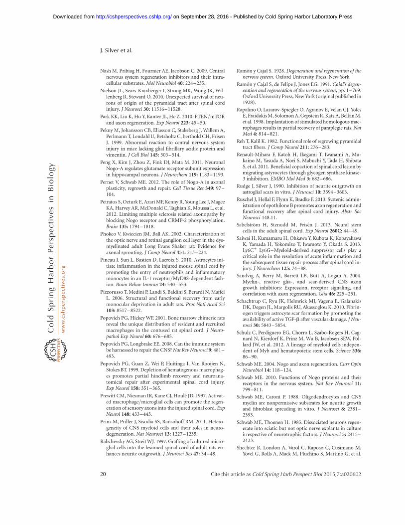
Nash M, Pribiag H, Fournier AE, Jacobson C. 2009. Centralnervous system regeneration inhibitors and their intra-cellular substrates. Mol Neurobiol 40: 224–235.
Nielson JL, Sears-Kraxberger I, Strong MK, Wong JK, Wil-lenberg R, Steward O. 2010. Unexpected survival of neu-rons of origin of the pyramidal tract after spinal cordinjury. J Neurosci 30: 11516–11528.
Park KK, Liu K, Hu Y, Kanter JL, He Z. 2010. PTEN/mTORand axon regeneration. Exp Neurol 223: 45–50.
Pekny M, Johansson CB, Eliasson C, Stakeberg J, Wallem A,Perlmann T, Lendahl U, Betsholtz C, berthold CH, FrisenJ. 1999. Abnormal reaction to central nervous systeminjury in mice lacking glial fibrillary acidic protein andvimentin. J Cell Biol 145: 503–514.
Peng X, Kim J, Zhou Z, Fink DJ, Mata M. 2011. NeuronalNogo-A regulates glutamate receptor subunit expressionin hippocampal neurons. J Neurochem 119: 1183–1193.
Pernet V, Schwab ME. 2012. The role of Nogo-A in axonalplasticity, regrowth and repair. Cell Tissue Res 349: 97–104.
Petratos S, Ozturk E, Azari MF, Kenny R, Young Lee J, MageeKA, Harvey AR, McDonald C, Taghian K, Moussa L, et al.2012. Limiting multiple sclerosis related axonopathy byblocking Nogo receptor and CRMP-2 phosphorylation.Brain 135: 1794–1818.
Phokeo V, Kwiecien JM, Ball AK. 2002. Characterization ofthe optic nerve and retinal ganglion cell layer in the dys-myelinated adult Long Evans Shaker rat: Evidence foraxonal sprouting. J Comp Neurol 451: 213–224.
Pineau I, Sun L, Bastien D, Lacroix S. 2010. Astrocytes ini-tiate inflammation in the injured mouse spinal cord bypromoting the entry of neutrophils and inflammatorymonocytes in an IL-1 receptor/MyD88-dependent fash-ion. Brain Behav Immun 24: 540–553.
Pizzorusso T, Medini P, Landi S, Baldini S, Berardi N, MaffeiL. 2006. Structural and functional recovery from earlymonocular deprivation in adult rats. Proc Natl Acad Sci103: 8517–8522.
Popovich PG, Hickey WF. 2001. Bone marrow chimeric ratsreveal the unique distribution of resident and recruitedmacrophages in the contused rat spinal cord. J Neuro-pathol Exp Neurol 60: 676–685.
Popovich PG, Longbrake EE. 2008. Can the immune systembe harnessed to repair the CNS? Nat Rev Neurosci 9: 481–493.
Popovich PG, Guan Z, Wei P, Huitinga I, Van Rooijen N,Stokes BT. 1999. Depletion of hematogenous macrophag-es promotes partial hindlimb recovery and neuroana-tomical repair after experimental spinal cord injury.Exp Neurol 158: 351–365.
Prewitt CM, Niesman IR, Kane CJ, Houle JD. 1997. Activat-ed macrophage/microglial cells can promote the regen-eration of sensory axons into the injured spinal cord. ExpNeurol 148: 433–443.
Prinz M, Priller J, Sisodia SS, Ransohoff RM. 2011. Hetero-geneity of CNS myeloid cells and their roles in neuro-degeneration. Nat Neurosci 13: 1227–1235.
Rabchevsky AG, Streit WJ. 1997. Grafting of cultured micro-glial cells into the lesioned spinal cord of adult rats en-hances neurite outgrowth. J Neurosci Res 47: 34–48.
Ramon y Cajal S. 1928. Degeneration and regeneration of thenervous system. Oxford University Press, New York.
Ramon y Cajal S, de Felipe J, Jones EG. 1991. Cajal’s degen-eration and regeneration of the nervous system, pp. 1–769.Oxford University Press, New York (original published in1928).
Rapalino O, Lazarov-Spiegler O, Agranov E, Velan GJ, YolesE, Fraidakis M, Solomon A, Gepstein R, Katz A, Belkin M,et al. 1998. Implantation of stimulated homologous mac-rophages results in partial recovery of paraplegic rats. NatMed 4: 814–821.
Reh T, Kalil K. 1982. Functional role of regrowing pyramidaltract fibers. J Comp Neurol 211: 276–283.
Renault-Mihara F, Katoh H, Ikegami T, Iwanami A, Mu-kaino M, Yasuda A, Nori S, Mabuchi Y, Tada H, ShibataS, et al. 2011. Beneficial copaction of spinal cord lesion bymigrating astyrocytes through glycogen synthase kinase-3 inhibition. EMBO Mol Med 3: 682–686.
Rudge J, Silver J, 1990. Inhibition of neurite outgrowth onastroglial scars in vitro. J Neurosci 10: 3594–3603.
Ruschel J, Hellal F, Flynn K, Bradke F. 2013. Systenic admin-istration of epothilone B promotes axon regeneration andfunctional recovery after spinal cord injury. Abstr SocNeurosci 148.11.
Sabelstrom H, Stenudd M, Frisen J. 2013. Neural stemcells in the adult spinal cord. Exp Neurol 260C: 44–49.
Saiwai H, Kumamaru H, Ohkawa Y, Kubota K, KobayakawaK, Yamada H, Yokomizo T, Iwamoto Y, Okada S. 2013.Ly6Cþ Ly6G–Myeloid-derived suppressor cells play acritical role in the resolution of acute inflammation andthe subsequent tissue repair process after spinal cord in-jury. J Neurochem 125: 74–88.
Sandvig A, Berry M, Barrett LB, Butt A, Logan A. 2004.Myelin-, reactive glia-, and scar-derived CNS axongrowth inhibitors: Expression, receptor signaling, andcorrelation with axon regeneration. Glia 46: 225–251.
Schachtrup C, Ryu JK, Helmrick MJ, Vagena E, GalanakisDK, Degen JL, Margolis RU, Akassoglou K. 2010. Fibrin-ogen triggers astrocyte scar formation by promoting theavailability of active TGF-b after vascular damage. J Neu-rosci 30: 5843–5854.
Schulz C, Perdiguero EG, Chorro L, Szabo-Rogers H, Cag-nard N, Kierdorf K, Prinz M, Wu B, Jacobsen SEW, Pol-lard JW, et al. 2012. A lineage of myeloid cells indepen-dent of Myb and hematopoietic stem cells. Science 336:86–90.
Schwab ME. 2004. Nogo and axon regeneration. Curr OpinNeurobiol 14: 118–124.
Schwab ME. 2010. Functions of Nogo proteins and theirreceptors in the nervous system. Nat Rev Neurosci 11:799–811.
Schwab ME, Caroni P. 1988. Oligodendrocytes and CNSmyelin are nonpermissive substrates for neurite growthand fibroblast spreading in vitro. J Neurosci 8: 2381–2393.
Schwab ME, Thoenen H. 1985. Dissociated neurons regen-erate into sciatic but not optic nerve explants in cultureirrespective of neurotrophic factors. J Neurosci 5: 2415–2423.
Shechter R, London A, Varol C, Raposo C, Cusimano M,Yovel G, Rolls A, Mack M, Pluchino S, Martino G, et al.
J. Silver et al.
20 Cite this article as Cold Spring Harb Perspect Biol 2015;7:a020602
on September 28, 2016 - Published by Cold Spring Harbor Laboratory Press http://cshperspectives.cshlp.org/Downloaded from

2009. Infiltrating blood-derived macrophages are vitalcells playing an anti-inflammatory role in recoveryfrom spinal cord injury in mice. PLoS Med 6: e1000113.
Shen Y, Tenney AP, Busch SA, Horn KP, Cuascut FX, Liu K,He Z, Silver J, Flanagan JG. 2009. PTPs is a receptor forchondroitin sulfate proteoglycan, an inhibitor of neuralregeneration. Science 326: 592–596.
Silver J, Miller J. 2004. Regeneration beyond the glial scar.Nat Rev Neurosci 5: 146–156.
Silver J, Ogawa MY. 1983. Postnatally induced formation ofthe corpus callosum in acallosal mice on glial coatedcellulose bridges. Science 202: 1067–1069.
Silver DJ, Silver J. 2014. Contributions of chondroitin sulfateproteoglycans to neurodevelopment, injury and cancer.Curr Opin Neurobiol 27: 171–178.
Silver DJ, Steindler DA. 2009. Common astrocytic programsduring brain development, injury and cancer. TrendsNeurosci 32: 303–311.
Silver J, Lorenz SE, Wahlsten D, Coughlin J. 1982. Axonalguidance during development of the great cerebral com-missures: Descriptive and experimental studies, in vivo,on the role of preformed glial pathways. J Comp Neurol210: 10–29.
Silver J, Edwards M, Levitt P. 1993. Immunocytochemicaldemonstration of specialized astroglial structures thatform boundaries and pathways along axon tracts in thefetal brain. J Comp Neurol 328: 415–436.
Singer M, Nordlander RH, Egar M. 1979. Axonal guidanceduring embryogenesis and regeneration in the spinalcord of newt: The blueprint hypothesis of neuronal path-way patterning. J Comp Neurol 185: 1–22.
Sobel RA, Chen M, Maeda A, Hinojoza JR. 1995. Vitronec-tin and integrin vitronectin receptor localization in mul-tiple sclerosis lesions. J Neuropathol Exp Neurol 54: 202–213.
Soderblom C, Luo X, Blumenthal E, Bray E, Lyapichev K,Ramos J, Krishnan V, Lai-Hsu C, Park KK, Tsoulfas P,et al. 2013. Perivascular fibroblasts form the fibroticscar after contusive spinal cord injury. J Neurosci 33:13882–13887.
Steinmetz MP, Horn KP, Tom VJ, Miller JH, Busch SA, NairD, Silver DJ, Silver J. 2005. Chronic enhancement of theintrinsic growth capacity of sensory neurons combinedwith the degradation of inhibitory proteoglycans allowsfunctional regeneration of sensory axons through thedorsal root entry zone in the mammalian spinal cord. JNeurosci 25: 8066–8076.
Stichel CC, Hermanns S, Luhmann HJ, Lausberg F, Nier-mann H, D’Urso D, Servos G, Hartwig HG, Muller HW.1999. Inhibition of collagen IV deposition promotes re-generation of injured CNS axons. Eur J Neurosci 11: 632–646.
Stout RD, Jiang C, Matta B, Tietzel I, Watkins SK, Suttles J.2005. Macrophages sequentially change their functionalphenotype in response to changes in microenvironmen-tal influences. J Immunol 175: 342–349.
Sun F, Park KK, Belin S, Wang D, Lu T, Chen G, Zhang K,Yeung C, Feng G, Yankner BA, et al. 2011. Sustained axonregeneration induced by co-deletion of PTEN andSOCS3. Nature 480: 372–375.
Swirski FK, Nahrendorf M, Etzrodt M, Wildgruber M, Cor-tez-Retamozo V, Panizzi P, Figueiredo J-L, Kohler RH,Chudnovskiy A, Waterman P, et al. 2009. Identificationof splenic reservoir monocytes and their deployment toinflammatory sites. Science 325: 612–616.
Takeuchi K, Onaga SH, Watanabe Y, Kudo C, Ohko K, SatoT, Oda K, Yokoyama M, Yoshioka N, Sakimura K, et al.2013. Chondroitin sulphate N-acetylgalactosaminyl-transferase-1 inhibits recovery from neural injury. NatCommun 4: 2740.
Tan AM, Colletti M, Rorai AT, Skene JH, Levine JM. 2006.Antobidies against the NG2 proteoglycan promote theregeneration of sensory axons within the dorsal columnsof the spinal cord. J Neurosci 26: 4729–4739.
Tang S, Shen YJ, DeBellard ME, Mukhopadhyay G, Salzer JL,Crocker PR, Filbin MT. 1997. Myelin-associated glyco-protein interacts with neurons via a sialic acid bindingsite at ARG118 and a distinct neurite inhibition site. J CellBiol 138: 1355–1366.
Tansey EM, Pessoa VF, Fleming S, Landon DN, Ikeda H.1985. Pattern and extent of demyelination in the opticnerves of mice infected with Semliki forest virus and thepossibility of axonal sprouting. Brain 108: 29–41.
Thawer SG, Mawhinney L, Chadwick K, de Chickera SN,Weaver LC, Brown A, Dekaban GA. 2013. Temporalchanges in monocyte and macrophage subsets and mi-croglial macrophages following spinal cord injury in theLys-Egfp-ki mouse model. J Neuroimmunol 261: 7–20.
Tsai SY, Papadopoulos CM, Schwab ME, Kartje GL. 2011.Delayed anti-Nogo-A therapy improves function afterchronic stroke in adult rats. Stroke 42: 186–190.
Wang D, Fawcett J. 2012. The perineuronal net and thecontrol of CNS plasticity. Cell Tissue Res 349: 147–160.
Wanner IB, Anderson MA, Song B, Levine J, Fernandez A,Gray-Thompson Z, Ao Y, Sofroniew MV. 2013. Glial scarborders are formed by newly proliferated, elongated as-trocytes that interact to corral inflammatory and fibroticcells via STAT-3 dependent mechanisms after spinal cordinjury. J Neurosci 33: 12870–12886.
Wilhelmsson U, Li L, Pekna M, Berthold C-H, Blom S,Eliasson C, Renner O, Bushong E, Ellisman M, MorganTE, et al. 2004. Absence of glial fibrillary acidin proteinand vimentin prevents hypertrophy of astrocytic processand improves post-traumatic regeneration. J Neurosci 24:5016–5021.
Windle WF, Chambers WW. 1950. Regeneration in the spi-nal cord of the cat and dog. J Comp Neurol 93: 241–257.
Xu K, Messing A, Silver J. 1999. Glial fibrillary acidic proteinis necessary for mature astrocytes to react to b-amyloidsubstrates in vitro. Glia 25: 390–403.
Xue Y-X, Xue L-F, Liu J-F, He J, Deng J-H, Sun S-C, Han H-B, Luo YX, Xu L-Z, Wu P, et al. 2014. Depletion of peri-neuronal nets in the amygdala to enhance the erasure ofdrug memories. J Neurosci 34: 6647–6658.
Yamada H, Fredette B, Shitara K, Hagihara K, Miura R,Ranscht B, Stallcup WB, Yamaguchi Y. 1997. The brainchondroitin sulfate proteoglycan brevican associates withastrocytes ensheathing cerebellar glomeruli and inhibitsneurite outgrowth from granule neurons. J Neurosci 17:7784–7795.
Yamaguchi Y. 2000. Lecticans: Organizers of the brain extra-cellular matrix. Cell Mol Life Sci 57: 276–289.
CNS Regenerative Failure
Cite this article as Cold Spring Harb Perspect Biol 2015;7:a020602 21
on September 28, 2016 - Published by Cold Spring Harbor Laboratory Press http://cshperspectives.cshlp.org/Downloaded from

Yang Z, Suzuki R, Daniels SB, Brunquell CB, Sala CJ, Ni-shiyama A. 2006. NG2 glial cells provide a favorable sub-strate for growing axons. J Neurosci 26: 3829–3839.
Yang Y, Liu Y, Wei P, Peng H, Winger R, Hussain RZ, Ben LH,Cravens PD, Gocke AR, Puttaparthi K, et al. 2010. Silenc-ing Nogo-A promotes functional recovery in demyelinat-ing disease. Ann Neurol 67: 498–507.
Yin Y, Cui Q, Li Y, Irwin N, Fischer D, Harvey AR, BenowitzLI. 2003. Macrophage-derived factors stimulate opticnerve regeneration. J Neurosci 23: 2284–2293.
Yin Y, Henzl MT, Lorber B, Nakazawa T, Thomas TT, Jiang F,Langer R, Benowitz LI. 2006. Oncomodulin is a macro-phage-derived signal for axon regeneration in retinal gan-glion cells. Nat Neurosci 9: 843–852.
Ylera B, Erturk A, Hellaf F, Nadrigny F, Hurtado A, TahirovicS, Oudega M, Kirchhoff F, Bradke F. 2009. ChronicallyCNS-injured adult sensory neurons gain regenerativecompetence upon a lesion of their peripheral axon.Curr Biol 19: 930–936.
Zai LJ, Wrathall JR. 2005. Cell proliferation and replace-ment following contusive spinal cord injury. Glia 50:247–257.
Zhang Z, Guth L. 1997. Experimental spinal cord injury:Wallerian degeneration in the dorsal column is followedby revascularization, glial proliferation, and nerve regen-eration. Exp Neurol 147: 159–171.
Zhang Y, Tohyama K, Winterbottom JK, Haque NS, Schah-ner M, Lieberman AR, Anderson PN. 2001. Correlationbetween putative inhibitory molecules at the dorsal rootentry zone and failure of dorsal root axonal regeneration.Mol Cell Neurosci 17: 444–459.
Zhou L, Baumgartner BJ, Hill-Felberg SJ, McGowen LR,Shine HD. 2003. Neurotrophin-3 expressed in situ in-duces axonal plasticity in the adult injured spinal cord.J Neurosci 23: 1424–1431.
Zimmer M, Palmer A, Kohler J, Klein R. 2003. EphB-ephrinBbi-directional endocytosis terminates adhesion allowingcontact mediated repulsion. Nat Cell Biol 5: 869–878.
Zorner B, Schwab ME. 2010. Anti-Nogo on the go: Fromanimal models to a clinical trial. Ann NY Acad Sci 1198:E22–E34.
Zukor K, Belin S, Wang C, Keelan N, Wang X, He Z. 2013Short hairpin RNA against PTEN enhances regenerativegrowth of corticospinal tract axons after spinal cord in-jury. J Neurosci 33: 15350–15361.
J. Silver et al.
22 Cite this article as Cold Spring Harb Perspect Biol 2015;7:a020602
on September 28, 2016 - Published by Cold Spring Harbor Laboratory Press http://cshperspectives.cshlp.org/Downloaded from

December 4, 20142015; doi: 10.1101/cshperspect.a020602 originally published onlineCold Spring Harb Perspect Biol
Jerry Silver, Martin E. Schwab and Phillip G. Popovich Oligodendrocytes, Astrocytes, and MicrogliaCentral Nervous System Regenerative Failure: Role of
Subject Collection Glia
MaintenanceThe Nodes of Ranvier: Molecular Assembly and
Matthew N. Rasband and Elior Peles
Oligodendrocyte Development and PlasticityDwight E. Bergles and William D. Richardson
Microglia in Health and DiseaseRichard M. Ransohoff and Joseph El Khoury Support
Oligodendrocytes: Myelination and Axonal
Mikael Simons and Klaus-Armin NaveThe Astrocyte: Powerhouse and Recycling Center
Bruno Weber and L. Felipe Barros Central Nervous System GliaDrosophila
Marc R. Freeman
Development and PlasticityMicroglia Function in Central Nervous System
Dorothy P. Schafer and Beth StevensSynapse: Adaptable, Multitasking Glial CellsPerisynaptic Schwann Cells at the Neuromuscular
Chien-Ping Ko and Richard Robitaille
the Central Nervous SystemOligodendrocyte Development and Myelination in Transcriptional and Epigenetic Regulation of
Ben Emery and Q. Richard Lu
and EliminationAstrocytes Control Synapse Formation, Function,
Won-Suk Chung, Nicola J. Allen and Cagla Eroglu
ControversiesOrigin of Microglia: Current Concepts and Past
Florent Ginhoux and Marco Prinz
Schwann Cell MyelinationJames L. Salzer
Remyelination−−Glia Disease and RepairRobin J.M. Franklin and Steven A. Goldman Repair
Schwann Cells: Development and Role in Nerve
LloydKristján R. Jessen, Rhona Mirsky and Alison C.
Astrocytes in Neurodegenerative DiseaseHemali Phatnani and Tom Maniatis
Perineurial GliaSarah Kucenas
http://cshperspectives.cshlp.org/cgi/collection/ For additional articles in this collection, see
Copyright © 2015 Cold Spring Harbor Laboratory Press; all rights reserved
on September 28, 2016 - Published by Cold Spring Harbor Laboratory Press http://cshperspectives.cshlp.org/Downloaded from