center for molecular medicine, national heart lung and
TRANSCRIPT
The Rockefeller University PressJ. Cell Biol. Vol. 194 No. 1 7–15www.jcb.org/cgi/doi/10.1083/jcb.201102095 JCB �
JCB: Review
IntroductionVery early in his career, while working in a remote marine bio-logical laboratory in Italy, the young Otto Warburg observed that fertilization of sea urchin eggs resulted in a rapid and nearly sixfold increase in oxygen consumption (Warburg, 1908). The notion that oxygen consumption was dynamic and seemingly tied to cellular proliferation would have a profound effect on the young scientist. Later, these ideas would be refined and focused not on normal fertilization but rather on the metabolic abnor-malities of cancer cells. Some 23 years after his observations in Italy, Warburg was awarded the Nobel Prize for his discovery of the “nature and mode of action of the respiratory enzyme.” Interestingly, although Warburg’s oocyte observations have been confirmed by many others, his hypothesis that this repre-sented a burst of mitochondrial oxidative phosphorylation is undoubtedly incorrect. Indeed, nearly 100 years after his initial observation, it was established that the surge of oxygen con-sumption after fertilization is not, as originally envisioned, some primitive metabolic wakeup call by the young zygote. Rather, it would seem, oxygen is instead used by a specific NADPH oxi-dase on the egg’s surface for the purposeful production of nano-molar concentrations of hydrogen peroxide (Wong et al., 2004). Surprisingly, this burst of hydrogen peroxide production does not damage the nascent organism, but instead is used as part of
an enzymatic reaction that ultimately results in the development of a protective shell around the young fertilized egg. These ob-servations are in concert with a growing body of evidence that suggests that ROS can be purposefully made and harnessed to regulate a diverse array of physiological processes. In turn, ac-cumulating evidence also suggests that dysregulation of oxidant signaling may cause or accelerate a host of pathological condi-tions, including the rate that we age (Balaban et al., 2005). Thus, seemingly from life’s inception to its end, redox signaling acts as an important regulator of physiological and pathophysiologi-cal outcomes. Herein, I will try to selectively review some of the highlights and emerging trends in oxidant signaling.
Oxidants and their cellular targetsThere are numerous potential sources of ROS within the cell. As mentioned above, one important generator of intracellular oxidants is a family of membrane-bound enzymes that rely on NADPH for their activity. Although the expression of these enzymes was ini-tially thought to be confined to phagocytic cells, it now appears that this seven-member family (Nox1–5 and Duox1–2) is in fact widely expressed and evolutionarily conserved (Brown and Griendling, 2009; Aguirre and Lambeth, 2010). To date, the only clear function of these NADPH-dependent oxidases is the regulated generation of ROS. Mitochondria represent another source for intracellular oxi-dant production. Most evidence suggests that mitochondrial oxi-dants are formed predominantly at complex I or complex III of the cytochrome chain when electrons initially derived from NADH or FADH2 can react with oxygen to produce superoxide anions (Fig. 1). Although one-electron reactions predominate, two-electron reac-tions that allow the direct reduction of molecular oxygen to hydro-gen peroxide do exist within the mitochondria (Giorgio et al., 2005). The fraction of total oxygen consumption that is diverted into mitochondrial ROS production is a difficult number to accu-rately estimate. Although in isolated mitochondria under nonphysi-ological conditions this fraction can approach two percent or more, in the in vivo situation, the fraction of oxygen diverted to ROS production is presumably significantly less (Balaban et al., 2005). In addition to the mitochondria and NADPH oxidases, additional cellular sources of ROS production include a host of other intra-cellular enzymes such as xanthine oxidase, cyclooxygenases, cytochrome p450 enzymes, and lipoxygenases that produce oxi-dants as part of their normal enzymatic function.
Although historically viewed as purely harmful, recent evi-dence suggests that reactive oxygen species (ROS) func-tion as important physiological regulators of intracellular signaling pathways. The specific effects of ROS are modu-lated in large part through the covalent modification of specific cysteine residues found within redox-sensitive target proteins. Oxidation of these specific and reactive cysteine residues in turn can lead to the reversible modifi-cation of enzymatic activity. Emerging evidence suggests that ROS regulate diverse physiological parameters rang-ing from the response to growth factor stimulation to the generation of the inflammatory response, and that dys-regulated ROS signaling may contribute to a host of human diseases.
Signal transduction by reactive oxygen species
Toren Finkel
Center for Molecular Medicine, National Heart Lung and Blood Institute, National Institutes of Health, Bethesda, MD 20892
This article is distributed under the terms of an Attribution–Noncommercial–Share Alike–No Mirror Sites license for the first six months after the publication date (see http://www.rupress.org/terms). After six months it is available under a Creative Commons License (Attribution–Noncommercial–Share Alike 3.0 Unported license, as described at http://creativecommons .org/licenses/by-nc-sa/3.0/).Correspondence to Toren Finkel: [email protected]
TH
EJ
OU
RN
AL
OF
CE
LL
BIO
LO
GY
Dow
nloaded from http://rupress.org/jcb/article-pdf/194/1/7/1352587/jcb_201102095.pdf by guest on 03 April 2022

JCB • VOLUME 194 • NUMBER 1 • 2011 �
sulphonic (RSO3H) species can be created (Fig. 2). Other pos-sibilities for post-translational cysteine modifications include nitrosylation (RSNO), glutathionylation (RSSG), or the forma-tion of an inter- or intramolecular disulfide bond (RSSR). Although reactive cysteine residues have long been identified experimentally using individual purified proteins, the ability to predict these residues based on either computational means (Fomenko et al., 2007) or through large-scale proteomic approaches (Weerapana et al., 2010) is a relatively recent development. These more re-cent analysis has suggested that reactive and potentially modu-latory cysteine residues might exist in well over 500 individual proteins, thereby extending this form of redox regulation to a wide range of enzymatic activities.
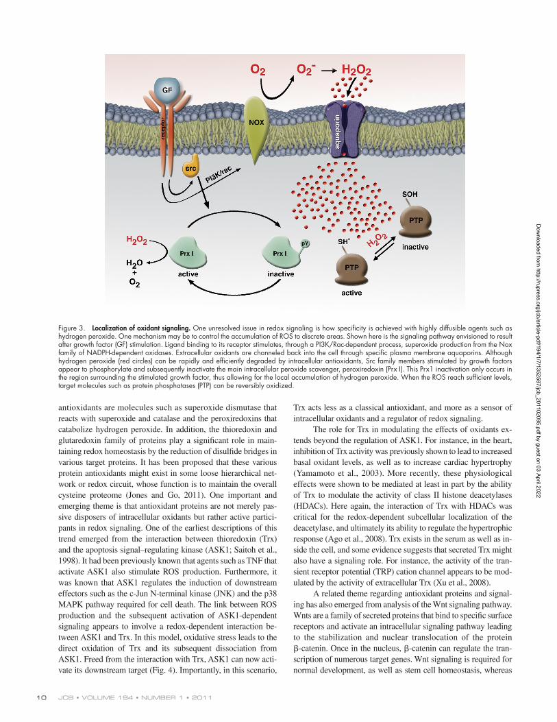
Although such analysis has established a role for intracellu-lar oxidants in signal transduction, given the wide range of puta-tive targets and the general reactivity of hydrogen peroxide, how can any specificity be achieved? At this point, this remains an open and potentially vexing concern. Part of the answer may however result from the colocalization of the source of oxidant production nearby to the intended target. One recent report demonstrated this principle by analyzing the antioxidant peroxi-redoxin 1 (Prx1), which was transiently phosphorylated on a ty-rosine residue after growth factor stimulation (Woo et al., 2010). This phosphorylation only occurred on a relatively small fraction
Although it had been appreciated for some time that mito-chondria produced ROS as part of aerobic respiration and that phagocytic cells could produce oxidants as part of a host de-fense mechanism, the role of oxidants within cells began to shift with the observation that the production of hydrogen peroxide was essential for normal growth factor signaling (Sundaresan et al., 1995; Bae et al., 1997). These studies demonstrated that for growth factors such as PDGF and EGF, ligand binding stim-ulated a burst of ROS production. Inhibiting this rise in ROS levels was shown to block the normal tyrosine kinase signaling induced by growth factor addition. These initial reports specu-lated that the intracellular target of oxidants might be the family of tyrosine and dual specific phosphatases, a hypothesis that was subsequently elegantly established (Lee et al., 1998; Salmeen et al., 2003). The basis for this regulation of phosphatase activ-ity centers on the unique chemistry of certain reactive cysteine residues. Although the pKa of thiol group on free cysteine is between 8 and 9, in some tyrosine phosphatases or similar tar-get molecules, the surrounding amino acid microenvironment of the cysteine can be substantially modified to result in pKas as low as 4 to 5. These reactive cysteine residues are easily oxi-dized to a sulfenic form (RSOH). The sulfenic form is unstable and can undergo further oxidation via disproportionation to a sulfinic (RSO2H) species. Under greater oxidative stress, the
Figure 1. Reactive oxygen species generation and disposal in the mitochondria. Primary sources of ROS occur from the transfer of electrons (e) to molecular oxygen at either Complex I or III. Superoxide produced at Complex I is thought to form only within the matrix, whereas at Complex III superoxide is released both into the matrix and the inner mitochondrial space (IMS). In addition to the cytochrome chain, ROS can be formed by enzymatic action of numerous enzymes in-cluding monoamine oxidase (MAO) and cytochrome b5 reductase (Cb5R) located on the outer mitochondrial membrane (OMM), as well as glycerol-3-phosphate dehydrogenase (GPDH) and in some cell types, various cytochrome P450 enzymes located in the inner mitochondrial membrane (IMM). There are also several matrix enzymes and complexes (box) including aconitase, pyruvate dehydrogenase (PDH), and -ketoglutarate dehydrogenase (KGDH) that can generate superoxide. Although one-electron reactions predominate, two-electron reactions leading to direct hydrogen peroxide production can occur as when, for instance, cytochrome c (Cyt C) and p66shc interact within the IMS. Once generated, superoxide is dismutated spontaneously or enzymatically by manganese superoxide dismutase (MnSOD). The hydrogen peroxide that is formed is further catabolized by the action of enzymes such as catalase (CAT), glutathione peroxidase (GPx), and peroxiredoxin 3 (Prx3). For further details see the text, as well as other recent reviews (Lin and Beal, 2006; Brand, 2010). CoQ, Coenzyme Q.
Dow
nloaded from http://rupress.org/jcb/article-pdf/194/1/7/1352587/jcb_201102095.pdf by guest on 03 April 2022

9Signal transduction by reactive oxygen species • Finkel
feedback regulation after metabolic excess (Nemoto et al., 2000), as well as the regulation of the hypoxia-inducible factor 1 (HIF-1a) during low oxygen conditions (Brunelle et al., 2005; Guzy et al., 2005; Mansfield et al., 2005). There also appears to be an important role for mitochondria oxidants in the regulation of autophagy through the direct regulation of Atg4 activity (Scherz-Shouval et al., 2007). Finally, there is growing recogni-tion that mitochondrial oxidants are important signaling mole-cules that regulate the inflammatory response. In particular, several recent reports have identified a critical role for mitochon-drial oxidants in the formation of the NLRP3 (NOD-like receptor pyrin domain-containing 3) inflammasome (Bulua et al., 2011; Nakahira et al., 2011; Zhou et al., 2011). In addition, as recog-nized by the above examples, phosphatases represent only a frac-tion of the presumably important redox targets. For instance, other signaling molecules such as p21ras (Lander et al., 1995; Clavreul et al., 2006) and certain 14-3-3 isoforms (Kim et al., 2009) have been shown to be direct targets of reactive oxygen and nitrogen species. Another recently identified target is the ataxia-telangiectasia mutated (ATM) protein kinase that is activated after certain stresses, most notably after double-stranded DNA breaks. In this study, hydrogen peroxide was shown to catalyze the formation of a disulfide ATM dimer through the oxidation of specific reactive cysteine residues on the C-terminal region of ATM (Guo et al., 2010). As opposed to the example of dual spe-cific and tyrosine phosphatases, oxidation of the cysteine residue in this case led to enzymatic activation rather than inactivation. These results are particularly intriguing because patients with mutated ATM suffer from a crippling and debilitating disease characterized by ataxia, immunodeficiency, premature aging, and cancer predisposition. Cells lacking ATM exhibit constitutively high levels of ROS, as do certain tissues obtained from knockout mice (Kamsler et al., 2001; Reichenbach et al., 2002). Indeed, treatment of Atm/ mice with antioxidants delays the develop-ment of tumors (Reliene and Schiestl, 2006), as well as rescuing the defect in self-renewal observed in hematopoietic stem cells derived from Atm-deficient animals (Ito et al., 2004). The mecha-nism through which ATM regulates the intracellular redox state is complex and may involve regulation of mitochondria biogene-sis (Eaton et al., 2007), altering mTOR-dependent autophagy (Alexander et al., 2010) or modulating the synthesis of NADPH through the pentose phosphate shunt (Cosentino et al., 2011). Nonetheless, it is intriguing that ATM appears to be regulated directly by oxidants while at the same time serving as a major regulator of intracellular redox status. It is unlikely that such a homeostatic feedback loop is unique for ATM. Taken together with the recently described studies on the inflammasome, these studies suggest that aerobic metabolism, stress responses, and oxidant generation will be tightly coupled.
Antioxidants and signalingThe maintenance of intracellular redox homeostasis is dependent on a complex web of antioxidant molecules. These antioxidants include low molecular weight molecules such as glutathione, present in millimolar concentrations within cells, as well as an array of protein antioxidants that each have specific subcellular localizations and chemical reactivities. Included among the protein
of Prx1 molecules, especially that fraction of Prx1 located near the membrane oxidant source and nearby various membrane- associated signaling intermediates. Phosphorylation of Prx1 actually inhibited the antioxidant function of the protein, thereby allowing for the local accumulation of ROS near the membrane and presumably allowing for cysteine modification confined to this local area beneath the growth factor receptor. Another recent study using a zebrafish model visually identified a gradient of hydrogen peroxide levels within tissue that was essential for signaling (Niethammer et al., 2009). It should be noted that in both preceding examples, the source of oxidants appeared to be a member of the Nox family of membrane-bound NADPH oxi-dases. These enzymes produce extracellular superoxide anions. It is generally believed that for signaling purposes, the superoxide anion dismutates spontaneously to hydrogen peroxide. Although it had been generally assumed that once generated, hydrogen per-oxide could simply diffuse back across the plasma membrane, re-cent evidence suggests that hydrogen peroxide might preferentially enter the cell through specific plasma membrane aquaporin chan-nels (Bienert et al., 2007; Miller et al., 2010). This regulated entry provides another potential mechanism through which oxidants could be channeled to an intended target and thereby achieve some measure of overall signaling specificity (Fig. 3).
It is important to note that the Nox-dependent oxidant burst that leads to the transient inactivation of phosphatases represents only one of many mechanisms through which oxidants elicit spe-cific responses in cells. First, mitochondrial oxidants also appear to participate in signaling events. Examples are varied and include
Figure 2. Cysteine biochemistry allows for redox-dependent signaling. Specific reactive cysteine (Cys) residues within target proteins can be covalently modified by oxidative stress. Much like phosphorylation on serine or threonine residues, alteration of the thiol group can in turn modify enzymatic activity. Although the sulfenic form (SOH) is readily reversible, higher states of oxidation generally, but not always, lead to irreversible modification.
Dow
nloaded from http://rupress.org/jcb/article-pdf/194/1/7/1352587/jcb_201102095.pdf by guest on 03 April 2022
JCB • VOLUME 194 • NUMBER 1 • 2011 10
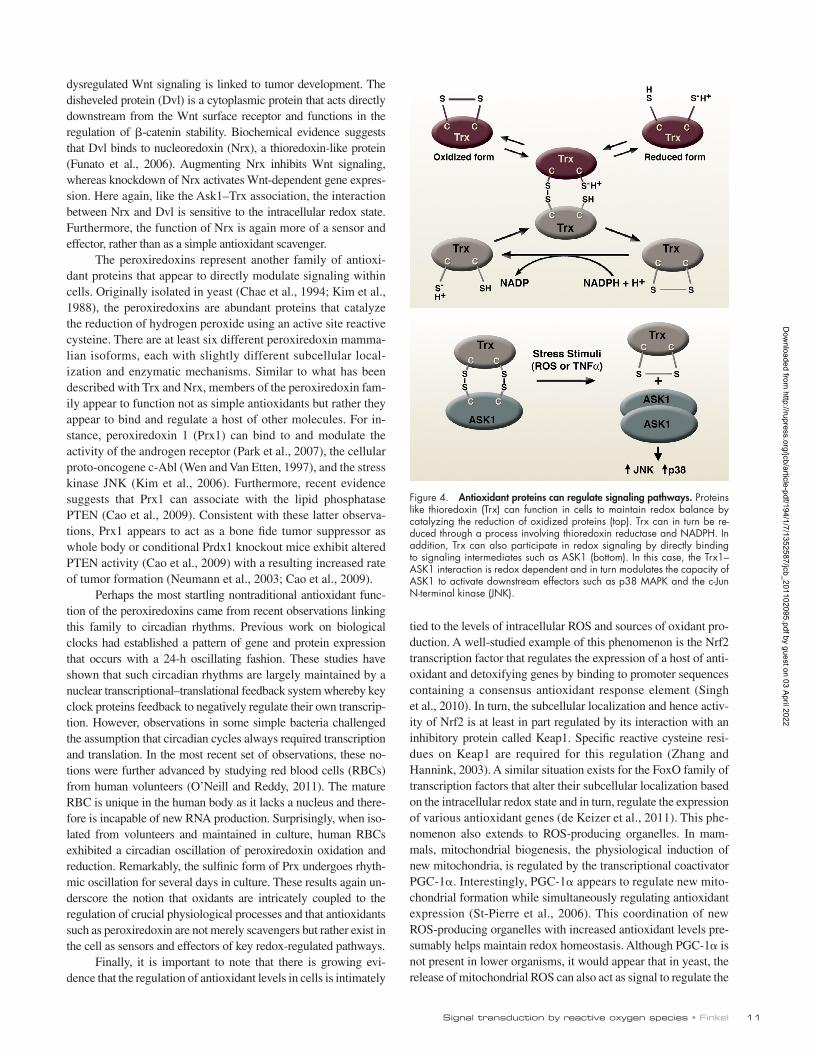
Trx acts less as a classical antioxidant, and more as a sensor of intracellular oxidants and a regulator of redox signaling.
The role for Trx in modulating the effects of oxidants ex-tends beyond the regulation of ASK1. For instance, in the heart, inhibition of Trx activity was previously shown to lead to increased basal oxidant levels, as well as to increase cardiac hypertrophy (Yamamoto et al., 2003). More recently, these physiological effects were shown to be mediated at least in part by the ability of Trx to modulate the activity of class II histone deacetylases (HDACs). Here again, the interaction of Trx with HDACs was critical for the redox-dependent subcellular localization of the deacetylase, and ultimately its ability to regulate the hypertrophic response (Ago et al., 2008). Trx exists in the serum as well as in-side the cell, and some evidence suggests that secreted Trx might also have a signaling role. For instance, the activity of the tran-sient receptor potential (TRP) cation channel appears to be mod-ulated by the activity of extracellular Trx (Xu et al., 2008).
A related theme regarding antioxidant proteins and signal-ing has also emerged from analysis of the Wnt signaling pathway. Wnts are a family of secreted proteins that bind to specific surface receptors and activate an intracellular signaling pathway leading to the stabilization and nuclear translocation of the protein -catenin. Once in the nucleus, -catenin can regulate the tran-scription of numerous target genes. Wnt signaling is required for normal development, as well as stem cell homeostasis, whereas
antioxidants are molecules such as superoxide dismutase that reacts with superoxide and catalase and the peroxiredoxins that catabolize hydrogen peroxide. In addition, the thioredoxin and glutaredoxin family of proteins play a significant role in main-taining redox homeostasis by the reduction of disulfide bridges in various target proteins. It has been proposed that these various protein antioxidants might exist in some loose hierarchical net-work or redox circuit, whose function is to maintain the overall cysteine proteome (Jones and Go, 2011). One important and emerging theme is that antioxidant proteins are not merely pas-sive disposers of intracellular oxidants but rather active partici-pants in redox signaling. One of the earliest descriptions of this trend emerged from the interaction between thioredoxin (Trx) and the apoptosis signal–regulating kinase (ASK1; Saitoh et al., 1998). It had been previously known that agents such as TNF that activate ASK1 also stimulate ROS production. Furthermore, it was known that ASK1 regulates the induction of downstream effectors such as the c-Jun N-terminal kinase (JNK) and the p38 MAPK pathway required for cell death. The link between ROS production and the subsequent activation of ASK1-dependent signaling appears to involve a redox-dependent interaction be-tween ASK1 and Trx. In this model, oxidative stress leads to the direct oxidation of Trx and its subsequent dissociation from ASK1. Freed from the interaction with Trx, ASK1 can now acti-vate its downstream target (Fig. 4). Importantly, in this scenario,
Figure 3. Localization of oxidant signaling. One unresolved issue in redox signaling is how specificity is achieved with highly diffusible agents such as hydrogen peroxide. One mechanism may be to control the accumulation of ROS to discrete areas. Shown here is the signaling pathway envisioned to result after growth factor (GF) stimulation. Ligand binding to its receptor stimulates, through a PI3K/Rac-dependent process, superoxide production from the Nox family of NADPH-dependent oxidases. Extracellular oxidants are channeled back into the cell through specific plasma membrane aquaporins. Although hydrogen peroxide (red circles) can be rapidly and efficiently degraded by intracellular antioxidants, Src family members stimulated by growth factors appear to phosphorylate and subsequently inactivate the main intracellular peroxide scavenger, peroxiredoxin (Prx I). This Prx1 inactivation only occurs in the region surrounding the stimulated growth factor, thus allowing for the local accumulation of hydrogen peroxide. When the ROS reach sufficient levels, target molecules such as protein phosphatases (PTP) can be reversibly oxidized.
Dow
nloaded from http://rupress.org/jcb/article-pdf/194/1/7/1352587/jcb_201102095.pdf by guest on 03 April 2022
11Signal transduction by reactive oxygen species • Finkel
tied to the levels of intracellular ROS and sources of oxidant pro-duction. A well-studied example of this phenomenon is the Nrf2 transcription factor that regulates the expression of a host of anti-oxidant and detoxifying genes by binding to promoter sequences containing a consensus antioxidant response element (Singh et al., 2010). In turn, the subcellular localization and hence activ-ity of Nrf2 is at least in part regulated by its interaction with an inhibitory protein called Keap1. Specific reactive cysteine resi-dues on Keap1 are required for this regulation (Zhang and Hannink, 2003). A similar situation exists for the FoxO family of transcription factors that alter their subcellular localization based on the intracellular redox state and in turn, regulate the expression of various antioxidant genes (de Keizer et al., 2011). This phe-nomenon also extends to ROS-producing organelles. In mam-mals, mitochondrial biogenesis, the physiological induction of new mitochondria, is regulated by the transcriptional coactivator PGC-1. Interestingly, PGC-1 appears to regulate new mito-chondrial formation while simultaneously regulating antioxidant expression (St-Pierre et al., 2006). This coordination of new ROS-producing organelles with increased antioxidant levels pre-sumably helps maintain redox homeostasis. Although PGC-1 is not present in lower organisms, it would appear that in yeast, the release of mitochondrial ROS can also act as signal to regulate the
dysregulated Wnt signaling is linked to tumor development. The disheveled protein (Dvl) is a cytoplasmic protein that acts directly downstream from the Wnt surface receptor and functions in the regulation of -catenin stability. Biochemical evidence suggests that Dvl binds to nucleoredoxin (Nrx), a thioredoxin-like protein (Funato et al., 2006). Augmenting Nrx inhibits Wnt signaling, whereas knockdown of Nrx activates Wnt-dependent gene expres-sion. Here again, like the Ask1–Trx association, the interaction between Nrx and Dvl is sensitive to the intracellular redox state. Furthermore, the function of Nrx is again more of a sensor and effector, rather than as a simple antioxidant scavenger.
The peroxiredoxins represent another family of antioxi-dant proteins that appear to directly modulate signaling within cells. Originally isolated in yeast (Chae et al., 1994; Kim et al., 1988), the peroxiredoxins are abundant proteins that catalyze the reduction of hydrogen peroxide using an active site reactive cysteine. There are at least six different peroxiredoxin mamma-lian isoforms, each with slightly different subcellular local-ization and enzymatic mechanisms. Similar to what has been described with Trx and Nrx, members of the peroxiredoxin fam-ily appear to function not as simple antioxidants but rather they appear to bind and regulate a host of other molecules. For in-stance, peroxiredoxin 1 (Prx1) can bind to and modulate the activity of the androgen receptor (Park et al., 2007), the cellular proto-oncogene c-Abl (Wen and Van Etten, 1997), and the stress kinase JNK (Kim et al., 2006). Furthermore, recent evidence suggests that Prx1 can associate with the lipid phosphatase PTEN (Cao et al., 2009). Consistent with these latter observa-tions, Prx1 appears to act as a bone fide tumor suppressor as whole body or conditional Prdx1 knockout mice exhibit altered PTEN activity (Cao et al., 2009) with a resulting increased rate of tumor formation (Neumann et al., 2003; Cao et al., 2009).
Perhaps the most startling nontraditional antioxidant func-tion of the peroxiredoxins came from recent observations linking this family to circadian rhythms. Previous work on biological clocks had established a pattern of gene and protein expression that occurs with a 24-h oscillating fashion. These studies have shown that such circadian rhythms are largely maintained by a nuclear transcriptional–translational feedback system whereby key clock proteins feedback to negatively regulate their own transcrip-tion. However, observations in some simple bacteria challenged the assumption that circadian cycles always required transcription and translation. In the most recent set of observations, these no-tions were further advanced by studying red blood cells (RBCs) from human volunteers (O’Neill and Reddy, 2011). The mature RBC is unique in the human body as it lacks a nucleus and there-fore is incapable of new RNA production. Surprisingly, when iso-lated from volunteers and maintained in culture, human RBCs exhibited a circadian oscillation of peroxiredoxin oxidation and reduction. Remarkably, the sulfinic form of Prx undergoes rhyth-mic oscillation for several days in culture. These results again un-derscore the notion that oxidants are intricately coupled to the regulation of crucial physiological processes and that antioxidants such as peroxiredoxin are not merely scavengers but rather exist in the cell as sensors and effectors of key redox-regulated pathways.
Finally, it is important to note that there is growing evi-dence that the regulation of antioxidant levels in cells is intimately
Figure 4. Antioxidant proteins can regulate signaling pathways. Proteins like thioredoxin (Trx) can function in cells to maintain redox balance by catalyzing the reduction of oxidized proteins (top). Trx can in turn be re-duced through a process involving thioredoxin reductase and NADPH. In addition, Trx can also participate in redox signaling by directly binding to signaling intermediates such as ASK1 (bottom). In this case, the Trx1–ASK1 interaction is redox dependent and in turn modulates the capacity of ASK1 to activate downstream effectors such as p38 MAPK and the c-Jun N-terminal kinase (JNK).
Dow
nloaded from http://rupress.org/jcb/article-pdf/194/1/7/1352587/jcb_201102095.pdf by guest on 03 April 2022

JCB • VOLUME 194 • NUMBER 1 • 2011 12
antioxidant glutathione peroxidase showed higher ROS levels, leading to the inactivation of the lipid phosphatase PTEN. This phosphatase inactivation resulted in higher PI3-K/Akt signaling in these animals and improved insulin sensitivity (Loh et al., 2009). In these animals, treatment with an antioxidant actually made the glucose metabolism worse. Analysis of rare individuals with selenoprotein deficiency suggests that these observations may potentially extend to humans as well (Schoenmakers et al., 2010). Presently, it is far from clear as to why in certain cellular and ani-mal models, insulin resistance is associated with increased ROS levels and scavenging these oxidants improves metabolic homeo-stasis, whereas in other models high ROS levels are associated with improved insulin sensitivity and antioxidants make things worse. Possible explanations include differences in the intensity and sources of oxidants, a change in the function of oxidants de-pending on the stage of the disease, as well as specific but poten-tially important differences in the various metabolic models used. In addition, because redox homeostasis presumably has a narrow biological window, it is conceivable that too much or too little oxidants could produce similar pathological effects.
Aging is another arena in which the role of oxidants has been often implicated but never definitively established. There is a long history, first formally articulated by Denham Harman in the 1950s, suggesting that the production of reactive oxygen spe-cies was linked to the rate of aging (Harman, 1956). Indeed, there is a large body of mostly correlative literature suggesting that aging is accompanied by an increase in the steady-state level of oxidants and oxidatively modified proteins, lipids, and DNA (Finkel and Holbrook, 2000; Balaban et al., 2005). Consistent with a causal role for oxidants, in some lower organisms and mammalian models, increasing the level of antioxidants results in life span extension (Sun and Tower, 1999; Schriner et al., 2005). In addition, certain long-lived mouse models such as dele-tion of the p66shc gene appear to extend life by modulating ROS levels (Migliaccio et al., 1999; Nemoto and Finkel, 2002; Giorgio et al., 2005). In contrast, there are other models in lower organisms whereby the addition of low levels of ROS-producing compounds actually extends life (Schulz et al., 2007; Heidler et al., 2010; Lee et al., 2010; Yang and Hekimi, 2010), and where the genetic reduction of antioxidant defenses actually promotes a longer lifespan (Van Raamsdonk and Hekimi, 2009). These effects have also been observed at the cellular level (Bell et al., 2007) and are generally ascribed to the concept of mitochondrial hormesis (Ristow and Zarse, 2010), the biological equivalent of Fredrick Nietzsche dictum, “what won’t kill you, will make you strong.” Finally, it should be noted that in some mouse models, re-ducing or increasing scavenging capacity has no impact on aging (Jang et al., 2009; Zhang et al., 2009), whereas in other models, impaired mitochondrial function can accelerate aging in an ROS-independent fashion (Trifunovic et al., 2004; Kujoth et al., 2005).
Although the evidence for ROS contributing to organismal aging is therefore mixed, there is a growing body of work suggest-ing that oxidants might regulate age-related regenerative capacity by being an important determinant of stem cell biology. Perhaps the first description of this phenomenon came from the analysis briefly mentioned in the preceding section involving mice deficient in the ATM kinase. These mice were known to develop a high rate
formation of new mitochondria (Chevtzoff et al., 2010). Such results again suggest a coupling between mitochondrial number, oxidant production, and the antioxidant network.
Oxidative stress and human diseaseAlthough the preceding discussion revolved mostly around the role of ROS in normal physiological signaling, growing evi-dence implicates alterations in redox signaling as a contributor to many disease processes. Nonetheless, the precise role that oxidants play remains controversial and often beset with the problem as to whether a rise in ROS causes or merely correlates with the disease state. Although an exhaustive description of the involvement of redox signaling in human disease is beyond the scope of this review, I will briefly highlight a few illustrative examples that exemplify some of the general principles.
The development of insulin resistance and subsequent type 2 diabetes mellitus (T2DM) is one area where oxidants have been consistently implicated in disease pathogenesis. Cellular models of insulin resistance are characterized by persistently elevated ROS levels (Houstis et al., 2006). The source of this dysregulated oxidant production appears to be the mitochondria (Anderson et al., 2009), and by unknown mechanisms, high glu-cose conditions can cause alterations in mitochondrial morphol-ogy including the rapid fragmentation of mitochondria (Yu et al., 2006; Bonnard et al., 2008). Similarly, both animal models and human samples suggest that T2DM is accompanied in particular by increased mitochondrial hydrogen peroxide production (Anderson et al., 2009). In these various animal models, pharma-cological or genetic strategies that increase mitochondrial anti-oxidant levels at least partially reverse the observed metabolic defects (Anderson et al., 2009; Hoehn et al., 2009). The link be-tween constitutive ROS production and insulin resistance has been ascribed to alterations in various intracellular signaling pathways. There is evidence, for instance, that activation of the JNK pathway is important for maintaining insulin resistance (Kaneto et al., 2004). The intracellular redox state in turn regu-lates JNK activity through multiple mechanisms including the inactivation of certain key phosphatases (Kamata et al., 2005), the modulation of ASK1 as previously described (Saitoh et al., 1998), as well as the ability of ROS to alter GST Pi, a known in-hibitor of JNK activity (Adler et al., 1999). Furthermore, through multiple direct and indirect means, oxidants can regulate the ac-tivity of the NF-B transcription family that can also play a role in the pathogenesis of diabetes (Gloire and Piette, 2009).
Although these data suggest that high levels of ROS con-tribute to insulin resistance, there are other datasets that suggest the complete opposite might be true. For instance, there is some evidence that mitochondrial oxidants are required for the glucose-induced insulin secretion by pancreatic -cells (Leloup et al., 2009). Furthermore, like many other growth factors, insu-lin binding to its cell surface receptor stimulates production of hydrogen peroxide (Mahadev et al., 2004; Seo et al., 2005). This rise in ROS levels has been shown to be necessary for the transient inactivation of protein tyrosine phosphatases such as PTP1B and TC45 (Meng et al., 2004). As such, ROS are also necessary to maintain normal insulin sensitivity. This point was underscored recently by the demonstration that mice lacking the
Dow
nloaded from http://rupress.org/jcb/article-pdf/194/1/7/1352587/jcb_201102095.pdf by guest on 03 April 2022

13Signal transduction by reactive oxygen species • Finkel
Over a century has passed since a young Otto Warburg measured the rapid burst of oxygen consumption that occurs as life begins. Few scientists in his or subsequent generations would have contemplated that at the moment of conception, a fertilized zygote would purposely make prodigious amounts of hydrogen peroxide. Nonetheless, the last two decades have convincingly demonstrated that oxidants can be regulated in their production and specific in their effects. Furthermore, the fact that bacteria and plants also heavily rely on aspects of oxidant signaling sug-gests that many of these mechanisms are ancient and evolution-arily conserved (Imlay, 2008; Wong and Shimamoto, 2009). A further understanding of these pathways promises to reveal to us many more secrets regarding how life begins, why it ends, and all the myriad complexities that make up the middle.
Due to space limitations, I was unable to review all relevant work and apolo-gize to those individuals whose contributions were not cited. I am grateful to members of my own laboratory for helpful discussions and to I. Rovira for help in preparing the manuscript.
This work was supported by National Institutes of Health Intramural funds and a grant from the Ellison Medical Foundation.
Submitted: 18 February 2011Accepted: 12 May 2011
ReferencesAdler, V., Z. Yin, S.Y. Fuchs, M. Benezra, L. Rosario, K.D. Tew, M.R. Pincus, M.
Sardana, C.J. Henderson, C.R. Wolf, et al. 1999. Regulation of JNK sig-naling by GSTp. EMBO J. 18:1321–1334. doi:10.1093/emboj/18.5.1321
Ago, T., T. Liu, P. Zhai, W. Chen, H. Li, J.D. Molkentin, S.F. Vatner, and J. Sadoshima. 2008. A redox-dependent pathway for regulating class II HDACs and cardiac hypertrophy. Cell. 133:978–993. doi:10.1016/j.cell .2008.04.041
Aguirre, J., and J.D. Lambeth. 2010. Nox enzymes from fungus to fly to fish and what they tell us about Nox function in mammals. Free Radic. Biol. Med. 49:1342–1353. doi:10.1016/j.freeradbiomed.2010.07.027
Alexander, A., S.L. Cai, J. Kim, A. Nanez, M. Sahin, K.H. MacLean, K. Inoki, K.L. Guan, J. Shen, M.D. Person, et al. 2010. ATM signals to TSC2 in the cytoplasm to regulate mTORC1 in response to ROS. Proc. Natl. Acad. Sci. USA. 107:4153–4158. doi:10.1073/pnas.0913860107
Anderson, E.J., M.E. Lustig, K.E. Boyle, T.L. Woodlief, D.A. Kane, C.T. Lin, J.W. Price III, L. Kang, P.S. Rabinovitch, H.H. Szeto, et al. 2009. Mitochondrial H2O2 emission and cellular redox state link excess fat intake to insulin resistance in both rodents and humans. J. Clin. Invest. 119:573–581. doi:10.1172/JCI37048
Bae, Y.S., S.W. Kang, M.S. Seo, I.C. Baines, E. Tekle, P.B. Chock, and S.G. Rhee. 1997. Epidermal growth factor (EGF)-induced generation of hydrogen peroxide. Role in EGF receptor-mediated tyrosine phosphorylation. J. Biol. Chem. 272:217–221. doi:10.1074/jbc.272.51.32071
Balaban, R.S., S. Nemoto, and T. Finkel. 2005. Mitochondria, oxidants, and aging. Cell. 120:483–495. doi:10.1016/j.cell.2005.02.001
Bell, E.L., T.A. Klimova, J. Eisenbart, P.T. Schumacker, and N.S. Chandel. 2007. Mitochondrial reactive oxygen species trigger hypoxia-inducible factor-dependent extension of the replicative life span during hypoxia. Mol. Cell. Biol. 27:5737–5745. doi:10.1128/MCB.02265-06
Bienert, G.P., A.L. Møller, K.A. Kristiansen, A. Schulz, I.M. Møller, J.K. Schjoerring, and T.P. Jahn. 2007. Specific aquaporins facilitate the diffu-sion of hydrogen peroxide across membranes. J. Biol. Chem. 282:1183–1192. doi:10.1074/jbc.M603761200
Bonnard, C., A. Durand, S. Peyrol, E. Chanseaume, M.A. Chauvin, B. Morio, H. Vidal, and J. Rieusset. 2008. Mitochondrial dysfunction results from oxidative stress in the skeletal muscle of diet-induced insulin-resistant mice. J. Clin. Invest. 118:789–800.
Brand, M.D. 2010. The sites and topology of mitochondrial superoxide produc-tion. Exp. Gerontol. 45:466–472. doi:10.1016/j.exger.2010.01.003
Brown, D.I., and K.K. Griendling. 2009. Nox proteins in signal transduction. Free Radic. Biol. Med. 47:1239–1253. doi:10.1016/j.freeradbiomed .2009.07.023
Brunelle, J.K., E.L. Bell, N.M. Quesada, K. Vercauteren, V. Tiranti, M. Zeviani, R.C. Scarpulla, and N.S. Chandel. 2005. Oxygen sensing requires
of tumors; however, it was observed that mice that are tumor free develop an age-dependent decline in hematopoietic function. This defect turned out to be the result in an accelerated decline in self-renewal capacity for the ATM-deficient hematopoietic stem cell (HSC; Ito et al., 2004). Self-renewal is a unique property of stem cells wherein a stem cell can divide and give rise to a daughter stem cell. This property is essential for the maintenance of the overall stem cell pool. Before the observed failure of ATM-deficient HSCs, it was noted that these cells had high levels of ROS. Indeed, treatment with an antioxidant rescued the self-renewal defect ob-served in this model. The link between high levels of ROS and stem cell defects was extended by examining the HSC derived from mice that were deficient in three members of the FoxO family of transcription factors. Again, as previously mentioned, the activity of the FoxO family is sensitive to intracellular oxidants, and in turn, FoxO transcription factors can specifically induce the expression of key intracellular antioxidants. HSCs lacking FoxO1, FoxO3, and FoxO4 had reduced expression of antioxidant proteins such as superoxide dismutase and catalase and corresponding increase in ROS levels within the HSC compartment (Tothova et al., 2007). As observed with Atm/ HSCs, FoxO-deficient HSCs demonstrated a marked impairment in self-renewal that could be corrected by an-tioxidant administration. Similar findings were observed in FoxO-deficient neural stem cells (Paik et al., 2009; Renault et al., 2009). Finally, mice deficient in the polycomb repressor Bmi1 also exhibit a defect in self-renewal and a corresponding rise in ROS levels (Liu et al., 2009). Taken together, these studies suggest that the age- dependent maintenance of the stem cell pool is intricately con-nected to regulation of the intracellular redox state. It is tempting to speculate that an age-dependent rise in ROS might contribute to the observed age-dependent decline in stem cell function. However, as we have learned from the previous examples the relationship be-tween a rise in oxidants and the observed pathology is often com-plex. Echoing this notion, at least in Drosophila, oxidants play an essential role in the normal differentiation of HSCs into mature progeny (Owusu-Ansah and Banerjee, 2009).
ConclusionsAlthough not exhaustive, the examples discussed highlight the emerging and rapidly expanding role of redox signaling in biology. These studies have implicated the unique chemistry of reactive cysteine residues within certain target proteins. Such redox reac-tions allows for the covalent modification of protein function in a fashion not unlike the well-established phosphorylation of serine, threonine, and tyrosine residues. Significant challenges in the field exist, however, including a more precise understanding of how signaling specificity occurs. Often, oxidants are implicated in events in which the levels of ROS may change but where their precise function is inferred and not proven. This is particularly true in disease states where a rise in oxidants is sometimes taken as a causal rather than a correlative event. Progress is also ham-pered by technical limitations in providing high-throughput means to identify the modification of reactive cysteine residues, as well as accurate and quantitative means of measuring intra-cellular ROS levels. Newer approaches to address the latter defi-ciency appear, however, to hold promise (Miller and Chang, 2007; Dickinson et al., 2010; Cochemé et al., 2011).
Dow
nloaded from http://rupress.org/jcb/article-pdf/194/1/7/1352587/jcb_201102095.pdf by guest on 03 April 2022

JCB • VOLUME 194 • NUMBER 1 • 2011 14
Ito, K., A. Hirao, F. Arai, S. Matsuoka, K. Takubo, I. Hamaguchi, K. Nomiyama, K. Hosokawa, K. Sakurada, N. Nakagata, et al. 2004. Regulation of oxi-dative stress by ATM is required for self-renewal of haematopoietic stem cells. Nature. 431:997–1002. doi:10.1038/nature02989
Jang, Y.C., V.I. Pérez, W. Song, M.S. Lustgarten, A.B. Salmon, J. Mele, W. Qi, Y. Liu, H. Liang, A. Chaudhuri, et al. 2009. Overexpression of Mn super-oxide dismutase does not increase life span in mice. J. Gerontol. A Biol. Sci. Med. Sci. 64:1114–1125. doi:10.1093/gerona/glp100
Jones, D.P., and Y.M. Go. 2011. Mapping the cysteine proteome: analysis of redox-sensing thiols. Curr. Opin. Chem. Biol. 15:103–112. doi:10.1016/ j.cbpa.2010.12.014
Kamata, H., S. Honda, S. Maeda, L. Chang, H. Hirata, and M. Karin. 2005. Reactive oxygen species promote TNFalpha-induced death and sustained JNK activation by inhibiting MAP kinase phosphatases. Cell. 120:649–661. doi:10.1016/j.cell.2004.12.041
Kamsler, A., D. Daily, A. Hochman, N. Stern, Y. Shiloh, G. Rotman, and A. Barzilai. 2001. Increased oxidative stress in ataxia telangiectasia evi-denced by alterations in redox state of brains from Atm-deficient mice. Cancer Res. 61:1849–1854.
Kaneto, H., Y. Nakatani, T. Miyatsuka, D. Kawamori, T.A. Matsuoka, M. Matsuhisa, Y. Kajimoto, H. Ichijo, Y. Yamasaki, and M. Hori. 2004. Possible novel therapy for diabetes with cell-permeable JNK-inhibitory peptide. Nat. Med. 10:1128–1132. doi:10.1038/nm1111
Kim, K., I.H. Kim, K.Y. Lee, S.G. Rhee, and E.R. Stadtman. 1988. The isola-tion and purification of a specific “protector” protein which inhibits en-zyme inactivation by a thiol/Fe(III)/O2 mixed-function oxidation system. J. Biol. Chem. 263:4704–4711.
Kim, Y.J., W.S. Lee, C. Ip, H.Z. Chae, E.M. Park, and Y.M. Park. 2006. Prx1 suppresses radiation-induced c-Jun NH2-terminal kinase signaling in lung cancer cells through interaction with the glutathione S-transferase Pi/c-Jun NH2-terminal kinase complex. Cancer Res. 66:7136–7142. doi:10.1158/0008-5472.CAN-05-4446
Kim, J.S., T.Y. Huang, and G.M. Bokoch. 2009. Reactive oxygen species regu-late a slingshot-cofilin activation pathway. Mol. Biol. Cell. 20:2650–2660. doi:10.1091/mbc.E09-02-0131
Kujoth, G.C., A. Hiona, T.D. Pugh, S. Someya, K. Panzer, S.E. Wohlgemuth, T. Hofer, A.Y. Seo, R. Sullivan, W.A. Jobling, et al. 2005. Mitochondrial DNA mutations, oxidative stress, and apoptosis in mammalian aging. Science. 309:481–484. doi:10.1126/science.1112125
Lander, H.M., J.S. Ogiste, K.K. Teng, and A. Novogrodsky. 1995. p21ras as a common signaling target of reactive free radicals and cellular redox stress. J. Biol. Chem. 270:21195–21198. doi:10.1074/jbc.270.35.20677
Lee, S.R., K.S. Kwon, S.R. Kim, and S.G. Rhee. 1998. Reversible inactivation of pro-tein-tyrosine phosphatase 1B in A431 cells stimulated with epidermal growth factor. J. Biol. Chem. 273:15366–15372. doi:10.1074/jbc.273.25.15366
Lee, S.J., A.B. Hwang, and C. Kenyon. 2010. Inhibition of respiration extends C. elegans life span via reactive oxygen species that increase HIF-1 activ-ity. Curr. Biol. 20:2131–2136. doi:10.1016/j.cub.2010.10.057
Leloup, C., C. Tourrel-Cuzin, C. Magnan, M. Karaca, J. Castel, L. Carneiro, A.L. Colombani, A. Ktorza, L. Casteilla, and L. Pénicaud. 2009. Mitochondrial reactive oxygen species are obligatory signals for glucose-induced insulin secretion. Diabetes. 58:673–681. doi:10.2337/db07-1056
Lin, M.T., and M.F. Beal. 2006. Mitochondrial dysfunction and oxidative stress in neurodegenerative diseases. Nature. 443:787–795. doi:10.1038/nature05292
Liu, J., L. Cao, J. Chen, S. Song, I.H. Lee, C. Quijano, H. Liu, K. Keyvanfar, H. Chen, L.Y. Cao, et al. 2009. Bmi1 regulates mitochondrial func-tion and the DNA damage response pathway. Nature. 459:387–392. doi:10.1038/nature08040
Loh, K., H. Deng, A. Fukushima, X. Cai, B. Boivin, S. Galic, C. Bruce, B.J. Shields, B. Skiba, L.M. Ooms, et al. 2009. Reactive oxygen species enhance insulin sensitivity. Cell Metab. 10:260–272. doi:10.1016/j.cmet.2009.08.009
Mahadev, K., H. Motoshima, X. Wu, J.M. Ruddy, R.S. Arnold, G. Cheng, J.D. Lambeth, and B.J. Goldstein. 2004. The NAD(P)H oxidase homolog Nox4 modulates insulin-stimulated generation of H2O2 and plays an in-tegral role in insulin signal transduction. Mol. Cell. Biol. 24:1844–1854. doi:10.1128/MCB.24.5.1844-1854.2004
Mansfield, K.D., R.D. Guzy, Y. Pan, R.M. Young, T.P. Cash, P.T. Schumacker, and M.C. Simon. 2005. Mitochondrial dysfunction resulting from loss of cytochrome c impairs cellular oxygen sensing and hypoxic HIF-alpha activation. Cell Metab. 1:393–399. doi:10.1016/j.cmet.2005.05.003
Meng, T.C., D.A. Buckley, S. Galic, T. Tiganis, and N.K. Tonks. 2004. Regulation of insulin signaling through reversible oxidation of the protein-tyrosine phosphatases TC45 and PTP1B. J. Biol. Chem. 279:37716–37725. doi:10 .1074/jbc.M404606200
Migliaccio, E., M. Giorgio, S. Mele, G. Pelicci, P. Reboldi, P.P. Pandolfi, L. Lanfrancone, and P.G. Pelicci. 1999. The p66shc adaptor protein controls oxidative stress response and life span in mammals. Nature. 402:309–313. doi:10.1038/46311
mitochondrial ROS but not oxidative phosphorylation. Cell Metab. 1:409–414. doi:10.1016/j.cmet.2005.05.002
Bulua, A.C., A. Simon, R. Maddipati, M. Pelletier, H. Park, K.Y. Kim, M.N. Sack, D.L. Kastner, and R.M. Siegel. 2011. Mitochondrial reactive oxy-gen species promote production of proinflammatory cytokines and are elevated in TNFR1-associated periodic syndrome (TRAPS). J. Exp. Med. 208:519–533. doi:10.1084/jem.20102049
Cao, J., J. Schulte, A. Knight, N.R. Leslie, A. Zagozdzon, R. Bronson, Y. Manevich, C. Beeson, and C.A. Neumann. 2009. Prdx1 inhibits tumori-genesis via regulating PTEN/AKT activity. EMBO J. 28:1505–1517. doi:10.1038/emboj.2009.101
Chae, H.Z., S.J. Chung, and S.G. Rhee. 1994. Thioredoxin-dependent peroxide reductase from yeast. J. Biol. Chem. 269:27670–27678.
Chevtzoff, C., E.D. Yoboue, A. Galinier, L. Casteilla, B. Daignan-Fornier, M. Rigoulet, and A. Devin. 2010. Reactive oxygen species-mediated regula-tion of mitochondrial biogenesis in the yeast Saccharomyces cerevisiae. J. Biol. Chem. 285:1733–1742. doi:10.1074/jbc.M109.019570
Clavreul, N., T. Adachi, D.R. Pimental, Y. Ido, C. Schöneich, and R.A. Cohen. 2006. S-glutathiolation by peroxynitrite of p21ras at cysteine-118 mediates its direct activation and downstream signaling in endothelial cells. FASEB J. 20:518–520.
Cochemé, H.M., C. Quin, S.J. McQuaker, F. Cabreiro, A. Logan, T.A. Prime, I. Abakumova, J.V. Patel, I.M. Fearnley, A.M. James, et al. 2011. Measurement of H2O2 within living Drosophila during aging using a ratiometric mass spectrometry probe targeted to the mitochondrial ma-trix. Cell Metab. 13:340–350. doi:10.1016/j.cmet.2011.02.003
Cosentino, C., D. Grieco, and V. Costanzo. 2011. ATM activates the pentose phosphate pathway promoting anti-oxidant defence and DNA repair. EMBO J. 30:546–555. doi:10.1038/emboj.2010.330
de Keizer, P.L., B.M. Burgering, and T.B. Dansen. 2011. Forkhead box o as a sensor, mediator, and regulator of redox signaling. Antioxid. Redox Signal. 14:1093–1106. doi:10.1089/ars.2010.3403
Dickinson, B.C., C. Huynh, and C.J. Chang. 2010. A palette of fluorescent probes with varying emission colors for imaging hydrogen peroxide signaling in living cells. J. Am. Chem. Soc. 132:5906–5915. doi:10.1021/ja1014103
Eaton, J.S., Z.P. Lin, A.C. Sartorelli, N.D. Bonawitz, and G.S. Shadel. 2007. Ataxia-telangiectasia mutated kinase regulates ribonucleotide reductase and mitochondrial homeostasis. J. Clin. Invest. 117:2723–2734. doi:10 .1172/JCI31604
Finkel, T., and N.J. Holbrook. 2000. Oxidants, oxidative stress and the biology of ageing. Nature. 408:239–247. doi:10.1038/35041687
Fomenko, D.E., W. Xing, B.M. Adair, D.J. Thomas, and V.N. Gladyshev. 2007. High-throughput identification of catalytic redox-active cysteine residues. Science. 315:387–389. doi:10.1126/science.1133114
Funato, Y., T. Michiue, M. Asashima, and H. Miki. 2006. The thioredoxin-related redox-regulating protein nucleoredoxin inhibits Wnt-beta-catenin signalling through dishevelled. Nat. Cell Biol. 8:501–508. doi:10.1038/ncb1405
Giorgio, M., E. Migliaccio, F. Orsini, D. Paolucci, M. Moroni, C. Contursi, G. Pelliccia, L. Luzi, S. Minucci, M. Marcaccio, et al. 2005. Electron transfer between cytochrome c and p66Shc generates reactive oxygen species that trigger mitochondrial apoptosis. Cell. 122:221–233. doi:10 .1016/j.cell.2005.05.011
Gloire, G., and J. Piette. 2009. Redox regulation of nuclear post-translational modifications during NF-kappaB activation. Antioxid. Redox Signal. 11:2209–2222. doi:10.1089/ars.2009.2463
Guo, Z., S. Kozlov, M.F. Lavin, M.D. Person, and T.T. Paull. 2010. ATM activation by oxidative stress. Science. 330:517–521. doi:10.1126/science.1192912
Guzy, R.D., B. Hoyos, E. Robin, H. Chen, L. Liu, K.D. Mansfield, M.C. Simon, U. Hammerling, and P.T. Schumacker. 2005. Mitochondrial complex III is required for hypoxia-induced ROS production and cellular oxygen sensing. Cell Metab. 1:401–408. doi:10.1016/j.cmet.2005.05.001
Harman, D. 1956. Aging: a theory based on free radical and radiation chemistry. J. Gerontol. 11:298–300.
Heidler, T., K. Hartwig, H. Daniel, and U. Wenzel. 2010. Caenorhabditis elegans lifespan extension caused by treatment with an orally active ROS-generator is dependent on DAF-16 and SIR-2.1. Biogerontology. 11:183–195. doi:10 .1007/s10522-009-9239-x
Hoehn, K.L., A.B. Salmon, C. Hohnen-Behrens, N. Turner, A.J. Hoy, G.J. Maghzal, R. Stocker, H. Van Remmen, E.W. Kraegen, G.J. Cooney, et al. 2009. Insulin resistance is a cellular antioxidant defense mechanism. Proc. Natl. Acad. Sci. USA. 106:17787–17792. doi:10.1073/pnas.0902380106
Houstis, N., E.D. Rosen, and E.S. Lander. 2006. Reactive oxygen species have a causal role in multiple forms of insulin resistance. Nature. 440:944–948. doi:10.1038/nature04634
Imlay, J.A. 2008. Cellular defenses against superoxide and hydrogen perox-ide. Annu. Rev. Biochem. 77:755–776. doi:10.1146/annurev.biochem.77 .061606.161055
Dow
nloaded from http://rupress.org/jcb/article-pdf/194/1/7/1352587/jcb_201102095.pdf by guest on 03 April 2022

15Signal transduction by reactive oxygen species • Finkel
response to insulin stimulation is phosphatase and tensin homolog and not phosphoinositide-3 kinase (PI-3 kinase) in the PI-3 kinase/Akt pathway. Mol. Biol. Cell. 16:348–357. doi:10.1091/mbc.E04-05-0369
Singh, S., S. Vrishni, B.K. Singh, I. Rahman, and P. Kakkar. 2010. Nrf2-ARE stress response mechanism: a control point in oxidative stress-mediated dysfunc-tions and chronic inflammatory diseases. Free Radic. Res. 44:1267–1288. doi:10.3109/10715762.2010.507670
St-Pierre, J., S. Drori, M. Uldry, J.M. Silvaggi, J. Rhee, S. Jäger, C. Handschin, K. Zheng, J. Lin, W. Yang, et al. 2006. Suppression of reactive oxygen spe-cies and neurodegeneration by the PGC-1 transcriptional coactivators. Cell. 127:397–408. doi:10.1016/j.cell.2006.09.024
Sun, J., and J. Tower. 1999. FLP recombinase-mediated induction of Cu/Zn- superoxide dismutase transgene expression can extend the life span of adult Drosophila melanogaster flies. Mol. Cell. Biol. 19:216–228.
Sundaresan, M., Z.X. Yu, V.J. Ferrans, K. Irani, and T. Finkel. 1995. Requirement for generation of H2O2 for platelet-derived growth factor signal transduc-tion. Science. 270:296–299. doi:10.1126/science.270.5234.296
Tothova, Z., R. Kollipara, B.J. Huntly, B.H. Lee, D.H. Castrillon, D.E. Cullen, E.P. McDowell, S. Lazo-Kallanian, I.R. Williams, C. Sears, et al. 2007. FoxOs are critical mediators of hematopoietic stem cell resistance to physiologic oxidative stress. Cell. 128:325–339. doi:10.1016/j.cell.2007.01.003
Trifunovic, A., A. Wredenberg, M. Falkenberg, J.N. Spelbrink, A.T. Rovio, C.E. Bruder, M. Bohlooly-Y, S. Gidlöf, A. Oldfors, R. Wibom, et al. 2004. Premature ageing in mice expressing defective mitochondrial DNA poly-merase. Nature. 429:417–423. doi:10.1038/nature02517
Van Raamsdonk, J.M., and S. Hekimi. 2009. Deletion of the mitochondrial super-oxide dismutase sod-2 extends lifespan in Caenorhabditis elegans. PLoS Genet. 5:e1000361. doi:10.1371/journal.pgen.1000361
Warburg, O. 1908. Beobachtungen über die Oxydationsprozesse im Seeigelei. Z. Physiol. Chem. 57:1–16. doi:10.1515/bchm2.1908.57.1-2.1
Weerapana, E., C. Wang, G.M. Simon, F. Richter, S. Khare, M.B. Dillon, D.A. Bachovchin, K. Mowen, D. Baker, and B.F. Cravatt. 2010. Quantitative reactivity profiling predicts functional cysteines in proteomes. Nature. 468:790–795. doi:10.1038/nature09472
Wen, S.T., and R.A. Van Etten. 1997. The PAG gene product, a stress-induced protein with antioxidant properties, is an Abl SH3-binding protein and a physiological inhibitor of c-Abl tyrosine kinase activity. Genes Dev. 11:2456–2467. doi:10.1101/gad.11.19.2456
Wong, H.L., and K. Shimamoto. 2009. Sending ROS on a bullet train. Sci. Signal. 2:pe60. doi:10.1126/scisignal.290pe60
Wong, J.L., R. Créton, and G.M. Wessel. 2004. The oxidative burst at fertilization is dependent upon activation of the dual oxidase Udx1. Dev. Cell. 7:801–814. doi:10.1016/j.devcel.2004.10.014
Woo, H.A., S.H. Yim, D.H. Shin, D. Kang, D.Y. Yu, and S.G. Rhee. 2010. Inactivation of peroxiredoxin I by phosphorylation allows localized H(2)O(2) accumula-tion for cell signaling. Cell. 140:517–528. doi:10.1016/j.cell.2010.01.009
Xu, S.Z., P. Sukumar, F. Zeng, J. Li, A. Jairaman, A. English, J. Naylor, C. Ciurtin, Y. Majeed, C.J. Milligan, et al. 2008. TRPC channel activation by extra-cellular thioredoxin. Nature. 451:69–72. doi:10.1038/nature06414
Yamamoto, M., G. Yang, C. Hong, J. Liu, E. Holle, X. Yu, T. Wagner, S.F. Vatner, and J. Sadoshima. 2003. Inhibition of endogenous thioredoxin in the heart increases oxidative stress and cardiac hypertrophy. J. Clin. Invest. 112:1395–1406.
Yang, W., and S. Hekimi. 2010. A mitochondrial superoxide signal triggers in-creased longevity in Caenorhabditis elegans. PLoS Biol. 8:e1000556. doi:10.1371/journal.pbio.1000556
Yu, T., J.L. Robotham, and Y. Yoon. 2006. Increased production of reactive oxy-gen species in hyperglycemic conditions requires dynamic change of mito-chondrial morphology. Proc. Natl. Acad. Sci. USA. 103:2653–2658. doi:10 .1073/pnas.0511154103
Zhang, D.D., and M. Hannink. 2003. Distinct cysteine residues in Keap1 are re-quired for Keap1-dependent ubiquitination of Nrf2 and for stabilization of Nrf2 by chemopreventive agents and oxidative stress. Mol. Cell. Biol. 23:8137–8151. doi:10.1128/MCB.23.22.8137-8151.2003
Zhang, Y., Y. Ikeno, W. Qi, A. Chaudhuri, Y. Li, A. Bokov, S.R. Thorpe, J.W. Baynes, C. Epstein, A. Richardson, and H. Van Remmen. 2009. Mice defi-cient in both Mn superoxide dismutase and glutathione peroxidase-1 have increased oxidative damage and a greater incidence of pathology but no reduction in longevity. J. Gerontol. A Biol. Sci. Med. Sci. 64:1212–1220. doi:10.1093/gerona/glp132
Zhou, R., A.S. Yazdi, P. Menu, and J. Tschopp. 2011. A role for mitochon-dria in NLRP3 inflammasome activation. Nature. 469:221–225. doi:10 .1038/nature09663
Miller, E.W., and C.J. Chang. 2007. Fluorescent probes for nitric oxide and hy-drogen peroxide in cell signaling. Curr. Opin. Chem. Biol. 11:620–625. doi:10.1016/j.cbpa.2007.09.018
Miller, E.W., B.C. Dickinson, and C.J. Chang. 2010. Aquaporin-3 mediates hydro-gen peroxide uptake to regulate downstream intracellular signaling. Proc. Natl. Acad. Sci. USA. 107:15681–15686. doi:10.1073/pnas.1005776107
Nakahira, K., J.A. Haspel, V.A. Rathinam, S.J. Lee, T. Dolinay, H.C. Lam, J.A. Englert, M. Rabinovitch, M. Cernadas, H.P. Kim, et al. 2011. Autophagy proteins regulate innate immune responses by inhibiting the release of mito-chondrial DNA mediated by the NALP3 inflammasome. Nat. Immunol. 12:222–230. doi:10.1038/ni.1980
Nemoto, S., and T. Finkel. 2002. Redox regulation of forkhead proteins through a p66shc-dependent signaling pathway. Science. 295:2450–2452. doi:10 .1126/science.1069004
Nemoto, S., K. Takeda, Z.X. Yu, V.J. Ferrans, and T. Finkel. 2000. Role for mi-tochondrial oxidants as regulators of cellular metabolism. Mol. Cell. Biol. 20:7311–7318. doi:10.1128/MCB.20.19.7311-7318.2000
Neumann, C.A., D.S. Krause, C.V. Carman, S. Das, D.P. Dubey, J.L. Abraham, R.T. Bronson, Y. Fujiwara, S.H. Orkin, and R.A. Van Etten. 2003. Essential role for the peroxiredoxin Prdx1 in erythrocyte antioxidant defence and tumour suppression. Nature. 424:561–565. doi:10.1038/nature01819
Niethammer, P., C. Grabher, A.T. Look, and T.J. Mitchison. 2009. A tissue-scale gradient of hydrogen peroxide mediates rapid wound detection in zebra-fish. Nature. 459:996–999. doi:10.1038/nature08119
O’Neill, J.S., and A.B. Reddy. 2011. Circadian clocks in human red blood cells. Nature. 469:498–503. doi:10.1038/nature09702
Owusu-Ansah, E., and U. Banerjee. 2009. Reactive oxygen species prime Drosophila haematopoietic progenitors for differentiation. Nature. 461:537– 541. doi:10.1038/nature08313
Paik, J.H., Z. Ding, R. Narurkar, S. Ramkissoon, F. Muller, W.S. Kamoun, S.S. Chae, H. Zheng, H. Ying, J. Mahoney, et al. 2009. FoxOs cooperatively regulate diverse pathways governing neural stem cell homeostasis. Cell Stem Cell. 5:540–553. doi:10.1016/j.stem.2009.09.013
Park, S.Y., X. Yu, C. Ip, J.L. Mohler, P.N. Bogner, and Y.M. Park. 2007. Peroxiredoxin 1 interacts with androgen receptor and enhances its trans-activation. Cancer Res. 67:9294–9303. doi:10.1158/0008-5472.CAN- 07-0651
Reichenbach, J., R. Schubert, D. Schindler, K. Müller, H. Böhles, and S. Zielen. 2002. Elevated oxidative stress in patients with ataxia telangiectasia. Antioxid. Redox Signal. 4:465–469. doi:10.1089/15230860260196254
Reliene, R., and R.H. Schiestl. 2006. Antioxidant N-acetyl cysteine reduces inci-dence and multiplicity of lymphoma in Atm deficient mice. DNA Repair (Amst.). 5:852–859. doi:10.1016/j.dnarep.2006.05.003
Renault, V.M., V.A. Rafalski, A.A. Morgan, D.A. Salih, J.O. Brett, A.E. Webb, S.A. Villeda, P.U. Thekkat, C. Guillerey, N.C. Denko, et al. 2009. FoxO3 regulates neural stem cell homeostasis. Cell Stem Cell. 5:527–539. doi:10.1016/j.stem.2009.09.014
Ristow, M., and K. Zarse. 2010. How increased oxidative stress promotes longev-ity and metabolic health: The concept of mitochondrial hormesis (mito-hormesis). Exp. Gerontol. 45:410–418. doi:10.1016/j.exger.2010.03.014
Saitoh, M., H. Nishitoh, M. Fujii, K. Takeda, K. Tobiume, Y. Sawada, M. Kawabata, K. Miyazono, and H. Ichijo. 1998. Mammalian thioredoxin is a direct inhibitor of apoptosis signal-regulating kinase (ASK) 1. EMBO J. 17:2596–2606. doi:10.1093/emboj/17.9.2596
Salmeen, A., J.N. Andersen, M.P. Myers, T.C. Meng, J.A. Hinks, N.K. Tonks, and D. Barford. 2003. Redox regulation of protein tyrosine phospha-tase 1B involves a sulphenyl-amide intermediate. Nature. 423:769–773. doi:10.1038/nature01680
Scherz-Shouval, R., E. Shvets, E. Fass, H. Shorer, L. Gil, and Z. Elazar. 2007. Reactive oxygen species are essential for autophagy and spe-cifically regulate the activity of Atg4. EMBO J. 26:1749–1760. doi:10 .1038/sj.emboj.7601623
Schoenmakers, E., M. Agostini, C. Mitchell, N. Schoenmakers, L. Papp, O. Rajanayagam, R. Padidela, L. Ceron-Gutierrez, R. Doffinger, C. Prevosto, et al. 2010. Mutations in the selenocysteine insertion sequence-binding protein 2 gene lead to a multisystem selenoprotein deficiency disorder in humans. J. Clin. Invest. 120:4220–4235. doi:10.1172/JCI43653
Schriner, S.E., N.J. Linford, G.M. Martin, P. Treuting, C.E. Ogburn, M. Emond, P.E. Coskun, W. Ladiges, N. Wolf, H. Van Remmen, et al. 2005. Extension of murine life span by overexpression of catalase targeted to mitochon-dria. Science. 308:1909–1911. doi:10.1126/science.1106653
Schulz, T.J., K. Zarse, A. Voigt, N. Urban, M. Birringer, and M. Ristow. 2007. Glucose restriction extends Caenorhabditis elegans life span by inducing mitochondrial respiration and increasing oxidative stress. Cell Metab. 6:280–293. doi:10.1016/j.cmet.2007.08.011
Seo, J.H., Y. Ahn, S.R. Lee, C. Yeol Yeo, and K. Chung Hur. 2005. The major target of the endogenously generated reactive oxygen species in
Dow
nloaded from http://rupress.org/jcb/article-pdf/194/1/7/1352587/jcb_201102095.pdf by guest on 03 April 2022