cellular sources of cyclooxygenase-1 and -2 up-regulation in the spinal dorsal horn after spinal...
TRANSCRIPT

1 This article is protected by copyright. All rights reserved.
NAN-2013-0004.R1
Original Article
Cellular sources of cyclooxygenase-1 and -2 up-regulation in the
spinal dorsal horn after spinal nerve ligation1
by
Yee Man Lau, Shing Chau Wong, Sin Wah Tsang, Wai Kit Lau, Ai Ping Lu and
HongQi Zhang*
School of Chinese Medicine Hong Kong Baptist University, Hong Kong SAR
Running title: Spinal cellular sources of COX
No. of pages: 29; figure: 5
Word counts: Abstract: 153; main test 3,586;
References: 52; Legends (5)414.
* Corresponding author: Dr. H. Q. Zhang Director /Asso Prof School of Chinese Medicine Hong Kong Baptist University, Kowloon Tong, Hong Kong SAR This article has been accepted for publication and undergone full peer review but has not been through the copyediting, typesetting, pagination and proofreading process, which may lead to differences between this version and the Version of Record. Please cite this article as doi: 10.1111/nan.12078 Acc
epte
d A
rticl
e

2 This article is protected by copyright. All rights reserved.
Fax: 852-34112461 Tel: 852-34112431 E-mail: [email protected]
Key words: Neuropathic pain; spinal cord; microglia; astrocyte; neuron; COX
Abstract
Aims: Recent studies suggested that the development of neuropathic pain associated
with neural injury may be partly due to up-regulation of cyclooxygenase (COX) in the
CNS. However, the cellular sources of COX-1 and COX-2 up-regulation following
nerve injury are unclear. Methods: We investigated the spinal cellular sources of
COX-1 and COX-2 in association with allodynia following L5 spinal nerve ligation
(SNL). Results: Post-SNL pain-related behaviour was shown by increased sensitivity
to mechanical stimulation. There was a significant increase in both COX-1 and
COX-2 immunoreactivity (p<0.01) on the ipsilateral side of spinal dorsal horn.
Double immunofluorescence labeling demonstrated that COX-1 immunoreactive cells
co-localized chiefly with dorsal horn neuronal nuclei and microglia, whereas COX-2
was expressed in neuronal cytoplasm. Conclusion: These findings demonstrate that
while spinal dorsal horn neurons are important source of COX-1 and COX-2 after
nerve injury, microglia also contribute to the pathogenesis of neuropathic pain, partly
by producing additional COX-1. Acc
epte
d A
rticl
e

3 This article is protected by copyright. All rights reserved.
List of abbreviations
CNS central nervous system
COX cyclooxygenase
DAPI 4’,6-diamidino-2-phenylindole
IR immunoreactivity
PGs prostaglandins
PGD2 prostaglandin D2
PLP periodate-lysine-paraformaldehyde
P2X4 purinergic ionotropic receptor
PWT paw withdrawal threshold
SNL spinal nerve ligation
Acc
epte
d A
rticl
e

4 This article is protected by copyright. All rights reserved.
Introduction
Neuropathic pain caused by damage to or dysfunction of the nervous system is
characterized by spontaneous pain and allodynia in which normally innocuous tactile
stimuli can cause pain. Accumulating evidence in recent studies shows that glial cells
in the central nervous system (CNS) in particular in the spinal cord play somewhat
important roles in the development of neuropathic pain [1]. After nerve injury, glial
cells in particular microglia may proliferate and become activated [2, 3], and they
may release inflammatory mediators such as cytokines and prostanoids, which may in
turn activate and/or enhance the sensitivity of primary afferents and spinal cord
neurons [4]. Among other molecules and mediators, cyclooxygenases COX-1 and
COX-2 that are the enzymes involved in the production of prostaglandins (PGs) via
arachidonic acid pathways appear to play certain functional roles in inflammation and
pain processing [5, 6].
COX-2 may play an important role in neuropathic pain. COX-2 immunoreactivity
increases at the site of nerve injury in the partial sciatic nerve ligation model [7] and
in the spinal dorsal horn in the spinal nerve ligation (SNL) model [8]. COX-2 can
enhance the conversion of PGs from arachidonic acid to increase the excitability of
nociceptive neurons to peripheral stimulation, resulting in central sensitization [9, 10]. Acc
epte
d A
rticl
e
5 This article is protected by copyright. All rights reserved.
Relatively little is known about the involvement of COX-1 in neuropathic pain.
Nevertheless, the classical view that COX-1 is constitutive, in contrast to COX-2
induced in inflammation, has now been challenged, as both isoforms are present in the
CNS, and both can be induced and/or up-regulated in the spinal cord after nerve
injuries [8, 11-15]. The involvement of COX-1 in neuropathic pain was indirectly
supported by the fact that COX-1 selective inhibitor (SC-560) administration in the
early stages could significantly reverse the development of mechanical allodynia after
spinal nerve injury [16].
Despite the clear up-regulation of COX-1 and COX-2 after nerve injuries and their
roles in the pathogenesis of neuropathic pain, a question that remains to be answered
is where COX-1 and COX-2 are produced. Microglia (and some other glial cells) may
be a culprit since many previous studies have revealed microglial involvement in
neuropathic pain [17].
In this study we set out to identify the cellular sources for the up-regulation of COX in
the rats following spinal nerve injury by measuring COX-1- and COX-2
immunoreactivities (IRs) in the spinal dorsal horn. The cellular and subcellular Acc
epte
d A
rticl
e

6 This article is protected by copyright. All rights reserved.
co-localization of COX-1 and COX-2 proteins was determined by double
immunofluorescence labeling of COX and specific cellular markers. The hypothesis
to be tested was that glial cells, in particular microglia, may be an important
additional source of COX up-regulation in neuropathic pain.
Materials and Methods
Animal model of mechanical allodynia
Twenty male Sprague-Dawley (SD) rats were randomly divided into two groups to
serve as SNL or sham control. L5 spinal nerve was ligated according to the procedure
described [18]. Briefly, under deep anaesthesia with sodium pentobarbital (Nembutal,
50mg/kg, i.p., Boehringer Mannheim), the rats’ L5 spinal nerve was isolated and
tightly ligated with 6-0 silk thread in half (5) of the animals. Similar procedure for
sham control was performed for the remaining half (5) animals but their spinal nerve
was not ligated. All the experimental protocols and animal handling procedures were
approved by the ethics committee on the use of human and animal subjects in
teaching and research ethics, Hong Kong Baptist University, and all effects were
made to ensure that both animal numbers and suffering were minimal.
Neuropathic mechanical hypersensitivity Acc
epte
d A
rticl
e
7 This article is protected by copyright. All rights reserved.
Behavioural tests were performed to assess mechanical allodynia by measuring paw
withdrawal threshold (PWT) one day before SNL as baseline control and then the
measurement was repeated on day 3 and 7 after surgery. The detailed methods were
described elsewhere [8, 19].
Immunohistochemistry
At the end of experiment (post-surgery Day 7), the rats were overdosed with
pentobarbital and perfused intracardially with cold saline, followed by
paraformaldehyde-lysine-periodate (PLP) solution that consisted of 2%
paraformaldehyde, 75mM L-lysine and 0.01mM periodate in phosphate buffer (pH
7.4). The lumbar enlargement around L5 spinal segment was collected from the
perfused animals. Tissues were postfixed overnight in PLP fixative, cryopreserved in
30% sucrose for at least 48 h, snap frozen in dry ice-cooled isopentane and stored at
-80oC until used.
Transverse sections (30µm) were cut by cryostat and processed for
immunohistochemical staining as previously described [8]. Sections were blocked
with 5% goat serum in 0.3% Triton X-100 for 1 hour at room temperature and then
incubated overnight at 4oC with primary antibody against COX-1 (1:300, Cayman
Chemical, Ann Arbor, MI, USA) or COX-2 (1:150, Cayman Chemical, Ann Arbor, Acc
epte
d A
rticl
e

8 This article is protected by copyright. All rights reserved.
MI, USA). The sections were then incubated with Alexa Fluor 488 goat anti-rabbit
IgG secondary antibody (1:1000, Invitrogen, Carlsbad, CA, USA) for 1 hour at room
temperature.
For double immunofluoresence staining, spinal sections were incubated overnight at
4oC with a mixture of rabbit anti-COX-1 or anti-COX-2 antibody and mouse
anti-neuronal nuclei marker (NeuN, 1:1000, Chemicon Millipore, Temecula, CA,
USA), mouse astrocyte marker GFAP (1:1000, Chemicon Millipore, Temecula, CA,
USA) or mouse microglia marker anti-CD11b/OX-42 (1:100, Serotec, Oxford, UK)
antibodies. Afterwards the sections were incubated with a mixture of Alexa Fluor
488 goat anti-rabbit IgG secondary antibody (1:1000, Invitrogen, Carlsbad, CA, USA)
and Alexa Fluor 568 goat anti-mouse IgG secondary antibody (1:1000, Invitrogen,
Carlsbad, CA, USA). The sections were mounted with fluorescent mounting medium
(DakoCytomation, Carpinteria, CA, USA) that contains 4,6-diamino-2-phenylindole
(DAPI, Biotium, Hayward, CA, USA) for nuclear staining. Slides where primary
antibody was omitted served as a negative control to ensure no autofluorescence from
the secondary antibody in the reaction sequences of the labeling experiments.
Image analysis Acc
epte
d A
rticl
e
9 This article is protected by copyright. All rights reserved.
Similar to previously described [19], five slides (30µm) of L5 segments were selected
randomly from all animals in each group for the measurement of immunoreactivity in
the dorsal horn. To determine the levels of immunoreactivity of COX-1-IR and
COX-2-IR, digital images were captured by fluorescence microscope with same
parameters. The intensity of immunoreactivity per section was measured and analyzed
using Image J NIH software, with an average derived from 5 sections for each animal
and expressed as mean ± S.E.M.
Statistical analysis
Data were compared by means of Student’s paired t test or one-way ANOVA
followed by Dunnett’s post hoc comparisons to assess group differences. P < 0.05
was considered significant.
Results
Mechanical allodynia
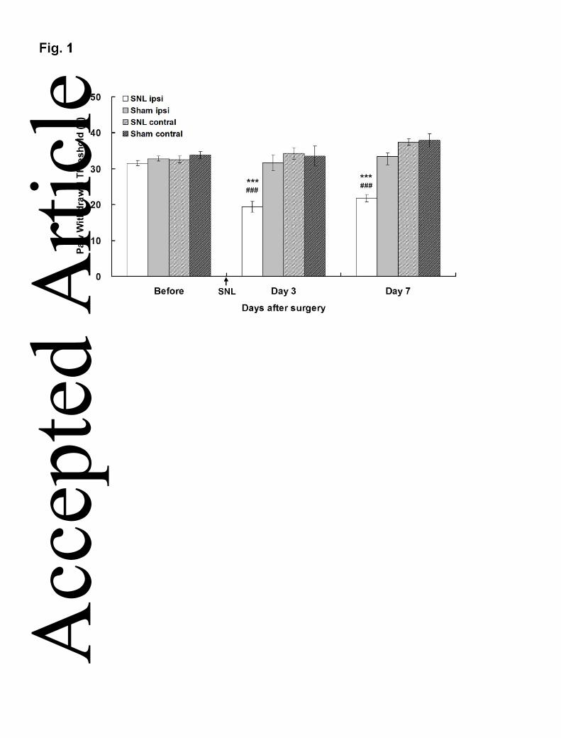
Baseline measurement of paw withdrawal thresholds (PWTs) on both hind-paws did
not show any significant difference between sham-control and SNL groups prior to Acc
epte
d A
rticl
e

10 This article is protected by copyright. All rights reserved.
the operation. In naïve control, no significant difference was found (data not
shown).After SNL, mechanical hypersensitivity was observed since the PWTs on the
ipsilateral hind-paw significantly decreased at 3 (19.46±1.51g) and 7 (21.77±1.04g)
days after SNL in comparison with those of the sham-control group at corresponding
days and site (31.59±2.28g and 33.38±2.25g, respectively; ***p<0.001) (Fig. 1; Table
1), as well as when compared with the PWTs of the corresponding pre-operation
baseline (31.53±0.57g, ###p<0.001). These results demonstrated that the ipsilateral
hind-paw of SNL rats developed a marked allodynia to mechanical stimulation after
SNL.
Up-regulation of COX-1 and COX-2 in dorsal horn
The immunoreactivities (IR) for COX-1 and COX-2 were examined in the L5 spinal
dorsal horn collected 7 days after SNL (Fig. 2). In the superficial (particularly laminae
I-II) dorsal horn of SNL rats, there was an increase in COX-1-IR (Fig. 2A) and
COX-2-IR (Fig 2B) in the ipsilateral side when compared with that of the
contralateral side. Both COX-1 and COX-2 IR were most prevalent in the superficial
dorsal horn instead of the deeper laminae. The semi-quantitative analysis results
illustrated that both COX-1-IR and COX-2-IR increased significantly in the spinal
dorsal horn ipsilateral to SNL (p<0.01, Student’s paired t test). To confirm that
Fig. 2 near here
Acc
epte
d A
rticl
e
11 This article is protected by copyright. All rights reserved.
antibody labeling was specific, control experiments where the primary antibody was
omitted were carried out. No immunofluoresence was detected when the primary
antibody was absent (Data not shown).
Cellular and subcellular sources of COX-1 and COX-2
To identify the specific cell types that expressed COX-1 and COX-2 after SNL,
double immunostaining was performed for the cells expressing COX-1-IR or
COX-2-IR with cell-specific markers: NeuN for neurons, GFAP for astrocytes and
OX-42 for microglia.
COX-1 immunoreactive cells in the spinal dorsal horn at Day 7 post-SNL were
predominantly co-localized with neuronal marker NeuN (Fig. 3c) and microglia
marker OX-42 (Fig. 3f) but not the astrocyte marker GFAP (Fig. 3g-i). Although the
methods we used did not allow us to quantify the COX-1 source separately from
neurons and microglia, it appears that neurons in particular their nuclei were a
dominant source (Fig. 3a-c) whereas microglia cells were an additional important
source of COX-1 (Fig. 3d-f).
Fig. 3 Fig. 4 Fig. 5 near here
Acc
epte
d A
rticl
e

12 This article is protected by copyright. All rights reserved.
For COX-2, almost all COX-2 immunoreactive cells in the spinal dorsal horn were
co-localized with the neuronal marker NeuN (Fig. 4c) but not with OX-42-positive
(Fig. 4f) nor with GFAP-positive cells (Fig. 4i). In contrast to COX-1 that appeared to
be expressed in both neurons and microglia, COX-2 could only be observed in
neurons.
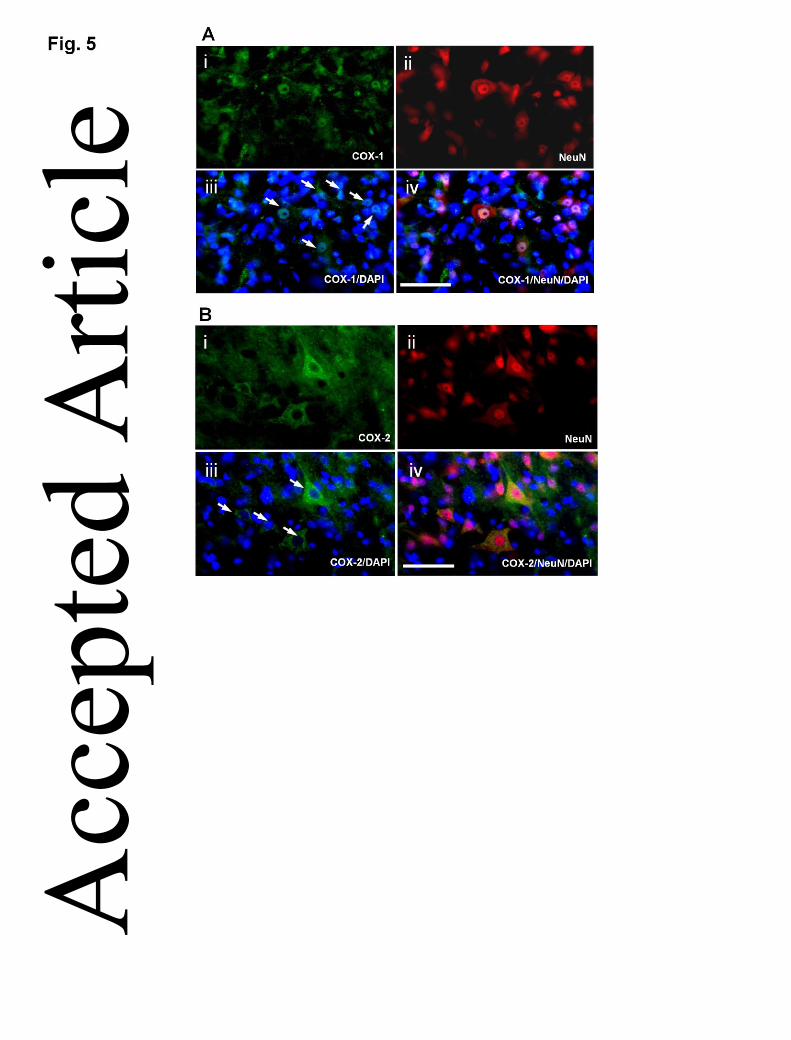
Since the double immunostaining demonstrated that COX-1 was particularly
expressed by neuronal nuclei (Fig. 3a-c), nuclear marker DAPI was then added to
confirm subcellular localization of COX-1. The results revealed that COX-1 was
indeed predominantly expressed in the nucleus of neurons (Fig. 5A), with only trace
amount detectable in the cytoplasm. In contrast, COX-2 was expressed only in the
cytoplasm of neurons as there was no co-localization with DAPI (Fig. 5B).
Discussion
In this study, we examined COX up-regulation and its sources in the spinal dorsal
horn after SNL. Up-regulation of COX-1 and COX-2 was found in the ipsilateral
lumbar superficial dorsal horn 7 days after SNL along with the development of
mechanical allodynia. For the first time, we demonstrated that COX-2 was Acc
epte
d A
rticl
e

13 This article is protected by copyright. All rights reserved.
prominently in neuronal cytoplasm, whereas COX-1 was from neuronal nuclei and
microglia in the superficial dorsal horn.
This study is in line with previous findings in that both COX-1 and COX-2 can be
induced and/or up-regulated in the spinal cord after nerve injuries [8, 11-15]. In
contrast to COX-2 that has been well shown to be involved in the development of
mechanical allodynia [8, 11-15], relatively little is known about the role of COX-1 in
neuropathic pain. Nevertheless, COX-1 must be involved in the pathogenesis of
neuropathic pain, as evidenced by the facts that prolonged COX-1 expression has
been observed in spinal microglia during central sensitization [20], COX-1 deficient
mice show an increased threshold to nociceptive stimuli, and that the COX-1 selective
inhibitor (SC-560) could significantly reverse the development of mechanical
allodynia after spinal nerve injury [16]. A recent study from Kanda demonstrated that
prostaglandin D2 (PGD2), a potent inflammatory mediator, produced by microglia is
COX-1 dependent, and neurons in the spinal cord can receive PGD2 from microglia
following peripheral nerved injury. The intrathecal injection of COX-1 inhibitor
significantly attenuated the mechanical allodynia in nerve injury [21]. The fact that
COX-1, in contrast to COX-2, is found predominantly in neuronal nuclei and in
microglia is perhaps a reflection that COX-1 is more constitutional while COX-2 is Acc
epte
d A
rticl
e

14 This article is protected by copyright. All rights reserved.
more induced, and COX-1 up-regulation in the spinal cord is largely attributable to
microglia activation.
Previously Durrenberger et al. observed an increase in COX-2 expression in human
injured nerve and in rat chronic constriction injury (CCI) model, and later showed the
time course of COX-2 up-regulation [22, 23]. Interestingly, in contrast to the injured
nerve, spinal COX-2 in CCI rats was found to be decreased at all time points in their
latter study. They suggested that the COX-2 up-regulation was due to macrophage
infiltration as there was an increase in the number of microglial-like COX-2
immunoreactive cells in the peripheral nerve and dorsal root ganglia. In this study, we
used the SNL model and demonstrated that spinal dorsal horn neurons are important
source of COX-1 and COX-2 while microglia also contribute to the pathogenesis of
neuropathic pain, partly by producing additional COX-1. In our study, COX-2 was
found to be co-localized predominantly in the spinal dorsal horn neurons after SNL.
Although this finding is not consistent with some previous reports [22, 23], several
previous studies also demonstrated the expression of COX-2 protein in rat spinal
dorsal and ventral horn neurons [24-26], while some other studies also showed the
presence of COX-2 in radial glia [27], astrocytes [24], or endothelia [28]. Though we
also observed trace COX-2 in some of the radial glia in the white matter, its role in
neuropathic pain remains unknown and remains to be further invested. Furthermore, it Acc
epte
d A
rticl
e

15 This article is protected by copyright. All rights reserved.
remains to be investigated whether treatment with COX inhibitors, including specific
and non-selective COX inhibitors, could inhibit spinal nerve ligation-induced
mechanical hypersensitivity.
One of the important findings of this study is COX-1 up-regulation in the spinal
dorsal horn. We found that COX-1 is not only from neurons but also from microglia.
However, due to the limitation of the methods used in this study, we could not
quantify the COX-1 up-regulation from neurons and microglia separately, neither
could we identify whether COX-1 up-regulation was attributable to activation of the
endogenous microglia, newly proliferated cells, or macrophages migrated into the
nervous tissue, since most microglia were clustered together with their processes
tangled (Fig. 3f). However, a previous study [3] by means of more sophisticated
methods has shown that axonal injuries could induce microglia proliferation, and
lesion-reactive microglia accounted for the vast majority of proliferating cells in the
dendate gyrus after damaging projecting axons to the region, and 24% of all
microglial cells were proliferating 3 days post-lesion. As they found that actively
proliferating microglial cells are clustered, and our study also showed that COX-1
expression was particularly strong on clustered OX-42 positive microglial cells (Fig.
3f), it appears that COX-1 up-regulation is chiefly from these proliferating microglia Acc
epte
d A
rticl
e

16 This article is protected by copyright. All rights reserved.
apart from neurons, and microglia may contribute to the pathogenesis by COX-1
expression apart from making other new cytokines and proteins [17].
In this study, we found that the dorsal horn neurons were a major source for both
COX-1 and COX-2. It appears that damaged neurons may play an important role and
interplay with microglia to up-regulate COX in the pathogenesis of neuropathic pain.
The damaged neurons may perhaps work as an initiator by releasing prostagladins,
nitric oxide, substance P, excitatory amino acids etc for other chain reactions
including activation of glial cells in the CNS. A number of studies have shown some
functional changes of spinal dorsal horn neurons in mechanical allodynia caused by
traumatic nerve injury [29-31]. Takeda et al. [32] suggested that the spinal PG surge
following COX-2 up-regulation may directly or indirectly activate spinal glia, hence
contributing to the neuropathic hypersensitivity. After a peripheral nerve injury,
sensitization occurs which is characterized by increased spontaneous activity of
neurons, a lowered threshold and increased response to a given stimulus.
Whether and how the pathophysiological and functional changes are related to COX
up-regulation have been investigated. A previous study revealed that COX-2 may act
as a multifunctional neuronal modulator [33]. In rodents, COX-2 is up-regulated in
spinal cord and brain in response to nerve injury, leading to increased spinal cord
prostaglandin E2 (PGE2) synthesis, and hence to central sensitization, allodynia, and Acc
epte
d A
rticl
e

17 This article is protected by copyright. All rights reserved.
hypersensitivity, whereas COX-2 inhibition prevented the increase in spinal cord
PGE2 concentrations caused by the injury and attenuated the pain response [34-37].
The COX-2 inhibitor, meloxicam, exerts antiallodynic effects on established
neuropathic pain in diabetic mice, which indicates that COX-2 inhibitor can acts as a
prominent drug for treatment of diabetic neuropathic pain [38].
This study showed that microglia were an important source of COX-1 apart from
neurons (Fig. 3i,f). There is increasing evidence supporting a role for microglia in the
pathogenesis in pain and in the maintenance of neuronal homeostasis [17, 39]. In
animal models of mechanical allodynia, activation of microglia was consistently
observed. Spinal microglia contribute to the development and maintenance of
mechanical hypersensitivity after a variety of peripheral nerve injuries, including SNL
[40] and chronic constriction nerve injury [41]. A microglia inhibitor, minocycline,
may reduce mechanical allodynia, possibly by suppressing p38 activation [42].
Recently, the injection of activated microglia into the dorsal horn has been shown to
induce mechanical allodynia, while depletion of activated microglia has been found to
block pain induction [43, 44].
In this study we only selected 7th day after SNL for COX investigation. This was
based on our hypothesis that indigenous glia cells are involved in neuropathic pain by
producing COXs, and our preliminary investigation showed that the activation of glial Acc
epte
d A
rticl
e

18 This article is protected by copyright. All rights reserved.
cells reaches a peak at Day 7 after SNL. This was consistent with some previous
findings. For example, Tanga et al showed the markers of microglial activation
(ITGAM, TLR4, and CD14) increased significantly after nerve transaction, peaked at
4 to 7 days and began to decline afterward [45]. In the study by Zhu et al. (2003), the
number of COX-1-IR cells increased in cells with glial morphology in the superficial
laminae of the ipsilateral spinal cord 4 days after SNL but decreased in the rest of the
ipsilateral spinal cord 4 weeks after PSNT and 2 weeks after SNL. However, different
studies have presented rather confusing results previously. For example, in spared
nerve injury (SNI) model, an increase in COX-2 protein was observed in the dorsal
horn at 24 hours post surgery which then returned to sham levels at 72 hours [46].
Similar findings were also reported by Zhao [14]. In rat chronic constriction injury
(CCI) model, in contrast to the up-regulation of COX-2 in the injured peripheral nerve,
spinal COX-2 was found to be decreased at all time points at days 4, 21, and 30 after
the nerve injury in the rats [22, 23]. In L5 SNL model, COX-2 positive cells were
co-expressed with the Schwann cell marker at Day 1 and followed by co-expression
with macrophage marker 7 days post-surgery [47]. COX-1 protein was found to be
increased 4 days after SNL and decreased 2 weeks after SNL [15]. In contrast, COX-1
protein was not detectable on 1, 3 and 14 days after L5/L6 ligation [14]. Acc
epte
d A
rticl
e

19 This article is protected by copyright. All rights reserved.
It was puzzling that in the study by Durrenberger et al (2006) that, in contrast to their
earlier report [22], COX-2 was down-regulated at all time points 4, 21 and 30 days
after constrictive nerve injury in the rat, but our previous studies showed that at least
COX-2 was increased and still significantly up-regulated 38 days after SNL [8]. It still
remains to be studied how long this COX-2 (and COX-1) up-regulation may last and
whether it is associated with neuropathic pain development and maintenance in long
term. In our opinion, dynamic COX changes in the spinal cord could be due to
recruitment of macrophages or fluctuation of a cocktail of inflammatory mediators
that can be influenced by many factors.
Experimental data on the cellular localization of COX-1 and COX-2 remain
controversial. Early studies indicated, in different tissue types though, that in 3T3
(fibroblast) cells, both COX-1 and COX-2 localized in the nuclear envelope and
endoplasmic reticulum [48]. In vascular endothelial cell, both COX-1 and COX-2
have nuclear and cytoplasmic localization, and COX-2 travels between the nucleus
and the cytoplasm upon IL-1β stimulation [49]. This study revealed, for the first time
in the spinal cord, that COX-1 up-regulation in the spinal dorsal horn is chiefly from
neuronal nuclei and microglia, whereas COX-2 up-regulation is from neuronal
cytoplasm. The exact contribution of these two isoforms to neuropathic pain remains
unclear, but the distinct distribution pattern of the two suggests that COX-1 and Acc
epte
d A
rticl
e
20 This article is protected by copyright. All rights reserved.
COX-2 may play somewhat different roles in the pathogenesis of neuropathic pain.
Despite COX-1 and COX-2 share similar structure and catalytic properties, they use
different pools of arachidonic acid [50] and have distinct function in cell proliferation
and differentiation [51-53]. Researchers have proposed that COX-1 and COX-2 may
acquire arachidonic acid from different phospholipases [54, 55]. It has also been
shown that COX-1 and COX-2 exhibit differences in their subcellular localization and
ability to metabolize arachidonic acid by working in independent prostanoid
biosynthetic systems [56], and each COX enzyme has been reported to have a distinct
subcellular localization and a functional coupling to constitutive and inducible
membrane-associated prostaglandin E2 synthase enzymes, the enzymes responsible
for the final conversion of PGH2 to PGE2 [57, 58]. Thus, it appears that COX-1 and
COX-2 may play somewhat different roles in pain mechanisms at different locations.
Nevertheless, COX-1 in neuropathic pain is relatively less investigated, perhaps partly
due to its constitutive nature in other body tissues, such as the gastrointestinal tract,
and inhibition of it could be associated with severe side effects. It is noted that in Fig
5 that COX-1 is present in the cytoplasm of some but not all of the neurons. This was
perhaps due to existence of different subgroups of neurons and COX-1 up-regulation
may be related to receptors in different subpopulations of neurons. In this connection,
Chopra et al. [59] reported that COX-1 is a marker for a subpopulation of putative Acc
epte
d A
rticl
e

21 This article is protected by copyright. All rights reserved.
nociceptive neurons in rat dorsal root ganglia. In this regard, it is perhaps worth
investigating further in future studies what types of neurons (e.g. small vs big) at what
locations (e.g. superficial vs deep laminae) would have the potential for what subtypes
of COX up-regulation, and what receptors are involved, respectively.
In summary, our findings demonstrated that there was an up-regulation of COX-1 and
COX-2 expression in the spinal cord of rats along with the development mechanical
allodynia after SNL. The SNL induced up-regulation of spinal COX-1 and COX-2
proteins was co-localized in different cell types. COX-2 was predominantly expressed
in the cytoplasm of dorsal horn neurons, whereas COX-1 up-regulation appeared
chiefly from the nuclei of dorsal horn neurons but also from microglia that appeared
to be activated and proliferating after the nerve injury.
Competing interests
We declare that there are no competing interests with other people or organizations
Acknowledgement: Supported by Hong Kong Baptist University FRG05-06/II-55 &
FRG/04-05/I-16, and SCM postdoctoral fellowship. We thank Nickie Chan for her
invaluable technical support. Authors’ contributions: YM, HQ and WK contributed
to the conception and design of the study. SW as an honors project student Acc
epte
d A
rticl
e
22 This article is protected by copyright. All rights reserved.
participated the behavioral and immunohistochemical experiments. YM conducted
most parts of the experiment and SC participated the image acquisition and statistical
analysis. YM, AL and HQ interpreted the data and wrote the manuscript. All authors
read and approved the final manuscript.
References
1 Zhuo M. Neuronal mechanism for neuropathic pain. Mol Pain 2007; 3: 14 2 Colburn RW, Rickman AJ, DeLeo JA. The effect of site and type of nerve injury on spinal glial activation and neuropathic pain behavior. Exp Neurol 1999; 157: 289-304 3 Dissing-Olesen L, Ladeby R, Nielsen HH, Toft-Hansen H, Dalmau I, Finsen B. Axonal lesion-induced microglial proliferation and microglial cluster formation in the mouse. Neuroscience 2007; 149: 112-22 4 Tsuda M, Beggs S, Salter MW, Inoue K. Microglia and intractable chronic pain. Glia 2013; 61: 55-61 5 Smith WL, DeWitt DL, Garavito RM. Cyclooxygenases: structural, cellular, and molecular biology. Annu Rev Biochem 2000; 69: 145-82 6 Kawabata A. Prostaglandin E2 and pain--an update. Biological & pharmaceutical bulletin 2011; 34: 1170-3 7 Ma W, Du W, Eisenach JC. Role for both spinal cord COX-1 and COX-2 in maintenance of mechanical hypersensitivity following peripheral nerve injury. Brain Res 2002; 937: 94-9 8 Lau WK, Chan WK, Zhang JL, Yung KK, Zhang HQ. Electroacupuncture inhibits cyclooxygenase-2 up-regulation in rat spinal cord after spinal nerve ligation. Neuroscience 2008; 155: 463-8 9 Pertusi RM. Selective cyclooxygenase inhibition in pain management. J Am Osteopath Assoc 2004; 104: S19-24 10 Telleria-Diaz A, Schmidt M, Kreusch S, Neubert AK, Schache F, Vazquez E, Vanegas H, Schaible HG, Ebersberger A. Spinal antinociceptive effects of cyclooxygenase inhibition during inflammation: Involvement of prostaglandins and endocannabinoids. Pain 2010; 148: 26-35 Acc
epte
d A
rticl
e

23 This article is protected by copyright. All rights reserved.
11 Lau WK, Lau YM, Zhang HQ, Wong SC, Bian ZX. Electroacupuncture versus celecoxib for neuropathic pain in rat SNL model. Neuroscience 2010; 170: 655-61 12 O'Neill GP, Ford-Hutchinson AW. Expression of mRNA for cyclooxygenase-1 and cyclooxygenase-2 in human tissues. FEBS Lett 1993; 330: 156-60 13 Siegle I, Klein T, Backman JT, Saal JG, Nusing RM, Fritz P. Expression of cyclooxygenase 1 and cyclooxygenase 2 in human synovial tissue: differential elevation of cyclooxygenase 2 in inflammatory joint diseases. Arthritis Rheum 1998; 41: 122-9 14 Zhao Z, Chen SR, Eisenach JC, Busija DW, Pan HL. Spinal cyclooxygenase-2 is involved in development of allodynia after nerve injury in rats. Neuroscience 2000; 97: 743-8 15 Zhu X, Eisenach JC. Cyclooxygenase-1 in the spinal cord is altered after peripheral nerve injury. Anesthesiology 2003; 99: 1175-9 16 Hefferan MP, O'Rielly DD, Loomis CW. Inhibition of spinal prostaglandin synthesis early after L5/L6 nerve ligation prevents the development of prostaglandin-dependent and prostaglandin-independent allodynia in the rat. Anesthesiology 2003; 99: 1180-8 17 Tsuda M, Inoue K, Salter MW. Neuropathic pain and spinal microglia: a big problem from molecules in "small" glia. Trends Neurosci 2005; 28: 101-7 18 Kim SH, Chung JM. An experimental model for peripheral neuropathy produced by segmental spinal nerve ligation in the rat. Pain 1992; 50: 355-63 19 Lau WK, Lau YM, Zhang HQ, Wong SC, Bian ZX. Electroacupuncture versus celecoxib for neuropathic pain in rat SNL model. Neuroscience 2010; 170: 655-61 20 Zhang FY, Wan Y, Zhang ZK, Light AR, Fu KY. Peripheral formalin injection induces long-lasting increases in cyclooxygenase 1 expression by microglia in the spinal cord. J Pain 2007; 8: 110-7 21 Kanda H, Kobayashi K, Yamanaka H, Noguchi K. COX-1-dependent prostaglandin D2 in microglia contributes to neuropathic pain via DP2 receptor in spinal neurons. Glia 2013; 61: 943-56 22 Durrenberger PF, Facer P, Gray RA, Chessell IP, Naylor A, Bountra C, Banati RB, Birch R, Anand P. Cyclooxygenase-2 (Cox-2) in injured human nerve and a rat model of nerve injury. J Peripher Nerv Syst 2004; 9: 15-25 23 Durrenberger PF, Facer P, Casula MA, Yiangou Y, Gray RA, Chessell IP, Day NC, Collins SD, Bingham S, Wilson AW, Elliot D, Birch R, Anand P. Prostanoid receptor EP1 and Cox-2 in injured human nerves and a rat model of nerve injury: a time-course study. BMC Neurol 2006; 6: 1 Acc
epte
d A
rticl
e

24 This article is protected by copyright. All rights reserved.
24 Beiche F, Klein T, Nusing R, Neuhuber W, Goppelt-Struebe M. Localization of cyclooxygenase-2 and prostaglandin E2 receptor EP3 in the rat lumbar spinal cord. J Neuroimmunol 1998; 89: 26-34 25 Samad TA, Moore KA, Sapirstein A, Billet S, Allchorne A, Poole S, Bonventre JV, Woolf CJ. Interleukin-1beta-mediated induction of Cox-2 in the CNS contributes to inflammatory pain hypersensitivity. Nature 2001; 410: 471-5 26 Willingale HL, Gardiner NJ, McLymont N, Giblett S, Grubb BD. Prostanoids synthesized by cyclo-oxygenase isoforms in rat spinal cord and their contribution to the development of neuronal hyperexcitability. British journal of pharmacology 1997; 122: 1593-604 27 Ghilardi JR, Svensson CI, Rogers SD, Yaksh TL, Mantyh PW. Constitutive spinal cyclooxygenase-2 participates in the initiation of tissue injury-induced hyperalgesia. J Neurosci 2004; 24: 2727-32 28 Resnick DK, Graham SH, Dixon CE, Marion DW. Role of cyclooxygenase 2 in acute spinal cord injury. Journal of neurotrauma 1998; 15: 1005-13 29 Chapman V, Suzuki R, Dickenson AH. Electrophysiological characterization of spinal neuronal response properties in anaesthetized rats after ligation of spinal nerves L5-L6. J Physiol 1998; 507 ( Pt 3): 881-94 30 Laird JM, Bennett GJ. An electrophysiological study of dorsal horn neurons in the spinal cord of rats with an experimental peripheral neuropathy. J Neurophysiol 1993; 69: 2072-85 31 Takaishi K, Eisele JH, Jr., Carstens E. Behavioral and electrophysiological assessment of hyperalgesia and changes in dorsal horn responses following partial sciatic nerve ligation in rats. Pain 1996; 66: 297-306 32 Takeda K, Sawamura S, Tamai H, Sekiyama H, Hanaoka K. Role for cyclooxygenase 2 in the development and maintenance of neuropathic pain and spinal glial activation. Anesthesiology 2005; 103: 837-44 33 Bazan NG. COX-2 as a multifunctional neuronal modulator. Nat Med 2001; 7: 414-5 34 Yaksh TL, Dirig DM, Conway CM, Svensson C, Luo ZD, Isakson PC. The acute antihyperalgesic action of nonsteroidal, anti-inflammatory drugs and release of spinal prostaglandin E2 is mediated by the inhibition of constitutive spinal cyclooxygenase-2 (COX-2) but not COX-1. J Neurosci 2001; 21: 5847-53 35 Samad TA, Sapirstein A, Woolf CJ. Prostanoids and pain: unraveling mechanisms and revealing therapeutic targets. Trends Mol Med 2002; 8: 390-6 36 Svensson CI, Yaksh TL. The spinal phospholipase-cyclooxygenase-prostanoid cascade in nociceptive processing. Annu Rev Pharmacol Toxicol 2002; 42: 553-83 Acc
epte
d A
rticl
e

25 This article is protected by copyright. All rights reserved.
37 Staniaszek LE, Norris LM, Kendall DA, Barrett DA, Chapman V. Effects of COX-2 inhibition on spinal nociception: the role of endocannabinoids. British journal of pharmacology 2010; 160: 669-76 38 Kimura S, Kontani H. Demonstration of antiallodynic effects of the cyclooxygenase-2 inhibitor meloxicam on established diabetic neuropathic pain in mice. Journal of pharmacological sciences 2009; 110: 213-7 39 Mika J, Zychowska M, Popiolek-Barczyk K, Rojewska E, Przewlocka B. Importance of glial activation in neuropathic pain. European journal of pharmacology 2013 Mar 13: 40 Jin SX, Zhuang ZY, Woolf CJ, Ji RR. p38 mitogen-activated protein kinase is activated after a spinal nerve ligation in spinal cord microglia and dorsal root ganglion neurons and contributes to the generation of neuropathic pain. J Neurosci 2003; 23: 4017-22 41 Stuesse SL, Cruce WL, Lovell JA, McBurney DL, Crisp T. Microglial proliferation in the spinal cord of aged rats with a sciatic nerve injury. Neurosci Lett 2000; 287: 121-4 42 Hua XY, Svensson CI, Matsui T, Fitzsimmons B, Yaksh TL, Webb M. Intrathecal minocycline attenuates peripheral inflammation-induced hyperalgesia by inhibiting p38 MAPK in spinal microglia. Eur J Neurosci 2005; 22: 2431-40 43 Hains BC, Waxman SG. Activated microglia contribute to the maintenance of chronic pain after spinal cord injury. J Neurosci 2006; 26: 4308-17 44 Tsuda M, Shigemoto-Mogami Y, Koizumi S, Mizokoshi A, Kohsaka S, Salter MW, Inoue K. P2X4 receptors induced in spinal microglia gate tactile allodynia after nerve injury. Nature 2003; 424: 778-83 45 Tanga FY, Raghavendra V, DeLeo JA. Quantitative real-time RT-PCR assessment of spinal microglial and astrocytic activation markers in a rat model of neuropathic pain. Neurochem Int 2004; 45: 397-407 46 Broom DC, Samad TA, Kohno T, Tegeder I, Geisslinger G, Woolf CJ. Cyclooxygenase 2 expression in the spared nerve injury model of neuropathic pain. Neuroscience 2004; 124: 891-900 47 Takahashi M, Kawaguchi M, Shimada K, Konishi N, Furuya H, Nakashima T. Cyclooxygenase-2 expression in Schwann cells and macrophages in the sciatic nerve after single spinal nerve injury in rats. Neuroscience letters 2004; 363: 203-6 48 Reiger MK, DeWitt DL, Schindler MS, Smith WL. Subcellular localization of prostaglandin endoperoxide synthase-2 in murine 3T3 cells. Arch Biochem Biophys 1993; 301: 439-44 Acc
epte
d A
rticl
e

26 This article is protected by copyright. All rights reserved.
49 Parfenova H, Parfenov VN, Shlopov BV, Levine V, Falkos S, Pourcyrous M, Leffler CW. Dynamics of nuclear localization sites for COX-2 in vascular endothelial cells. Am J Physiol Cell Physiol 2001; 281: C166-78 50 Reddy ST, Herschman HR. Prostaglandin synthase-1 and prostaglandin synthase-2 are coupled to distinct phospholipases for the generation of prostaglandin D2 in activated mast cells. J Biol Chem 1997; 272: 3231-7 51 Smith WL, Garavito RM, DeWitt DL. Prostaglandin endoperoxide H synthases (cyclooxygenases)-1 and -2. J Biol Chem 1996; 271: 33157-60 52 O'Banion MK. Cyclooxygenase-2: molecular biology, pharmacology, and neurobiology. Crit Rev Neurobiol 1999; 13: 45-82 53 Hla T, Ristimaki A, Appleby S, Barriocanal JG. Cyclooxygenase gene expression in inflammation and angiogenesis. Ann N Y Acad Sci 1993; 696: 197-204 54 Murakami M, Matsumoto R, Austen KF, Arm JP. Prostaglandin endoperoxide synthase-1 and -2 couple to different transmembrane stimuli to generate prostaglandin D2 in mouse bone marrow-derived mast cells. J Biol Chem 1994; 269: 22269-75 55 Reddy ST, Herschman HR. Ligand-induced prostaglandin synthesis requires expression of the TIS10/PGS-2 prostaglandin synthase gene in murine fibroblasts and macrophages. J Biol Chem 1994; 269: 15473-80 56 Morita I, Schindler M, Regier MK, Otto JC, Hori T, DeWitt DL, Smith WL. Different intracellular locations for prostaglandin endoperoxide H synthase-1 and -2. J Biol Chem 1995; 270: 10902-8 57 Murakami M, Naraba H, Tanioka T, Semmyo N, Nakatani Y, Kojima F, Ikeda T, Fueki M, Ueno A, Oh S, Kudo I. Regulation of prostaglandin E2 biosynthesis by inducible membrane-associated prostaglandin E2 synthase that acts in concert with cyclooxygenase-2. J Biol Chem 2000; 275: 32783-92 58 Tanioka T, Nakatani Y, Semmyo N, Murakami M, Kudo I. Molecular identification of cytosolic prostaglandin E2 synthase that is functionally coupled with cyclooxygenase-1 in immediate prostaglandin E2 biosynthesis. J Biol Chem 2000; 275: 32775-82 59 Chopra B, Giblett S, Little JG, Donaldson LF, Tate S, Evans RJ, Grubb BD. Cyclooxygenase-1 is a marker for a subpopulation of putative nociceptive neurons in rat dorsal root ganglia. Eur J Neurosci 2000; 12: 911-20
Acc
epte
d A
rticl
e
27 This article is protected by copyright. All rights reserved.
Figure legends
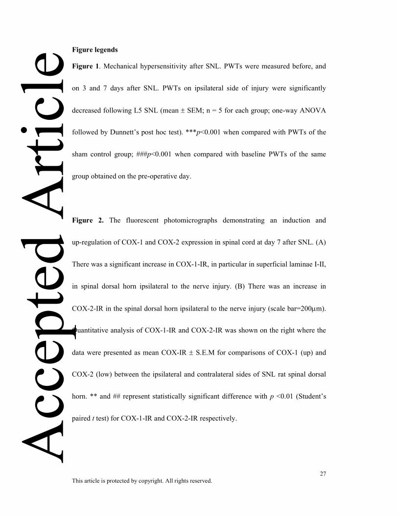
Figure 1. Mechanical hypersensitivity after SNL. PWTs were measured before, and
on 3 and 7 days after SNL. PWTs on ipsilateral side of injury were significantly
decreased following L5 SNL (mean ± SEM; n = 5 for each group; one-way ANOVA
followed by Dunnett’s post hoc test). ***p<0.001 when compared with PWTs of the
sham control group; ###p<0.001 when compared with baseline PWTs of the same
group obtained on the pre-operative day.
Figure 2. The fluorescent photomicrographs demonstrating an induction and
up-regulation of COX-1 and COX-2 expression in spinal cord at day 7 after SNL. (A)
There was a significant increase in COX-1-IR, in particular in superficial laminae I-II,
in spinal dorsal horn ipsilateral to the nerve injury. (B) There was an increase in
COX-2-IR in the spinal dorsal horn ipsilateral to the nerve injury (scale bar=200µm).
Quantitative analysis of COX-1-IR and COX-2-IR was shown on the right where the
data were presented as mean COX-IR ± S.E.M for comparisons of COX-1 (up) and
COX-2 (low) between the ipsilateral and contralateral sides of SNL rat spinal dorsal
horn. ** and ## represent statistically significant difference with p <0.01 (Student’s
paired t test) for COX-1-IR and COX-2-IR respectively.
Acc
epte
d A
rticl
e

28 This article is protected by copyright. All rights reserved.
Figure 3. Co-labeling to identify the cellular sources of COX-1 in the spinal dorsal
horn of SNL rats. Double immunofluorescence for COX-1 (a, d, g, green) and NeuN,
a neuronal marker (b, red), OX-42, a microglia marker (e, red) and GFAP, an
astrocyte marker (h, red). Double immunofluorescence indicated COX-1 positive cells
co-localized with NeuN and OX-42 (arrows) in the spinal dorsal horn 7 days after
SNL (magnification x400; scale bar=50µm applies to all).
Figure 4. Double immunofluorescence showing COX-2 (a, d, g, green) co-localized
with NeuN (b-c, red) but not with OX-42 (e-f, red) or GFAP (h-i, red), in the spinal
dorsal horn 7 days after SNL. Arrows in (c) point to COX-2/NeuN co-localized
neurons (magnification x400; scale bar=50µm applies to all).
Figure 5. Nuclear staining with DAPI to identify the subcellular sources of COX-1
and COX-2 in the neurons of spinal dorsal horn. (A) COX-1 (i, green) was localized
predominantly within the nuclei (iii, iv, blue) of NeuN-positive cells (ii, red). The
arrows indicated the location of COX-1-positive nuclei in neurons. (B) COX-2 (i,
green) was absent in the nucleus (iii, iv, blue), but localized mainly in the cytoplasm
of neuron (ii, iv, red). The arrows indicated the location of COX-2 in the cytoplasm of Acc
epte
d A
rticl
e
29 This article is protected by copyright. All rights reserved.
neurons. Nuclei were counterstained with DAPI (blue) (magnification x600; scale
bar=50µm applies to all).
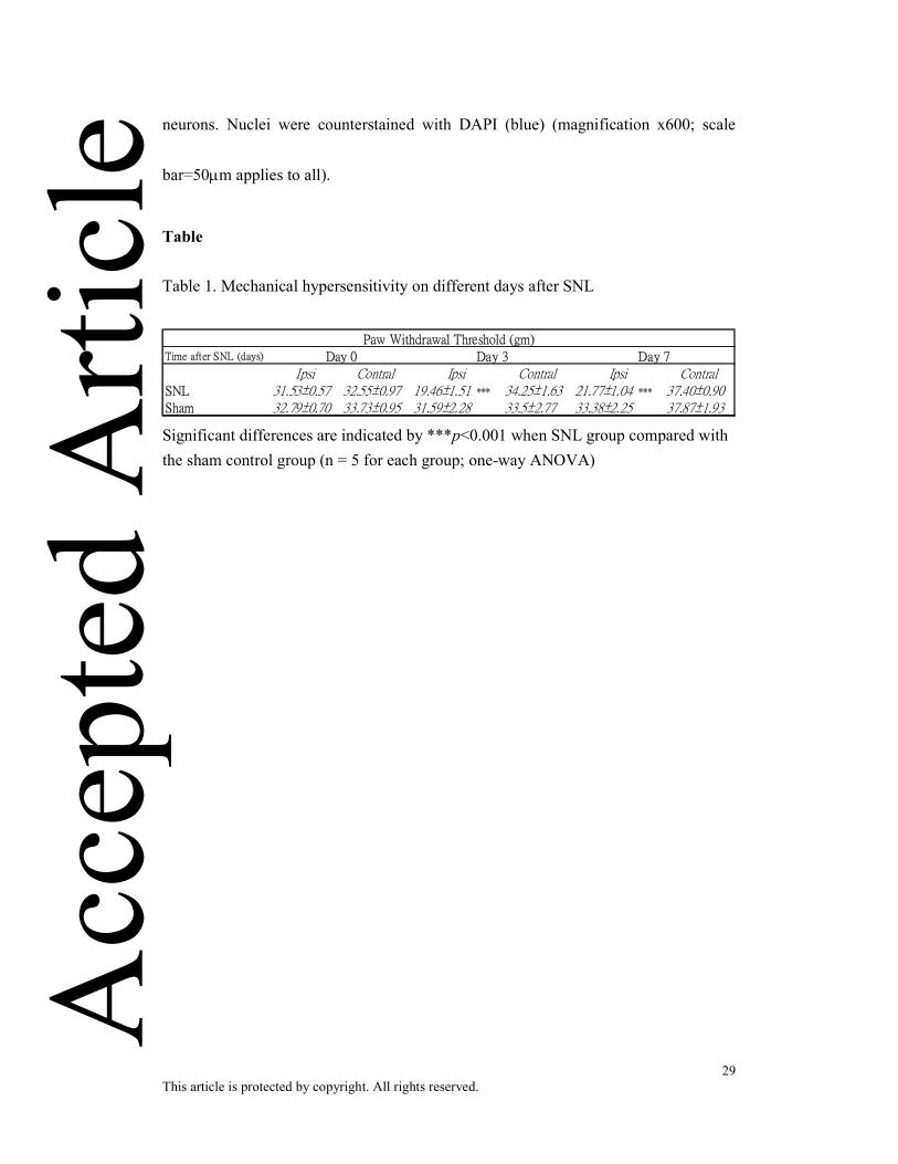
Table Table 1. Mechanical hypersensitivity on different days after SNL
Time after SNL (days)
Ipsi Contral Ipsi Contral Ipsi ContralSNL 31.53±0.57 32.55±0.97 19.46±1.51 *** 34.25±1.63 21.77±1.04 *** 37.40±0.90Sham 32.79±0.70 33.73±0.95 31.59±2.28 33.5±2.77 33.38±2.25 37.87±1.93
Paw Withdrawal Threshold (gm)
Day 0 Day 3 Day 7
Significant differences are indicated by ***p<0.001 when SNL group compared with the sham control group (n = 5 for each group; one-way ANOVA)
Acc
epte
d A
rticl
e
Acc
epte
d A
rticl
e

Acc
epte
d A
rticl
e

Acc
epte
d A
rticl
e

Acc
epte
d A
rticl
e
Acc
epte
d A
rticl
e