cellular oxidation of low density lipoprotein is caused by thiol production in media containing
TRANSCRIPT

Cellular oxidation of low density lipoprotein is caused by thiol production in media containing transition metal ions
Carl P. Sparrow’ and Joanne Olszewski
Department of Atherosclerosis Research, Merck Research Laboratories, P.O. Box 2000, 126 E. Lincoln Avenue, Rahway, NJ 07065
Abstract The oxidation of low density lipoprotein (LDL) may be important in atherosclerosis. LDL can be oxidized by cul- tured cells, including macrophages and endothelial cells. This cellular oxidation is dependent on transition metal ions in the medium. We now report that LDL oxidation by endothelial cells and macrophages is caused by cell-dependent appearance of thiol in the medium (“thiol production”). Thiol appeared in medium when cells were incubated under standard serum-free culture conditions. L-Cystine in the medium was required for thiol production and also for LDL oxidation. Cell-dependent appearance of thiol was inhibited by glutamate (which blocks cystine uptake) and by diethylmaleate (which reacts with thiols). Both compounds also blocked cellular LDL oxidation, even though neither compound had antioxidant activity. Finally, we designed an enzymatic system, based on glutathione reductase, that mimicked cellular thiol production. This enzymatic system caused LDL oxidation, and showed the same dependency for transition metal ions as did cellular LDL oxidation. We conclude that in media containing transition metal ions, cellular oxidation of LDL can be explained by the cell-dependent ap- pearance of thiol in the medium. A very similar mechanism was proposed in 1987 by Heinecke et al. u Bid. C h . 262: 10098-10103). Under other conditions, however, cellular oxida- tion of LDL may occur by other mechanisms.- Sparrow, C. P., and J. Olszewski. Cellular oxidation of low density lipoprotein is caused by thiol production in media containing transition me- tal ions. J. Lipid Res. 1993. 34: 1219-1228.
Supplementary key words cholesterol endothelial cells macrophage atherosclerosis
oxidized LDL free radicals cysteine
Recent evidence strongly suggests that an important part of the pathogenesis of atherosclerosis is the oxidative modification of low density lipoprotein (LDL) (1-4). Oxi- dized LDL probably participates in the formation of “foam cells,” the cholesterol-loaded macrophage-derived cells of the atherosclerotic lesion (5). Foam cell formation can be modeled in vitro: oxidized LDL is taken up avidly by macrophages via scavenger receptors (6), leading to cholesterol accumulation (1). Native LDL, however, is not recognized by scavenger receptors and thus does not cause
cholesterol accumulation in macrophages (7). Monocytes, the precursors of macrophages, are attracted towards oxi- dized LDL by chemotaxis (B), and therefore oxidized LDL may recruit monocytes into the subendothelial space. Oxidized LDL has been reported to be cytotoxic (9), which may explain the endothelial damage that oc- curs during atherogenesis (10). Most studies of the effects of oxidized LDL on cultured cells have used LDL sub- jected to strong oxidizing conditions, but even “minimally modified” LDL has potent biological activity (11).
LDL can be oxidatively modified in vitro by copper ions and by various cultured cells (reviewed in ref. 1). To the extent that LDL oxidation occurs in vivo, it is believed to occur within arterial tissue (l), and this oxidation is presumably catalyzed by the cells of the artery. Because of the pathophysiological importance of oxidized LDL, it would be valuable to fully understand the mechanism by which cells oxidize LDL.
LDL oxidation by most cultured cells requires a medium containing transition metal ions (12-14). The medium most commonly used to support cellular LDL oxidation is F-10 (14-18). GIBCO F-10 is formulated with 9 nM CuSO, and 3 p M FeSO,; this level of transition me- tal ions causes only minimal oxidation of LDL in the ab- sence of cells (14). Proposed mechanisms for cellular LDL oxidation include superoxide release (19, 20) and 15-lipoxygenase activity (15, 21, 22). Recent evidence sug- gests, however, that lipoxygenases are not required for cel- lular oxidation of LDL (16).
Two observations suggest a role for sulfur-containing molecules in cellular oxidation of LDL in medium con-
Abbreviations: DEM, diethihaleate; DTNB, 5,5’-dithio-bir(2-nitro- benzoic acid); LDL, low density lipoprotein; TBARS, thiobarbituric acid-reactive substances; TNB, 5-thio-2-nitrobenzoic acid.
‘To whom correspondence should be addressed.
Journal of Lipid Rese* Volume 34, 1993 1219
by guest, on April 10, 2019
ww
w.jlr.org
Dow
nloaded from
taining transition metal ions. Parthasarathy (23) showed that the addition of millimolar concentrations of thiols to F-10 caused LDL oxidation. Heinecke et al. (12) showed that oxidative modification of LDL by smooth muscle cells was dependent on cystine in the medium, and they proposed that cellular LDL oxidation was dependent on cellular production of thiol. In the present work, we have tested whether thiol metabolism is important in cellular oxidation of LDL in media containing transition metal ions.
MATERIALS AND METHODS
Materials
Tissue culture supplies were from GIBCO (Grand Is- land, NY), except fetal bovine serum was from HyClone (Logan, UT). F-10 medium lacking cysteine was obtained from CIBCO by special order. 5,5‘-Dithio-bu(2-nitrobenzoic acid) (DTNB) was from Pierce Chemical Co., Rockford, IL. Yeast glutathione reductase was obtained from Boehrifiger Mannheim as a suspension in 3.2 M ammo- nium kulfate at 600 unit/ml. Yeast glucose-6-phosphate dehydrogenase was obtained from Sigma (Type XI) as a suspension in 2.6 M ammonium sulfate at 306 unit/ml. For both enzyrries, 1 unit converts 1 pmol per min at 25OC under optimum conditions.
Cells and cell culture The cell line RECB4, derived from rabbit aortic en-
dothelial cells (24), was obtained from D. Steinberg (University of California, San Diego), and grown as described (6). RECB4 endothelial cells were seeded in 6-well plates and allowed to reach confluency prior to use. Resident mouse peritoneal macrophages were obtained from female Swiss-Webster mice by peritoneal lavage with phosphate-buffered saline. Macrophages were plated in 6-well cell culture plates at 3 x 10⁶ cells/well (for modification) or in 24-well plates at 0.6 x 10⁶ cells/well (for uptake assays) in DMEM containing 10% fetal bo- vine serum. Macrophages were used 16-20 h after plating.
Lipoprotein Plasma was obtained from fasted normal volunteers,
and LDL was isolated by standard procedures (25). LDL was dialyzed against phosphate-buffered saline containing 270 pM EDTA, then sterile-filtered and stored at 4OC. Concentrations of LDL are expressed on the basis of pro- tein, measured using the micro BCA (bicinchoninic acid) protein reagent (Pierce Chemical Co., Rockford, IL), with bovine serum albumin as the standard. LDL was io- dinated with l*5I using the “trapped label” tyramine cel- lobiose (26). No significant differences were found in preliminary experiments comparing the oxidative modification of LDL labeled with 1251-tyramine cellobiose and LDL labeled conventionally (6) (data not shown).
Assays of oxidative modification of LDL Cells were washed three times with serum-free F-10
medium and then incubated with serum-free F-10 medium containing 100 pg/ml LDL with or without other additions. Some experiments used LDL radioiodinated with tyramine cellobiose (20-50 cpm/ng). Small amounts of EDTA that were present in the LDL solution carried over into the incubations (< 2 pM); this level of EDTA had no effect on the results, as judged by experiments in which LDL was dialyzed against F-10 prior to incubation with cells (data not shown). Controls for LDL oxidation assays were performed by incubating LDL in F-10 in the absence of cells, or in the presence of 10 pM CuS04. In- cubations, usually in duplicate, had a total volume of 1.25 ml, and were performed in 35-mm wells (6-well plates) at 37°C in 93% air/7% COP. After 24 h, media were har- vested and adjusted to 20 pM butylated hydroxytoluene. Oxidative modification of LDL was assayed by measure- ment of thiobarbituric acid-reactive substances (TBARS) (27) and/or by macrophage uptake. Uptake was measured by incubating macrophages in DMEM containing 1 mg of protein per ml of lipoprotein-deficient serum and 10 pglml of radiolabeled LDL for 5 h, then washing the cells 3 times with PBS. The washed cells were dissolved in 0.2 N NaOH, and cell-associated lZ5I was quantitated using a gamma counter. Under our assay conditions, the uptake of oxidized radiolabeled LDL was inhibited about 90% by 10 pg/ml polyinosinic acid, indicating that the uptake was via scavenger receptors (6, 7). Macrophage uptake of un- oxidized radiolabeled LDL was unaffected or slightly stimulated by polyinosinic acid.
The amount of TBARS produced by incubating LDL without cells was variable, but was always significantly less than incubations in the presence of cells. We calcu- lated ‘‘% of control” TBARS values as follows. If X equals the TBARS generated by LDL incubated with cells, and Y equals the TBARS generated by the cell-free incuba- tion, then the ‘‘% of control” TBARS for experimental condition Z equals:
(100) ([TBARS from Z] - Y)/(X - Y).
Other assays Thiol concentration was measured using the thiol-
specific reagent 5,5‘-dithio-bis(2-nitrobenzoic acid) (DTNB) (28). Medium was removed from cells and 900 pl was mixed with 100 p1 of 1 mM DTNB dissolved in 200 mM Napi (pH 8), and then absorbance was measured at 412 nm. The time from media harvest to performing the thiol assay was never more than 10 min, to prevent loss of thiol to autoxidation. Medium and unreacted DTNB each have a small absorbance at 412 nm; these were subtracted from experimental media samples. Standard curves were produced using freshly prepared solutions of cysteine.
1220 Journal of Lipid Research Volume 34, 1993
by guest, on April 10, 2019
ww
w.jlr.org
Dow
nloaded from
15-Lipoxygenase activity was measured in intact macro- phages as previously described (16). Cytotoxicity was as- sessed by measuring the uptake of ~-[4,5-3H]leucine. Cells were incubated with [3H]leucine (1 pCi/ml in F-10 medium) with or without test compounds. After 24 h, the medium was harvested and the cells were washed three times with saline. The cells were dissolved in 0.2 M NaOH, and radioactivity in the dissolved cells was meas- ured. Under these conditions, 10 pM cycloheximide in- hibited [3H]leucine uptake by at least 90% (data not shown).
100
cn K K a .- .- E 2 10
f
- 0 E I-
RESULTS
Characterization of F-10 medium and the medium components necessary for LDL oxidation
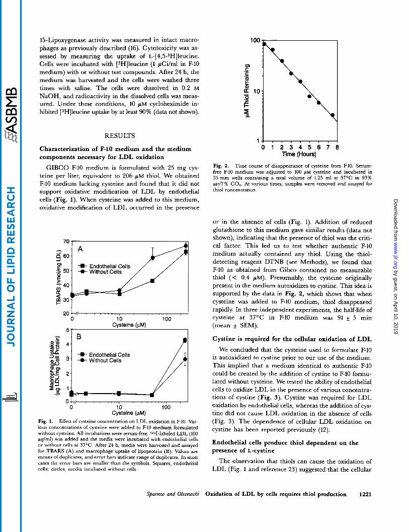
GIBCO F-10 medium is formulated with 25 mg cys- teine per liter, equivalent to 206 pM thiol. We obtained F-10 medium lacking cysteine and found that it did not support oxidative modification of LDL by endothelial cells (Fig. 1). When cysteine was added to this medium, oxidative modification of LDL occurred in the presence
1 0 1 2 3 4 5 6 7
Time (Hours)
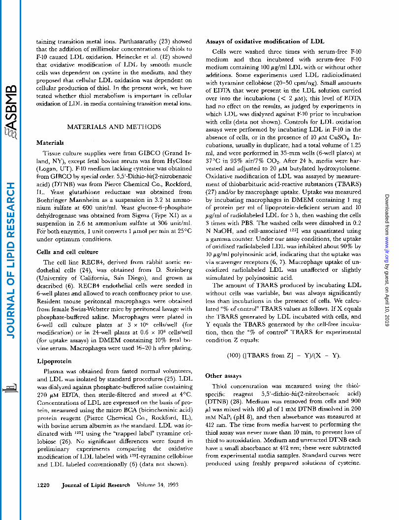
Fig. 2. Time course of disappearance of cysteine from F-10. Serum- free F-10 medium was adjusted to 100 p~ cysteine and incubated in 35" wells containing a total volume of 1.25 ml at 37OC in 93% air/7% C 0 2 . At various times, samples were removed and assayed for thiol concentration.
or in the absence of cells (Fig. 1). Addition of reduced glutathione to this medium gave similar results (data not shown), indicating that the presence of thiol was the criti- cal factor. This led us to test whether authentic F-10 $'':I detecting medium actually reagent contained DTNB (see any Methods), thiol. Using we found the thiol- that
E10 as obtained from Gibco contained no measurable thiol (< 0.4 p ~ ) . Presumably, the cysteine originally present in the medium autoxidizes to cvstine. This idea is -40
50 -0- Without Cells
E
2 0 k l . . , , I 0 10 100
Cysteine (pM) 51 I
4 - 1 -W- Endothelial Cells -0- Without Cells
0 10 100 Cysteine (pM)
supported by the data in Fig. 2, which shows that when cysteine was added to F-10 medium, thiol disappeared rapidly. In three independent experiments, the half-life of cysteine at 37OC in F-10 medium was 91 f 5 min (mean f SEM).
Cystine is required for the cellular oxidation of LDL
We concluded that the cysteine used to formulate E10 is autoxidized to cystine prior to our use of the medium. This implied that a medium identical to authentic F-10 could be created by the addition of cystine to F-10 formu- lated without cysteine. We tested the ability of endothelial cells to oxidize LDL in the presence of various concentra- tions of cystine (Fig. 3). Cystine was required for LDL oxidation by endothelial cells, whereas the addition of cys- tine did not cause LDL oxidation in the absence of cells
Fig. 1. Effect of cysteine concentration on LDL oxidation in F-10. Var- (Fig. 3). The dependence of cellular LDL oxidation on ious concentrations of cysteine were added to F-10 medium formulated without cysteine. All incubations were serum-free. 1251-labeled LDL (100 pg/ml) was added and the media were incubated with endothelial cells or without cells at 37OC. After 24 h, media were harvested and assaved
cystine has been reported previously (12).
Endothelial cells produce thiol dependent on the for TBARS (A) and macrophage uptake of lipoprotein (B). Values are means of duplicates, and error bars indicate range of duplicates. In most cases the error bars are smaller than the symbols. Squares, endothelial cells; circles, media incubated without cells.
presence Of L-cystine
The observation that thiols can cause the oxidation of LDL (Fig. 1 and reference 23) suggested that the cellular
Sparrow and Olszewski Oxidation of LDL by cells requires thiol production 1221
by guest, on April 10, 2019
ww
w.jlr.org
Dow
nloaded from

Endothelial Cells /
Without Cells
- ., 0 10
Cystine (pM) 100
Fig. 3. Cellular oxidation of LDL requires cystine. Various concentra- tions of L-cystine were added to F-10 medium formulated without cys- teine (see text for description of cysteine and cystine content of media). LDL was added and the media were incubated with endothelial cells or without cells at 37OC. All incubations were Serum-free. After 24 h, me- dia were harvested and assayed for TBARS. Values are means of dupli- cates, and error bars indicate range of duplicates. In most cases the error bars are smaller than the symbols. Squares, endothelial cells; circles, me- dia incubated without cells. The extent of cell-dependent LDL oxidation in the absence of cystine was variable, and sometimes was not different from LDL incubated without cells.
mechanism of LDL oxidation might involve cell- dependent appearance of thiol in the medium. Fig. 4 shows that endothelial cells caused the appearance of significant amounts of thiol in the medium. Hereafter we will refer to this cell-dependent appearance of thiol as “thiol production.” Thiol production was dependent on the presence of L-cystine in the medium (Fig. 4). A simi- lar observation has been described previously (29).
Disulfides other than L-cystine did not support cellular thiol production. Endothelial cells were incubated in the presence of various disulfides (150 p ~ ) , and after 2 h thiol in the medium was measured. Levels of thiol produced were (means and range of duplicates, in pM): from L- cystine, 33 k 4; D-cystine, 5 1; L-homocystine, 4 f 1, and from glutathione disulfide 3 k 1. In the same experi- ment, endothelial cells incubated without disulfide produced 3 f 1 p M thiol.
A thiol-producing enzymatic system causes the oxidation of LDL
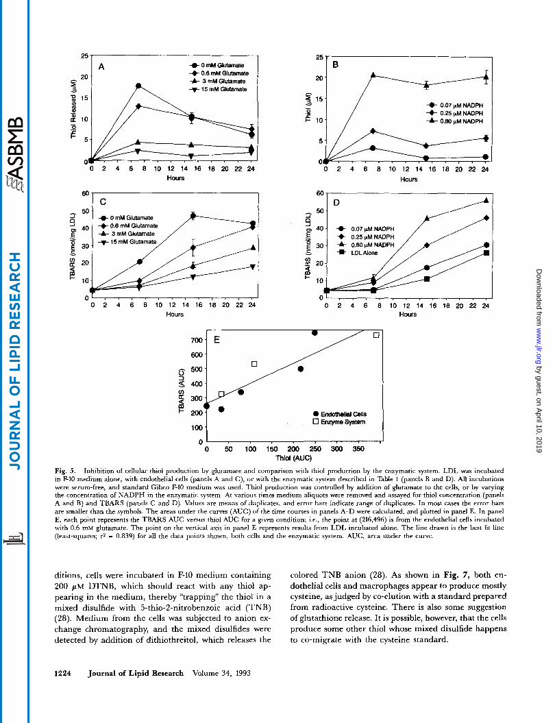
To determine whether thiol production alone could oxi- dize LDL, we developed an enzymatic system that produced thiols. The system used glutathione reductase to reduce glutathione disulfide to the free thiol, with NADPH as reductant. The NADP byproduct was recon- verted to NADPH using glucose-6-phosphate plus glucose- 6-phosphate dehydrogenase. The rate of thiol production was controlled by varying the concentration of NADPH.
Table 1 shows that thiol production by this enzymatic system causes LDL oxidation in F-10 medium. Both TBARS and macrophage uptake were increased (Table l),
and the macrophage uptake of the enzymatically oxidized LDL was inhibited by polyinosinic acid (data not shown), indicating scavenger receptor recognition. The oxidation is dependent on the presence of both substrate and en- zymes, and is responsive to the concentration of NADPH. This shows that the production of thiol in F-10 medium is sufficient to cause LDL oxidation.
Similarities between cellular and enzymatic oxidation of LDL
LDL oxidation by cells requires transition metal ions in the medium (12-14). Table 2 compares the ability of en- dothelial cells and the enzymatic system to oxidize LDL in various media. The dependency on transition metal ions was very similar for cells and for the enzymatic system.
Glutamate blocks cellular thiol production and LDL oxidation
Glutamate blocks cystine uptake and also lowers cystine-dependent thiol production by cells (29). Endothelial cell thiol production and LDL oxidation were inhibited by glutamate in a dose-dependent manner (Fig. 5, panels A and C). In the same experiment, thiol production and LDL oxidation by the enzymatic system were quantitated (Fig. 5 , panels B and D). This allowed a direct compari- son between the cells and the enzymatic system for the efficiency of LDL oxidation at various rates of thiol production. Fig. 5E shows that the relationship between LDL oxidation and thiol production was independent of whether the thiol was produced by the enzymatic system or by the cells. This strongly suggests that thiol produc- tion is the major mechanism of cellular LDL oxidation under these conditions.
25 I
20 4 F-10 Medium
I
0 Y! 5 10 15 20 1
Hours
Fig. 4. Endothelial cells produce thiol only in medium containing cys- tine. Endothelial cells were incubated in complete F-10 medium or F-10 lacking cystine (see text for description of cysteine and cystine content of media). At various times medium aliquots were removed and assayed for thiol. Values are means of duplicates, and error bars indicate range of duplicates. In most cases the error bars are smaller than the symbols. Squares, authentic F-10 medium; circles, F-10 medium formulated without cystine.
1222 Journal of Lipid Research Volume 34, 1993
by guest, on April 10, 2019
ww
w.jlr.org
Dow
nloaded from

TABLE 1 . Enzymatic production of thiol in F-10 causes oxidative modification of LDL
Incubation Conditions TBARS Macrophage Uptake
nmol/mg LDL Fg L D L / m g macrophage protein
Enzyme system 2.0 p~ NADPH 0.67 p~ NADPH 0.23 p~ NADPH 0.07 PM NADPH 2 p~ NADPH; omit enzymes 2 p~ NADPH; omit GSSG
71 * 0 61 i 1 41 f 1 25 f 2 15 i 1 12 f 1
7.2 + 1.6 4.2 f 1.0 1.0 f 0.02 1.0 f 0.01 1.0 f 0.2 1.3 f 0.02
F-10, no additions 13 f 1 0.8 f 0.2 Endothelial cells 58 f 1 6.9 f 0.8 10 pM c u s o , 62 i 3 10.4 i 1.5
1Z51-labeled LDL was incubated in serum-free F-10 at 37OC under various conditions. After 24 h, the oxidative modification of LDL was assessed by measuring TBARS and macrophage uptake. The complete thiol-producing enzyme system included 100 PM glutathione disulfide (GSSG), 400 I(M glucose-6-phosphate, 0.05 U/ml glutathione reductase, 0.05 U/ml glucose-6-phosphate dehydrogenase, and various amounts of NADPH, in F-10 medium. Values given are means and range of duplicates
Two important controls were performed with gluta- mate. First, 15 mM glutamate had no effect on [3H]leu- cine uptake by endothelial cells, which shows that there was no cytotoxicity. Secondly, 15 mM glutamate blocked neither thiol production nor LDL oxidation by the en- zymatic system, which shows that the glutamate did not function as an antioxidant.
Diethylmaleate blocks cellular thiol production and LDL oxidation
The thiol-reactive compound diethylmaleate (DEM) has been shown to lower intracellular glutathione levels (30, 31). DEM decreased cellular thiol production and LDL oxidation by endothelial cells (Fig. 6). DEM did show some cytotoxicity at the highest level used (900 pM), but 300 pM DEM was sufficient to inhibit both thiol production and LDL oxidation with no apparent cytotox- icity (Fig. 6). DEM may have entered the cells and
TABLE 2. Comparison of LDL oxidation by endothelial cells and the enzymatic system in various media
TBARS after 24 h Incubation with:
Medium Endothelial Enzymatic Medium Alone Cells System
nmol/mg LDL
RPMI 1 + 1 1 i 2 1 i 1 DMEM O i l DMEM + 2 I(M CuSO, 5 f 1 3 4 f 4 l i l 1 5 f l DMEM + 5 p ~ C u S 0 , 15 i 2 53 f 3 52 i 1
LDL was incubated in various media at 37OC with endothelial cells, with the enzymatic system, or in medium alone. After 24 h, LDL oxida- tion was assessed by measuring TBARS. The enzymatic system was the same as in Table 1 , except 1 I(M NADPH was used. Values given are mean and range of duplicates.
depleted cellular thiol, and/or DEM may have merely reacted with thiol after its appearance in the medium. We cannot distinguish between these two possibilities, but it is clear that DEM blocked the appearance of $io1 in the medium and also blocked LDL oxidation. DEM has no intrinsic antioxidant activity as judged by the fact that 9 mM DEM had no effect on LDL oxidation mediated by 10 pM CuS04 (data not shown).
Oxidation of LDL by macrophages requires thiol product ion
Mouse peritoneal macrophages were similar to en- dothelial cells with respect to the critical role of thiol production in LDL oxidation. We found that macro- phages produced thiol: in one typical experiment, macro- phage medium contained 22 pM thiol at 4 h, 17 pM thiol at 8 h, and 6 pM thiol at 24 h. When macrophages were incubated in F-10 medium containing 9 mM glutamate, thiol production was inhibited by 85%, and TBARS generation was inhibited 83%, whereas [3H]leucine up- take was unaffected. In a separate experiment, macro- phages were incubated with 10 mM glutamate for 3 h. This treatment decreased thiol production by 80%, but decreased 15-lipoxygenase activity in the same cells by only 16%. Thus the ability of macrophages to oxidize LDL can be predicted from macrophage thiol production, but not from macrophage 15-lipoxygenase activity. This supports a previous report that 15-lipoxygenase is not in- volved in LDL oxidation (16). Finally, macrophages did not oxidize LDL in F-10 medium lacking cystine, generat- ing only 7 % of the TBARS generated in authentic F-10.
Endothelial cells and macrophages produce cysteine
Previous reports of cellular production of thiol have shown that cells release glutathione (32) andor cysteine (29, 33). To determine which thiol appears under our con-
Sparrow and Olstewski Oxidation of LDL by cells requires thiol production 1223
by guest, on April 10, 2019
ww
w.jlr.org
Dow
nloaded from
4 0 mM Glutamate 0.6 mM Glutamate
-& 3 mM Glutamate 9 Y A t 15 mM Glutamate
5 0 . 1
0 40 - E
9 L
0 2 4 6 8 10 12 14 16 18 20 22 24 Hours
60
50 ~
i- 4 0 mM Glutamate cD 40. -+0.6mMGlutamate
4- 3 mM Glutamate
C
Y
0 2 4 6 8 10 12 14 16 18 20 22 24 Hours
D
0.07 gM NADPH 0.25 jtM NADPH
20 2 1 - 2 1
0.07 pM NADPH 0.25pMNADPH
-&- 0.80 gM NADPH
-- 0 2 4 6 8 IO 12 14 16 18 20 22 24
Hours
60,
0 ’ I 1 I I 1 r I , I , I , ’ 0 2 4 6 8 10 12 14 16 18 20 22 24
Hours
0 0 700 E
””;.--’ 3 300 I- 200
/ 0
700- E 600-
0 oldotheli Cells 0 Enzyme system
100- I
0 50 100 150 200 250 300 350 Thiol (AUC)
Fig. 5. Inhibition of cellular thiol production by glutamate and comparison with thiol production by the enzymatic system. LDL was incubated in F-10 medium alone, with endothelial cells (panels A and C), or with the enzymatic system described in Table 1 (panels B and D). All incubations were serum-free, and standard Gibco F-10 medium was used. Thiol production was controlled by addition of glutamate to the cells, or by varying the concentration of NADPH in the enzymatic system. At various times medium aliquots were removed and assayed for thiol concentration (panels A and B) and TBARS (panels C and D). Values are means of duplicates, and error bars indicate range of duplicates. In most cases the error bars are smaller than the symbols. The areas under the curves (AUC) of the time courses in panels A-D were calculated, and plotted in panel E. In panel E, each point represents the TBARS AUC versus thiol AUC for a given condition; Le., the point at (216,496) is from the endothelial cells incubated with 0.6 mM glutamate. The point on the vertical axis in panel E represents results from LDL incubated alone. The line drawn is the best fit line (least-squares; r2 = 0.839) for all the data points shown, both cens and the enzymatic system. AUC, area under the curve.
ditions, cells were incubated in F-10 medium containing 200 /.iM DTNB, which should react with any thiol ap- pearing in the medium, thereby “trapping” the thiol in a mixed disulfide with 5-thio-2-nitrobenzoic acid (TNB) (28). Medium from the cells was subjected to anion ex- change chromatography, and the mixed disulfides were detected by addition of dithiothreitol, which releases the
colored TNB anion (28). As shown in Fig. 7, both en- dothelial cells and macrophages appear to produce mostly cysteine, as judged by co-elution with a standard prepared from radioactive cysteine. There is also some suggestion of glutathione release. It is possible, however, that the cells produce some other thiol whose mixed disulfide happens to co-migrate with the cysteine standard.
1224 Journal of Lipid Research Volume 34, 1993
by guest, on April 10, 2019
ww
w.jlr.org
Dow
nloaded from

125 -
- 75-
0
? 50-
25 - Thiol in Medium
O C / / , , , , I 0 1000
O0 Diethylmaleate (pM)
First, an enzymatic system that produces thiols causes LDL oxidation in media containing transition metal ions. This shows that thiol production is sufficient for LDL oxi- dation. Secondly, medium conditions that prevent cellular thiol production (withholding cysthe, adding glutamate or DEM) also prevent LDL oxidation. This shows that thiol production is necessary for LDL oxidation by cells. Taken together, our results show that LDL oxidation by cultured cells can be completely explained by cellular thiol production in media containing transition metal
Fig. 6. DEM decreased thiol appearance in medium and blocked LDL oxidation. Endothelial cells were incubated in serum-free F-10 medium containing LDL, ~-[4,5-~H]leucine, and various concentrations of DEM. The graph shows the effect of DEM on three different parameters: media thiol concentration was measured at 4, 9, and 24 h; TBARS were measured at 24 h; and cellular content of 'H was measured at 24 h. For thiol concentration, values are means f SEM for all time points. For TBARS and [3H]leucine uptake, means and range of dupli- cates are shown. In some cases the error bars are smaller than the sym- bols. At 100 p~ DEM, the points for TBARS and [SHIleucine uptake overlap.
Two approaches that failed Two other experimental approaches were tried but were
unsuccessful. First, we found that cells did not oxidize LDL in the presence of IYI"B, which reacts with thiols to yield the yellow TNB anion. Although this result sup- ports our conclusions, there was a confounding factor: the TNB anion is an antioxidant. Specifically, TNB blocked LDL oxidation by cells and by CuS04 with approximate ECS0 values of 40 p M and 80 p M , respectively. Secondly, we incubated cells with buthionine sulfoximine, which blocks glutathione synthesis and lowers cellular glutathione levels (34). Buthionine sulfoximine, however, had no effect on thiol production or LDL oxidation. It may be that the endothelial cells we used were resistant to buthionine sulfoximine. Alternatively, perhaps blocking glutathione synthesis does not significantly lower total cytoplasmic thiol levels, and thus cystine can be reduced even when glutathione levels are low. Although these two approaches were unsuccessful, the results do not weaken the hypothesis that cell-dependent appearance of thiol is required for LDL oxidation.
DISCUSSION
The cellular oxidative modification of LDL to a form recognized by the scavenger receptor requires the presence of transition metal ions in the medium (12-18). We believe that this cellular oxidation of LDL is caused by cellular production of thiol in the medium. There are two major lines of evidence that support this conclusion.
0.300
$! 0.200 2 1 0.100
0.000 0 1 2 3 4 5 6 7 8 9 1 0 1 1 1 2 1 3 1 4 1 5
Fraction Number
1750 B 0.300 x
0 1 2 3 4 5 6 7 8 9 1 0 1 1 1 2 1 3 1 4 1 5
$ - 0.200 5
f c f
-0.100 f n
- 0.000
Fraction Number
Fig. 7. Endothelial cells and macrophages produce cysteine. Mouse peritoneal macrophages and endothelial cells were incubated with F-10 medium containing LDL and 200 fiM DTNB. All incubations were serum-free, and standard Gibco F-10 medium was used. After 24 h, the media were harvested, and for each cell type, 400 p1 of medium was mixed with 4 ml column buffer plus radioactive standards generated by the reaction of excess DTNB with ['Hlglutathione and 'SS-labeled cys- teine. This mixture was loaded at 0.2 ml/min onto a DEAE-cellulose (DE52, Whatman) column (0.75 x 4.5 cm) ,pre-equilibrated with column buffer (10 mM imidazole hydrochloride (pH 6.5) containing 0.1 mM EDTA + 0.01% azide). After loading, the column was washed with 4 ml column buffer, then step-eluted with 1-ml aliquots of column buffer containing NaCl at the following concentrations (mM): 40, 50, 60, 70, 75, 80, 90, 100, 110, 125, 150, 200, and 400. Fractions #1 and #2 were collected during loading and washing, respectively, and fractions #3 and #15 were collected during the 40 and 400 mM salt step, respectively. Fractions were analyzed for the presence of mixed disulfides of DTNB by measuring absorbance at 412 nm before and after addition of excess dithiothreitol. This assay is based on the fact that mixed disulfides of TNB are colorless, whereas reduction releases the yellow TNB anion. Under the conditions of this experiment, TNB anion and unreacted DTNB in the medium remained on the column. Panel A, the column for media from endothelial cells; panel B, from macrophages. The 3H dpm values were divided by 10 to allow the use of the same scale as for the 35S dpm. GSH, glutathione.
Sparrow and Olrrcwski Oxidation of LDL by cells requires thiol production 1225
by guest, on April 10, 2019
ww
w.jlr.org
Dow
nloaded from

ions. Most studies of cellular LDL oxidation have used media containing transition metal ions (12-18).
How does thiol production cause LDL oxidation? Based on evidence in the literature, we suggest the follow- ing model: thiols in F-10 interact with transition metal ions and/or oxygen to produce thiyl radicals (RS ) (35-37). ThiyI radicals are capable of hydrogen atom ab- straction from biallylic C-H bonds of unsaturated fatty acids, which initiates lipid peroxidation (38, 39). Thiyl radicals could react with the unsaturated fatty acids in the phospholipids of LDL, thus initiating lipid peroxidation within the lipoprotein particle. Alternatively, thiyl radi- cals may produce active oxygen species, possibly via radi- cal disulfide anion intermediates (12, 36, 37, 40). It is also possible that thiols do not influence the initiation of oxida- tion but rather increase the rate of free radical chain branching by maintaining the transition metals in the re- dox active state. Although we have shown that cellular ox- idation of LDL is caused by thiol production, our data are insufficient to determine the molecular details of thiol- mediated LDL oxidation. Further research may clarify the mechanism by which thiols oxidize LDL.
Two previous studies of LDL oxidation have suggested a possible role for thiols. Most importantly, Heinecke et al. (12) showed that oxidative modification of LDL by smooth muscle cells was dependent upon cystine in the medium. They were the first to suggest that cellular thiol production might cause LDL oxidation, and they pro- posed a model very similar to the model described in this report (12). Heinecke et al. (12), however, demonstrated neither cellular production of thiols, nor the ability of thiols to oxidize LDL. Parthasarathy (23) showed that the addition of millimolar concentrations of thiols to F-10 caused LDL oxidation, but that work did not consider the possibility that cellular production of thiol was the mechanism of LDL oxidation by cells. Other workers have shown that cultured cells produce thiols: appearance of glutathione (32) and cysteine (33) has been documented.
The cell-dependent appearance of thiol may indicate cellular secretion of thiol, or it may merely represent reduction of cystine in the medium following secretion of a reducing agent. We favor the former hypothesis, for two reasons. First, glutamate blocks thiol production, and glutamate has previously been shown to compete with cystine for cellular uptake (29). This implies that cystine is taken up by cells, reduced intracellularly, and then some portion- of the cysteine is re-released. Secondly, disulfides other than L-cystine, including D-cystine, did not support thiol production (this work) or LDL oxida- tion (12); stereospecific reduction of cystine is inconsistent with cellular. release of a nonspecific reducing agent. Taken together, these observations are consistent with cel- lular secretion of thiol, but we have not proven that thiol secretion occurs.
Thiols are generally viewed as antioxidants. Cytoplas-
mic glutathione almost certainly has an antioxidant func- tion, as judged by the fact that depletion of intracellular glutathione causes cells to be more sensitive to ionizing radiation (32, 41). Glutathione probably also supports an- tioxidant activity in vivo by maintaining ascorbate levels (42); ascorbate can regenerate vitamin E and thereby pro- tect LDL from oxidation (43). Thiols can, however, act as pro-oxidants (38), especially in the presence of transition metal ions (23, 37). The pro-oxidant activity of thiols may explain the observed mutagenicity of cysteine and glutathione in the Ames test (44). Other compounds are known to have both antioxidant and pro-oxidant activity, including ascorbate (43, 45), and flavonoids (18, 46, 47).
A number of enzymes can act on LDL and alter subse- quent interactions with cultured cells, including sphin- gomyelinase (48), cholesterol oxidase (49), phospholipase C (501, lipoprotein lipase (51), soybean lipoxygenase (52), and lipoxygenase plus phospholipase A2 (27). Only the last enzymatic treatment produces a particle that is recog- nized by the scavenger receptor (27). The observations with soybean lipoxygenase led to the suggestion that cellu- lar lipoxygenases are required for LDL oxidation (1, 15, 22, 27). Although one of us (CPS) was involved in some of the work with lipoxygenases, we now believe that en- zymatic thiol production is a better model of cellular oxi- dation of LDL in media containing transition metal ions. There are four lines of evidence for this conclusion. 1) Cellular LDL oxidation is more closely mimicked by en- zymatic thiol production than by lipoxygenase enzymes as judged by dependency on transition metal ions and by the absolute level of TBARS produced (Table 2 and reference 27); 2) the amount of thiol required to cause LDL oxida- tion is comparable for the enzymatic system and the en- dothelial cells (Fig. 5); this comparison has never been made for lipoxygenase action (1, 15, 22, 27); 3) glutamate blocks macrophage thiol production and macrophage LDL oxidation, but does not block macrophage lipoxy- genase activity (see Results section); and 4) we recently showed that cellular LDL-oxidizing activity does not correlate with cellular lipoxygenase activity (16).
Although cellular production of thiol causes LDL oxi- dation in media containing transition metal ions, other mechanisms for LDL oxidation may be important in other settings. It is particularly important to note that ac- tivated monocytes can oxidize LDL in RPMI medium (21, 53), which does not support LDL oxidation by en- dothelial cells or our enzymatic system (Table 2). This im- plies that activated monocytes have a different mechanism for LDL oxidation. It should be noted, however, that the extent of LDL oxidation by activated monocytes in RPMI medium is less than the extent achieved by cells in F-10 medium (14, 21, 22, 27, 53).
LDL can be oxidized in vitro by a number of treat- ments, including copper ions in saline (54), thiols in F-10 medium but not in RPMI medium (Table 2 and reference
1226 Journal of Lipid Research Volume 34, 1993
by guest, on April 10, 2019
ww
w.jlr.org
Dow
nloaded from
23), activated monocytes in RPMI (21, 53), flavonoids (46, 47), soybean lipoxygenase (27, 52), and the free radical-generating compound 2,2'-azobis(2-amidinopropane) (55). The mechanism of LDL oxidation in vivo, however, remains unknown. The model for the role of oxidized LDL in atherosclerosis assumes that LDL oxidation oc- curs in the sequestered environment beneath the en- dothelial cells of the artery (1). If arterial cells release thiols and sufficient transition metal ions are present, then thiol-mediated oxidation of LDL might occur. Other mechanisms, however, may be more important.
In conclusion, we have investigated the mechanism of LDL oxidation by cultured endothelial cells and macro- phages. We used the standard media conditions that in- clude pM levels of free transition metal ions (12-18). we have found that, under these conditions, cellular LDL ox- idation is caused by cellular production of thiol, as shown by two lines of evidence: I ) agents that block cellular thiol production also block LDL oxidation, and 2) an en- zymatic system that mimics cellular thiol production also mimics cellular LDL oxidation. Our data do not, however, define the molecular details of LDL oxidation by thiols. Further study will be required to elucidate these details, and also to determine whether thiol metabolism contributes to LDL oxidation in vivo. I
We are grateful to Dr. Matt S. Anderson for helpful discussions concerning chromatography of thiol derivatives. We thank Dr. Y. S. Chao and Mr. A. W. Alberts for their support and advice. Manuscap receined 3 November 1992 and in revtsed form I February 1993
REFERENCES
1. Steinberg, D., S. Parthasarathy, T. E. Carew, J. C. Khoo, and J. L. Witztum. 1989. Beyond cholesterol: modifications of low-density lipoprotein that increase its atherogenicity. N. Engl. J Med. 320: 915-924.
2. Yla-Herttuala, S., W. Palinski, M. E. Rosenfeld, S. Par- thasarathy, T. E. Carew, s. Butler, J. L. Witztum, and D. Steinberg. 1989. Evidence for the presence of oxidatively modified low density lipoprotein in atherosclerotic lesions of rabbit and man. J Clin. Invest. 84: 1086-1095.
3. Palinski, W., M. Rosenfeld, S. Yla-Herttuala, G. C. Gurt- ner, S. S. Socher, S. W. Butler, S. Parthasarathy, T. E. Carew, D. Steinberg, and J. L. Witztum. 1989. Low density lipoprotein undergoes oxidative modification in vivo. Proc. Natl. Acad Sci. USA. 86: 1372-1376.
4. Yla-Herttuala, S., M. E. Rosenfeld, S. Parthasarathy,.C. K. Glass, E. Sigal, J. L. Witztum, .and D. Steinberg. 1990. Colocalization of 15-lipoxygenase mRNA and protein with epitopes of oxidized low density lipoprotein in macrophage- rich areas of atherosclerotic lesions. Proc. Natl. Acad. Sci.
5. Gown, A. M., T. Tsukada, and R. Ross. .1986. Human atherosclerosis. 11. Immunocytochemical analysis of the cel- lular composition of human atherosclerotic lesions. Am. J Pathol, 125: 191-207.
USA. 87: 6959-6963.
9.
10.
11.
12.
13.
14.
15.
16.
17.
18.
19.
20.
21.
Sparrow, C. P., S. Parthasarathy, and D. Steinberg. 1989. A macrophage receptor that recognizes oxidized low density lipoprotein but not acetylated low density lipoprotein. J. Biol. Chem 264: 2599-2604. Brown, M. S., and J. L. Goldstein. 1983. Lipoprotein metabolism in the macrophage: implications for cholesterol deposition in atherosclerosis. Annu. Review Biochem. 52:
Quinn, M. T., S. Parthasarathy, L. G. Fong, and D. Stein- berg. 1987. Oxidatively modified low density lipoproteins: a potential role in recruitment and retention of mono- cytelmacrophages during atherogenesis. Proc. Natl. Acad. Sci. USA. 84: 2995-2998. Cathcart, M. K., D. W. Morel, and G. M. Chisolm. 1985. Monocytes and neutrophilis oxidize low density lipoprotein making it cytotoxic. J LeukoEyte Biol. 38: 341-350. Rosenfeld, M. E., T. Tsukada, A. Chait, E. L. Bierman, A. M. Gown, and 'R . Ross: 1987. Fatty streak expansion and maturation in Watanabe heritable hyperlipemic and com- parably hypercholesterolemic fat-fed rabbits. Arteriosclerosis. 7: 24-34. 2 '
Cushing, S. D., J. A. Berliner,'A. J. Valente, M. C. Territo, M. Navab, E P ~ h a m i , R. Gerrity, C. J. Schwartz, and A. M. Fogelman. 1990. Minimally modified low density lipoprotein induces monocyte chemotactic protein 1 in hu- man endothelial cells and smooth muscle cells. Proc. Natl. Acad. Sci. USA. 87: 5134-5138. Heinecke, J. W., H. Rosen, L. A. Suzuki, and A. Chait. 1987. The role of sulfur-containing amino acids in superox- ide production and modification of low density lipoprotein by arterial smooth muscle cells. J. Biol. Chem. 262:
Heinecke, J. W., L. Baker, H. Rosen, and A. Chait. 1984. Iron and copper promote modification of low density lipoprotein by human arterial smooth muscle cells in cul- ture. J. Clin. Invest. 74: 1890-1894. Steinbrecher, U. P., S. Parthasarathy, D. S. Leake, J. L. Witztum, and D. Steinberg. 1984. Modification of low den- sity lipoprotein by endothelial cells involves lipid peroxida- tion and degradation of low density lipoprotein phos- pholipids. Proc. Natl. Acad. Sci. USA. 81: 3883-3887. Parthasarathy, S., E. Wieland, and D. Steinberg. 1989. A role for endothelial cell lipoxygenase in the oxidative modification of low density lipoprotein. Pmc. Natl. Acad. Sci.
Sparrow, C. P., and J. Olszewski. 1992. Cellular oxidative modification of low density lipoprotein does not require lipoxygenases. Proc. Natl. Acad. Sci. USA. 89: 128-131. Kugiyama, K., S. A. Kerns, J. D. Morrisett, R. Roberts, and P. D. Henry. 1990. Impairment of endothelium- dependent arterial relaxation by lysolecithin in modified low-density lipoproteins. Nature. 344: 160-162. DeWhalley, C. V., S. M. Rankin, J. R. S. Hoult, W. Jessup, and D. S. Leake. 1990. Flavonoids inhibit the oxidative modification of low density lipoproteins by macrophages. Biochem. Phamacol. 39: 1743 -1 750. Heinecke, J. W., L. Baker, H. Rosen, and A. Chait. 1986. Superoxide-mediated modification of low density lipoprotein by arterial smooth muscle cells. J Clin. Invest.
Steinbrecher, U. 1988. Role of superoxide in endothelial cell-modification of low density 'lipoprotein. Biochim. Bio-
McNally, A. K., G. M. Chisolm 111, D. W. Morel, and M. K.
223-261.
10098-10103.
USA. 86: 1046-1050.
77: 757-761.
phys. Acta. 959: 20-30.
Spamw and Ohzewski Oxidation of LDL by cells requires thiol production 1227
by guest, on April 10, 2019
ww
w.jlr.org
Dow
nloaded from

22.
23.
24.
25.
26.
27.
28.
29.
30.
31.
32.
33.
34.
35.
36.
37.
38.
39.
Cathcart. 1990. Activated human monocytes oxidize low- density lipoprotein by a lipoxygenase-dependent pathway. J. Immunol. 145: 254-259. Rankin, S. M., S. Parthasarathy, and D. Steinberg. 1991. Evidence for a dominant role of lipoxygenase(s) in the oxi- dation of LDL by mouse peritoneal macrophages. J. Lipid Res. 32: 449-456. Parthasarathy, S. 1987. Oxidation of low-density lipoprotein by thiol compounds leads to its recognition by the acetyl LDL receptor. Biochim Biophys. Acta. 917: 337-340. Buonassisi, V., and J. C. Venter. 1976. Hormone and neu- rotransmitter receptors in an established vascular en- dothelial cell line. Proc. Natl. Acad. Sci. USA. 73: 1612-1616. Havel, R. J., H. A. Eder, and J. H. Bragdon. 1955. The dis- tribution and chemical composition of ultracentrifugally separated lipoproteins in human serum. J. Clin. Invest. 34:
Pittman, R. C., and C. A. Taylor, Jr. 1986. Methods for as- sessment of tissue sites of lipoprotein degradation. Method Enzymol. 129: 613-628. Sparrow, C. P., S. Parthasarathy, and D. Steinberg. 1988. Enzymatic modification of low density lipoprotein by purified lipoxygenase plus phospholipase A2 mimics cell- mediated oxidative modification. J. Lipid Res. 29: 745-753. Anderson, W. L., and D. B. Wetlaufer. 1975. A new method for disulfide analysis of peptides. Anal. Biochem. 67:
Bannai, S., and T. Ishii. 1982. Transport of cystine and cys- teine and cell growth in cultured human diploid fibroblasts: effect of glutamate and homocysteate. J. Cell. Physiol. 112:
Reed, D. J., A. E. Brodie, and M. J. Meredith. 1983. Cellu- lar heterogeneity in the status and functions of cysteine and glutathione. In Functions of Glutathione: Biochemical, Physiological, Toxicological, and Clinical Aspects. A. Lars- son, ed Raven Press, New York. 39-52. Buckley, B. J., R. S. Kent, and A. R. Whorton. 1991. Regu- lation of endothelial cell prostaglandin synthesis by glutathione. J. Biol. Chem. 266: 16659-16666. Dethmers, J. K., and A. Meister. 1981. Glutathione export by human lymphoid cells: depletion of glutathione by inhi- bition of its synthesis decreases export and increases sensi- tivity to irradiation. Pmc. Natl. Acad. Sci. USA. 78: 7492-7496. Bannai, S., and T. Ishii. 1980. Formation of sulfhydryl groups in the culture medium by human diploid fibroblasts. J. Cell. Physiol. 104: 215-223. Meister, A. 1988. Glutathione metabolism and its selective modification. J. Biol. Chem. 263: 17205-17208. Saez, G., P. J. Thornalley, H. A. 0. Hill, R. Hems, and J. V. Bannister. 1982. The production of free radicals during the autoxidation of cysteine and their effect on isolated rat hepatocytes. Biochim. Biophys. Acta. 719: 24-31. Misra, H. P. 1974. Generation of superoxide free radical during the autoxidation of thiols. J. Biol. Chem. 249:
Miller, D. M., G. R. Buettner, and S. D. Aust. 1990. Transi- tion metals as catalysts of autoxidation reactions. Free Rudi- cal Biol. Med. 8: 95-108. Schoneich, C., U. Dillinger, F. von Bruchhausen, and K. D. Asmus. 1992. Oxidation of polyunsaturated fatty acids and lipids through thiyl and sulfonyl radicals: reaction kinetics and influence of oxygen and structure of thiyl radicals. Arch. Biochem. Biophys. 292: 456-467. Schoneich, C., K. D. Asmus, U. Dillinger, and F. von Bruchhausen. 1989. Thiyl radical attack on polyunsatu-
1345-1353.
493-502.
265-272.
2151-2155.
40.
41.
42.
43.
44.
45.
46.
47.
48.
49.
50.
51.
52.
53.
54.
55.
rated fatty acids: a possible route to lipid peroxidation. Bio- c h . Biophys. Res. Commun. 161: 113-120. Schafer, K., M. Bonifacic, D. Bahnemann, andK. D. Asmus. 1978. Addition of oxygen to organic sulfur radicals.J. Physi- cal Chem. 82: 2777-2780. Midander, J., P. J. Deschavanne, D. Debieu, E. P. Malaise, and L. Revesz. 1986. Reduced repair of potentially lethal radiation damage in gluthione synthetase-deficient human fibroblasts after x-irradiation. Int. J . Radiat. Biol. 49:
Martensson, J., and A. Meister. 1991. Glutathione deficiency decreases tissue ascorbate levels in newborn rats: ascorbate spares glutathione and protects. Pmc. Natl. Acad. Sci. USA.
Jialal, I., and S. M. Grundy. 1991. Preservation of the en- dogenous antioxidants in low density lipoprotein by ascor- bate but not probucol during oxidative modification. J. Clin. Inuest. 87: 597-601. Glatt, H., M. Protic-Sabljic, and E Oesch. 1983. Mutage- nicity of glutathione and cysteine in the Ames test. Science.
Halliwell, B., and J. M. C. Gutteridge. 1990. Role of free radicals and catalytic metal ions in human disease: an over- view. Method Enzymol. 186: 1-85. Laughton, M. J., B. Halliwell, P. J. Evans, and J. R. S. Hoult. 1989. Antioxidant and pro-oxidant actions of the plant phenolics quercetin, gossypol and myricetin. Biochem. Pharmacol. 38: 2859-2865. DeWhalley, C. V., S. M. Rankin, J. R. S. Hoult, W. Jessup, G. M. Wilkins, J. Collard, and D. S. Leake. 1990. Modification of low-density lipoproteins by flavonoids. Bzo- chem. Soc. Fans. 18: 1172-1173. Xu, X., and I. Tabas. 1991. Sphingomyelinase enhances low density lipoprotein uptake and ability to induce cholesteryl ester accumulation in macrophages. J. Biol. Chem. 266: 24849-24858. Aviram, M. 1992. Low density lipoprotein modification by cholesterol oxidase induces enhanced uptake and cholesterol accumulation in ce1ls.J. Biol. Chem. 267: 218-225. Suits, A. G., A. Chait, M. Aviram, and J. W. Heinecke. 1989. Phagocytosis of aggregated lipoprotein by macro- phages: low density lipoprotein receptor-dependent foam- cell formation. Proc. Natl. Acad. Sci. USA. 86: 2713-2717. Williams, K. J., G. M. Fless, K. A. Petrie, M. L. Snyder, R. W. Brocia, and T. L. Swenson. 1992. Mechanisms by which lipoprotein lipase alters cellular metabolism of lipoprotein[a], low density lipoprotein, and nascent lipoproteins. J. Biol. Chem. 267: 13284-13292. Cathcart, M. K., A. K. McNally, and G. M. Chisolm. 1991. Lipoxygenase-mediated transformation of human low den- sity lipoprotein to an oxidized and cytotoxic complex. J. Lipid Res. 32: 63-70. Cathcart, M. K., G. M. Chisolm 111, A. K. McNally, and D. W. Morel. 1988. Oxidative modification of low density lipoprotein (LDL) by activated human monocytes and the cell lines U937 and HL60. In Vitm Cell. C3 Deu. Biol. 24:
Thomas, C. E., and R. L. Jackson. 1991. Lipid hydroperox- ide involvement in copper-dependent and -independent ox- idation of low density 1ipoproteins.J Phann. Exp. The1 256:
Sato, K., E. Niki, and H. Shimasaki. 1990. Free radical- mediated chain oxidation of low density lipoprotein and its synergistic inhibition by vitamin E and vitamin C. Arch. Biochem. Biophys. 279: 402-405.
403-413.
88: 4656-4660.
220: 961-962.
1001-1008.
1182-1188.
1228 Journal of Lipid Research Volume 34, 1993
by guest, on April 10, 2019
ww
w.jlr.org
Dow
nloaded from