cellular cholesterol directly activates smoothened in
TRANSCRIPT

Article
Cellular Cholesterol Directly Activates Smoothened
in Hedgehog SignalingGraphical Abstract
Highlights
d Sterol-induced conformational change in Smoothened
triggers Hedgehog signaling
d Cholesterol is the endogenous activator of Smoothened
d Stimulation of Hedgehog pathway activates Smoothened
through cholesterol
Huang et al., 2016, Cell 166, 1176–1187August 25, 2016 ª 2016 Elsevier Inc.http://dx.doi.org/10.1016/j.cell.2016.08.003
Authors
Pengxiang Huang, Daniel Nedelcu,
Miyako Watanabe, Cindy Jao,
Youngchang Kim, Jing Liu, Adrian Salic
In Brief
Although Smoothened can bind to
several different sterols, cholesterol
serves as its endogenous activator,
driving a conformational change in the
protein that enables Hedgehog signaling.
Data Resources
5KZZ
5KZV
5KZY

Article
Cellular Cholesterol Directly ActivatesSmoothened in Hedgehog SignalingPengxiang Huang,1 Daniel Nedelcu,1 Miyako Watanabe,1 Cindy Jao,1 Youngchang Kim,2 Jing Liu,1 and Adrian Salic1,3,*1Department of Cell Biology, Harvard Medical School, 240 Longwood Avenue, Boston, MA 02115, USA2Structural Biology Center, Biosciences Division, Argonne National Laboratory, Argonne, Illinois 60439, USA3Lead Contact*Correspondence: [email protected]
http://dx.doi.org/10.1016/j.cell.2016.08.003
SUMMARY
In vertebrates, sterols are necessary for Hedgehogsignaling, a pathway critical in embryogenesisand cancer. Sterols activate the membrane proteinSmoothened by binding its extracellular, cysteine-rich domain (CRD). Major unanswered questionsconcern the nature of the endogenous, activatingsterol and the mechanism by which it regulatesSmoothened. We report crystal structures of CRDcomplexed with sterols and alone, revealing that ste-rols induce a dramatic conformational change ofthe binding site, which is sufficient for Smoothenedactivation and is unique among CRD-containing re-ceptors. We demonstrate that Hedgehog signalingrequires sterol binding to Smoothened and definekey residues for sterol recognition and activity. Wealso show that cholesterol itself binds and activatesSmoothened. Furthermore, the effect of oxysterolsis abolished in Smoothened mutants that retainactivation by cholesterol and Hedgehog. We pro-pose that the endogenous Smoothened activator ischolesterol, not oxysterols, and that vertebrateHedgehog signaling controls Smoothened by regu-lating its access to cholesterol.
INTRODUCTION
The Hedgehog (Hh) cell-cell signaling pathway controls key
events in the development of most animals. Insufficient Hh activ-
ity during embryogenesis causes birth defects such as holopro-
sencephaly and brachydactyly, while hyperactive Hh signaling
after birth is implicated in many cancers (Ingham and McMahon,
2001; Lum and Beachy, 2004), including basal cell carcinoma
and medulloblastoma.
The oncoprotein Smoothened (Smo) (Alcedo et al., 1996; van
den Heuvel and Ingham, 1996), a member of the Frizzled (Fz)
family of seven-transmembrane domain (7TM) proteins, is critical
for relaying Hh signals across the plasmamembrane. In unstimu-
lated cells, Smo is inhibited by the tumor suppressor membrane
protein Patched (Ptch) (Nakano et al., 1989), thus ensuring
that the Hh pathway is repressed. During Hh stimulation, the
1176 Cell 166, 1176–1187, August 25, 2016 ª 2016 Elsevier Inc.
secreted Hh ligand binds and inhibits Ptch, leading to activation
of Smo, which in turn, triggers the downstream signal transduc-
tion events of the Hh pathway, ultimately causing activation of
target gene transcription.
A central unresolved question in Hh signaling is how Smo ac-
tivity is controlled. Like other 7TM receptors, Smo functions as a
conformational switch, equilibrating between inactive and active
conformations. It has long been hypothesized this equilibrium is
controlled by an unknown endogenous small molecule, which in
turn, is regulated by Ptch (Taipale et al., 2002). Supporting this
hypothesis, Ptch belongs to the resistance-nodulation-division
(RND) family of small molecule pumps (Tseng et al., 1999), and
residues required for activity in bacterial RND homologs (Mura-
kami et al., 2006) are critical for Ptch function in suppressing
Hh signaling (Taipale et al., 2002).
Recently, sterols have emerged as candidate endogenous
activators of vertebrate Smo. Sterols are required for vertebrate
Hh signaling, which is inhibited by sterol depletion or by genetic
defects in cholesterol biosynthesis (Cooper et al., 2003). Further-
more, some oxysterols, compounds belonging to a poorly
understood class of metabolites generated by cholesterol oxida-
tion, activate Smo (Corcoran and Scott, 2006; Dwyer et al., 2007;
Nachtergaele et al., 2012) by binding to a site located in its extra-
cellular, cysteine-rich domain (CRD) (Myers et al., 2013; Nachter-
gaele et al., 2013; Nedelcu et al., 2013). Interestingly, vertebrate
Smo harbors an additional small-molecule-binding site in its 7TM
(Chen et al., 2002a; Wang et al., 2013), which binds synthetic ag-
onists (such as SAG [Chen et al., 2002b; Frank-Kamenetsky
et al., 2002] and purmorphamine [Sinha andChen, 2006]) and an-
tagonists (such as cyclopamine [Chen et al., 2002a] and SANT1
[Frank-Kamenetsky et al., 2002]); however, no endogenous
small molecule is known to bind the 7TM site, whose physiolog-
ical significance in Hh signaling thus remains unclear.
Naturally occurring oxysterols that activate Smo are 20(S)-hy-
droxycholesterol (20(S)-OHC, the most potent Hh-stimulating
oxysterol) (Kim et al., 2007; Nachtergaele et al., 2012), 25-hy-
droxycholesterol (25-OHC) (Dwyer et al., 2007), 7-keto-25-hy-
droxycholesterol (7-keto-25-OHC) and 7-keto-27-hydroxycho-
lesterol (7-keto-27-OHC) (Myers et al., 2013). It is unclear if any
of these oxysterols are physiological activators of Smo, given
their significantly lower endogenous levels than the EC50 for Hh
pathway activation (Myers et al., 2013). Additionally, oxysterols
such as 20(S)-OHC do not synergize with Hh ligand (Kim et al.,
2007; Nachtergaele et al., 2012), as would be expected if they
were involved in Smo regulation by Ptch. Together, these results

Figure 1. Structure of a SmoCRD-Oxysterol
Complex
(A) Ribbon model showing overall structure of
XSmoCRD (navy) in complex with 20(S)-OHC
(yellow). Disulfide bonds (green) and the four he-
lices are numbered.
(B) Contacts mediating 20(S)-OHC recognition.
Hydrogen bonds involving the two hydroxyls of
oxysterol (3b-OH bonded to D68 and 20-OH
bonded to E133) are shown as dashed lines.
See also Figure S2D and S2E for all Smo-sterol
contacts.
(C) Shape complementarity of Smo sterol-binding
site to 20(S)-OHC. Electron density of 20(S)-OHC
(yellow oxysterol surrounded by red mesh, Fo-Fcomit map contoured at 3s) is shown with the
molecular surface of the sterol-binding site (blue).
See also Figure S2F and S2H for binding site
volume.
(D) Comparison between recognition of 20(S)-
OHC by XSmoCRD and recognition of Xenopus
Wnt8 palmitoyl moiety by mouse Fz8CRD (PDB ID
4F0A). Close-up view of sterol-binding site of
XSmoCRD (navy), superimposed on palmitate-
binding site of mFz8CRD (cyan). 20(S)-OHC (yel-
low) and palmitate (orange) occupy topologically
equivalent sites. Two bulky residues in Helix 3
of mFz8, Y125 and F127, cause the palmitate-
binding groove to be significantly narrower than
the sterol-binding groove in XSmo. Additionally,
the two charged residues responsible for polar
recognition of oxysterols by XSmo, D68 and E133,
are absent in mFz8. See also Figure S2F–S2I
for comparison of ligand-binding site volume
between XSmoCRD and mFz8CRD.
suggest that, perhaps, another sterol functions as endogenous
ligand for Smo.
Three key unanswered questions are how Smo recognizes
sterols, how sterols activate Smo, and most importantly, which
endogenous sterol is involved in Smo activation during Hh
signaling. Here, we use X-ray crystallography and functional ap-
proaches to elucidate these questions. We begin by solving the
structure of SmoCRD bound to 20(S)-OHC, which we use to pre-
cisely define how Smo recognizes sterols. Smo mutants de-
signed on basis of the structure are then used to demonstrate
that sterol binding is essential for Hh signaling. We next solve
the structure of unliganded SmoCRD, revealing that 20(S)-OHC
induces a dramatic conformational change of the protein. We
demonstrate that this conformational change is sufficient for
Smo activation, revealing the mechanism by which sterol-bound
CRD activates allosterically the 7TM domain, to trigger down-
stream signaling. Strikingly, we discover that cholesterol directly
activates Smo and synergizes with Hh ligand. We also find
that cholesterol hydroxylation on C-25, C-26, or C-27 is not
absolutely required for Hh signaling, indicating that 25-OHC,
7-keto-25-OHC, or 7-keto-27-OHC are not essential for Smo
activation. Finally, we use structure-guided mutagenesis to
demonstrate that cholesterol, and not 20(S)-OHC, activates
Smo in cells. Together, our results suggest that the endogenous
activator of vertebrate Smo is cholesterol itself, providing the first
instance in which cholesterol plays a secondmessenger role in a
critical developmental signaling pathway. We propose that
vertebrate Hh signaling through Ptch controls Smo by regulating
its interaction with cholesterol.
RESULTS
Structure of SmoCRD Bound to OxysterolTo understand Smo regulation by sterols, we focused on the
interaction between SmoCRD and 20(S)-hydroxycholesterol
(20(S)-OHC; Figure S1) (Kim et al., 2007; Nachtergaele et al.,
2012). We purified the CRD of Xenopus laevis Smo (XSmoCRD),
which recapitulates the sterol-binding properties of the larger
XSmo ectodomain (Figure S2A–S2C). We then crystallized
XSmoCRD in complex with 20(S)-OHC and solved its structure
at 1.6-A resolution (Table S1).
XSmoCRD adopts a characteristic Frizzled CRD fold, consist-
ing of four a helices and a short two-strand b sheet, arranged in
globular shape and stabilized by five conserved disulfide bonds
(Figure 1A). The ligand occupies the proposed binding groove on
the CRD surface (Bazan and de Sauvage, 2009), formed primar-
ily by Helix 1 on one side, and Helix 30 and the following loop
region on the other (Figures 1B, S2D, and S2E). The A ring of
the sterol molecule faces the bottom portion of the a-helical
bundle (Figure 1A), while the isooctyl tail is positioned near the
groove opening at the top: this orientation is opposite to the
previous docking model (Nachtergaele et al., 2013) based on
Cell 166, 1176–1187, August 25, 2016 1177
the structure of zebrafish SmoCRD (zfSmoCRD). The smooth a
face of 20(S)-OHC is completely buried, while the rough b face
is largely exposed to solvent. The flat tetracyclic sterol core fits
closely the shape of the binding site (Figures 1C and S2F). Our
structure helps explain the mechanism of impaired oxysterol
binding by previous point mutations in mSmoCRD (Table S2).
A comparison with the structure of Frizzled-8 (Fz8) CRD bound
to Wnt8 (Janda et al., 2012) shows that 20(S)-OHC and the pal-
mitoyl moiety of Wnt8 occupy topologically equivalent sites in
CRD (Figures 1D, S2F, and S2G). The sterol 3b-OH points in
the same direction as the carboxyl head of palmitate, and the
sterol isooctyl tail aligns with the hydrocarbon tail of palmitate.
Though the CRD fold is conserved, the ligand-binding grooves
in Smo and Fz8 have unique size, shape, and chemical proper-
ties, allowing them to discriminate their cognate lipid ligands
(Figures S2F–S2I).
Oxysterol Recognition by SmoIn addition to sterol shape complementarity, XSmoCRD recog-
nizes the two hydroxyls of 20(S)-OHC via hydrogen bonding
(Figure 1B). The 3b-OH group, a salient feature of all sterols,
forms a hydrogen bond with the conserved D68 residue, itself
precisely positioned by a hydrogen bond network involving
one of its carboxylate oxygen atoms, the hydroxyl group of
Y103, and the indole nitrogen of W82. Mutating D99 in mouse
Smo (mSmo), which corresponds to D68 in XSmo, abolishes
sterol binding (Figure 2A), indicating that 3b-OH recognition
via hydrogen bonding is crucial. Consistent with this result,
D99 mutation selectively blocks activation of mSmo by the
20(S)-OHC analog, 20(S)-OHC-Pent (Nedelcu et al., 2013; Fig-
ure S1), while preserving robust activation by the synthetic
agonist SAG (Chen et al., 2002b), which binds the 7TM of
mSmo (Figure 2B). Crucially, mSmoD99A is unresponsive to
Sonic Hedgehog (Shh) stimulation (Figure 2C), demonstrating
that sterol binding to CRD is necessary for Smo activation
in response to upstream signaling triggered by Shh binding to
Patched1 (Ptch1).
The 20(S)-OH group is recognized via hydrogen bond with the
conserved E133 residue (Figure 1B). Mutating the corresponding
mSmo residue (mSmoE164L) greatly reduces 20(S)-OHC bind-
ing (Figure 2A) and significantly decreases activation by 20(S)-
OHC-Pent (Figure 2D). In addition to 20(S)-OH binding by
E133, the 20-methyl group contacts the backbone of residue
G84 (Figures 1B and S2E). Positioning of E133 andG84 on oppo-
site sides of the sterol binding groove suggests an explanation
for the exquisite diastereoselectivity of Smo for 20(S)-OHC
versus 20(R)-OHC (Nachtergaele et al., 2012; Nedelcu et al.,
2013): binding of 20(R)-OHC would be energetically costly, due
to loss of hydrogen bond with E133 and a clash between the
20-methyl group and E133 carboxylate. To test this hypothesis,
we generated the G84S mutation, reasoning that the Ser side
chain will hydrogen bond with the 20(R)-OH group, thereby
stabilizing 20(R)-OHC binding. Indeed, purified XSmoG84S ec-
todomain displayed reversed diastereoselectivity compared to
wild-type XSmo ectodomain, binding 20(R)-OHC preferentially
over 20(S)-OHC (Figure S3). Strikingly, the corresponding
mSmo mutant, mSmoG115S, was strongly activated by 20(R)-
OHC (Figure 2E), in contrast to wild-type mSmo (Figure 2F).
1178 Cell 166, 1176–1187, August 25, 2016
This result elucidates themechanism of Smo diastereoselectivity
and indicates that sterol configuration at C-20 is important for
Smo binding but not for activation.
Sterol Binding Induces Conformational Change inSmoCRDA comparison between our XSmoCRD-20(S)-OHC structure and
that of unliganded zfSmoCRD (Nachtergaele et al., 2013) re-
vealed no significant conformational change upon ligand binding
(Figure S4A). However, the sterol-binding groove of zfSmoCRD
is heavily involved in crystal packing (Figure S4B), suggesting
that zfSmoCRD might have been inadvertently captured in a
conformation similar to the sterol-bound state. We thus crystal-
ized unliganded XSmoCRD in a form in which the sterol-binding
site is not involved in crystal contacts and is completely solvent
exposed and determined its structure at 1.3-A resolution (Fig-
ures 3A and S4C; Table S1). A comparison between unliganded
XSmoCRD and XSmoCRD-20(S)-OHC reveals that sterol bind-
ing induces a dramatic yet highly localized conformational
change (Figure 3B; Movie S1), consisting of backbone rear-
rangement and side chain rotameric switches within the poly-
peptide segment anchored by Cys127 andCys142, two residues
involved in disulfide bonds 4 and 5. The most pronounced
change involves a cluster of hydrophobic residues underneath
the sterol rings (W136, P137, F139, and L140), with Ca dis-
placements of up to 7.0 A (Figure 3C). The end result of these
movements is formation of a complete binding cavity that
encloses the sterol. The ligand-induced conformational change
we observed in Smo is unique among CRD-containing proteins;
for example, mFz8CRD conformation does not change upon
Wnt8 binding (Janda et al., 2012).
SmoCRD Conformational Change Is Sufficient for SmoActivationThe sterol-induced conformational change in SmoCRD suggests
amechanism for Smo activation by oxysterols. We hypothesized
that CRD conformational change is relayed to the 7TM domain of
Smo, which then switches to an active conformation that triggers
downstream signaling. This model predicts that non-oxysterol
compounds that bind SmoCRD and change its conformation
should consequently activate Hh signaling. Currently, the only
non-oxysterol small molecule known to bind SmoCRD is the
plant alkaloid, cyclopamine (Nachtergaele et al., 2013). We ob-
tained crystals of the XSmoCRD-cyclopamine complex and
solved its structure at 2.5-A resolution (Figures 4A and S5A; Ta-
ble S1). Strikingly, in spite of the profound chemical differences
between cyclopamine and 20(S)-OHC (Figure S1), cyclop-
amine-bound XSmoCRD adopts a structure almost identical to
the XSmoCRD-20(S)-OHC complex (Figures 4B and S5B). We
therefore tested if cyclopamine binding to CRD can activate
Smo. However, cyclopamine also binds with high affinity to
the 7TM small-molecule-binding site, causing Smo inhibition
(Chen et al., 2002a). To determine the consequence of cyclop-
amine engaging only the CRD site, we generated the mutant
mSmoD477G/E522K, which combines two point mutations
that block cyclopamine binding to the 7TM site (Dijkgraaf
et al., 2011; Yauch et al., 2009). Indeed, mSmoD477G/E522K
did not bind the fluorescent derivative BODIPY-cyclopamine
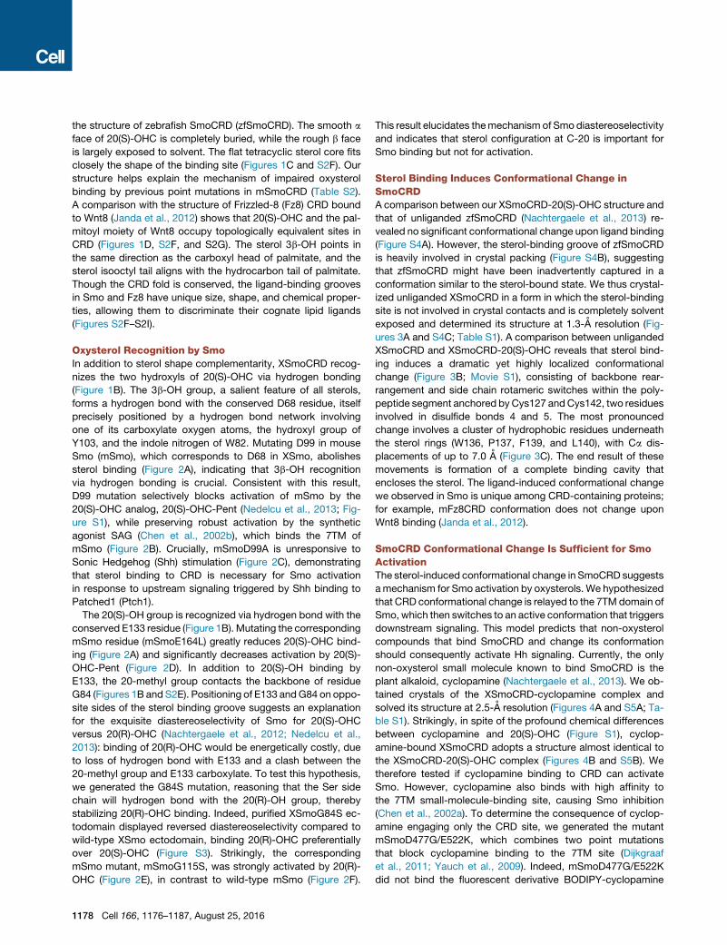
Figure 2. Oxysterol Recognition by Smo
(A) D99 and E164, the twomSmo residues that hydrogen bond with 3b-OH and 20(S)-OH, are critical for oxysterol binding. Full-length proteins expressed in 293T
cells were assayed by binding to 20(S)-OHC affinity matrix (Nedelcu et al., 2013), in the absence or presence of free 20(S)-OHC competitor.
(B) Smo-null cells rescued with mSmoD99A do not respond to 20(S)-OHC-Pent, in contrast to cells rescued with wild-type mSmo. Hh pathway activity was
assayed by qPCR for endogenous Gli1 and is shown normalized to activation elicited by saturating levels of SAG (0.5 mM). Error bars indicate SD (n = 3).
(C) As in (B), but treating cells with various concentrations of Shh. MSmoD99A does not respond to stimulation by Shh.
(D) As in (B), but with Smo-null cells rescued with mSmoE164L. The mutant has reduced responsiveness to oxysterols.
(E) mSmoG115S has reversed distereospecificity, responding stronger to 20(R)-OHC-Pent (EC50 = 1.3 mM) than to 20(S)-OHC-Pent (EC50 = 23.4 mM). See also
oxysterol binding assays in Figure S3.
(F) In contrast, wild-type mSmo is activated preferentially by 20(S)-OHC-Pent (EC50=0.84 mM) compared to 20(R)-OHC-Pent (EC50=26.3 mM). Experiments in
(B–F) were performed in parallel, and the curve for wild-type mSmo is shown in B, D and F.
(Figure S5C) and, furthermore, was not activated by SAG (Fig-
ure 4C), indicating that the 7TM small-molecule-binding site
is broadly disabled by the two point mutations. Binding to oxy-
sterols, however, was preserved in mSmoD477G/E522K (Fig-
ure S5D). Dramatically, cyclopamine activated mSmoD477G/
E522K (Figure 4C), in contrast to cyclopamine inhibition
of wild-type mSmo (Figure S5E). Importantly, mSmoD477G/
E522K also retained activation by Shh and 20(S)-OHC (Fig-
ure 4C). These results suggest that ligand-induced conforma-
tional change in the CRD is sufficient to activate Smo, and that
surprisingly, integrity of the 7TM small molecule-binding site of
Smo is not necessary for Hh signaling.
Cholesterol Binds SmoCRD and Activates Hh SignalingAlthough sterol binding to Smo is critical for Hh signaling, the
endogenous sterol ligand remains unknown. Oxysterols, and
in particular 20(S)-OHC, are unlikely candidates, being present
endogenously at much lower levels than their EC50 for Hh
pathway activation (Myers et al., 2013). We thus tested the pos-
sibility that cholesterol itself, by far the most abundant cellular
sterol, might be the endogenous Smo ligand in vertebrate Hh
signaling.
We first found that cholesterol binds SmoCRD, as demon-
strated by the following results: (1) in a fluorescence polarization
assay, cholesterol competed binding of BODIPY-cyclopamine
Cell 166, 1176–1187, August 25, 2016 1179

Figure 3. Sterol Binding Induces SmoCRD
Conformational Change
(A) Close-up view of the sterol-binding site in un-
liganded XSmoCRD. The indicated amino acids
are involved in sterol binding. See also Figure S4C
for overall structure of unliganded XSmoCRD.
(B) As in (A), but with unliganded XSmoCRD (cyan)
superimposed on 20(S)-OHC-bound XSmoCRD
(navy). In absence of sterol, the binding site is
in ‘‘open’’ state. 20(S)-OHC-induced conforma-
tional change results in ‘‘closed’’ state of the
binding site.
(C) Ribbon diagram showing overall structure
of unliganded XSmoCRD (cyan), superimposed
on 20(S)-OHC-bound XSmoCRD (navy). Binding
of 20(S)-OHC (yellow) induces inward rotation
of helix H30 (top right) and dramatic positional
swap of residues in following loop (bottom
right). See also Figure S4D for view from different
angle.
to XSmo ectodomain (Figure 5A), with an apparent IC50 of
2.4 mM, which is within the range of physiological cholesterol
concentration (Xie et al., 1999); (2) cholesterol enhanced the
thermal stability of XSmo ectodomain in a circular dichroism
(CD) melting assay (Figure S6A), indicative of binding—this
effect was similar to the thermostabilizing effect of known
CRD-binding molecules like 20(S)-OHC and cyclopamine (Fig-
ure S6B); and (3) we generated a novel cholesterol affinity resin,
in which the sterol moiety is attached to beads via a C-19 handle
(Figure S6C). This resin bound XSmo ectodomain specifically
(Figure S6D), demonstrating that the structural elements of
cholesterol are sufficient for Smo binding, while any sterol
hydroxylation or oxidation is not necessary. In all these binding
experiments, the closely related analog, cholestanol (Figure S1),
was inactive, indicating the high specificity of SmoCRD for
cholesterol.
Furthermore, exogenously added cholesterol activated Hh
signaling in a dose-dependent manner (Figure 5B), suggesting
that cholesterol binding is sufficient to activate Smo; as ex-
pected, cholestanol was inactive. Smo activation by cholesterol
occurred via binding to the CRD, as it was drastically reduced by
the D99A mutation (Figure S7A).
During vertebrate Hh signaling, Shh inhibits Ptch1, leading
to Smo activation. If cholesterol were the endogenous Smo-
activating ligand antagonized by Ptch1, one prediction is
that cholesterol should display synergy with Shh. Indeed, low
doses of Shh synergized with cholesterol in activating Hh
signaling (Figure 5C), and conversely, low doses of cholesterol,
1180 Cell 166, 1176–1187, August 25, 2016
but not cholestanol, displayed synergy
with Shh (Figure S7B). Importantly, in
agreement with previous reports (Nach-
tergaele et al., 2012), Shh did not
synergize with 20(S)-OHC-Pent (Fig-
ure 5D). These results are consistent
with cholesterol, but not the oxysterol,
being the endogenous Smo ligand. We
speculate that that the lack of synergy
between Shh and 20(S)-OHC indicates that oxysterols cannot
be inhibited by Ptch1, while cholesterol can.
Cholesterol as Endogenous Smo ActivatorWe used mutagenesis based on our XSmoCRD-20(S)-OHC
structure to further demonstrate that cholesterol and not 20(S)-
OHC is the endogenous ligand for Smo. To generate Smo
mutants that distinguish between cholesterol and 20(S)-OHC,
we focused on mSmo residues G115 and E164, which flank
C-20 of the sterol molecule. The mutants, mSmoG115F,
mSmoG115S, and mSmoG115S/E164L have abolished or
significantly reduced responsiveness to 20(S)-OHC-Pent (Fig-
ures 6A and S7C). Strikingly, these mutants retain responsive-
ness to cholesterol (Figures 6B and S7D) as well as to Shh
(Figures 6C and S7E). These results demonstrate that 20(S)-
OHC is not required for Hh signaling and are consistent with
cholesterol activating Smo during Shh stimulation.
We also asked whether the other oxysterols that can activate
Smo, 25-OHC, 7-keto-25-OHC, and 7-keto-27-OHC (Figure S1),
are required for Hh signaling. To address this question, we used
a strategy based on the fluorinated cholesterol derivatives,
25-fluorocholesterol and F7-cholesterol (Carroll et al., 1998)
(Figure S1). Since C-F bonds cannot be oxidized in cells, 25-flu-
orocholesterol cannot be converted into C-25-hydroxylated
oxysterols (25-OHC and 7-keto-25-OHC), while F7-cholesterol
cannot form oxysterols bearing C-25, C26, or C-27 hydroxyl
groups (25-OHC, 7-keto-25-OHC, and 7-keto-27-OHC) (Carroll
et al., 1998). Cells were depleted of sterols by incubation with

Figure 4. Sterol-Induced Conformational Change Activates Smo
(A) Cyclopamine bound in XSmoCRD sterol-binding site. Cyclopamine OH
group is recognized by the same network of hydrogen bonds (dashed lines) as
3b-OH group of 20(S)-OHC. See also Figure S5A for overall structure.
(B) Superimposition of cyclopamine-bound (green) and 20(S)-OHC-bound
(navy) XSmoCRD. Cyclopamine (orange) induces a protein conformation very
similar to that induced by 20(S)-OHC (yellow). See also Figure S5B for a close-
up view of binding site.
(C) Cyclopamine (100 mM, as soluble complex with methyl-b-cyclodextrin
[MCD]) binding to CRD activates mSmoD477G/E522K, a mutant in which the
cyclopamine-binding site in 7TM is destroyed. MSmoD477G/E522K also re-
sponds to 20(S)-OHC-Pent (10 mM), cholesterol (250 mM, as MCD complex),
and Shh, but not SAG (0.5 mM), which binds the 7TM site. Error bars indicate
SD (n = 3).
methyl-b-cyclodextrin (MCD), after which fluorinated sterols
were added back as soluble MCD complexes, and the cells
were stimulated with Shh, followed by assaying two Hh pathway
readouts: recruitment of endogenous Smo to primary cilia (Cor-
bit et al., 2005) and Hh transcriptional reporter assays. As
expected, sterol depletion blocked Smo recruitment to cilia (Fig-
ures 6D and 6E) and Hh pathway activation (Figure 6F), which
was rescued by pure cholesterol, but not by cholestanol. Inter-
estingly, 25-fluorocholesterol completely rescued Hh signaling
in sterol-depleted cells (Figures 6D–6F). Furthermore, F7-
cholesterol showed significant rescue, although less than pure
cholesterol (Figures 6D–6F). We speculate that the lower activity
of F7-cholesterol is due to its reduced solubility compared to
cholesterol and 25-fluorocholesterol. Together, these results
demonstrate that cholesterol hydroxylation on C-25, C-26, or
C-27 is not absolutely required for Hh signaling, indicating that
25-OHC, 7-keto-25-OHC, or 7-keto-27-OHC are not essential
for Smo activation.
Recently, based on experiments showing that mSmoDCRD is
inhibited by sterol depletion, it was proposed that Smo con-
tains an additional sterol-binding site, distinct from CRD (Myers
et al., 2013). To elucidate which site mediates the effect of ste-
rols on Smo, we compared the consequences of sterol deple-
tion on mSmo and mSmoDCRD activity (Figure 6G). Both
mSmo and mSmoDCRD activated Hh signaling; however, in
contrast to mSmo, mSmoDCRD activity was unaffected by ste-
rol depletion (Figure 6G), indicating that sterols exert their effect
on mSmo via the CRD and that basal activity of mSmoDCRD is
independent of sterols. We also measured ciliary localization of
mSmo and mSmoDCRD in response to Shh and to sterol
depletion (Figure 6H). As expected, sterol depletion blocked
mSmo recruitment to cilia in response to Shh; in contrast,
ciliary localization of mSmoDCRD was unaffected by either
Shh or by sterol depletion (Figure 6H). This suggests that
CRD is required for Smo regulation both by Shh and by sterols.
Finally, we examined whether sterols are required for direct
activation of Smo by SAG. Contrary to recent reports (Myers
et al., 2013), we find that sterol depletion has no effect on Hh
signaling triggered by SAG in Smo-null cells rescued with
mSmo or with mSmoDCRD (Figures 6G and 6H) or in wild-
type NIH 3T3 cells (Figure 6I). Thus, Smo requires sterols for
activation only during Shh stimulation, while sterols are no
longer required when Smo is forced into active conformation
by the SAG agonist.
DiscussionSmo is essential for relaying Hh signals across plasma mem-
brane. In absence of stimulation, Ptch represses Smo, ensuring
that Hh pathway is inhibited. Signaling is triggered by Hh ligand,
which inhibits Ptch, thus allowing Smo to adopt an active confor-
mation and transduce Hh signals to the cytoplasm. A critical
unanswered question has been how Smo is regulated. As 7TM
protein targeted by numerous synthetic agonists and antago-
nists, Smo was postulated be controlled by an unidentified
endogenous ligand. This hypothesis is also consistent with
Ptch belonging to the RND family of small molecule pumps,
suggesting that the Smo ligand is itself regulated by Ptch. Sterol
depletion inhibits vertebrate Smo, and furthermore, some
oxysterols activate Smo by binding to its CRD; together, these
findings suggested that Smo is regulated by a sterol activator.
However, the identity of the endogenous sterol has remained
unknown, as well as how Smo recognizes sterols and how
sterols activate Smo.
Here, we first elucidate how Smo recognizes sterols, by solv-
ing the crystal structure of SmoCRD in complex with 20(S)-OHC.
We then use structure-guided mutagenesis to demonstrate that
sterol binding is critical for Smo activation by upstream Shh
signaling through Ptch1, as well as to explain the basis for dia-
stereospecific Smo activation by oxysterols. Next, we solve
the structure of SmoCRD alone, revealing that sterol binding trig-
gers a conformational change. We also determine the structure
of SmoCRD in complex with the plant alkaloid, cyclopamine,
which shows a conformation almost identical to that of
Cell 166, 1176–1187, August 25, 2016 1181

Figure 5. Cholesterol Binds Smo and Acti-
vates Hh Signaling
(A) Fluorescence polarization assay showing
cholesterol competes binding of BODIPY-cy-
clopamine to purified XSmo ectodomain in dose-
dependent manner (IC50 = 2.4 mM). In contrast, the
saturated analog cholestanol, does not bind Smo.
See also Figure S6 for additional assays demon-
strating binding of cholesterol to XSmoCRD.
(B) Cholesterol, but not cholestanol, activates Hh
signaling in Smo-null cells rescued with wild-type
mSmo. Cholesterol and cholestanol were delivered
as MCD complexes. Error bars indicate SD (n = 3).
(C) Cholesterol (added as MCD complex) syner-
gizes with Shh to activate Hh signaling in NIH 3T3
cells. See also Figure S7B showing synergy of Shh
with cholesterol, but not cholestanol. Error bars
indicate SD (n = 3).
(D) Unlike cholesterol, the oxysterol 20(S)-OHC-
Pent does not synergize with Shh in NIH 3T3 cells.
oxysterol-bound SmoCRD: we exploit this similarity to show that
CRD conformational change is sufficient to activate Smo, sug-
gesting that it is a key event in allosteric Smo activation. Impor-
tantly, we discover that cholesterol itself binds and activates
Smo, as well as synergizes with Shh. Furthermore, we demon-
strate that the known Smo-activating oxysterols are not required
for Smo activation in cells. Taken together, our data support a
model in which cholesterol is the endogenous ligand responsible
for Smo activation during Hh signaling, and that Ptch1 inhibits
Smo by counteracting its activation by cholesterol (Figure 7).
Thus cholesterol plays a unique role as second messenger in
vertebrate Hh signaling, mediating the functional interaction
between Ptch1 and Smo.
Our structural analysis clarifies how Smo recognizes, and
how it is activated by, sterols. We show that sterols occupy
their site on SmoCRD in a head-to-tail orientation that matches
that of the palmitoyl residue of Wnt8 binding to Fz8CRD (Janda
et al., 2012), thus disproving the previous model based on
molecular docking of 20(S)-OHC to unliganded zfSmoCRD
(Nachtergaele et al., 2013). Reminiscent of steroid hormone re-
ceptors, SmoCRD discriminates sterols from other lipids by
exploiting both shape and amphipathic properties. The high de-
gree of shape complementarity between SmoCRD and 20(S)-
OHC suggests that the endogenous ligand for Smo is indeed
a sterol: this notion is further supported by the fact that recog-
1182 Cell 166, 1176–1187, August 25, 2016
nition of 3b-OH, a universally conserved
feature of sterols, is essential for both
Smo-sterol interaction and for Smo acti-
vation by Shh. Based on the SmoCRD-
20(S)-OHC structure, we also elucidate
the basis for diastereoselective recogni-
tion of 20(S)-OHC and validate our
model by generating a Smo mutant that
responds to 20(R)-OHC instead of
20(S)-OHC. Our data also highlights the
exquisite sterol specificity of Smo, which
discriminates between cholesterol and
the reduced analog, cholestanol. Inter-
estingly, while Smo does not bind cholestanol, we have shown
that it binds a C-20(S)-hydroxylated derivative of it (Nedelcu
et al., 2013). Perhaps, hydrogen bonding between 20(S)-OH
and the conserved E164 in mSmo is able to rescue cholestanol
binding to Smo.
Unexpectedly, sterol binding to SmoCRD triggers a conforma-
tional change, suggesting a mechanism for Smo activation.
Interestingly, such a ligand-induced conformational change
has not been seen in other CRDs. In the case of the related
Fz8 receptor, Wnt8 binding does not change Fz8CRD conforma-
tion (Janda et al., 2012): this is consistent withWnt-Fz interaction
alone not being sufficient for downstream pathway activation,
which requires recruitment of co-receptors, such as lipopro-
tein-receptor-related protein (LRP)-5/6 in the canonical pathway
or tyrosine kinase receptor ROR2 in the non-canonical pathway.
Together, these differences suggest that Smo and Fz proteins
have evolved distinct activation mechanisms.
How does oxysterol binding to SmoCRD activate the rest of
the protein? A likely possibility is that it occurs through commu-
nication with the extracellular loops (ECLs) of the 7TM domain of
Smo (Wang et al., 2013), from which conformational change
propagates to, and activates, the parts of Smo facing the
cytosol. We speculate that CRD, ECLs, and TM helices form
an integrated multi-domain convergence zone, in which ECLs
mediate communication between CRD and 7TM: this model is
(legend on next page)
Cell 166, 1176–1187, August 25, 2016 1183
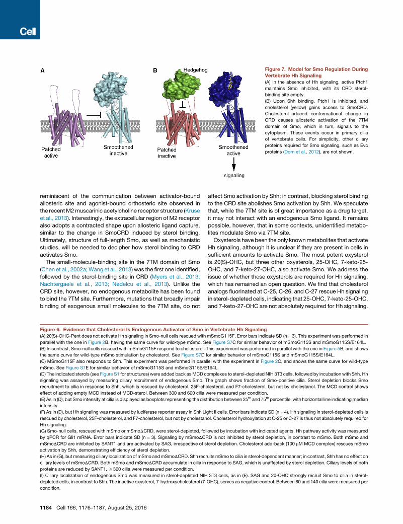
Figure 7. Model for Smo Regulation During
Vertebrate Hh Signaling
(A) In the absence of Hh signaling, active Ptch1
maintains Smo inhibited, with its CRD sterol-
binding site empty.
(B) Upon Shh binding, Ptch1 is inhibited, and
cholesterol (yellow) gains access to SmoCRD.
Cholesterol-induced conformational change in
CRD causes allosteric activation of the 7TM
domain of Smo, which in turn, signals to the
cytoplasm. These events occur in primary cilia
of vertebrate cells. For simplicity, other ciliary
proteins required for Smo signaling, such as Evc
proteins (Dorn et al., 2012), are not shown.
reminiscent of the communication between activator-bound
allosteric site and agonist-bound orthosteric site observed in
the recentM2muscarinic acetylcholine receptor structure (Kruse
et al., 2013). Interestingly, the extracellular region of M2 receptor
also adopts a contracted shape upon allosteric ligand capture,
similar to the change in SmoCRD induced by sterol binding.
Ultimately, structure of full-length Smo, as well as mechanistic
studies, will be needed to decipher how sterol binding to CRD
activates Smo.
The small-molecule-binding site in the 7TM domain of Smo
(Chen et al., 2002a;Wang et al., 2013) was the first one identified,
followed by the sterol-binding site in CRD (Myers et al., 2013;
Nachtergaele et al., 2013; Nedelcu et al., 2013). Unlike the
CRD site, however, no endogenous metabolite has been found
to bind the 7TM site. Furthermore, mutations that broadly impair
binding of exogenous small molecules to the 7TM site, do not
Figure 6. Evidence that Cholesterol Is Endogenous Activator of Smo in
(A) 20(S)-OHC-Pent does not activate Hh signaling in Smo-null cells rescued with
parallel with the one in Figure 2B, having the same curve for wild-type mSmo. S
(B) In contrast, Smo-null cells rescued with mSmoG115F respond to cholesterol. T
the same curve for wild-type mSmo stimulation by cholesterol. See Figure S7D f
(C) MSmoG115F also responds to Shh. This experiment was performed in paral
mSmo. See Figure S7E for similar behavior of mSmoG115S and mSmoG115S/E
(D) The indicated sterols (see Figure S1 for structures) were added back asMCD c
signaling was assayed by measuring ciliary recruitment of endogenous Smo. T
recruitment to cilia in response to Shh, which is rescued by cholesterol, 25F-cho
effect of adding empty MCD instead of MCD-sterol. Between 300 and 600 cilia w
(E) As in (D), but Smo intensity at cilia is displayed as boxplots representing the dist
intensity.
(F) As in (D), but Hh signaling was measured by luciferase reporter assay in Shh Li
rescued by cholesterol, 25F-cholesterol, and F7-cholesterol, but not by cholestan
Hh signaling.
(G) Smo-null cells, rescued with mSmo or mSmoDCRD, were sterol-depleted, fol
by qPCR for Gli1 mRNA. Error bars indicate SD (n = 3). Signaling by mSmoDCR
mSmoDCRD are inhibited by SANT1 and are activated by SAG, irrespective of s
activation by Shh, demonstrating efficiency of sterol depletion.
(H) As in (G), butmeasuring ciliary localization ofmSmo andmSmoDCRD. Shh recr
ciliary levels of mSmoDCRD. Both mSmo and mSmoDCRD accumulate in cilia in
proteins are reduced by SANT1. R300 cilia were measured per condition.
(I) Ciliary localization of endogenous Smo was measured in sterol-depleted NIH
depleted cells, in contrast to Shh. The inactive oxysterol, 7-hydroxycholesterol (7-
condition.
1184 Cell 166, 1176–1187, August 25, 2016
affect Smo activation by Shh; in contrast, blocking sterol binding
to the CRD site abolishes Smo activation by Shh. We speculate
that, while the 7TM site is of great importance as a drug target,
it may not interact with an endogenous Smo ligand. It remains
possible, however, that in some contexts, unidentified metabo-
lites modulate Smo via 7TM site.
Oxysterols have been the only knownmetabolites that activate
Hh signaling, although it is unclear if they are present in cells in
sufficient amounts to activate Smo. The most potent oxysterol
is 20(S)-OHC, but three other oxysterols, 25-OHC, 7-keto-25-
OHC, and 7-keto-27-OHC, also activate Smo. We address the
issue of whether these oxysterols are required for Hh signaling,
which has remained an open question. We find that cholesterol
analogs fluorinated at C-25, C-26, and C-27 rescue Hh signaling
in sterol-depleted cells, indicating that 25-OHC, 7-keto-25-OHC,
and 7-keto-27-OHC are not absolutely required for Hh signaling.
Vertebrate Hh Signaling
mSmoG115F. Error bars indicate SD (n = 3). This experiment was performed in
ee Figure S7C for similar behavior of mSmoG115S and mSmoG115S/E164L.
his experiment was performed in parallel with the one in Figure 5B, and shows
or similar behavior of mSmoG115S and mSmoG115S/E164L.
lel with the experiment in Figure 2C, and shows the same curve for wild-type
164L.
omplexes to sterol-depleted NIH 3T3 cells, followed by incubation with Shh. Hh
he graph shows fraction of Smo-positive cilia. Sterol depletion blocks Smo
lesterol, and F7-cholesterol, but not by cholestanol. The MCD control shows
ere measured per condition.
ribution between 25th and 75th percentile, with horizontal line indicating median
ght II cells. Error bars indicate SD (n = 4). Hh signaling in sterol-depleted cells is
ol. Cholesterol hydroxylation at C-25 or C-27 is thus not absolutely required for
lowed by incubation with indicated agents. Hh pathway activity was measured
D is not inhibited by sterol depletion, in contrast to mSmo. Both mSmo and
terol depletion. Cholesterol add-back (100 mM MCD complex) rescues mSmo
uitsmSmo to cilia in sterol-dependent manner; in contrast, Shh has no effect on
response to SAG, which is unaffected by sterol depletion. Ciliary levels of both
3T3 cells, as in (E). SAG and 20-OHC strongly recruit Smo to cilia in sterol-
OHC), serves as negative control. Between 80 and 140 cilia were measured per

Furthermore, 20(S)-OHC is also not required for Hh signaling,
as shown by mutants such as mSmoG115F, which does not
respond to 20(S)-OHC, but is still activated by Shh.
We find that cholesterol fulfills the criteria for being the endog-
enous Smo ligand. Cholesterol binds SmoCRD with micromolar
affinity, within the range of endogenous cholesterol levels (Xie
et al., 1999). We note that affinity for cholesterol might be higher
for full-length Smo, as other domains of the protein might coop-
erate to enhance binding, possibly by shielding the b face of
the sterol molecule, which is not engaged in CRD binding.
Cholesterol also activates Smo, including mutants that do not
respond to 20(S)-OHC, like mSmoG115F. Furthermore, choles-
terol synergizes with Shh to activate Smo, as expected for an
endogenous Smo ligand that is negatively regulated by Ptch1.
In contrast to cholesterol, 20(S)-OHC does not synergize with
Shh. We speculate that this difference is due to Ptch1 inability
to inhibit 20(S)-OHC. Perhaps Ptch1 antagonizes cholesterol at
the membrane bilayer level, and 20(S)-OHC, by virtue of its
much higher water solubility compared to cholesterol, can
escape inhibition by Ptch1.
Our results also clarify the role of CRD in Smo regulation. In
contrast to a recent report (Myers et al., 2013), we find that ste-
rol depletion does not block activation of Hh signaling by
SmoDCRD, or by SAG: this demonstrates that CRD mediates
the effect of sterols on Smo and that Smo requires sterols only
when activated by upstream Shh, while SAG, by directly acti-
vating Smo, bypasses the sterol requirement. We also find that
SmoDCRD levels in cilia are not affected by Shh, suggesting
that CRD is critical for Smo regulation by Shh-Ptch1: this view
is consistent with results showing that SmoDCRD is not respon-
sive to Shh (Aanstad et al., 2009) and that replacing CRD and the
first two TM domains of Smowith corresponding portions of Fz5,
results in a protein no longer inhibited by Ptch1 (Murone et al.,
1999). We have previously observed a small effect of Shh on
overexpressed SmoDCRD in qPCR assays (Nedelcu et al.,
2013), so it is possible that SmoDCRD retains some residual
regulation by Ptch1.
How does Ptch1 inhibit Smo? Since Ptch1 acts at the primary
cilium (Rohatgi et al., 2007), a possibility is that Ptch1 maintains
low levels and/or chemical activity of cholesterol in the ciliary
membrane, thereby ensuring that Smo will not be activated.
Upon Ptch1 inhibition by Shh, cholesterol activity in cilia in-
creases, leading to Smo activation: testing this hypothesis will
require developing methods to image cholesterol in cilia. In this
model, Smo arrives at cilia in an unliganded, inactive form.
Perhaps cholesterol activity in compartments through which
Smo traffics on its way to the cilium is too low for binding to
Smo. Alternatively, Smo binds cholesterol before reaching the
cilium, but the low cholesterol maintained by Ptch1 in cilia favors
dissociation, thereby inhibiting Smo. Importantly, ciliary resident
proteins that interact with Smo, such as EVC proteins, are
required for turning on signaling downstream of Smo (Dorn
et al., 2012): this ensures that, even if cholesterol binds Smo
and induces it to adopt an active conformation outside the
cilium, this will not trigger Hh signal transduction. A detailed anal-
ysis of where the interaction between Smo and cholesterol takes
place in the cell will be needed to answer these questions, aswell
as determining if and how Ptch1 regulates cholesterol in cilia.
STAR+METHODS
Detailed methods are provided in the online version of this paper
and include the following:
d KEY RESOURCES TABLE
d CONTACT FOR REAGENT AND RESOURCE SHARING
d EXPERIMENTAL MODEL AND SUBJECT DETAILS
B Cell Culture and Generation of Stable Cell Lines
d METHOD DETAILS
B Reagents
B Antibodies
B Hh Pathway Assays
B Sterol Depletion and Rescue Experiments
B Immunofluorescence and Measurements of Smo
Ciliary Localization
B BODIPY-Cyclopamine Binding Assays
B Sterol Affinity Matrices
B Sterol Affinity Assays
B Recombinant Protein Expression and Purification
B Crystallization, Data Collection, and Structure
Determination
B Fluorescence Polarization-Based Ligand Binding
Assays
B Circular Dichroism Temperature Melting Experiments
B Chemical Synthesis
d QUANTIFICATION AND STATISTICAL ANALYSIS
d DATA AND SOFTWARE AVAILABILITY
B Data Resources
SUPPLEMENTAL INFORMATION
Supplemental Information includes seven figures, two tables, and one
movie and can be found with this article at http://dx.doi.org/10.1016/j.cell.
2016.08.003.
AUTHOR CONTRIBUTIONS
P.H., D.N., M.W., and A.S. performed cellular and biochemical experiments.
J.L. synthesized, purified, and characterized BODIPY probes. C.J. synthe-
sized, purified, and characterized the C19-position cholesterol analog. Y.K.
collected diffraction data sets. P.H. solved the structures. P.H., D.N., M.W.,
and A.S. analyzed data. A.S. and P.H. wrote the manuscript, with input from
the other authors.
ACKNOWLEDGMENTS
This work was supported by NIH grants RO1 GM092924 and GM110041 to
A.S. We thank members of the Salic lab for helpful discussions and members
of the Structural Biology Center at Argonne National Laboratory for help with
data collection at the 19-ID beam line.
Received: April 23, 2016
Revised: June 14, 2016
Accepted: July 30, 2016
Published: August 18, 2016
REFERENCES
Aanstad, P., Santos, N., Corbit, K.C., Scherz, P.J., Trinh, A., Salvenmoser, W.,
Huisken, J., Reiter, J.F., and Stainier, D.Y. (2009). The extracellular domain of
Cell 166, 1176–1187, August 25, 2016 1185

Smoothened regulates ciliary localization and is required for high-level Hh
signaling. Curr. Biol. 19, 1034–1039.
Alcedo, J., Ayzenzon, M., Von Ohlen, T., Noll, M., and Hooper, J.E. (1996). The
Drosophila smoothened gene encodes a seven-pass membrane protein, a
putative receptor for the hedgehog signal. Cell 86, 221–232.
Bazan, J.F., and de Sauvage, F.J. (2009). Structural ties between cholesterol
transport and morphogen signaling. Cell 138, 1055–1056.
Braberg, H., Webb, B.M., Tjioe, E., Pieper, U., Sali, A., and Madhusudhan,
M.S. (2012). SALIGN: a web server for alignment of multiple protein sequences
and structures. Bioinformatics 28, 2072–2073.
Carroll, J.N., Pinkerton, F.D., Su, X., Gerst, N., Wilson, W.K., and Schroepfer,
G.J., Jr. (1998). Sterol synthesis. Synthesis of 3 beta-hydroxy-25,26,26,
26,27,27,27-heptafluorocholest-5-en-7-one and its effects on HMG-CoA
reductase activity in Chinese hamster ovary cells, on ACAT activity in rat jejunal
microsomes, and serum cholesterol levels in rats. Chem. Phys. Lipids 94,
209–225.
Chen, J.K., Taipale, J., Cooper, M.K., and Beachy, P.A. (2002a). Inhibition of
Hedgehog signaling by direct binding of cyclopamine to Smoothened. Genes
Dev. 16, 2743–2748.
Chen, J.K., Taipale, J., Young, K.E., Maiti, T., and Beachy, P.A. (2002b). Small
molecule modulation of Smoothened activity. Proc. Natl. Acad. Sci. USA 99,
14071–14076.
Cooper, M.K., Wassif, C.A., Krakowiak, P.A., Taipale, J., Gong, R., Kelley,
R.I., Porter, F.D., and Beachy, P.A. (2003). A defective response to Hedge-
hog signaling in disorders of cholesterol biosynthesis. Nat. Genet. 33,
508–513.
Corbit, K.C., Aanstad, P., Singla, V., Norman, A.R., Stainier, D.Y., and Reiter,
J.F. (2005). Vertebrate Smoothened functions at the primary cilium. Nature
437, 1018–1021.
Corcoran, R.B., and Scott, M.P. (2006). Oxysterols stimulate Sonic hedgehog
signal transduction and proliferation of medulloblastoma cells. Proc. Natl.
Acad. Sci. USA 103, 8408–8413.
Dijkgraaf, G.J., Alicke, B., Weinmann, L., Januario, T., West, K., Modru-
san, Z., Burdick, D., Goldsmith, R., Robarge, K., Sutherlin, D., et al.
(2011). Small molecule inhibition of GDC-0449 refractory smoothened mu-
tants and downstream mechanisms of drug resistance. Cancer Res. 71,
435–444.
Dorn, K.V., Hughes, C.E., and Rohatgi, R. (2012). A Smoothened-Evc2 com-
plex transduces the Hedgehog signal at primary cilia. Dev. Cell 23,
823–835.
Dwyer, J.R., Sever, N., Carlson, M., Nelson, S.F., Beachy, P.A., and Par-
hami, F. (2007). Oxysterols are novel activators of the hedgehog signaling
pathway in pluripotent mesenchymal cells. J. Biol. Chem. 282, 8959–
8968.
Emsley, P., and Cowtan, K. (2004). Coot: model-building tools for molecular
graphics. Acta Crystallogr. D Biol. Crystallogr. 60, 2126–2132.
Frank-Kamenetsky, M., Zhang, X.M., Bottega, S., Guicherit, O., Wichterle, H.,
Dudek, H., Bumcrot, D., Wang, F.Y., Jones, S., Shulok, J., et al. (2002). Small-
molecule modulators of Hedgehog signaling: identification and characteriza-
tion of Smoothened agonists and antagonists. J. Biol. 1, 10.
Gimpl, G., Klein, U., Reilander, H., and Fahrenholz, F. (1995). Expression of the
human oxytocin receptor in baculovirus-infected insect cells: high-affinity
binding is induced by a cholesterol-cyclodextrin complex. Biochemistry 34,
13794–13801.
Greenfield, N.J. (2006). Using circular dichroism spectra to estimate protein
secondary structure. Nat. Protoc. 1, 2876–2890.
Ingham, P.W., and McMahon, A.P. (2001). Hedgehog signaling in animal
development: paradigms and principles. Genes Dev. 15, 3059–3087.
Janda, C.Y., Waghray, D., Levin, A.M., Thomas, C., and Garcia, K.C. (2012).
Structural basis of Wnt recognition by Frizzled. Science 337, 59–64.
Jao, C.Y., Nedelcu, D., Lopez, L.V., Samarakoon, T.N., Welti, R., and Salic, A.
(2015). Bioorthogonal probes for imaging sterols in cells. ChemBioChem 16,
611–617.
1186 Cell 166, 1176–1187, August 25, 2016
Kim, W.K., Meliton, V., Amantea, C.M., Hahn, T.J., and Parhami, F. (2007).
20(S)-hydroxycholesterol inhibits PPARgamma expression and adipogenic
differentiation of bone marrow stromal cells through a hedgehog-dependent
mechanism. J. Bone Miner. Res. 22, 1711–1719.
Kruse, A.C., Ring, A.M., Manglik, A., Hu, J., Hu, K., Eitel, K., Hubner, H.,
Pardon, E., Valant, C., Sexton, P.M., et al. (2013). Activation and allo-
steric modulation of a muscarinic acetylcholine receptor. Nature 504,
101–106.
Lum, L., and Beachy, P.A. (2004). The Hedgehog response network: sensors,
switches, and routers. Science 304, 1755–1759.
McCoy, A.J., Grosse-Kunstleve, R.W., Adams, P.D., Winn, M.D., Storoni, L.C.,
and Read, R.J. (2007). Phaser crystallographic software. J. Appl. Cryst. 40,
658–674.
Minor, W., Cymborowski, M., Otwinowski, Z., and Chruszcz, M. (2006). HKL-
3000: the integration of data reduction and structure solution–from diffraction
images to an initial model in minutes. Acta Crystallogr. D Biol. Crystallogr. 62,
859–866.
Murakami, S., Nakashima, R., Yamashita, E., Matsumoto, T., and Yamaguchi,
A. (2006). Crystal structures of a multidrug transporter reveal a functionally
rotating mechanism. Nature 443, 173–179.
Murone, M., Rosenthal, A., and de Sauvage, F.J. (1999). Sonic hedgehog
signaling by the patched-smoothened receptor complex. Curr. Biol. 9,
76–84.
Myers, B.R., Sever, N., Chong, Y.C., Kim, J., Belani, J.D., Rychnovsky, S., Ba-
zan, J.F., and Beachy, P.A. (2013). Hedgehog pathway modulation by multiple
lipid binding sites on the smoothened effector of signal response. Dev. Cell 26,
346–357.
Nachtergaele, S., Mydock, L.K., Krishnan, K., Rammohan, J., Schlesinger,
P.H., Covey, D.F., and Rohatgi, R. (2012). Oxysterols are allosteric activators
of the oncoprotein Smoothened. Nat. Chem. Biol. 8, 211–220.
Nachtergaele, S., Whalen, D.M., Mydock, L.K., Zhao, Z., Malinauskas, T.,
Krishnan, K., Ingham, P.W., Covey, D.F., Siebold, C., and Rohatgi, R. (2013).
Structure and function of the Smoothened extracellular domain in vertebrate
Hedgehog signaling. eLife 2, e01340.
Nakano, Y., Guerrero, I., Hidalgo, A., Taylor, A., Whittle, J.R., and Ingham,
P.W. (1989). A protein with several possible membrane-spanning domains
encoded by the Drosophila segment polarity gene patched. Nature 341,
508–513.
Nedelcu, D., Liu, J., Xu, Y., Jao, C., and Salic, A. (2013). Oxysterol binding to
the extracellular domain of Smoothened in Hedgehog signaling. Nat. Chem.
Biol. 9, 557–564.
Rohatgi, R., Milenkovic, L., and Scott, M.P. (2007). Patched1 regulates hedge-
hog signaling at the primary cilium. Science 317, 372–376.
Sinha, S., and Chen, J.K. (2006). Purmorphamine activates the Hedgehog
pathway by targeting Smoothened. Nat. Chem. Biol. 2, 29–30.
Taipale, J., Chen, J.K., Cooper, M.K., Wang, B., Mann, R.K., Milenkovic, L.,
Scott, M.P., and Beachy, P.A. (2000). Effects of oncogenic mutations in
Smoothened and Patched can be reversed by cyclopamine. Nature 406,
1005–1009.
Taipale, J., Cooper, M.K., Maiti, T., and Beachy, P.A. (2002). Patched
acts catalytically to suppress the activity of Smoothened. Nature 418,
892–897.
Tseng, T.T., Gratwick, K.S., Kollman, J., Park, D., Nies, D.H., Goffeau, A., and
Saier, M.H., Jr. (1999). The RND permease superfamily: an ancient, ubiquitous
and diverse family that includes human disease and development proteins.
J. Mol. Microbiol. Biotechnol. 1, 107–125.
Tukachinsky, H., Lopez, L.V., and Salic, A. (2010). A mechanism for vertebrate
Hedgehog signaling: recruitment to cilia and dissociation of SuFu-Gli protein
complexes. J. Cell Biol. 191, 415–428.
Tukachinsky, H., Kuzmickas, R.P., Jao, C.Y., Liu, J., and Salic, A. (2012). Dis-
patched and scube mediate the efficient secretion of the cholesterol-modified
hedgehog ligand. Cell Rep. 2, 308–320.

van den Heuvel, M., and Ingham, P.W. (1996). smoothened encodes a recep-
tor-like serpentine protein required for hedgehog signalling. Nature 382,
547–551.
Voss, N.R., andGerstein,M. (2010). 3V: cavity, channel and cleft volume calcu-
lator and extractor. Nucleic Acids Res. 38, W555–W562.
Wang, C., Wu, H., Katritch, V., Han, G.W., Huang, X.P., Liu, W., Siu, F.Y.,
Roth, B.L., Cherezov, V., and Stevens, R.C. (2013). Structure of the hu-
man smoothened receptor bound to an antitumour agent. Nature 497,
338–343.
Xie, C., Turley, S.D., and Dietschy, J.M. (1999). Cholesterol accumulation in
tissues of the Niemann-pick type Cmouse is determined by the rate of lipopro-
tein-cholesterol uptake through the coated-pit pathway in each organ. Proc.
Natl. Acad. Sci. USA 96, 11992–11997.
Yauch, R.L., Dijkgraaf, G.J., Alicke, B., Januario, T., Ahn, C.P., Holcomb, T.,
Pujara, K., Stinson, J., Callahan, C.A., Tang, T., et al. (2009). Smoothened mu-
tation confers resistance to a Hedgehog pathway inhibitor in medulloblas-
toma. Science 326, 572–574.
Cell 166, 1176–1187, August 25, 2016 1187
STAR+METHODS
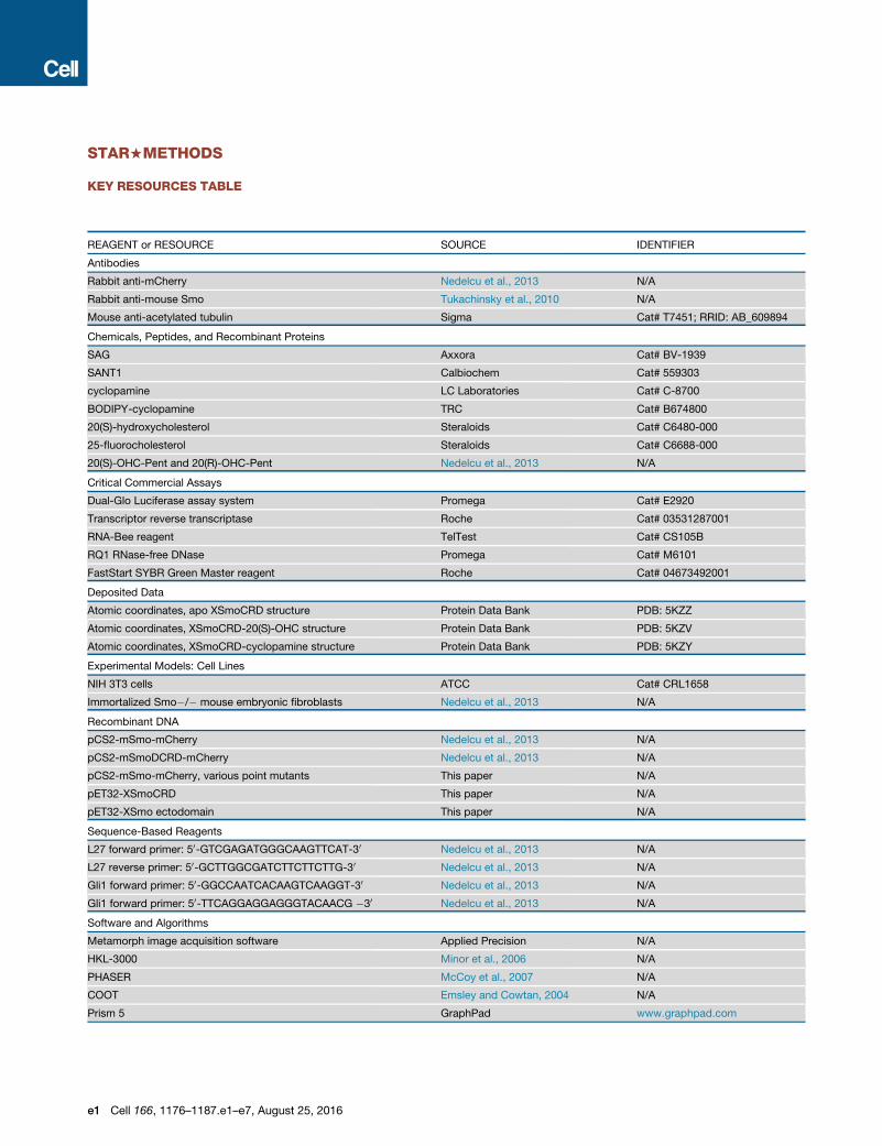
KEY RESOURCES TABLE
REAGENT or RESOURCE SOURCE IDENTIFIER
Antibodies
Rabbit anti-mCherry Nedelcu et al., 2013 N/A
Rabbit anti-mouse Smo Tukachinsky et al., 2010 N/A
Mouse anti-acetylated tubulin Sigma Cat# T7451; RRID: AB_609894
Chemicals, Peptides, and Recombinant Proteins
SAG Axxora Cat# BV-1939
SANT1 Calbiochem Cat# 559303
cyclopamine LC Laboratories Cat# C-8700
BODIPY-cyclopamine TRC Cat# B674800
20(S)-hydroxycholesterol Steraloids Cat# C6480-000
25-fluorocholesterol Steraloids Cat# C6688-000
20(S)-OHC-Pent and 20(R)-OHC-Pent Nedelcu et al., 2013 N/A
Critical Commercial Assays
Dual-Glo Luciferase assay system Promega Cat# E2920
Transcriptor reverse transcriptase Roche Cat# 03531287001
RNA-Bee reagent TelTest Cat# CS105B
RQ1 RNase-free DNase Promega Cat# M6101
FastStart SYBR Green Master reagent Roche Cat# 04673492001
Deposited Data
Atomic coordinates, apo XSmoCRD structure Protein Data Bank PDB: 5KZZ
Atomic coordinates, XSmoCRD-20(S)-OHC structure Protein Data Bank PDB: 5KZV
Atomic coordinates, XSmoCRD-cyclopamine structure Protein Data Bank PDB: 5KZY
Experimental Models: Cell Lines
NIH 3T3 cells ATCC Cat# CRL1658
Immortalized Smo�/� mouse embryonic fibroblasts Nedelcu et al., 2013 N/A
Recombinant DNA
pCS2-mSmo-mCherry Nedelcu et al., 2013 N/A
pCS2-mSmoDCRD-mCherry Nedelcu et al., 2013 N/A
pCS2-mSmo-mCherry, various point mutants This paper N/A
pET32-XSmoCRD This paper N/A
pET32-XSmo ectodomain This paper N/A
Sequence-Based Reagents
L27 forward primer: 50-GTCGAGATGGGCAAGTTCAT-30 Nedelcu et al., 2013 N/A
L27 reverse primer: 50-GCTTGGCGATCTTCTTCTTG-30 Nedelcu et al., 2013 N/A
Gli1 forward primer: 50-GGCCAATCACAAGTCAAGGT-30 Nedelcu et al., 2013 N/A
Gli1 forward primer: 50-TTCAGGAGGAGGGTACAACG �30 Nedelcu et al., 2013 N/A
Software and Algorithms
Metamorph image acquisition software Applied Precision N/A
HKL-3000 Minor et al., 2006 N/A
PHASER McCoy et al., 2007 N/A
COOT Emsley and Cowtan, 2004 N/A
Prism 5 GraphPad www.graphpad.com
e1 Cell 166, 1176–1187.e1–e7, August 25, 2016

CONTACT FOR REAGENT AND RESOURCE SHARING
Further information and requests for reagents may be directed to, and will be fulfilled by the corresponding author Adrian Salic
EXPERIMENTAL MODEL AND SUBJECT DETAILS
Cell Culture and Generation of Stable Cell LinesNIH 3T3 cells were grown in Dulbecco’s Modified Eagle’s Medium (DMEM) with 10% bovine calf serum, penicillin, and streptomycin.
Smo null (Smo�/�) mouse embryonic fibroblasts (MEFs) were grown in DMEM supplemented with 10% fetal bovine serum, peni-
cillin, and streptomycin. To generate stable lines, mouse Smoothened (mSmo) constructs were subcloned into a vector for lentiviral
production, bearing a C-terminal mCherry tag. Replication-defective lentiviruses were packaged in 293T cells, using standard pro-
tocols. Supernatants containing the virus were used to infect Smo�/� cells, followed by antibiotic selection beginning 48 hr post-
infection (50 mg/mL blasticidin). After selection for 2 days, the cell cultures were expanded, and cells expressing low levels of
mCherry-tagged mSmo were obtained by fluorescence-activated cell sorting.
METHOD DETAILS
ReagentsThe following reagents were purchased: SAG (R98%) from Axxora; SANT1 (R95%) fromCalbiochem; cyclopamine (> 99%) from LC
Laboratories; BODIPY-cyclopamine from Toronto Research Chemicals; BODIPY-FL N-hydroxysuccinimide ester from Thermo
Fisher; pravastatin (R98%), cholesterol, cholesteryl hemisuccinate and methyl-b-cyclodextrin (MCD) from Sigma; cholestanol
(98%) from Alfa Aesar; 20(S)-hydroxycholesterol (R98%) and 25-fluorocholesterol (R98%) from Steraloids; F7-Cholesterol
(> 99%) from Avanti Polar Lipids. The oxysterol analogs, 20(S)-OHC-Pent and 20(R)-OHC-Pent (Nedelcu et al., 2013), were synthe-
sized byGrignard reaction of pregnenolone with n-pentylmagnesium bromide. The product was purified by chromatography on silica
gel (gradient elution, 0%–70% EtOAc/hexane), to provide a mixture of the R and S diasteromers. This mixture was subjected to
normal phase chiral HPLC purification (6% i-PrOH/Hexane, on a RegisCell column), to yield the pure diastereomers. To prepare wa-
ter-soluble sterol-MCD complexes (Gimpl et al., 1995), a sterol solution (40 mM in ethanol) was added, in portions, to a solution of
MCD (40 mM in water). The mix was filter-sterilized, and the solvent was removed by evaporation under reduced pressure. The dried
sterol-MCD complexes were then dissolved in sterile water, to a final concentration of 2.5mMsterol. For cholesterol, cholestanol, 25-
fluorocholesterol, cyclopamine, and 20(S)-hydroxycholesterol themolar ratio of MCD to sterol was 10:1, while for F7-cholesterol, due
to its reduced solubility, the ratio was 25:1.
AntibodiesRabbit anti-mCherry (Nedelcu et al., 2013) and anti-mSmo (Tukachinsky et al., 2010) antibodies were described before. The mono-
clonal mouse anti-acetylated tubulin antibody was purchased from Sigma.
Hh Pathway AssaysFor qPCR assays, confluent cultures of NIH 3T3 cells or MEFs were starved overnight in DMEM, after which they were incubated for
24 hr in DMEM supplemented with the desired agents. Sterols were added as soluble MCD complexes, while more soluble
compounds (oxysterols or SAG) were added from DMSO stocks. Cyclopamine was added from DMSO stock, except in the exper-
iment testing activation of mSmoD477G/E522K, in which it was added as MCD complex. As Shh source, we used serum-free condi-
tioned media from 293T cells transiently transfected with an expression construct encoding amino acids 1-197 of human Shh
(Nedelcu et al., 2013). Ligand thus generated was added to cells diluted in fresh DMEM. Following incubation, cells were harvested
and total RNA was isolated with RNA-Bee reagent (TelTest). After treatment with RNase-free DNase (Promega) and a second round
of RNA-Bee purification, the RNA was reverse transcribed using Transcriptor reverse transcriptase and random hexamers (Roche).
Transcription of mouse Gli1 gene was measured by qPCR, using FastStart SYBR Green Master reagent (Roche) on a Rotor-Gene
6000 (Corbett Robotics), as described (Nedelcu et al., 2013). Relative gene expression was calculated using a Two Standard Curve
method in which the gene-of-interest was normalized to the Ribosomal Protein L27 gene. The sequences for gene-specific primers
are: L27: 50-GTCGAGATGGGCAAGTTCAT-30 and 50-GCTTGGCGATCTTCTTCTTG-30, Gli1: 50-GGCCAATCACAAGTCAAGGT-30
and 50-TTCAGGAGGAGGGTACAACG �30. All qPCR experiments were done in triplicate starting from three cell cultures, with error
bars indicating SD.
Luciferase reporter assays were performed in Shh Light II cells (Taipale et al., 2000), as described (Tukachinsky et al., 2012).
Confluent cell cultures were starved overnight in DMEM, after which they were incubated for 36 hr in DMEM supplemented with
the desired compounds, followed by luciferase activity measurements. Each luciferase experiment was performed in quadruplicate
starting from four biological replicates, and error bars represent the SD.
Cell 166, 1176–1187.e1–e7, August 25, 2016 e2

Sterol Depletion and Rescue ExperimentsTo deplete sterols, confluent cultures were first starved in DMEM overnight, after which they were incubated for 30 min with 1.5%
MCD in DMEM. All subsequent incubations were done in DMEM supplemented with 40 mM pravastatin (to block sterol synthesis),
with or without the indicated additives. For rescue experiments, sterols were delivered by incubating the cells for 1 hr with water-
soluble MCD-sterol complexes (unless otherwise indicated, at a concentration of 100 mM in DMEM supplemented with 40 mM pra-
vastatin). The cells were then incubated overnight with the desired agents, and were processed for immunofluorescence, qPCR or
luciferase assay.
Immunofluorescence and Measurements of Smo Ciliary LocalizationCells were grown on glass coverslips and immunofluorescence was performed as described (Nedelcu et al., 2013). The primary
antibodies used were: mouse anti-acetylated tubulin monoclonal antibody (cilia marker, 1:5000 dilution) and rabbit anti-mSmo
polyclonal antibody (final concentration 1 mg/mL). Ciliary intensity of endogenous Smo was measured using custom automated
image analysis software implemented in MATLAB (Nedelcu et al., 2013). Images used for automated analysis were acquired on
a Nikon TE2000E microscope controlled by Metamorph software (Applied Precision), using a 40x PlanApo 0.95NA air objective
(Nikon). Between 300 and 1000 cilia were analyzed per condition. Ciliary intensities are represented as boxplots, with the lower
and upper bounds corresponding to the 25th and 75th quantiles respectively, and the horizontal line indicating the median
intensity.
BODIPY-Cyclopamine Binding AssaysBinding of mSmo-mCherry fusions to BODIPY-cyclopamine was performed as described(Nedelcu et al., 2013). Briefly, mSmo
constructs were expressed in 293T cells by transient transfection. The cells were washed with serum-free media (OptiMEM,
Thermo Fisher) and were incubated for 1 hr in OptiMEM supplemented with 20 nM BODIPY-cyclopamine, in the presence or
absence of competitor drug. The cells were fixed in PBS with 3.6% formaldehyde for 30 min at room temperature, followed by
several washes with TBST (10 mM Tris [pH 7.5], 150 mM NaCl, 0.2% Triton X-100). The cells were then imaged by epifluorescence
microscopy.
Sterol Affinity MatricesPreparation of Affigel-10 coupled to 20(S)-hydroxycholesterol and Affigel-10 control beads was described before (Nedelcu et al.,
2013). Similar method was used to prepare other sterol affinity matrices. Affigel-10 beads (BioRad) were converted to primary amine
beads, by reaction with 4,7,10-trioxa-1,13-tridecanediamine (0.5 M in dry isopropanol), for 3 hr at room temperature. The amine
beads were washed extensively with isopropanol, to remove excess unreacted diamine, and were resuspended in dry isopropanol.
N-hydroxysuccinimide (NHS) esters of either C19-position carboxylic acid derivative of cholesterol, or of cholesteryl hemisuccinate,
were dissolved in dry DMSO and were added to the beads (20 mM final concentration), followed by addition of dry triethylamine
(100 mM). The beads were incubated overnight at room temperature, with tumbling. The beads were washed with DMSO and iso-
propanol, to remove unreacted NHS esters. Before use, the beadswerewashedwith distilled water, and thenwith lysis buffer (20mM
HEPES [pH 7.5], 150 mM NaCl, 0.5% dodecyl-b-maltoside).
Sterol Affinity AssaysSterol affinity assays were performed using either mSmo-mCherry fusions expressed in 293T cells (Nedelcu et al., 2013), or XSmo
ectodomain expressed and purified from bacteria. Briefly, mSmo-mCherry fusions were expressed in 293T cells, either stably or
by transient transfection. The cells were lysed for 30 min on ice in lysis buffer supplemented with protease inhibitors (Roche),
and the detergent extract was clarified by centrifugation at 21,000 g. The extract was then incubated for 30 min at room
temperature with the desired competitor compound dissolved in DMSO, or just DMSO control. Sterol beads or control beads
were added to extracts, and the mix was incubated for 1 hr at 4�C, with tumbling. The beads were pelleted and were washed
3 times with wash buffer (20 mM HEPES [pH 7.5], 150 mM NaCl, 0.2% dodecyl-b-maltoside). Bound material was eluted in sample
buffer with DTT, separated by SDS-PAGE and subjected to immunoblotting with anti-mCherry antibodies. Some sterol affinity
assays employed XSmo ectodomain (amino acids 35-189) expressed and purified from bacteria (see below). XSmo ectodomain
was incubated with competitor compounds in binding buffer (20 mM Tris-HCl [pH 7.5], 150 mM NaCl, 0.2% Triton X-100)
for 30 min at room temperature, followed by addition of beads and incubation at room temperature for 1 hr, with tumbling.
The beads were washed 3 times with binding buffer, and bound protein was analyzed by SDS-PAGE followed by Coomassie
staining.
Recombinant Protein Expression and PurificationFragments of Xenopus laevis Smoothened (XSmo) comprising only the cysteine-rich domain (CRD, residues 35-154) or the
ectodomain (residues 35-189) were subcloned into pET-32a bacterial expression vector (Millipore). The final constructs contained
an N-terminal thioredoxin-His6-S-tag, followed by enterokinase and TEV protease cleavage sites, followed by the XSmo sequence.
The fusions were expressed as soluble proteins in Rosetta-gami 2 E. coli cells (Millipore), by overnight induction with 0.1 mM
IPTG, at 16�C. Induced bacterial cells were harvested and lysed in 20 mM Tris-HCl (pH 8.0), 500 mM NaCl, 20 mM imidazole,
e3 Cell 166, 1176–1187.e1–e7, August 25, 2016

10% glycerol and 0.1% Triton X-100. The clarified supernatant was loaded on a 5 mL Ni-NTA agarose column (QIAGEN) pre-equil-
ibrated with lysis buffer, followed by washing with 20 column volumes of lysis buffer. Triton X-100 was then removed by
washing with 5 column volumes of buffer containing 20 mM Tris-HCl (pH 8.0), 500mM NaCl, 50mM imidazole, 10% glycerol
and 0.5% n-octyl-b-D-glucoside. The protein was then eluted in 30 mL elution buffer, consisting of 20 mM Tris-HCl (pH 8.0),
500 mM NaCl, 250 mM imidazole, 10% glycerol and 0.5% n-octyl-b-D-glucoside. The eluate was diluted into 200 mL of
redox buffer containing 20 mM Tris-HCl (pH 8.5), 200 mM NaCl, 5% glycerol, 10 mM EDTA, 0.05% n-octyl-b-D-glucoside,
5 mM reduced glutathione and 0.5 mM oxidized glutathione. After stirring at room temperature for 12 hr to promote disulfide
bond formation, the protein was concentrated and fractionated by size exclusion chromatography on a HiLoadTM 16/60 Super-
dexTM 200 prep grade column (GE Healthcare). The peak corresponding to monomeric protein was collected, and was digested
overnight with TEV protease. The cleaved thioredoxin fusion tag was removed by passage through a Ni-NTA column, and un-
tagged XSmoCRD was further purified by size exclusion chromatography in a buffer containing 20 mM HEPES (pH 7.5) and
100 mM NaCl.
Crystallization, Data Collection, and Structure DeterminationPurified XSmoCRD, with or without added 20(S)-OHC or cyclopamine (20 mM), was concentrated to 10-15 mg/mL in 20 mM
HEPES (pH 7.5) and 100 mM NaCl. Crystals were grown in hanging drops at 22�C, by mixing protein samples 1:1 with reservoir
solution. Apo XSmoCRD crystals were grown in 0.2 M zinc acetate and 20% PEG 3350. Co-crystals of XSmoCRD with 20(S)-
OHC were grown in 0.15 M potassium bromide and 30% PEG MME 2000. XSmoCRD-cyclopamine co-crystals were grown in
0.1M HEPES (pH 7.5) and 25% PEG 3350. Crystals were harvested and flash frozen in liquid nitrogen, using crystallization
reservoir buffer supplemented with 20% glycerol as cryoprotective solution. X-ray diffraction data were collected using the
APS beamline SBC-CAT 19-ID at the Argonne National Laboratory, and were processed with HKL-3000 (Minor et al., 2006). All
structures were solved by molecular replacement in the program PHASER (McCoy et al., 2007). ZfSmoCRD structure (PDB ID:
4C79) was used as search model to find the solution for the structure of XSmoCRD bound to 20(S)-OHC. The initial model
was built automatically using the program AutoBuild in PHENIX and the ligand was located by LigandFit. The complex model
thus generated was manually rebuilt in COOT (Emsley and Cowtan, 2004) and refined using PHENIX. This structure, with
20(S)-OHC removed, subsequently served as search model to determine the structures of apo XSmoCRD and XSmoCRD-cyclop-
amine, following similar procedures, except that the CCP4 program ARP/wARP was used to build the automatic model for apo
XSmoCRD structure. All structure figures were prepared in PyMol. Data collection and refinement statistics are summarized in
Table S1.
Fluorescence Polarization-Based Ligand Binding AssaysBODIPY FL-labeled 20(S)-OHC, cyclopamine or 22-azacholesterol (5nM) were added to purified XSmoCRD or XSmo ectodomain, in
buffer containing 20 mM HEPES (pH 7.5) and 100 mM NaCl, and were incubated for 1 hr at room temperature. For competition ex-
periments, purified XSmo ectodomain (1.5 mM) was incubated with BODIPY-cyclopamine (5 nM), in the presence or absence of vary-
ing concentrations of unlabeled compounds, for 1 hr at room temperature. Fluorescence polarization was then measured using a
SpectraMax M5 microplate reader, and was converted to anisotropy values. All measurements were performed in triplicate, and
Kd and IC50 values were calculated by fitting the binding curves in GraphPad Prism 5. For figure preparation, anisotropy data
were normalized and plotted as fraction bound values.
Circular Dichroism Temperature Melting ExperimentsPurified XSmo ectodomain was incubated overnight in the presence of the indicated small molecules (25 mM final concentration,
added from 10mM stocks in DMSO). The protein samples were then purified by size exclusion using a G-25 column. The final protein
concentration was adjusted to 0.2 mg/mL, in a buffer consisting of 10 mM Na phosphate (pH = 7.5) and 100 mM NaCl. Circular di-
chroism (CD)melting experiments were carried out on a Jasco J-815CD spectrometer using a 1mmcuvette. TheCD signal at 225 nm
was continuously recorded in 1�C increments between 25�Cand 95�C,with 1�C/min ramp rate and 1min equilibration time. To calcu-
late melting temperatures (Tm), the data points were converted to the corresponding denatured fractions and were fitted in Kaleida-
Graph, as described(Greenfield, 2006).
Chemical SynthesisGeneral Methods for Chemical Synthesis
All solvents and reagents were obtained from commercial sources and were used as such. NMR spectra were recorded on a Varian
Oxford 600MHz NMR spectrometer. NMR chemical shifts are expressed in ppm relative to internal solvent peaks, and coupling con-
stants are measured in Hz. LC/MS was performed on a Waters Micromass ZQ instrument using an ESI source coupled to a Waters
2525 HPLC system operating in reverse mode with a Waters SunfireTM C185 uM 4.63 50 mm column. Flash chromatography was
performed on silica gel columns using a Biotage Isolera One flash purification system.
Cell 166, 1176–1187.e1–e7, August 25, 2016 e4

Synthesis of BODIPY-22-azacholesterol
Triethylamine (3.6 mL, 51.4 mmol) andBODIPY-FLN-hydroxysuccinimide ester (10.0mg, 25.7 mmol) were added to a solution of amine
1 (Nedelcu et al., 2013) (16.1 mg, 30.8 mmol) in dichloromethane (800 mL). The reaction mixture was stirred at room temperature for
12 hr and then evaporated to dryness under a stream of nitrogen gas. Purification by flash chromatography (SiO2, stepwise gradient
from 100:1 to 85:15 CH2Cl2/MeOH) yielded the desired product 2 as a dark red solid (19.8 mg, 96.9%).
Compound 2: 1H NMR (600 MHz, CDCl3): d 7.08 (s, 1H), 6.87 (d, J = 4.0 Hz, 1H), 6.53-6.49, 6.40-6.35 (1:1.8; m, 1H), 6.31-6.27
(m, 1H), 6.10 (s, 1H), 5.33-5.30 (m, 1H), 3.63-3.51 (m, 9H), 3.51-3.46 (m, 3H), 3.33-3.28 (m, 2H), 3.26 (t, J = 7.2 Hz, 2H), 3.18-2.97
(m, 2H), 2.94-2.83 (m, 1H), 2.74-2.67 (m, 1H), 2.62-2.57 (m, 2H), 2.54 (s, 3H), 2.30-2.17 (m, 5H), 2.02-1.76 (m, 7H), 1.74-1.69
(m, 2H), 1.69-1.40 (m, 7H), 1.40-1.20 (m, 4H), 1.20-1.00 (m, 5H), 1.00-0.78 (m, 5H), 0.70, 0.67 (2.5:1; s, 3H); 13C NMR
(150 MHz, CDCl3) (as the major diasteromer): d 171.8, 160.1, 158.1, 143.9, 141.0, 135.2, 133.6, 128.6, 124.0, 121.6, 120.5,
117.6, 71.8, 70.7, 70.7, 70.5, 70.5, 70.4, 70.3, 70.3, 69.9, 69.8, 56.3, 55.8, 50.0, 44.3, 42.4, 42.2, 37.8, 37.4, 36.7, 36.0, 31.9,
31.9, 31.8, 29.3, 25.0, 24.2, 21.2, 19.6, 19.6, 15.1, 12.5, 12.2, 11.5; LC/MS: (ESI, m/z) calculated [M+H]+ for C45H69BF2N4O5:
795.5, found 795.67.
Synthesis of BODIPY-20(S)-hydroxycholesterol
Diisopropylethylamine (11.2 mL, 64.2 mmol) and BODIPY FL N-hydroxysuccinimide ester (5.0 mg, 12.8 mmol) were added to
a solution of amine 3 (Nedelcu et al., 2013) (13.1 mg, 15.4 mmol) in dichloromethane (600 mL). The reaction mixture was
e5 Cell 166, 1176–1187.e1–e7, August 25, 2016

stirred at room temperature for 2 hr and then evaporated to dryness under a stream of nitrogen gas. Purification by flash
chromatography (SiO2, stepwise gradient from 100:1 to 85:15 CH2Cl2/MeOH) yielded the desired amide 4 as a dark red solid
(9 mg, 47%).
Compound 4: 1H NMR (600 MHz, CDCl3): d 7.09 (s, 1H), 6.88 (d, J = 4.2 Hz, 1H), 6.62-6.57 (m, 1H,), 6.31 (d, J = 4.2 Hz, 1H),
6.10 (s, 1H), 5.35-5.33 (m, 1H), 3.62-3.53 (m, 8H), 3.53-3.47 (m, 2H), 3.33-3.29 (m, 2H), 3.26 (t, J = 7.8 Hz, 2H), 3.07-3.03
(m, 2H), 2.90-2.85 (m, 2H), 2.62 (t, J = 7.8 Hz, 2H), 2.60-2.53 (m, 3H), 2.30-2.18 (m, 5H), 2.08-2.05 (m, 1H), 2.01-1.93
(m, 3H), 1.85-1.80 (m, 2H), 1.75-1.66 (m, 4H), 1.65-1.55 (m, 3H), 1.55-1.44 (m, 5H), 1.44-1.36 (m, 3H), 1.35-1.15 (m, 6H),
1.15-1.05 (m, 5H), 1.01-0.95 (m, 4H), 0.94-0.78 (m, 9H), 0.78-0.69 (m, 2H); 13C NMR (150 MHz, CDCl3) (as the major dia-
steromer): d172.0, 160.0, 158.1, 143.8, 140.9, 135.1, 133.5, 128.5, 123.9, 121.7, 120.4, 117.6, 75.2, 71.9, 70.6, 70.2, 70.1,
70.1, 69.6, 69.6, 58.0, 57.0, 50.2, 47.8, 46.7, 43.8, 42.4, 40.3, 37.5, 37.4, 36.6, 35.8, 32.1, 31.9, 31.8, 31.5, 29.8, 29.3, 26.9,
26.4, 25.9, 25.0, 24.1, 23.9, 22.8, 22.5, 21.1, 19.5, 15.1, 13.7, 11.5; LC/MS: (ESI, m/z) calculated [M+H]+ for C51H81BF2N4O6:
895.6, found 896.
Synthesis of Cholesterol Analog with Handle Attached to C19
Compound 5 (19-hydroxycholesteryl acetate) was prepared as
described (Jao et al., 2015). To a stirred solution of compound5 (2 g, 4.5 mmol) in dry dichloromethane (50 mL) was added Rh(II)(OAc)2 (20 mg, 0.01 equivalents). Ethyl diazoacetate
(0.6 mL, 1.05 equivalents) was then added drop-wise over one hour. The reaction mix was stirred overnight at room temperature,
under nitrogen. The solvent was evaporated under reduced pressure, and the product (TLC: 20% ethyl acetate/hexane,
Rf = 0.38, 2% H2SO4/EtOH stain) was purified by flash chromatography (5%–40% linear gradient ethyl acetate/hexane).
Compound 6 (3b-acetyl, 19-cholesteryl ethyl acetate ether) was obtained as clear oil, which formed clear needles upon standing
(1.65 g, 69%).
1H NMR (600 MHz, CDCl3): d 5.58 (1H, d, 6-H, vinylic), 4.60 (1H, m, 3a-H), 4.17 (2H, q), 4.02 (2H, s), 3.73 (1H, d), 3.46 (1H, d), 2.36
(2H, m), 2.08 (1H, m), 1.98 (3H, s), 1.25 (3H, t), 0.69 (3H, s); 13C NMR (600 MHz, CDCl3): d 170.52, 170.43, 135.61, 126.25, 73.70,
72.77, 68.85, 60.68, 57.36, 56.18, 50.39, 42.52, 40.80, 40.10, 39.58, 38.42, 36.26, 35.86, 33.34, 32.86, 31.63, 28.29, 28.08, 28.05,
24.24, 23.89, 22.87, 22.61, 21.77, 21.41, 18.75, 14.28, 11.99.
Compound 6 (220 mg, 0.41 mmol) was added to a solution o
f KOH (1.12 g) in 95% ethanol (20 mL). The mix was stirred at85�C for 2 hr, after which it was chilled on ice and was neutralized with hydrochloric acid. The crude product was extracted
twice with dichloromethane, the organic layers were combined, dried over Na2SO4, filtered, and the solvent was evaporated
under reduced pressure. The product (TLC: 10% MeOH/CHCl3 + 0.5% AcOH, Rf = 0.17, 2% H2SO4/EtOH) was purified by
flash chromatography (2% MeOH/CHCl3 + 0.5% AcOH), to yield compound 7 (19-cholesteryl acetic acid ether) as a white
solid (123 mg, 65%). A second, larger scale reaction started with 1.65 g (3.1 mmol) compound 6 and yielded 1.3 g compound
7 (91%).
Cell 166, 1176–1187.e1–e7, August 25, 2016 e6

Compound 7 (123 mg, 0.27 mmol) was dissolved in dry DMF (3
mL), followed by addition of TSTU (101.3 mg) and triethylamine(0.74 mL). The reaction was stirred overnight under nitrogen, after which the solvent was evaporated under reduced pressure.
The product (TLC: 80% ethyl acetate/hexane, Rf = 0.32, 2% H2SO4/EtOH) was purified by flash chromatography (linear gradient
40%–80% ethyl acetate/hexane), to yield 8 (19-cholesteryl N-hydroxysuccinimide acetic acid ether) as white crystals (33 mg,
22%). A second, scaled-up reaction started with 1.27 g (2.76 mmol) compound 7 and resulted in 755 mg compound 8 (49%).
1H NMR (400 MHz, CDCl3): d 5.59 (1H, d, 6-H, vinylic), 4.41 (2H, s), 3.80 (1H, d), 3.59 (3H, m), 3.10 (2H, s), 3.00 (2H, s), 0.69 (3H, s).
13CNMR (400MHz, CDCl3): d 168.93, 166.04, 147.38, 134.23, 125.68, 102.68, 76.84, 73.33, 71.65, 66.78, 57.52, 56.30, 50.43, 42.61,
40.85, 40.20, 39.68, 36.35, 35.95, 33.54, 33.02, 32.02, 31.70, 28.39, 28.17, 25.75, 25.64, 24.32, 23.99, 22.97, 22.71, 21.99, 19.69,
18.85, 12.10.
QUANTIFICATION AND STATISTICAL ANALYSIS
Statistical parameters are reported in Figure Legends and inMethodDetails. Q-PCR experiments were done in triplicate starting from
three cell cultures, with error bars indicating SD. Luciferase experiments were performed in quadruplicate starting from four biological
replicates, and error bars represent the SD. For measuring Smo localization to cilia, at least 300 cilia were analyzed for each treat-
ment. Smo intensity at cilia is displayed as boxplots; the lower and upper bounds of the box represent the 25th and 75th percentile of
intensity distribution, and the horizontal line represents the median intensity. In graphs showing fraction of Smo-positive cilia, error
bars represent the sub-sampling SD of the fraction of positive cilia; this is calculated by dividing the cilia analyzed for each condition
into five random non-overlapping sub-samples, calculating the fraction of positive cilia in each sub-sample, and then calculating SD
of the fraction of positive cilia across sub-samples.
DATA AND SOFTWARE AVAILABILITY
Data ResourcesAccession numbers for the atomic coordinates for apo XSmoCRD, XSmoCRD-20(S)-OHC, and XSmoCRD-cyclopamine structures
reported in this paper are PDB: 5KZZ, 5KZV, and 5KZY, respectively.
e7 Cell 166, 1176–1187.e1–e7, August 25, 2016

Supplemental Figures
Figure S1. SmoCRD Ligands, Related to Figures 1–6
Shown are chemical structures of sterols, oxysterols, cyclopamine, and fluorinated sterols used in this study.

Figure S2. Sterol Recognition by XSmoCRD and Comparison with Palmitate Recognition by mFz8CRD, Related to Figure 1
(A–C) Sterol binding to XSmoCRD and XSmo ectodomain measured by fluorescence polarization (FP). Purified XSmoCRD (amino acids 35-154) or ectodomain
(amino acids 35-189) were mixed with small molecule-BODIPY probes, followed by FP measurement. (A) Increased amounts of XSmoCRD were incubated with
the indicated BODIPY probes. XSmoCRD binds BODIPY-20(S)-OHC (Kd = 225 nM), BODIPY-cyclopamine (Kd = 389 nM), and BODIPY-22-NHC (Kd = 1.36 mM).
(B) As in (A), but with XSmo ectodomain. XSmo ectodomain binds BODIPY-20(S)-OHC (Kd = 138 nM), BODIPY-cyclopamine (Kd = 434 nM), and BODIPY-22-
NHC (Kd = 1.4 mM). XSmoCRD and XSmo ectodomain have very similar sterol-binding properties. (C) XSmo ectodomain was incubated with BODIPY-cy-
clopamine in the presence of the indicated concentrations of competitors. XSmo ectodomain binding to BODIPY-cyclopamine is competed by 20(S)-OHC (IC50 =
1.8 mM) and cyclopamine (IC50 = 4.5 mM), and is competed by 20(S)-OHC-Pent diastereomer (IC50 = 8 mM) preferentially over 20(R)-OHC-Pent diastereomer
(IC50 = 34 mM).
(D and E) Amino acid contacts involved in recognition of sterols by Smo. (D) Structural alignment of XSmo CRD (this study), mSmoCRD, zfSmoCRD (PDB ID
4C79), Drosophila SmoCRD (PDB ID 2MAH), and mFz8CRD (PDB ID 4F0A). Blue circles indicate XSmo residues that interact with 20(S)-OHC, while cyan circles
indicate interactions with cyclopamine. Red triangles indicate mFz8CRD residues that contact the palmitoyl moiety of XWnt8(Janda et al., 2012). Secondary
structure of XSmoCRD is shown above the alignment. The sequences were aligned using SALIGN (Braberg et al., 2012). (E) XSmo amino acids that contact 20(S)-
OHC. Residues are color coded to indicate the secondary structure element in XSmoCRD that they belong to.
(F–I) Comparison between structure of XSmoCRD-20(S)-OHC andmFz8CRD-palmitate. (F) Surface representation of XSmoCRD bound to 20(S)-OHC (shown as
space-filling model). (G) Surface representation of mFz8CRD bound to the palmitoyl moiety of XWnt8. (H) The total volume of the sterol-binding site in XSmoCRD
is 604A3, compared to a volume of 527 A3 for 20(S)-OHC. The total buried surface area for the XSmoCRD-20(S)-OHC interaction is 788 A2 (454 A2 from the ligand
and 334 A2 from the CRD). The exposed hydrophobic regions of the ligand (172 A2, including the b face and distal portion of the isooctyl tail) may interact with
other parts of full-length XSmo, to provide additional shielding from aqueous environment. Volumes were calculated using the 3V program (Voss and Gerstein,
2010). (I) The total volume of the binding site in mFz8CRD is 375A3, compared to a volume of 331 A3 for palmitate. The small volume of the ligand-binding site in
mFz8 makes it incompatible with sterol binding.
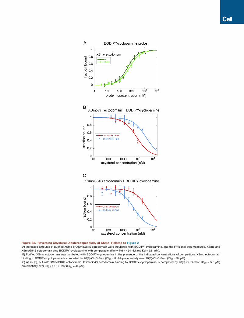
Figure S3. Reversing Oxysterol Diastereospecificity of XSmo, Related to Figure 2
(A) Increased amounts of purified XSmo or XSmoG84S ectodomain were incubated with BODIPY-cyclopamine, and the FP signal was measured. XSmo and
XSmoG84S ectodomain bind BODIPY-cyclopamine with comparable affinity (Kd = 434 nM and Kd = 621 nM).
(B) Purified XSmo ectodomain was incubated with BODIPY-cyclopamine in the presence of the indicated concentrations of competitors. XSmo ectodomain
binding to BODIPY-cyclopamine is competed by 20(S)-OHC-Pent (IC50 = 8 mM) preferentially over 20(R)-OHC-Pent (IC50 = 34 mM).
(C) As in (B), but with XSmoG84S ectodomain. XSmoG84S ectodomain binding to BODIPY-cyclopamine is competed by 20(R)-OHC-Pent (IC50 = 5.5 mM)
preferentially over 20(S)-OHC-Pent (IC50 = 44 mM).

Figure S4. Sterol-Induced Conformational Change in XSmoCRD, Related to Figure 3
(A) Comparison between XSmoCRD-20(S)-OHC and apo zfSmoCRD structures. Unliganded zfSmoCRD (green) (PDB ID 4C79) superimposed on XSmoCRD
(navy) bound to 20(S)-OHC (yellow), showing that the two proteins adopt very similar conformations.
(B) Crystal packing of unliganded zfSmoCRD (PDB ID 4C79). Helix 30 and the subsequent loop (red), which together constitute half of the sterol-binding groove,
are involved in contacts with the same portion of the neighboring protein molecule, forming a 2-fold symmetric dimer. This suggests that the ligand-binding site in
apo zfSmoCRD might artifactually adopt a ‘‘closed’’ conformation, mimicking the ligand-bound state.
(C) Ribbon diagram showing the overall structure of unliganded XSmoCRD.
(D) Superimposition of unliganded XSmoCRD (cyan) and 20(S)-OHC-bound XSmoCRD (navy). The protein undergoes a dramatic conformational change upon
oxysterol (yellow) binding, involving the polypeptide stretch between disulfide bonds 4 and 5.

Figure S5. Cyclopamine-Induced Conformational Change in XSmoCRD, Related to Figure 4
(A) Overall structure of XSmoCRD (green) in complex with the plant alkaloid, cyclopamine (orange).
(B) Close up view of cyclopamine (orange) bound to XSmoCRD (green), superimposed on 20(S)-OHC (yellow) bound to XSmoCRD (navy). Cyclopamine and 20(S)-
OHC induce very similar conformations of the sterol-binding site.
(C) The mSmoD477G/E522K mutant does not bind BODIPY-cyclopamine. Cultured 293T cells expressing mCherry-tagged mSmo wild-type or mSmoD477G/
E522Kwere incubated with BODIPY-cyclopamine (50 nM), in the presence or absence of SANT1 competitor (10 mM). The cells were fixed, washed, and BODIPY-
cyclopamine and mCherry fusions were imaged by fluorescence microscopy. The mSmoD477G/E522K mutant does not bind BODIPY-cyclopamine, in contrast
to wild-type mSmo.
(D) The mSmoD477G/E522K mutant still binds oxysterols. MCherry-tagged mSmoD477G/E522K was expressed in 293T cells, and was assayed for binding to
20(S)-OHC affinity matrix (Nedelcu et al., 2013), in the absence or presence of free 20(S)-OHC competitor (100 mM). The mutant protein binds 20(S)-OHC beads
and is competed by free 20(S)-OHC, but does not bind control beads (see Figure S6C for schematic of the beads used in this assay).
(E) Smo null MEFs rescued by stable expression of mCherry-taggedmSmowere incubated overnight in the absence of presence of cyclopamine (10 mM), and Hh
pathway activity was assayed by qPCR for endogenous Gli1 mRNA. Error bars indicate SD (n = 3). As expected, cyclopamine inhibits wild-type mSmo.

Figure S6. Smo Binds Cholesterol, Related to Figure 5
(A and B) Binding of XSmo ectodomain to sterols measured by circular dichroism (CD) melting. Purified XSmo ectodomain was incubated in the absence or
presence of the indicated small molecules, and thermal denaturation of the protein was measured by recording the CD signal at 225 nm as a function of tem-
perature. (A) The melting temperature of XSmo ectodomain (Tm = 62.1�C) is increased by cholesterol (Tm = 65.3�C), but not by cholestanol (Tm = 62.9�C),indicative of cholesterol binding. (B) XSmo ectodomain melting temperature is increased by binding to 20(S)-OHC (Tm = 66.5�C) or cyclopamine (Tm = 66.6�C).(C) Schematic of affinity resins used in this study. Sterol molecules are attached to Affigel beads via a linker with three ethylene glycol units. Attachment is through
C-19 for 19-cholesterol beads, through the 3b-OH group for 3-cholesterol beads, and through the sterol tail for 20(S)-OHC beads. Beads derivatized with the
ethylene glycol linker alone serve as negative control.
(D) XSmo ectodomain binds to 19-cholesterol affinity resin. Purified XSmo ectodomain was incubated with 19-cholesterol beads, in the presence of the indicated
compounds (500 mM, added from 20 mM DMSO stocks). The beads were washed, and bound protein was eluted and analyzed by SDS-PAGE and Coomassie
staining. Beads without attached sterols served as negative control. XSmo ectodomain does not bind 3-cholesterol beads, consistent with the requirement of a
free 3b-OH group for XSmo binding (Nedelcu et al., 2013).

Figure S7. Cholesterol as Endogenous Activator of Smo, Related to Figure 6
(A) Hh pathway activation by cholesterol is defective in Smo null cells rescued with mSmoD99A, in contrast to cells rescued with wild-type mSmo. Hh signaling
was assayed by qPCR for endogenous Gli1. Error bars indicate SD (n = 3). This experiment was performed in parallel with the ones in Figure 5B and Figure 6B, and
shows the same curve for stimulation of wild-type mSmo by cholesterol.
(B) Shh synergizes with cholesterol, but not with cholestanol, to activate Hh signaling in Smo null cells rescued with wild-typemSmo. Sterols were added to 5 mM,
as soluble MCD complexes.
(C–E) Responsiveness to 20(S)-OHC-Pent is reduced in Smo null cells rescued with mSmoG115S, compared to wild-type mSmo. Cells rescued with
mSmoG115S/E164L do not respond to 20(S)-OHC-Pent. In contrast, cholesterol (D) and Shh (E) activate wild-type mSmo, mSmoG115S and mSmoG115S/
E164L to a similar extent. Experiments in (C–E) were performed in parallel with experiments shown in Figures 6A–6C, thus the curves for wild-type mSmo are the
same.